Article

Ectopic Expression of Neurod1 Is Sufficient for Functional Recovery following a Sensory–Motor Cortical Stroke

by
Jessica M. Livingston
1,2,
Tina T. Lee
1,
Tom Enbar
1,2,3,
Emerson Daniele
1,3,
Clara M. Phillips
1,2,
Alexandra Krassikova
1,
K. W. Annie Bang
4,
Ines Kortebi
1,3,
Brennan W. Donville
1,2,
Omadyor S. Ibragimov
1,
Nadia Sachewsky
1,2,
Daniela Lozano Casasbuenas
1,
Arman Olfat
1 and
Cindi M. Morshead
1,2,3,5,*
1
Department of Surgery, Division of Anatomy, 1 King’s College Circle, University of Toronto, Toronto, ON M5S 1A8, Canada
2
Donnelly Centre for Cellular and Biomolecular Research, University of Toronto, 160 College Street, Toronto, ON M5S 3E1, Canada
3
Institute of Medical Science, 1 King’s College Circle, University of Toronto, Toronto, ON M5S1A8, Canada
4
Lunenfeld-Tanenbaum Research Institute, 600 University Ave., Toronto, ON M5G 1X7, Canada
5
Institute of Biomedical Engineering, 1 King’s College Circle, University of Toronto, Toronto, ON M5S 1A8, Canada
*
Author to whom correspondence should be addressed.
Biomedicines 2024, 12(3), 663; https://doi.org/10.3390/biomedicines12030663
Submission received: 31 January 2024 / Revised: 5 March 2024 / Accepted: 11 March 2024 / Published: 15 March 2024

Abstract

:
Stroke is the leading cause of adult disability worldwide. The majority of stroke survivors are left with devastating functional impairments for which few treatment options exist. Recently, a number of studies have used ectopic expression of transcription factors that direct neuronal cell fate with the intention of converting astrocytes to neurons in various models of brain injury and disease. While there have been reports that question whether astrocyte-to-neuron conversion occurs in vivo, here, we have asked if ectopic expression of the transcription factor Neurod1 is sufficient to promote improved functional outcomes when delivered in the subacute phase following endothelin-1-induced sensory–motor cortex stroke. We used an adeno-associated virus to deliver Neurod1 from the short GFAP promoter and demonstrated improved functional outcomes as early as 28 days post-stroke and persisting to at least 63 days post-stroke. Using Cre-based cell fate tracking, we showed that functional recovery correlated with the expression of neuronal markers in transduced cells by 28 days post-stroke. By 63 days post-stroke, the reporter-expressing cells comprised ~20% of all the neurons in the perilesional cortex and expressed markers of cortical neuron subtypes. Overall, our findings indicate that ectopic expression of Neurod1 in the stroke-injured brain is sufficient to enhance neural repair.

1. Introduction

Stroke is a leading cause of death and disability worldwide, and many survivors experience long-lasting impairments [1]. Approximately 850,000 individuals in North America suffer from strokes annually [2]. Survivors often experience motor impairments, particularly affecting sensory-motor function in the upper limbs [2]. This can lead to chronic disability that significantly diminishes quality of life and increases the risk of post-stroke depression [3]. With an aging population and increasing projected incidence of stroke [4], there is an urgent need for alternative options to improve post-stroke impairments.
Numerous approaches to treat stroke have been explored, including activation of endogenous neural stem cells [5,6,7] and cell transplantation [8,9]. Since the discovery that ectopic transcription factor (TF) expression can drive cell fate [10], attention has been focused on developing interventions to replace lost cells in models of injury or disease. Many publications have since identified effective methods to convert somatic cells to other cell types [11,12], including neuronal cells, both in vitro and in vivo [13,14,15,16,17,18,19,20,21,22]. In the context of the central nervous system (CNS), a number of groups have examined the ectopic expression of TFs in the CNS and demonstrated that proneural TFs such as Ngn2 [23] and Ascl1 [20,24] and the neuronal differentiation TF Neurod1 [25,26] can convert glial cells to neurons [27]. Further, studies have shown that induced neurons from the ectopic expression of TFs have regionally appropriate [14,28,29] and functional neuronal subtype identities [15,30,31,32,33]. Some recent studies in the field have brought into question the efficacy of in vivo conversion [34,35], and this has served to highlight the importance of addressing potential caveats to further advance the field [19,36,37]. Wang et al. [34] reported off-target neuronal expression of adeno-associated virus (AAV)-based delivery strategies driving TF expression from the short human GFAP promoter (shGFAP). While the mechanism is not entirely understood, the implication is that transduced endogenous neurons could mistakenly be interpreted as astrocyte-to-neuron reprogramming [34,35]. Nevertheless, the report by Wang et al. [34] does not negate the capacity of glia to be converted to neurons [27,38,39].
Of particular interest in developing therapeutics to promote neural repair, a number of groups have validated the feasibility of ectopic TF expression in preclinical models of injury and disease, including Alzheimer’s disease [40], Parkinson’s disease [41], and stroke [26,42]. These studies have focused on cellular outcomes, and a considerable gap exists in understanding the resulting functional outcomes of the AAV-mediated ectopic expression of neural TFs. Acknowledging that endogenous neurons may be targeted to overexpress TFs delivered by AAVs underscores the possibility that ectopic expression of TFs in neurons may underlie any improved outcomes, making this approach promising to treat neurological disease. Hence, it is important to study the functional outcomes of forced TF expression-based therapy. To this end, in a study of Parkinson’s disease, the ectopic expression of a cocktail of TFs that promote dopaminergic cell fate in astrocytes improved some aspects of motor function [41]. Further, improved functional outcomes were recently reported using the TF Neurod1 in a rodent ischemia model [42]. While these studies highlight the promising potential of gene therapy for neurorepair and functional recovery there is caution when advancing the translation of preclinical studies. For example, translating preclinical stroke research has not proven successful, and a number of recommendations and guidelines have been put forth by the Stroke Therapy Academic Industry Roundtable (STAIR) to overcome this challenge, including the rigorous testing and replication of potential therapeutics in more than one preclinical model [43].
Herein, we aimed to understand the longer-term functional outcomes of ectopic TF expression. We used AAV-mediated delivery of Neurod1 to GFAP-expressing cells in a well-established preclinical model of sensory-motor ischemic stroke. The endothelin-1 model [44,45] has the advantage of generating highly reproducible lesions and controlled and persistent deficits in upper limb function, making it useful for studying post-stroke recovery strategies. Different from the published stroke studies of Chen and colleagues [42], we chose a different AAV serotype to deliver Neurod1 based on reports suggesting enhanced astrocyte targeting with AAV5 [46]. Further, the previous stroke study used the ubiquitous CAG promotor to drive TF expression. Herein, Neurod1 expression was under the control of the short GFAP (gfaAVC(1)D) promotor to further target astrocyte-driven expression. We performed our experiments in a conditional reporter mouse that enabled us to track transduced cells and performed functional assays up to 9 weeks post-stroke. We found that ectopic expression of Neurod1 delivered in a sub-acute phase post-cortical stroke led to persistent improved motor outcomes. Virally transduced cells expressed cortical layer-appropriate neuronal markers. These findings provide further support that ectopic expression of Neurod1 is a viable approach to improve stroke recovery.

2. Materials and Methods

2.1. Mice

Adult (8–12 week old) male and female R26R-YFP (Jackson Labs, Bar Harbor, ME, USA: B6.129X1-Gt(ROSA)26Sortm1(EYFP)Cos/J) and R26R-tdTomato (Jackson Labs: Gt(ROSA)26Sor tm9(CAG-tdTomato)Hze) mice were used in this study [47]. Mice were group-housed in a barrier facility with a 12 h light/12 h dark cycle with ad libitum access to food and water. Experiments were conducted according to protocols approved by the Institutional Animal Care Committee and performed in accordance with guidelines published by the Canadian Council for Animal Care.

2.2. Endothelin-1 Stroke

The vasoconstricting peptide endothelin-1 (ET-1; Calbiochem, San Diego, CA, USA) was used to induce focal stroke to the sensory–motor cortex as previously described [9,48]. Mice were anesthetized using isofluorane and mounted onto a stereotaxic apparatus. The scalp was incised, and a small hole was drilled into the skull at the injection location. A total of 1 μL of 400 picomolar ET-1 dissolved in sterile PBS was injected into the sensory–motor cortex at +0.6 AP, +2.20 ML, and −1.0 DV from bregma. A 26-gauge Hamilton syringe with a 45-degree beveled tip was used to inject ET-1 at a rate of 0.1 μL/min. The needle was left in place for 10 min after completion of the ET-1 injection to prevent backflow, then slowly withdrawn. Body temperature was maintained at 37 °C using a heating pad, and animals recovered under a heat lamp. Ketoprofen (5.0 mg/kg; s.c.) was administered for post-surgery analgesia.

2.3. Adeno-Associated Virus (AAV) Injections

AAV5-GFAP(0.7)promoter(gfaABC(1)D)::Neurod1-T2A-Cre-WPRE (AAV5::Neurod1; 5.0 × 10¹² GC/mL) and AAV5-GFAP(0.7)promoter(gfaABC(1)D)-Cre-WPRE (AAV5::Cre; 1.1 × 10¹³ GC/mL) were generated and packaged by Vector BioLabs (Malvern, PA, USA).
Mice were anesthetized with isofluorane and mounted onto a stereotaxic apparatus. A total of 1 μL of AAV5::Neurod1 or AAV5::Cre was injected into the ipsilesional cortex 7 days post-stroke or -sham injury, at a rate of 0.1 μL/min at each the following coordinates: [+0.6 AP, +2.2 ML, −1.0 DV], [+1.6 AP, +2.2 ML −1.0 DV], and [−0.4 AP, +2.2 ML, −1.0 DV] mm from bregma, representing regions encompassing the anterior-to-posterior extent of the stroke-lesioned brain. The needle was left in place for 10 min after each AAV injection to prevent backflow and then slowly withdrawn. Body temperature was maintained at 37 °C using a heating pad, and animals recovered under a heat lamp. Ketoprofen (5.0 mg/kg; s.c.) was administered for post-surgery analgesia.

2.4. Tissue Processing, Immunohistochemistry, Imaging, and Quantification

Mice were anesthetized with tribromoethanol (Avertin; 250 mg/kg; i.p.) and perfused transcardially with saline, followed by 4% paraformaldehyde (PFA). Brains were removed, post-fixed for 4 h with 4% PFA, and then placed in 30% sucrose to cryopreserve. Brains were frozen, sectioned (20 µm) using a cryostat, mounted onto Superfrost Plus slides, and stored at −20 °C.
For immunohistochemistry, slides were sectioned, blocked with 10% normal goat serum (NGS) and 0.3% Triton x-100 in PBS, and labeled with primary antibodies (for astrocytes, rabbit-anti-GFAP was used, 1:3000, DAKO, Santa Clara, CA, USA, 2016-04; or chicken-anti-GFAP, 1:1000, Aves, GFP-1020. For neurons, rabbit-anti-NeuN was used, 1:100, Millipore, Oakville, ON, Canada, ABN78. For layer-specific markers, the upper-layer marker rabbit-anti-CUX1 was used, 1:100, Abcam, Baltimore, MA, USA, AB54583; and the lower-layer marker rat-anti-CTIP2, 1:300, Abcam, AB18465. To detect evidence of activity, the early immediate gene protein mouse-anti-cFOS was used, 1:200, Santa Cruz, Dallas, TX, USA, Sc-166940. To determine the cell phenotype as oligodendrocyte, microglia, or migratory neuroblasts, Olig 2, 1:500, Millipore, Iba1, 1:500, Wako, Richmond, VA, USA were used) in PBS overnight at 4 °C, followed by incubation with secondary antibodies (1:400, from Life Technologies (Carlsbad, CA, USA): goat-anti-rabbit 568; goat-anti-rabbit 647; goat-anti-chicken 488; goat-anti-mouse 488) and DAPI (1:10,000; Invitrogen, Waltham, MA, USA) in PBS for 1 h at room temperature. Slides were then cover-slipped using Dako fluorescent mounting media (ThermoFisher, Waltham, MA, USA).
Imaging was performed using Zen 2011 (v.1.0) software and a Zeiss confocal microscope (LSM880, v.14, Zeiss, Oberkochen, Germany). Linear contrast/brightness adjustments were performed using Zen 2011 software or Adobe Photoshop (v.20). Colocalization analysis was performed by z-stack analysis to confirm overlapping expression in confocal images in three images from three different coronal sections (located at +1.6, +0.6, and −1.0 AP from bregma) per animal viewed at 20× magnification. Identical linear adjustments of contrast and brightness were made to micrographs using the respective microscope software or Photoshop using the levels function, as follows: CUX1 (linear brightness adjustment, all channels), TBR1 (linear brightness adjustment, red channel), CTIP2 (linear brightness adjustment, red channel), and c-FOS (linear brightness adjustment, green channel).

2.5. Lesion Volume Analysis

Lesion volume was calculated from NeuN-stained sections and defined as areas devoid of NeuN+ stain. Image J (v.2, National Institute of Health, New York, NY, USA) was used to measure this area in 5 × 20 µm thick coronal sections (160 µm apart) spanning the anterior–posterior extent of the injury site. The software was calibrated using a scale bar, and the area measurement was obtained using the freehand selection tool. Area was then multiplied by the distance between sections to estimate the total infarct volume.

2.6. Astrocyte Sorting and ImageStream Analysis

Mice were anesthetized with isofluorane and cervically dislocated. The brain and meninges were removed, and coronal slices were obtained using a scalpel blade. The cortex was collected from slices that included the lesioned cortex, taking care to avoid the corpus callosum. The tissue was enzymatically and mechanically dissociated into a single-cell suspension using the magnetically activated cell-sorting (MACS) Neural Tissue Dissociation Kit (Miltenyi Biotec, Bergisch Gladbach, Germany, 130-093-231). Myelin was removed using the MACS Myelin Removal Beads (Miltenyi Biotec 130-096-733). Dissociated cells were resuspended in 500 μL of PBS (azide and serum/protein free) and stained with a fixable cell viability dye (FVD eFluor 780; eBioscience, San Diego, CA, USA, 65-0865-14) according to the manufacturer’s instructions (1 μL FVD/mL cell suspension). Cells were washed with 10% FBS in PBS, then resuspended in 500 μL of MACS buffer (Ca++ and Mg++ free PBS with 2% FBS and 1 mM EDTA), stained with anti-GLAST-PE (1:50; Miltenyi Biotec 130-098-804), and sorted using the MACS anti-PE MicroBeads UltraPure Kit (Miltenyi Biotec 130-105-639) according to the manufacturer’s instructions. Sorted cells were stained with Hoescht (1 μg/mL, BD Biosciences, San Diego, CA, USA, 33342) for 30 min at room temperature, then resuspended in MACS sorting buffer.
Cells were analyzed with an Amnis ImageStream Mark II imaging flow cytometer (AMNIS). Cells from the MACS sorted fractions and column flow-through were analyzed. Single-stained controls were collected for the compensation matrix to determine the level of spectral overlap (live nucleated cells were examined for PE-GLAST signal and YFP). The resulting compensated image files were analyzed using IDEAS analysis software (AMNIS, v.6.0). For the analysis, focused single cells were selected based on viability (excluding FVD e780 dye) and the presence of a nucleus (Hoescht-positive). The live nucleated cells were examined for PE-GLAST signal, and the specific and punctate GLAST-PE binding to cells was described using the Bright Detail Intensity feature in IDEAS software v.6.0). The antibody binding patterns were confirmed in composite cell images of bright field and PE fluorescence generated from ImageStream data.

2.7. Foot Fault Analysis

Mice were placed onto a metal grid (1 cm spacing) that was suspended 12 inches above a table surface. Animals were allowed to walk around the grid for 3 min and were recorded from below. The number of steps and paw slips made with each forelimb were analyzed, and the difference in foot faults was calculated as follows: %slippage = [(#slips/#stepsispilesional) − (#slips/#stepscontralesional)]. The following inclusion criteria were applied: (i) demonstrated deficit following stroke (defined as ±2 standard deviations from the mean of uninjured controls at PSD 4), (ii) >50 steps taken during the test. These criteria resulted in the exclusion of a total of 5 mice from the study.

2.8. Gait Analysis

Gait analysis was performed using the automated Noldus CatWalk XT system (Noldus, Wageningen, The Netherlands). Mice were allowed to traverse a glass walkway illuminated with a light, and a number of gait parameters were measured. Mice were pre-trained to cross the platform for 2–3 consecutive days prior to stroke. A minimum of 3 successful trials was required, wherein the speed during crossing did not vary more than 60%. Footprints were recorded using a high-speed video camera and analyzed using CatWalk XT software (v.10.6). Parameters were normalized to the performance of uninjured mice and compared between both stroke groups and Cre-injected shams. Parameters that were significantly different between groups at baseline were excluded from analysis.

2.9. Data Acquisition and Statistical Analysis

All experiments were conducted by an experimenter blind to injury and treatment conditions. Data were tested for normality using the D’Angostino and Pearson omnibus normality test or the Kolmogorov–Smirnov test. Unpaired one-tailed t-tests were used to compare two groups. One-factor analysis of variance (ANOVAs) followed by post hoc analysis (Tukey’s or Dunnet’s test) was used to compare 3 or more groups. Foot fault outcomes were analyzed using 2-way repeated measures ANOVA, followed by Bonferroni post hoc tests when appropriate. Lesion volume was analyzed using Mann–Whitney U test. Differences were considered significant at p < 0.05. Values are presented as mean ± SEM.

3. Results

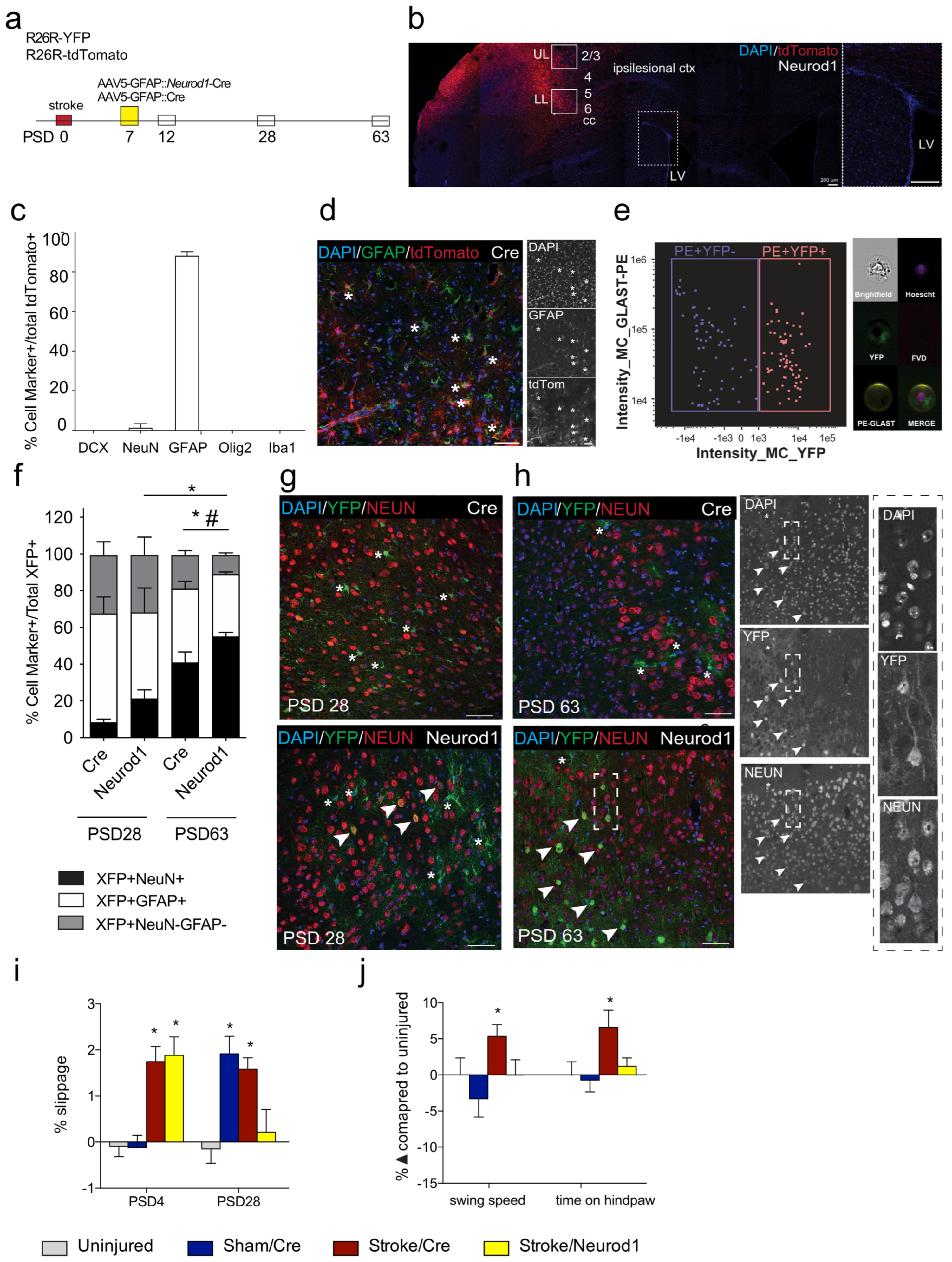
3.1. Transduced Cells Persist Long Term

We designed a strategy to deliver Neurod1 to GFAP+ cells in the stroke-injured cortex. AAV5::Neurod1 or AAV5::Cre control was injected into the injured cortex of Cre-conditional reporter mice (R26R-YFP or R26R-tdTomato) on post-stroke day 7 (PSD7) (Figure 1a,b), a time that reflects the subacute phase of injury. To first determine the profile of transduced cells using this delivery strategy, mice were sacrificed at day 5 post-AAV5::Cre injection (PSD12). As predicted, the vast majority of transduced (tdTom+) cells expressed GFAP (Figure 1c,d); a small percentage of cells co-expressed the neuronal marker NeuN. A rare tdTom+ cell co-localized with the microglia/macrophage marker Iba1. None of the tdTom+ cells were neuroblasts (DCX+) or oligodendrocytes (Oligo2+) at 5 days post-AAV delivery.
We predicted that perilesional reactive astrocytes would be transduced following stroke and Neurod1 treatment. To test this, we isolated GLAST+ astrocytes from the cortex of R26R-YFP mice at 3 days post-AAV (PSD10). The number of transduced (YFP+) cells within this population was analyzed using magnetically activated cell sorting. As shown in Figure 1e, 50% of perilesional GLAST+ astrocytes expressed YFP. We next examined the ectopic expression of Neurod1 over time in the stroke-injured brain. Using both R26R-YFP and R26R-tdTomato mice, we visualized the colocalization of neural phenotypes in fluorescent positive cells (XFP+) at early (PSD28) and late (PSD63) times post-stroke. We quantified the proportion of transduced cells that co-expressed the NeuN and GFAP markers after Neurod1 or Cre was delivered. On PSD 28, following Neurod1 injection, we observed 21.2 ± 4.9% of all XFP+ cells expressed NeuN, and 46.9 ± 13.4% expressed GFAP (Figure 1f,g). There were significantly fewer XFP+NeuN+ cells on PSD28 in the AAV5::Cre-injected mice (8.3 ± 1.7%, p < 0.05) and similar numbers of XFP+GFAP+ cells (59.1 ± 9.1%) (Figure 1f,g). Most interesting, by PSD 63, the relative percentage of NeuN+XFP+ cells in Neurod1 injected mice was 55.0 ± 2.4%, significantly greater than that of Cre-injected brains (40.9 ± 5.7%; p = 0.044; Figure 1f,h) and this was significantly increased from PSD28 (p = 0.0009). No change was seen in the relative percentage of XFP+GFAP+ astrocytes at PSD28 and PSD63 in any of the groups. Notably, there was a significant decrease in the relative fraction of XFP+GFAP-NeuN- cells at PSD63 in Neurod1-injected mice when compared to Cre controls (stroke/Cre_63d, 19.0 ± 2.7 vs. stroke/Neurod1_63d, 11.1 ± 1.5; p = 0.022). Hence, while the percentage of NeuN+ cells expressing Cre is increasing over time, the relative percentage of XFP+NeuN+ cells is significantly greater with ectopic expression of Neurod1.

3.2. Functional Recovery Is Observed following Ectopic Expression of Neurod1

We investigated the impact of ectopic Neurod1 expression on post-stroke recovery. Stroke-injured mice that received Neurod1 or Cre in the stroke-injured cortex on PSD7 were assessed for motor function using the grid walk task at PSD28 and CatWalk for gait analysis at PSD63. Stroke-injured mice displayed a significant motor deficit on the grid walking task at PSD4 prior to treatment with Cre or Neurod1 (p = 0.003, p = 0.002, respectively) (Figure 1i). As predicted, uninjured mice and sham controls did not show deficits (Figure 1i). By PSD28, only mice that received Cre were recovered to baseline performance [stroke/Neurod1 = 0.22 ± 0.53 slips (p = 0.846); stroke/Cre = 1.58 ± 0.26 slips (p = 0.005); sham/Cre, 1.91 ± 0.37 slips (p = 0.0004); uninjured controls, −0.15 ± 0.34 slips] (Figure 1i).
Since foot fault deficits are not sustained at longer survival times [49,50], and because an impairment was detected at PSD28 in sham mice that received cortical injections of Cre (Figure 1i), we performed gait analysis at PSD63 to compare long-term outcomes of ectopic Neurod1 expression [51]. Significant deficits were detected in stroke/Cre mice in swing speed (5.5 ± 1.63% change; p = 0.026) and time spent on the hindpaw (10.60 ± 3.79% change; p = 0.025) (Figure 1j). The gait of Neurod1-treated mice was indistinguishable from that of uninjured controls, changing 0.00 ± 2.15% in swing speed (p = 0.67) and 2.02 ± 1.79% in time spent on the hindpaw (p = 0.87) (Figure 1j). Taken together, these findings reveal that ectopic expression of Neurod1 is sufficient to improve functional outcomes following stroke as early as 3 weeks post-transduction and maintain recovery to at least 8 weeks post-intervention.

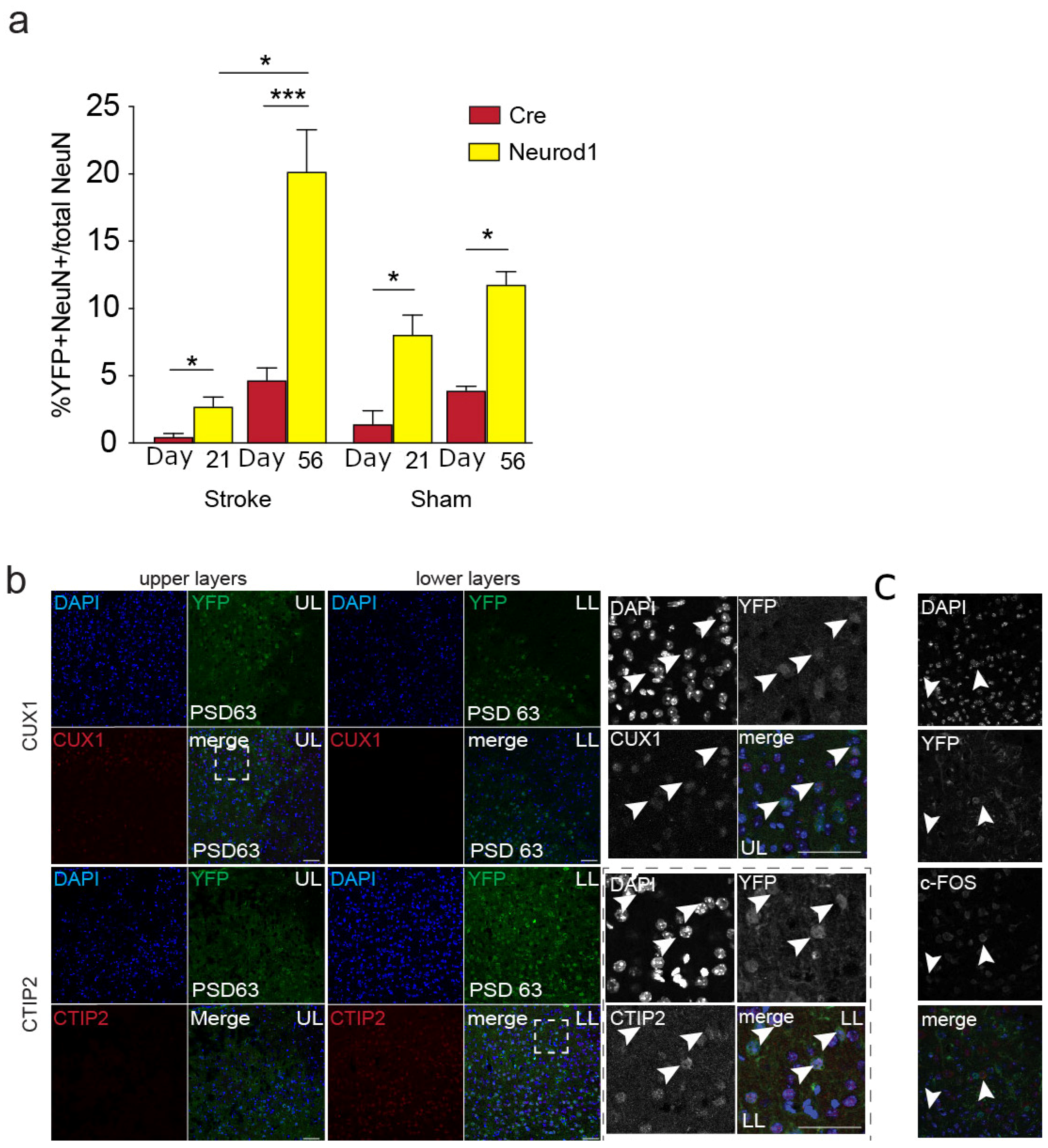
3.3. The Percent of Transduced Neurons in the Perilesional Cortex Increases over Time

We predicted that improved functional outcomes would be correlated with increased numbers of XFP+NeuN+ cells in the perilesional cortex. We quantified the relative percent of transduced perilesional neurons (NeuN+XFP+/NeuN+) at 21 and 56 days post-AAV transduction in stroke-injured and sham-injected mice. In the stroke-injured mice, there were significantly more XFP+ neurons in Neurod1 (2.78 ± 0.7% and 20.2 ± 3.1%, at day 21 and day 56, respectively) compared to Cre-injected brains (0.47 ± 0.26% and 4.67 ± 0.91%, at day 21 and day 56, respectively) (p = 0.031 for day 28 and p < 0.0001 for day 56) (Figure 2a). Further, the relative percentage of NeuN+XFP+/NeuN+ cells in the stroke-injured mice was significantly greater at PSD63 compared to PSD28 (p = 0.030). These findings indicate that the relative percentage of transduced neurons in the perilesional cortex is significantly greater in stroke-injured mice following the ectopic expression of Neurod1.
To compare the proportions of transduced neurons in the absence of injury, we delivered Neurod1 and Cre to sham animals and quantified the transduced cells that co-expressed NeuN in the equivalent cortical area and time points examined above. We predicted that sham animals that received Neurod1 would have more XFP+NeuN+ cells following treatment. As predicted, those mice that received AAV5::Neurod1 had more XFP+NeuN+ cells compared to controls that did not receive Neurod1 (Neurod1, 8.1 ± 1.5% and 11.8 ± 1.0 % of total neurons at day 21 and day 56, respectively; Cre, 1.4 ± 1.0% and 3.9 ± 0.3% at day 21 and day 56, respectively) (p < 0.05) (Figure 2a).
We sought to determine the cellular phenotypes associated with the observed long-term motor recovery. We analyzed the expression of neuronal subtype-specific transcription factors CUX1 (upper-layer neurons, layers 2/3 [52]) and CTIP2 (lower-layer projection neurons, layers 5/6 [53]) at PSD63 in R26R-YFP mice that received Neurod1 post-stroke (Figure 2b). We observed CUX1 exclusively in YFP+NeuN+ cells in the upper layers. Similarly, CTIP2 was only present in YFP+NeuN+ neurons in the lower layers. We performed immunohistochemistry for c-FOS as an indicator of neuronal activity and found that a subpopulation of the YFP+NeuN+ neurons co-expressed c-FOS within the cortex (Figure 2c). Hence, neurons expressing Neurod1 are active within the cortex at a time when functional recovery is observed.
We next asked whether the improved outcomes following Neurod1 treatment were associated with a change in lesion volume. We compared the brains of stroke-injured Neurod1 and Cre-injected animals at PSD63 and found similar lesion volumes (stroke/Neurod1 = 0.06 ± 0.03 mm3; stroke/Cre = 0.07 ± 0.01 mm3; p = 0.62) (Figure S1). Hence, ectopic expression of Neurod1 does not lead to a change in lesion volume at long survival times and when functional recovery is observed.

4. Discussion

Here, we demonstrate that ectopic expression of Neurod1 is sufficient for functional improvement following sensory–motor stroke. Previous studies have used AAV delivery to ectopically express TFs in vivo [11,14,15,28,32,40,41,54,55]; however, few have demonstrated improved functional outcomes following injury [41,42]. Using the grid walk task and gait analysis, we demonstrate that ectopic expression of Neurod1 in the stroke-injured brain leads to functional improvement as early as three weeks post Neurod1 delivery and that the recovery is sustained long-term.
Studying functional outcomes in rodent stroke models is important for determining the preclinical efficacy of interventions. However, this is difficult due to two main elements: (1) the equivalent cellular processes thought to underlie post-stroke neuroregenerative and neuroplastic processes (thought to recapitulate processes that occur in development to some degree) occur in an abbreviated time frame compared to those observed in the clinic (reviewed by [56]); and (2) spontaneous recovery of most widely validated post-stroke behavioral tests (including those used here) can occur within weeks in rodents models, as opposed to the persistent, attenuated timeframes seen in human stroke survivors. As such, the Stroke Therapy Academic Industry Roundtable Preclinical Recommendations [43] suggest that rodent stroke models include long-term outcomes measuring at least 2–3 weeks after stroke. In this study, our 9-week timeline exceeding this recommendation is considered chronic in terms of rodent stroke models [57], and importantly, it was still able to detect deficits in untreated controls that were significantly improved in Neurod1-treated animals. This finding has important implications for preclinical stroke research.
Chen and colleagues [42] examined the effects of ectopic expression of Neurod1 following stroke and reported similar outcomes while highlighting some important considerations for stroke studies. First, Chen et al. [42] used a dual AAV9 system (where Neurod1 expression is driven by a CAG promoter) following ET-1 stroke-injured mice and found that 70% of all transduced cells expressed the neuronal marker NeuN at 14 days post-stroke. We observed NeuN expression in 21% of transduced cells over a similar time course using a single AAV5 system, where the expression of Neurod1 was driven by the GFAP promoter. Further, our studies did not reveal a difference in the lesion volume in our stroke-injured mice that received Neurod1, which was significantly reduced following Neurod1 expression in the previous work. Different strains of mice, the severity and location of the injury, the time course for intervention, concentration, and serotype of the AAV are also likely to play a role in the outcomes. Importantly, we showed that a lower conversion efficiency also results in long-term recovery. A comparison of these studies is critical when developing therapeutics to treat stroke and reflects the complexity of the model and the importance of demonstrating success in more than one pre-clinical model of injury to improve the potential for translational success of novel therapeutics.
Interestingly, we found that the proportion of transduced neurons in Neurod1-treated mice was significantly increased compared to Cre controls at PSD28, a time at which functional recovery was already observed. Similarly, we show that transduced neurons comprise almost 20% of the perilesional population in Neurod1-treated mice and express markers of activity and regional specificity. When interpreting our findings, it is important to note that we also observed reporter-positive neurons in Cre-injected mice (i.e., in the absence of Neurod1 delivery). This is interesting and consistent with previous work showing a degree of non-specific expression from the GFAP promoter [28,34,35,58]. This reveals the importance of future studies aimed at discerning whether improved recovery is mediated by the conversion of astrocytes to neurons or other mechanisms, such as enhanced neuronal cell survival and/or neuroprotection as a result of ectopic Neurod1 expression. An eloquent study has recently demonstrated that the ablation of induced neurons generated from microglia/macrophages following a stroke that encompasses the striatum (middle cerebral artery occlusion) leads to the loss of functional improvements [59]. As we move forward in the field, a recent position paper that outlines the obligatory controls for demonstrating in vivo neuronal reprogramming will provide important guidance. Bocchi et al. [60] include lineage tracing (neuronal and glial), single-cell transcriptomics studies, and functional assessment of induced neurons as important next steps.
Recent papers are drawing attention to the poorly understood mechanism of action when reprogramming TFs are expressed in the in situ brain [34,60,61]. We demonstrated that the number of transduced neurons increases over time, and this is correlated with improved functional outcomes. This temporal increase could be the result of astrocyte-to-neuron reprogramming, as well as delayed expression of the AAV in neurons due to leakiness of the promoter. These possibilities are consistent with the observation that at early times post-AAV delivery, the vast majority of transduced cells did not express neuronal markers, and over 50% of the cortical astrocytes in the perilesional stroke area were transduced with the AAV. Studies revealing the extent of astrocyte heterogeneity raise the possibility that functional recovery could also be due to a shift in astrocyte cell state due to ectopic expression of TFs [62,63]. Indeed, specifically blocking a subset of reactive astrocytes was shown to be neuroprotective in a model of Parkinson’s disease [64], and ectopic expression of Neurod1 in astrocytes may specifically decrease this reactive astrocyte population [65].
We chose to use AAV5 to deliver Neurod1 based on previous work suggesting enhanced astrocyte targeting with this serotype [46]. A number of different viral delivery methods have been used for in vivo reprogramming (e.g., lentivirus, adenovirus, and AAV9 [66]. These have shown varied transduction and reprogramming efficiencies. Different delivery methods may preferentially transduce different astrocyte subtypes or other neural cell types, highlighting the importance of choosing an appropriate strategy. In our study, AAV5 was used to drive Neurod1 expression under the control of the short GFAP (gfaABC(1)D) promoter. We performed reprogramming at 7 days post-stroke in order to preferentially target cortical astrocytes and not neural stem cells that also express GFAP [67,68,69,70], as this is a time we have previously demonstrated a rapid decline in the number of migratory neural stem cells present in the cortex by this time point [71].
With the goal of realizing the translational potential of gene therapies that deliver neurogenic TFs to treat the injured or diseased brain, a comprehensive understanding of the cellular and molecular basis of functional recovery is an important consideration for future studies exploring gene therapy-based approaches for stroke repair. Herein, we have demonstrated that the AAV5-mediated expression of Neurod1 driven by the shGFAP promoter leads to post-stroke functional recovery. Our work highlights the promise of this strategy for stroke repair.

Supplementary Materials

The following supporting information can be downloaded at: https://www.mdpi.com/article/10.3390/biomedicines12030663/s1, Figure S1. Lesion volume. Volume (mm3) of the lesion was similar between stroke animals that received Neurod1 (stroke_nd1) and Cre (stroke_cre) injections. p > 0.05. n = 6–8/group. Data are expressed as mean ± SEM.

Author Contributions

Conceptualization, C.M.M.; Methodology, J.M.L., T.T.L., T.E., C.M.P., A.K., K.W.A.B., B.W.D., O.S.I., N.S., D.L.C. and A.O.; Formal analysis, J.M.L., T.T.L., T.E., E.D., C.M.P., A.K., K.W.A.B., I.K., O.S.I., N.S. and C.M.M.; Writing—original draft, J.M.L. and C.M.M.; Writing—review & editing, J.M.L. and C.M.M.; Visualization, C.M.M.; Supervision, C.M.M.; Funding acquisition, C.M.M. All authors have read and agreed to the published version of the manuscript.

Funding

This work was supported by grants to C.M.M. from the Heart and Stroke Foundation, Ontario Institute of Regenerative Medicine, Canada First Research Excellence Fund (Medicine by Design, MbD), CIHR, and the National Sciences and Engineering Research Council (C.M.P. studentship; C.M.M). J.M.L. held a postdoctoral fellowship (Medicine by Design, C.M.M.). The funders had no role in study design, data collection and analysis, decision to publish, or preparation of the manuscript.

Institutional Review Board Statement

The study was conducted in accordance with the Canadian Council for Animal Care, and approved by the Animal Care Committee of University of Toronto.

Informed Consent Statement

Not applicable.

Data Availability Statement

The data presented in this study are available on request from the corresponding author due to privacy.

Acknowledgments

The authors are grateful to Maryam Faiz for contributions to all aspects of the project and Ilan Vonderwalde for assistance with behavioral analyses.

Conflicts of Interest

The authors declare no conflict of interest.

References

  1. Virani, S.S.; Alonso, A.; Benjamin, E.J.; Bittencourt, M.S.; Callaway, C.W.; Carson, A.P.; Chamberlain, A.M.; Chang, A.R.; Cheng, S.; Delling, F.N.; et al. Heart Disease and Stroke Statistics—2020 Update: A Report from the American Heart Association. Circulation 2020, 141, E139–E596. [Google Scholar] [CrossRef] [PubMed]
  2. Tsao, C.W.; Aday, A.W.; Almarzooq, Z.I.; Alonso, A.; Beaton, A.Z.; Bittencourt, M.S.; Boehme, A.K.; Buxton, A.E.; Carson, A.P.; Commodore-Mensah, Y.; et al. Heart Disease and Stroke Statistics-2022 Update: A Report From the American Heart Association. Circulation 2022, 145, E153–E639. [Google Scholar] [CrossRef] [PubMed]
  3. Burvill, P.; Johnson, G.; Jamrozik, K.; Anderson, C.; Stewart-Wynn, E. Risk Factors for Post-Stroke Depression. Int. J. Geriatr. Psychiatry 1997, 12, 219–226. [Google Scholar] [CrossRef]
  4. Krueger, H.; Koot, J.; Hall, R.; O’Callaghan, C.; Bayley, M.; Corbett, D. Prevalence of Individuals Experiencing the Effects of Stroke in Canada: Trends and Projections. Stroke 2015, 46, 2226–2231. [Google Scholar] [CrossRef] [PubMed]
  5. Venna, V.R.; Li, J.; Hammond, M.D.; Mancini, N.S.; McCullough, L.D. Chronic Metformin Treatment Improves Post-Stroke Angiogenesis and Recovery after Experimental Stroke. Eur. J. Neurosci. 2014, 39, 2129–2138. [Google Scholar] [CrossRef]
  6. Dadwal, P.; Mahmud, N.; Sinai, L.; Azimi, A.; Fatt, M.; Wondisford, F.E.; Miller, F.D.; Morshead, C.M. Activating Endogenous Neural Precursor Cells Using Metformin Leads to Neural Repair and Functional Recovery in a Model of Childhood Brain Injury. Stem Cell Rep. 2015, 5, 166–173. [Google Scholar] [CrossRef] [PubMed]
  7. Nusrat, L.; Livingston-Thomas, J.M.; Raguthevan, V.; Adams, K.; Vonderwalde, I.; Corbett, D.; Morshead, C.M. Cyclosporin A-Mediated Activation of Endogenous Neural Precursor Cells Promotes Cognitive Recovery in a Mouse Model of Stroke. Front. Aging Neurosci. 2018, 10, 93. [Google Scholar] [CrossRef] [PubMed]
  8. Boese, A.C.; Le, Q.-S.E.; Pham, D.; Hamblin, M.H.; Lee, J.-P. Neural Stem Cell Therapy for Subacute and Chronic Ischemic Stroke. Stem Cell Res. Ther. 2018, 9, 154. [Google Scholar] [CrossRef]
  9. Vonderwalde, I.; Azimi, A.; Rolvink, G.; Ahlfors, J.; Shoichet, M.; Morshead, C. Transplantation of Directly Reprogrammed Human Neural Precursor Cells Following Stroke Promotes Synaptogenesis and Functional Recovery. Transl. Stroke Res. 2020, 11, 93–107. [Google Scholar] [CrossRef]
  10. Takahashi, K.; Yamanaka, S. Induction of Pluripotent Stem Cells from Mouse Embryonic and Adult Fibroblast Cultures by Defined Factors. Cell 2006, 126, 663–676. [Google Scholar] [CrossRef]
  11. Li, H.; Chen, G. In Vivo Reprogramming for CNS Repair: Regenerating Neurons from Endogenous Glial Cells. Neuron 2016, 91, 728–738. [Google Scholar] [CrossRef]
  12. Bajohr, J.; Faiz, M. Direct Lineage Reprogramming in the CNS. Adv. Exp. Med. Biol. 2020, 1212, 31–48. [Google Scholar] [CrossRef]
  13. Graf, T.; Enver, T. Forcing Cells to Change Lineages. Nature 2009, 462, 587–594. [Google Scholar] [CrossRef]
  14. Heinrich, C.; Blum, R.; Gascón, S.; Masserdotti, G.; Tripathi, P.; Sánchez, R.; Tiedt, S.; Schroeder, T.; Götz, M.; Berninger, B. Directing Astroglia from the Cerebral Cortex into Subtype Specific Functional Neurons. PLoS Biol. 2010, 8, e1000373. [Google Scholar] [CrossRef]
  15. Karow, M.; Sánchez, R.; Schichor, C.; Masserdotti, G.; Ortega, F.; Heinrich, C.; Gascón, S.; Khan, M.; Lie, D.; Dellavalle, A.; et al. Reprogramming of Pericyte-Derived Cells of the Adult Human Brain into Induced Neuronal Cells. Cell Stem Cell 2012, 11, 471–476. [Google Scholar] [CrossRef] [PubMed]
  16. Xu, J.; Du, Y.; Deng, H. Direct Lineage Reprogramming: Strategies, Mechanisms, and Applications. Cell Stem Cell 2015, 16, 119–134. [Google Scholar] [CrossRef] [PubMed]
  17. Masserdotti, G.; Gascón, S.; Götz, M. Direct Neuronal Reprogramming: Learning from and for Development. Development 2016, 143, 2494–2510. [Google Scholar] [CrossRef] [PubMed]
  18. Gascón, S.; Masserdotti, G.; Russo, G.; Götz, M. Direct Neuronal Reprogramming: Achievements, Hurdles, and New Roads to Success. Cell Stem Cell 2017, 21, 18–34. [Google Scholar] [CrossRef] [PubMed]
  19. Matsuda, T.; Nakashima, K. Clarifying the Ability of NeuroD1 to Convert Mouse Microglia into Neurons. Neuron 2021, 109, 3912–3913. [Google Scholar] [CrossRef] [PubMed]
  20. Ghazale, H.; Park, E.J.; Vasan, L.; Mester, J.; Saleh, F.; Trevisiol, A.; Zinyk, D.; Chinchalongporn, V.; Liu, M.; Fleming, T.; et al. Ascl1 Phospho-Site Mutations Enhance Neuronal Conversion of Adult Cortical Astrocytes In Vivo. Front. Neurosci. 2022, 16, 917071. [Google Scholar] [CrossRef] [PubMed]
  21. Zhang, Y.; Li, B.; Cananzi, S.; Han, C.; Wang, L.L.; Zou, Y.; Fu, Y.X.; Hon, G.C.; Zhang, C.L. A Single Factor Elicits Multilineage Reprogramming of Astrocytes in the Adult Mouse Striatum. Proc. Natl. Acad. Sci. USA 2022, 119, e2107339119. [Google Scholar] [CrossRef] [PubMed]
  22. Talifu, Z.; Liu, J.Y.; Pan, Y.Z.; Ke, H.; Zhang, C.J.; Xu, X.; Gao, F.; Yu, Y.; Du, L.J.; Li, J.J. In Vivo Astrocyte-to-Neuron Reprogramming for Central Nervous System Regeneration: A Narrative Review. Neural Regen. Res. 2023, 18, 750–755. [Google Scholar] [CrossRef]
  23. Grande, A.; Sumiyoshi, K.; López-Juárez, A.; Howard, J.; Sakthivel, B.; Aronow, B.; Campbell, K.; Nakafuku, M. Environmental Impact on Direct Neuronal Reprogramming In Vivo in the Adult Brain. Nat. Commun. 2013, 4, 2373. [Google Scholar] [CrossRef]
  24. Chanda, S.; Ang, C.E.; Davila, J.; Pak, C.; Mall, M.; Lee, Q.Y.; Ahlenius, H.; Jung, S.W.; Südhof, T.C.; Wernig, M. Generation of Induced Neuronal Cells by the Single Reprogramming Factor ASCL1. Stem Cell Rep. 2014, 3, 282–296. [Google Scholar] [CrossRef]
  25. Puls, B.; Ding, Y.; Zhang, F.; Pan, M.; Lei, Z.; Pei, Z.; Jiang, M.; Bai, Y.; Forsyth, C.; Metzger, M.; et al. Regeneration of Functional Neurons After Spinal Cord Injury via in Situ NeuroD1-Mediated Astrocyte-to-Neuron Conversion. Front. Cell Dev. Biol. 2020, 8, 591883. [Google Scholar] [CrossRef] [PubMed]
  26. Ge, L.J.; Yang, F.H.; Li, W.; Wang, T.; Lin, Y.; Feng, J.; Chen, N.H.; Jiang, M.; Wang, J.H.; Hu, X.T.; et al. In Vivo Neuroregeneration to Treat Ischemic Stroke Through NeuroD1 AAV-Based Gene Therapy in Adult Non-Human Primates. Front. Cell Dev. Biol. 2020, 8, 590008. [Google Scholar] [CrossRef]
  27. Vasan, L.; Park, E.; David, L.A.; Fleming, T.; Schuurmans, C. Direct Neuronal Reprogramming: Bridging the Gap Between Basic Science and Clinical Application. Front. Cell Dev. Biol. 2021, 9, 681087. [Google Scholar] [CrossRef]
  28. Mattugini, N.; Bocchi, R.; Scheuss, V.; Russo, G.L.; Torper, O.; Lao, C.L.; Götz, M. Inducing Different Neuronal Subtypes from Astrocytes in the Injured Mouse Cerebral Cortex. Neuron 2019, 103, 1086–1095.e5. [Google Scholar] [CrossRef]
  29. Herrero-Navarro, Á.; Puche-Aroca, L.; Moreno-Juan, V.; Sempere-Ferràndez, A.; Espinosa, A.; Susín, R.; Torres-Masjoan, L.; Leyva-Díaz, E.; Karow, M.; Figueres-Oñate, M.; et al. Astrocytes and Neurons Share Region-Specific Transcriptional Signatures That Confer Regional Identity to Neuronal Reprogramming. Sci. Adv. 2021, 7, eabe8978. [Google Scholar] [CrossRef]
  30. Pfisterer, U.; Kirkeby, A.; Torper, O.; Wood, J.; Nelander, J.; Dufour, A.; Björklund, A.; Lindvall, O.; Jakobsson, J.; Parmar, M. Direct Conversion of Human Fibroblasts to Dopaminergic Neurons. Proc. Natl. Acad. Sci. USA 2011, 108, 10343–10348. [Google Scholar] [CrossRef]
  31. Heinrich, C.; Götz, M.; Berninger, B. Reprogramming of Postnatal Astroglia of the Mouse Neocortex into Functional, Synapse-Forming Neurons. Methods Mol. Biol. 2012, 814, 485–498. [Google Scholar] [CrossRef] [PubMed]
  32. Liu, Y.; Miao, Q.; Yuan, J.; Han, S.; Zhang, P.; Li, S.; Rao, Z.; Zhao, W.; Ye, Q.; Geng, J.; et al. Ascl1 Converts Dorsal Midbrain Astrocytes into Functional Neurons In Vivo. J. Neurosci. 2015, 35, 9336–9355. [Google Scholar] [CrossRef]
  33. Vadodaria, K.; Mertens, J.; Paquola, A.; Bardy, C.; Li, X.; Jappelli, R.; Fung, L.; Marchetto, M.; Hamm, M.; Gorris, M.; et al. Generation of Functional Human Serotonergic Neurons from Fibroblasts. Mol. Psychiatry 2016, 21, 49–61. [Google Scholar] [CrossRef] [PubMed]
  34. Wang, L.L.; Serrano, C.; Zhong, X.; Ma, S.; Zou, Y.; Zhang, C.L. Revisiting Astrocyte to Neuron Conversion with Lineage Tracing In Vivo. Cell 2021, 184, 5465–5481.e16. [Google Scholar] [CrossRef] [PubMed]
  35. Xie, Y.; Zhou, J.; Wang, L.L.; Zhang, C.L.; Chen, B. New AAV Tools Fail to Detect Neurod1-Mediated Neuronal Conversion of Müller Glia and Astrocytes In Vivo. EBioMedicine 2023, 90, 1–19. [Google Scholar] [CrossRef]
  36. Xiang, Z.; Xu, L.; Liu, M.; Wang, Q.; Li, W.; Lei, W.; Chen, G. Lineage Tracing of Direct Astrocyte-to-Neuron Conversion in the Mouse Cortex. Neural Regen. Res. 2021, 16, 750–756. [Google Scholar] [CrossRef]
  37. Götz, M.; Bocchi, R. Neuronal Replacement: Concepts, Achievements, and Call for Caution. Curr. Opin. Neurobiol. 2021, 69, 185–192. [Google Scholar] [CrossRef]
  38. Barker, R.A.; Götz, M.; Parmar, M. New Approaches for Brain Repair-from Rescue to Reprogramming. Nature 2018, 557, 329–334. [Google Scholar] [CrossRef]
  39. Sharif, N.; Calzolari, F.; Berninger, B. Direct In Vitro Reprogramming of Astrocytes into Induced Neurons. Methods Mol. Biol. 2021, 2352, 13–29. [Google Scholar] [CrossRef]
  40. Guo, Z.; Zhang, L.; Wu, Z.; Chen, Y.; Wang, F.; Chen, G. In Vivo Direct Reprogramming of Reactive Glial Cells into Functional Neurons after Brain Injury and in an Alzheimer’s Disease Model. Cell Stem Cell 2014, 14, 188–202. [Google Scholar] [CrossRef]
  41. Rivetti di Val Cervo, P.; Romanov, R.; Spigolon, G.; Masini, D.; Martín-Montañez, E.; Toledo, E.; La Manno, G.; Feyder, M.; Pifl, C.; Ng, Y.; et al. Induction of Functional Dopamine Neurons from Human Astrocytes In Vitro and Mouse Astrocytes in a Parkinson’s Disease Model. Nat. Biotechnol. 2017, 35, 444–452. [Google Scholar] [CrossRef]
  42. Chen, Y.C.; Ma, N.X.; Pei, Z.F.; Wu, Z.; Do-Monte, F.H.; Keefe, S.; Yellin, E.; Chen, M.S.; Yin, J.C.; Lee, G.; et al. A NeuroD1 AAV-Based Gene Therapy for Functional Brain Repair after Ischemic Injury through In Vivo Astrocyte-to-Neuron Conversion. Mol. Ther. 2020, 28, 217–234. [Google Scholar] [CrossRef] [PubMed]
  43. Lapchak, P.A.; Zhang, J.H.; Noble-Haeusslein, L.J. RIGOR Guidelines: Escalating STAIR and STEPS for Effective Translational Research. Transl. Stroke Res. 2013, 4, 279–285. [Google Scholar] [CrossRef] [PubMed]
  44. Roome, R.B.; Bartlett, R.F.; Jeffers, M.; Xiong, J.; Corbett, D.; Vanderluit, J.L. A Reproducible Endothelin-1 Model of Forelimb Motor Cortex Stroke in the Mouse. J. Neurosci. Methods 2014, 233, 34–44. [Google Scholar] [CrossRef] [PubMed]
  45. Horie, N.; Maag, A.-L.; Hamilton, S.A.; Shichinohe, H.; Bliss, T.M.; Steinberg, G.K. Mouse Model of Focal Cerebral Ischemia Using Endothelin-1. J. Neurosci. Methods 2008, 173, 286–290. [Google Scholar] [CrossRef] [PubMed]
  46. Merienne, N.; Le Douce, J.; Faivre, E.; Déglon, N.; Bonvento, G. Efficient Gene Delivery and Selective Transduction of Astrocytes in the Mammalian Brain Using Viral Vectors. Front. Cell Neurosci. 2013, 7, 54254. [Google Scholar] [CrossRef]
  47. Srinivas, S.; Watanabe, T.; Lin, C.S.; William, C.M.; Tanabe, Y.; Jessell, T.M.; Costantini, F. Cre Reporter Strains Produced by Targeted Insertion of EYFP and ECFP into the ROSA26 Locus. BMC Dev. Biol. 2001, 1, 4. [Google Scholar] [CrossRef]
  48. Tennant, K.A.; Jones, T.A. Sensorimotor Behavioral Effects of Endothelin-1 Induced Small Cortical Infarcts in C57BL/6 Mice. J. Neurosci. Methods 2009, 181, 18–26. [Google Scholar] [CrossRef]
  49. Murphy, T.; Corbett, D. Plasticity during Stroke Recovery: From Synapse to Behaviour. Nat. Rev. Neurosci. 2009, 10, 861–872. [Google Scholar] [CrossRef]
  50. Ito, M.; Aswendt, M.; Lee, A.; Ishizaka, S.; Cao, Z.; Wang, E.; Levy, S.; Smerin, D.; McNab, J.; Zeineh, M.; et al. RNA-Sequencing Analysis Revealed a Distinct Motor Cortex Transcriptome in Spontaneously Recovered Mice After Stroke. Stroke 2018, 49, 2191–2199. [Google Scholar] [CrossRef] [PubMed]
  51. Neumann, M.; Wang, Y.; Kim, S.; Hong, S.; Jeng, L.; Bilgen, M.; Liu, J. Assessing Gait Impairment Following Experimental Traumatic Brain Injury in Mice. J. Neurosci. Methods 2009, 176, 34–44. [Google Scholar] [CrossRef] [PubMed]
  52. Cubelos, B.; Briz, C.G.; Esteban-Ortega, G.M.; Nieto, M. Cux1 and Cux2 Selectively Target Basal and Apical Dendritic Compartments of Layer II-III Cortical Neurons. Dev. Neurobiol. 2015, 75, 163–172. [Google Scholar] [CrossRef] [PubMed]
  53. Arlotta, P.; Molyneaux, B.J.; Jabaudon, D.; Yoshida, Y.; Macklis, J.D. Ctip2 Controls the Differentiation of Medium Spiny Neurons and the Establishment of the Cellular Architecture of the Striatum. J. Neurosci. 2008, 28, 622–632. [Google Scholar] [CrossRef] [PubMed]
  54. Torper, O.; Pfisterer, U.; Wolf, D.A.; Pereira, M.; Lau, S.; Jakobsson, J.; Björklund, A.; Grealish, S.; Parmar, M. Generation of Induced Neurons via Direct Conversion In Vivo. Proc. Natl. Acad. Sci. USA 2013, 110, 7038–7043. [Google Scholar] [CrossRef] [PubMed]
  55. Tao, Y.; Ma, L.; Liao, Z.; Le, Q.; Yu, J.; Liu, X.; Li, H.; Chen, Y.; Zheng, P.; Yang, Z.; et al. Astroglial β-Arrestin1-Mediated Nuclear Signaling Regulates the Expansion of Neural Precursor Cells in Adult Hippocampus. Sci. Rep. 2015, 5, 15506. [Google Scholar] [CrossRef] [PubMed]
  56. Dromerick, A.W.; Edwardson, M.A.; Edwards, D.F.; Giannetti, M.L.; Barth, J.; Brady, K.P.; Chan, E.; Tan, M.T.; Tamboli, I.; Chia, R.; et al. Critical Periods after Stroke Study: Translating Animal Stroke Recovery Experiments into a Clinical Trial. Front. Hum. Neurosci. 2015, 9, 136450. [Google Scholar] [CrossRef]
  57. De Boer, A.; Storm, A.; Gomez-Soler, M.; Smolders, S.; Rué, L.; Poppe, L.; B Pasquale, E.; Robberecht, W.; Lemmens, R. Environmental Enrichment during the Chronic Phase after Experimental Stroke Promotes Functional Recovery without Synergistic Effects of EphA4 Targeted Therapy. Hum. Mol. Genet. 2020, 29, 605–617. [Google Scholar] [CrossRef]
  58. Lee, Y.; Messing, A.; Su, M.; Brenner, M. GFAP Promoter Elements Required for Region-Specific and Astrocyte-Specific Expression. Glia 2008, 56, 481–493. [Google Scholar] [CrossRef]
  59. Irie, T.; Matsuda, T.; Hayashi, Y.; Matsuda-Ito, K.; Kamiya, A.; Masuda, T.; Prinz, M.; Isobe, N.; Kira, J.I.; Nakashima, K. Direct Neuronal Conversion of Microglia/Macrophages Reinstates Neurological Function after Stroke. Proc. Natl. Acad. Sci. USA 2023, 120, e2307972120. [Google Scholar] [CrossRef]
  60. Bocchi, R.; Masserdotti, G.; Götz, M. Direct Neuronal Reprogramming: Fast Forward from New Concepts toward Therapeutic Approaches. Neuron 2022, 110, 366–393. [Google Scholar] [CrossRef]
  61. Rao, Y.; Du, S.; Yang, B.; Wang, Y.; Li, Y.; Li, R.; Zhou, T.; Du, X.; He, Y.; Wang, Y.; et al. NeuroD1 Induces Microglial Apoptosis and Cannot Induce Microglia-to-Neuron Cross-Lineage Reprogramming. Neuron 2021, 109, 4094–4108.e5. [Google Scholar] [CrossRef] [PubMed]
  62. Zamanian, J.; Xu, L.; Foo, L.; Nouri, N.; Zhou, L.; Giffard, R.; Barres, B. Genomic Analysis of Reactive Astrogliosis. J. Neurosci. 2012, 32, 6391–6410. [Google Scholar] [CrossRef]
  63. Liddelow, S.; Guttenplan, K.; Clarke, L.; Bennett, F.; Bohlen, C.; Schirmer, L.; Bennett, M.; Münch, A.; Chung, W.; Peterson, T.; et al. Neurotoxic Reactive Astrocytes Are Induced by Activated Microglia. Nature 2017, 541, 481–487. [Google Scholar] [CrossRef] [PubMed]
  64. Yun, S.; Kam, T.; Panicker, N.; Kim, S.; Oh, Y.; Park, J.; Kwon, S.; Park, Y.; Karuppagounder, S.; Park, H.; et al. Block of A1 Astrocyte Conversion by Microglia Is Neuroprotective in Models of Parkinson’s Disease. Nat. Med. 2018, 24, 931–938. [Google Scholar] [CrossRef] [PubMed]
  65. Zhang, L.; Lei, Z.; Guo, Z.; Pei, Z.; Chen, Y.; Zhang, F.; Cai, A.; Mok, G.; Lee, G.; Swaminathan, V.; et al. Development of Neuroregenerative Gene Therapy to Reverse Glial Scar Tissue Back to Neuron-Enriched Tissue. Front. Cell Neurosci. 2020, 1–19. [Google Scholar] [CrossRef]
  66. Chen, G.; Wernig, M.; Berninger, B.; Nakafuku, M.; Parmar, M.; Zhang, C.L. In Vivo Reprogramming for Brain and Spinal Cord Repair. eNeuro 2015, 2. [Google Scholar] [CrossRef]
  67. Lois, C.; Alvarez-Buylla, A. Long-Distance Neuronal Migration in the Adult Mammalian Brain. Science 1994, 264, 1145–1148. [Google Scholar] [CrossRef]
  68. Doetsch, F.; García-Verdugo, J.M.; Alvarez-Buylla, A. Regeneration of a Germinal Layer in the Adult Mammalian Brain. Proc. Natl. Acad. Sci. USA 1999, 96, 11619–11624. [Google Scholar] [CrossRef]
  69. Imura, T.; Kornblum, H.; Sofroniew, M. The Predominant Neural Stem Cell Isolated from Postnatal and Adult Forebrain but Not Early Embryonic Forebrain Expresses GFAP. J. Neurosci. 2003, 23, 2824–2832. [Google Scholar] [CrossRef]
  70. Morshead, C.; Garcia, A.; Sofroniew, M.; van Der Kooy, D. The Ablation of Glial Fibrillary Acidic Protein-Positive Cells from the Adult Central Nervous System Results in the Loss of Forebrain Neural Stem Cells but Not Retinal Stem Cells. Eur. J. Neurosci. 2003, 18, 76–84. [Google Scholar] [CrossRef]
  71. Faiz, M.; Sachewsky, N.; Gascón, S.; Bang, K.; Morshead, C.; Nagy, A. Adult Neural Stem Cells from the Subventricular Zone Give Rise to Reactive Astrocytes in the Cortex after Stroke. Cell Stem Cell 2015, 17, 624–634. [Google Scholar] [CrossRef] [PubMed]
Figure 1. Astrocyte transduction following AAV5-based gene delivery. (a) Experimental design and timeline. (b) Example image of a stroke-injured cortex 5 days after viral transduction. Dashed box shows enlarged image of the neurogenic subependymal zone (SEZ). (c) Quantification of cell type marker expression in transduced cells at 5 days post-Cre; n = 4/group. (d) Example image of transduced astrocytes in a Cre-injected brain. Asterisks indicate examples of GFAP+tdTomato+ cells. Scale bar = 100 µm. (e) ImageStream plot of MACS sorted GLAST-PE+ astrocytes (40× magnification). Example image of a single (brightfield), live [fixable viability dye (FVD-)], nucleated (Hoechst+), GLAST-PE+YFP+ astrocyte. (1e3 = 1 × 10³, 1e4 = 1 × 10⁴, 1e5 = 1 × 10⁵, 1e6 = 1 × 10⁶, −1e3 = 1 × 10⁻³, −1e3 = 1 × 10⁻³) (f) Quantification of the percentage of transduced cells that were NeuN+ (black bars), GFAP+ (gray bars), and neither (white bars). * p < 0.05, XFP+NeuN+ in Cre vs. Neurod1; # = p < 0.05 XFP+NeuN-GFAP- comparison in Cre vs. Neurod1; n = 3–9/group. Data are expressed as mean ± SEM. (g) Example images of Cre- and Neurod1-treated stroke-injured brains at PSD28 and (h) PSD63. Stars = transduced astrocytes; arrowheads = transduced astrocytes expressing NeuN. White dashed box shows enlarged image of transduced astrocytes expressing NeuN. Scale bars = 50 μm. (i) Percent slippage in the foot fault test revealed significant impairment in stroke-injured mice prior to reprogramming (PSD4) that was recovered by PSD28 in Neurod1-treated mice. A transient deficit was detected at PSD28 in Cre-injected sham control mice. n = 6–8/group. (j) Hindpaw swing speed and time on hindpaw were measured by Catwalk digital gait analysis. At PSD63, Cre-injected stroke mice displayed a significant impairment compared to Cre-injected sham controls in both parameters. Neurod1-treated mice were not significantly different from controls in either task. * p < 0.05. n = 22–30/group. Data are expressed as mean ± SEM.
Biomedicines 12 00663 g001
Figure 2. Characterization of cell types produced following AAV5 delivery of Neurod1. (a) Percentage of XFP+NeuN+ neurons of the total perilesional neurons following stroke or sham injury on day 28 and day 63. * p < 0.05, *** p < 0.001; n = 3–16/group. (b) Representative images of CUX1+ and YFP+CTIP2+ neurons in the upper (UL) and lower (LL) layers. White dashed box shows position of enlarged images of CUX1+ cells in the UL and CTIP2+ cells in the LL. Arrowheads indicate examples of layer marker+YFP+ neurons. (c) Example image of c-FOS expression in NeuN+YFP+ reprogrammed neurons. Arrowheads indicate examples of c-FOS+YFP+ neurons.
Biomedicines 12 00663 g002
Disclaimer/Publisher’s Note: The statements, opinions and data contained in all publications are solely those of the individual author(s) and contributor(s) and not of MDPI and/or the editor(s). MDPI and/or the editor(s) disclaim responsibility for any injury to people or property resulting from any ideas, methods, instructions or products referred to in the content.

