Correction: Shibly et al. Analysis of Cerebral Small Vessel Changes in AD Model Mice. Biomedicines 2023, 11, 50

 , , and
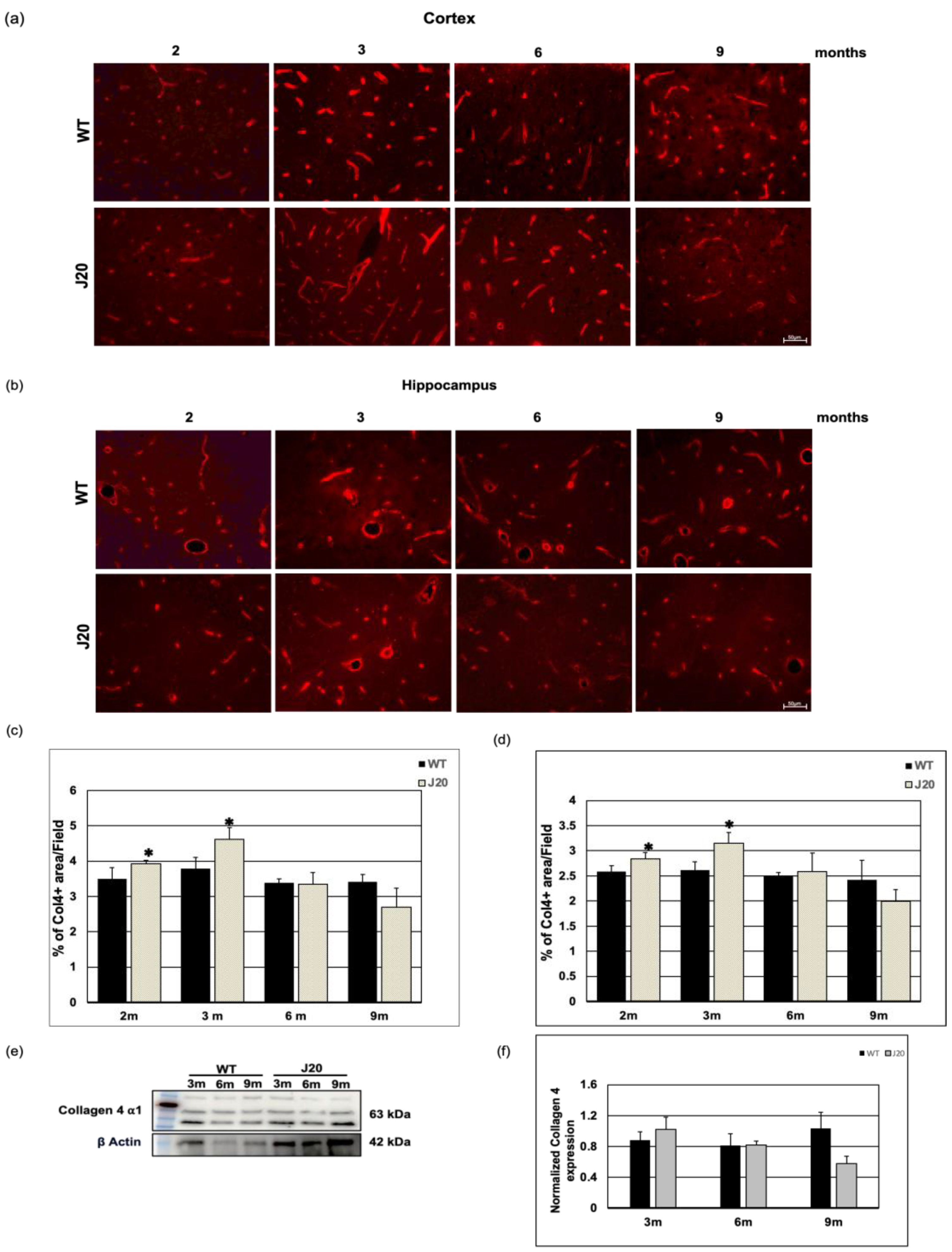
Error in Figure
Reference
- Shibly, A.Z.; Sheikh, A.M.; Michikawa, M.; Tabassum, S.; Azad, A.K.; Zhou, X.; Zhang, Y.; Yano, S.; Nagai, A. Analysis of Cerebral Small Vessel Changes in AD Model Mice. Biomedicines 2023, 11, 50. [Google Scholar] [CrossRef]
Disclaimer/Publisher’s Note: The statements, opinions and data contained in all publications are solely those of the individual author(s) and contributor(s) and not of MDPI and/or the editor(s). MDPI and/or the editor(s) disclaim responsibility for any injury to people or property resulting from any ideas, methods, instructions or products referred to in the content. |
© 2024 by the authors. Licensee MDPI, Basel, Switzerland. This article is an open access article distributed under the terms and conditions of the Creative Commons Attribution (CC BY) license (https://creativecommons.org/licenses/by/4.0/).
Share and Cite
Shibly, A.Z.; Sheikh, A.M.; Michikawa, M.; Tabassum, S.; Azad, A.K.; Zhou, X.; Zhang, Y.; Yano, S.; Nagai, A. Correction: Shibly et al. Analysis of Cerebral Small Vessel Changes in AD Model Mice. Biomedicines 2023, 11, 50. Biomedicines 2024, 12, 104. https://doi.org/10.3390/biomedicines12010104
Shibly AZ, Sheikh AM, Michikawa M, Tabassum S, Azad AK, Zhou X, Zhang Y, Yano S, Nagai A. Correction: Shibly et al. Analysis of Cerebral Small Vessel Changes in AD Model Mice. Biomedicines 2023, 11, 50. Biomedicines. 2024; 12(1):104. https://doi.org/10.3390/biomedicines12010104
Chicago/Turabian StyleShibly, Abu Zaffar, Abdullah Md. Sheikh, Makoto Michikawa, Shatera Tabassum, Abul Kalam Azad, Xiaojing Zhou, Yuchi Zhang, Shozo Yano, and Atsushi Nagai. 2024. "Correction: Shibly et al. Analysis of Cerebral Small Vessel Changes in AD Model Mice. Biomedicines 2023, 11, 50" Biomedicines 12, no. 1: 104. https://doi.org/10.3390/biomedicines12010104
APA StyleShibly, A. Z., Sheikh, A. M., Michikawa, M., Tabassum, S., Azad, A. K., Zhou, X., Zhang, Y., Yano, S., & Nagai, A. (2024). Correction: Shibly et al. Analysis of Cerebral Small Vessel Changes in AD Model Mice. Biomedicines 2023, 11, 50. Biomedicines, 12(1), 104. https://doi.org/10.3390/biomedicines12010104