Article

Effects of High Voltage Electrical Discharge (HVED) on Endogenous Hormone and Polyphenol Profile in Wheat

1
Faculty of Food Technology, Josip Juraj Strossmayer University of Osijek, Franje Kuhača 18, 31000 Osijek, Croatia
2
Agricultural Institute, Centre for Agricultural Research, ELKH, 2462 Martonvásár, Hungary
*
Author to whom correspondence should be addressed.
Plants 2023, 12(6), 1235; https://doi.org/10.3390/plants12061235
Submission received: 23 January 2023 / Revised: 15 February 2023 / Accepted: 3 March 2023 / Published: 8 March 2023

Abstract

:
High voltage electrical discharge (HVED) is an eco-friendly low-cost method based on the creation of plasma-activated water (PAW) through the release of electrical discharge in water which results in the formation of reactive particles. Recent studies have reported that such novel plasma technologies promote germination and growth but their hormonal and metabolic background is still not known. In the present work, the HVED-induced hormonal and metabolic changes were studied during the germination of wheat seedlings. Hormonal changes including abscisic acid (ABA), gibberellic acids (GAs), indol acetic acid (IAA) and jasmonic acid (JA) and the polyphenol responses were detected in the early (2nd day) and late (5th day) germination phases of wheat as well as their redistribution in shoot and root. HVED treatment significantly stimulated germination and growth both in the shoot and root. The root early response to HVED involved the upregulation of ABA and increased phaseic and ferulic acid content, while the active form of gibberellic acid (GA1) was downregulated. In the later phase (5th day of germination), HVED had a stimulatory effect on the production of benzoic and salicylic acid. The shoot showed a different response: HVED induced the synthesis of JA_Le_Ile, an active form of JA, and provoked the biosynthesis of cinnamic, p-coumaric and caffeic acid in both phases of germination. Surprisingly, in 2-day-old shoots, HVED decreased the GA20 levels, being intermediate in the synthesis of bioactive gibberellins. These HVED-provoked metabolic changes indicated a stress-related response that could contribute to germination in wheat.

1. Introduction

The major idea of agricultural sustainability is to fuse ecological and agronomic practice, farmer skills and knowledge to reduce the utilization of pesticides and fertilizers in the food production chain [1]. Today, in agricultural sustainability programs, low-cost beneficial innovative solutions with a minimum impact on the environment are receiving more attention. Numerous reports highlight an improvement in seed invigoration after the application of physical methods such as electromagnetic technologies (EMFs) and magnetic field technologies (MFs) [2,3], ionizing radiation (IR) [4,5] and various cold plasma technologies (CPTs) [6,7,8].
High voltage electrical discharge (HVED) is a physical, non-thermal, hydropriming technique that causes modification in the physicochemical structure of water during electrical discharge generation exposure [9]. An altered water structure is a result of ionization and dissociation reactions upon which reactive oxygen species (ROS) and reactive nitrogen species (RNS) may be created forming plasma-activated water (PAW) [10]. The chemical content of PAW is not always equal and the concentration of long-lifetime particles such as H2O2, NO2 and NO3 may depend on the experimental conditions of PAW preparation [11]. In addition, as observed via SEM micrographs, the HVED treatment induced ruptures on the seed coat [12]. These two events could be responsible for seed dormancy disruption and better germination properties [13,14,15]. Accordingly, the HVED treatment promoted the germination and growth of wheat seedlings, as we observed previously [12]. Furthermore, this effect could also be observed even under drought (15, 20 and 30% PEG) and salt (90, 160 and 230 mM NaCl) stress conditions, although the final success of HVED depended on the stress intensity [12]. These results indicated that the HVED affected and was connected to the stress response of plants.
Seed germination is a particularly important stage in plant development which is characterized by fast water uptake into dry seeds which activates several physiological and metabolic processes, resulting in radicle and bud protrusion [16]. Each event of germination is precisely regulated with multiple types of phytohormones such as auxin (IAA), abscisic acid (ABA), gibberellins (GAs), jasmonates (JAs) and salicylic acid (SA) [17]. GA and ABA are major seed dormancy and germination regulators in optimal and stressful environments. In the early stage of germination, seed hydration triggers gibberellin biosynthesis in embryo meristems, which is subsequently directed into the aleuronic layer and endosperm, whereas it induces the synthesis of α-amylase, a major hydrolyzation enzyme, leading to the disruption of seed coat dormancy [18]. GA induces the expansion of embryo cells, radicle protrusion and the elongation of young cells, and may also increase seed osmotic and water potential [19,20]. GAs synthesize through different intermediaries including GA53 and G12, which convert to GA9 and GA20. Finally, through 3-β-hydroxylation, GA1 and GA4 active compounds are generated [21,22]. Cui et al. [23] found that 1 min of exposure to atmospheric pressure cold plasma was enough to stimulate Arabidopsis growth and the production of GA.
ABA affects germination through the suppression of the events controlled by gibberellins. Phaseic acid (PA) and dihydrophaseic acid (dHPA) are compounds of ABA catabolism generated through the oxidation of 8′-hydroxy ABA which is the primary route of ABA decomposition [24]. Attri et al. [25] reported that non-thermal plasma treatment (NTP) caused a shift in the GA/ABA ratio which was reflected in the promotion of the germination of radish seeds. In heat-exposed rice seeds, NTP induced germination by suppressing ABA anabolic and inducing ABA catabolic gene expression [26]. One of the important regulators of seed dormancy is auxin (indole-3-acidic acid, IAA). Many studies have revealed a crosstalk between auxin and ABA signaling in keeping the dormancy [27,28,29,30]. The relationship between cell elongation, IAA content and plasma treatment has been observed in Arabidopsis [31]. The authors found that the active oxygen particles derived from air plasma promoted the expression of EXPA8 and EXPA20 expansin genes important in acid-induced growth [32]. Jasmonic acid, jasmonate isoleucine conjugate (JA-Ile) and methyl jasmonate (MeJA) belong to jasmonate (JA) pathways that originate from α-linolenic acid esterified in the galactolipids of thylakoids [33]. JA_Ile is formed by the conjugation of JA and isoleucine [34]. JA often operates in conjunction with other phytohormones and participates in complex regulatory network signaling pathways [35,36]. PAW watered tomato plants showed higher activity of 2-oxophytodienoic acid reductase (OPR1), phenylalanine ammonia-lyase (PAL) and allene oxidase synthase (AOS), key enzymes in SA and JA synthesis [37].
Phenylpropanoid metabolism plays a major role in the synthesis of a large number of compounds important for plant growth, development and stress response. Phenolic acids are separated into hydroxybenzoic and hydroxycinnamic acids [38]. Salicylic acid (SA, 2-hydroxy-BA) belongs to hydroxybenzoic acids and it is a plant growth regulator involved in the plant immunity response [39]. SA can be synthesized either from shikimate, converted into chorismate and isochorismate, or from p-coumaric acid (p-CA) via the phenylpropanoid pathway [38,40]. Phenylalanine (Phe), originating from the shikimate pathway, is a starting point in phenylpropanoid metabolism. The deamination of Phe creates cinnamic acid which via hydroxylation is converted into p-coumaric acid (p-CA) [40]. Ferulic acid (FA), caffeic acid (CFA) and p-CA are esters of hydroxycinnamic acids (C6-C3) naturally present in cell walls [40,41]. Jasim et al. [42] found that the application of DBD plasma on basil seeds promoted the expression of genes important in the phenylpropanoid pathway (4-coumarate coA ligase, 4CL, cinnamate 4-hydroxylase, C4H, and charicol o-methyl transferase, CVOMT), which indicated that the compounds of phenylpropanoid pathways may also play an important role in germination.
As indicated above, the HVED treatment stimulated the germination and growth of wheat under both control and stress conditions [12]. To better understand the mode of action of HVED treatment on germination and seedling development, the aim of this work was to follow the alterations in hormone profiles (ABA, GAs, IAA, JA) and their derivates, phenylpropanoids and benzoic acids in wheat. The effect was studied in a time-dependent manner, on the 2nd and 5th day of germination. Moreover, we wanted to compare the effect of HVED treatment on the spatial distribution of phytohormones and their substituents and to check whether HVED affects specific organ (root and shoot) responses. Since the evidence of the HVED impact on hormonal response and secondary metabolism has not been presented yet, the results of this study could contribute to understanding the role of HVED in hormonal balance in young wheat seedlings.

2. Results

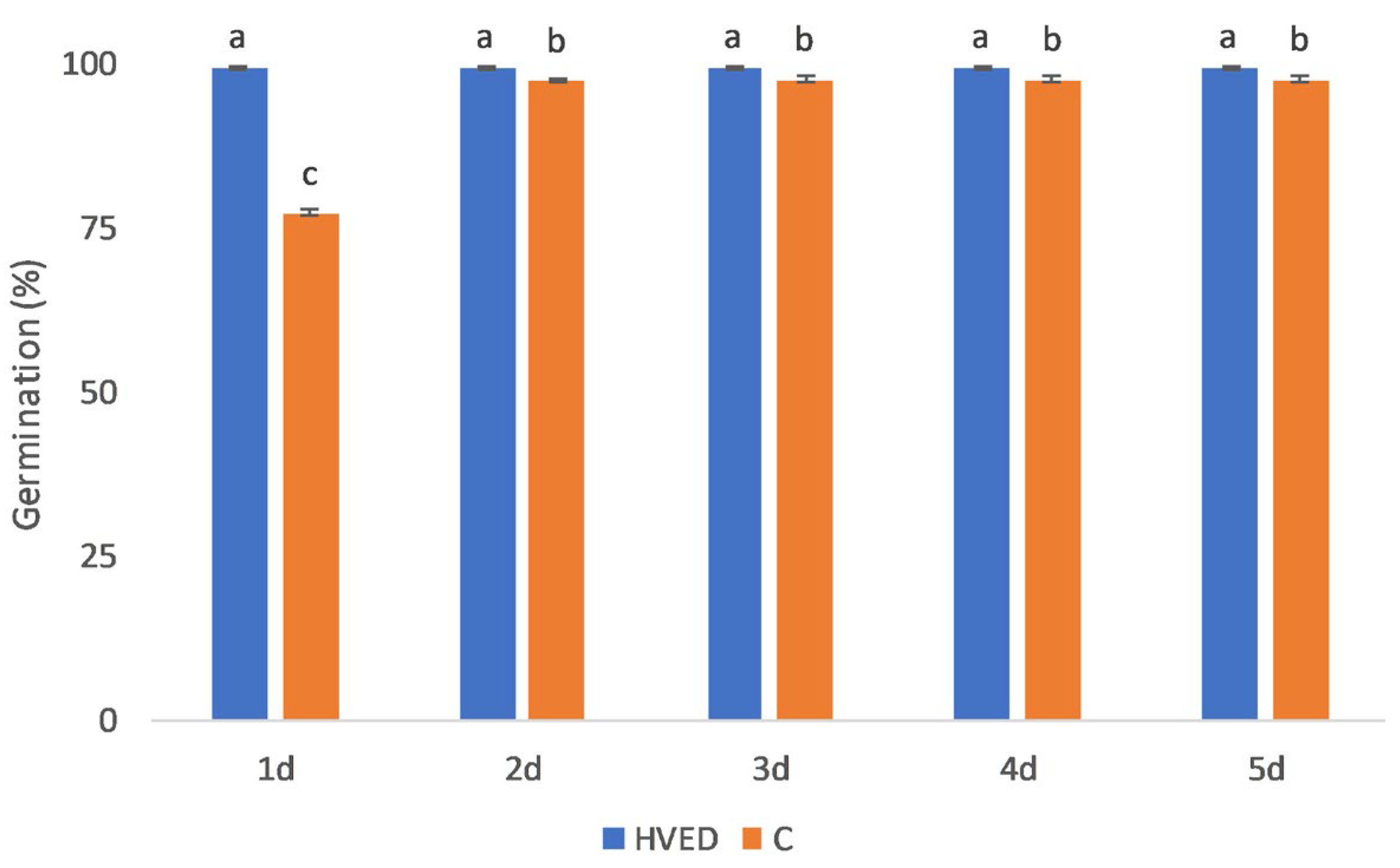
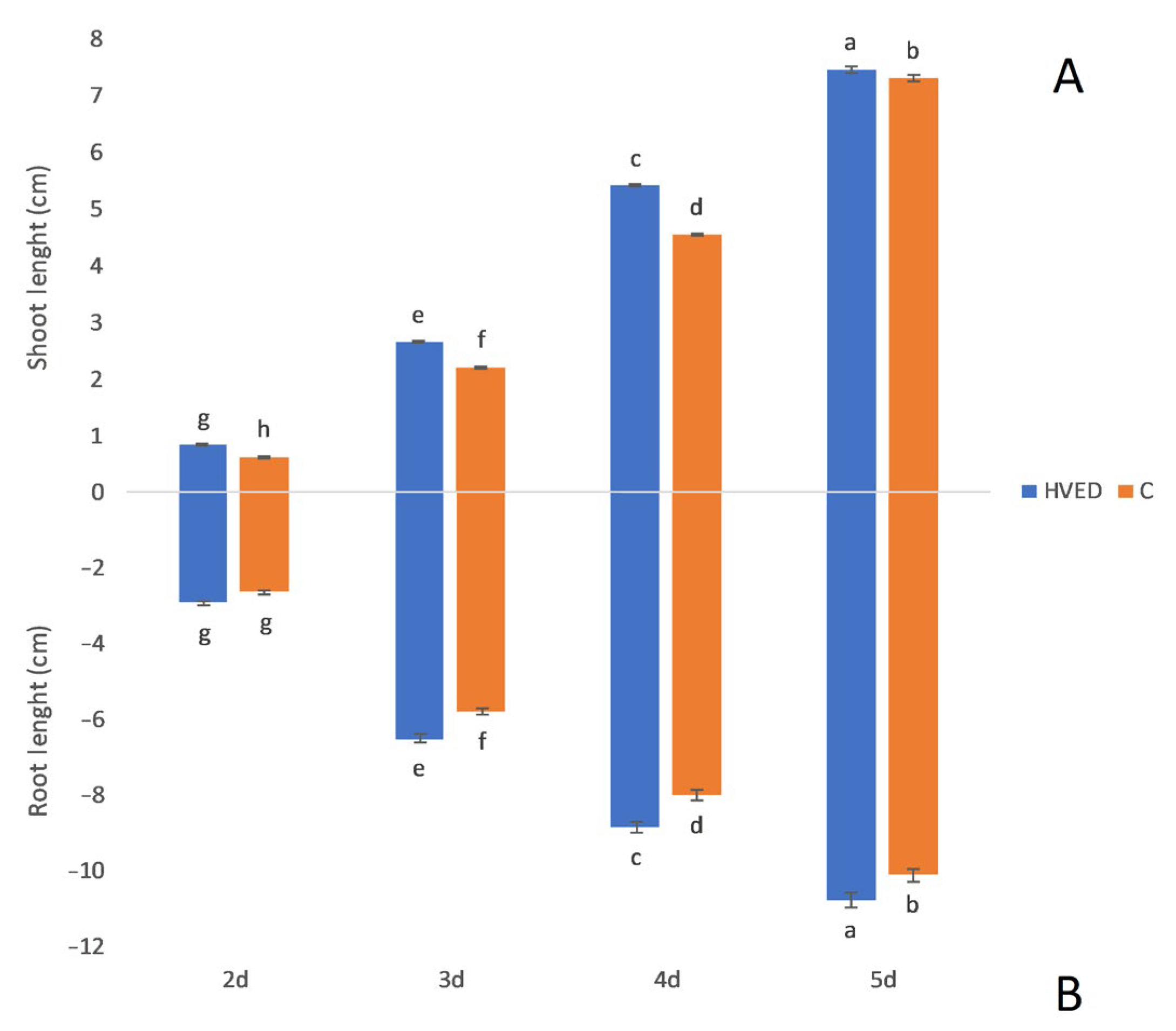
2.1. Effect of HVED Treatment on Germination and Growth

The HVED treatment promoted germination and growth in a time-dependent manner, as indicated by the results of statistical analysis (Table S1). The most notable changes in germination percentage were recorded on the 1st day in the HVED group in which germination was 28.4% higher than in the control (Figure 1). Although the differences in the germination percentage between the control and HVED groups became less pronounced with time, the germination % remained higher in HVED seeds as compared to the control seeds during the 5-day experimental period (Figure 1). Shoot and root growth were also improved in the HVED group (Figure 2A,B). Interestingly, the bigger differences in growth dynamics between the control and HVED group were observed within the 2–4 days, while differences in growth diminished on the 5th day. For instance, after the 2nd day, the HVED shoots were 31.2% higher than the control, while on the 5th day, the difference was only 2%. In the root, 2-day-old HVED roots were 10.6% longer than the untreated HVED, whilst after 5 days the HVED group had 6% longer roots compared to the control.

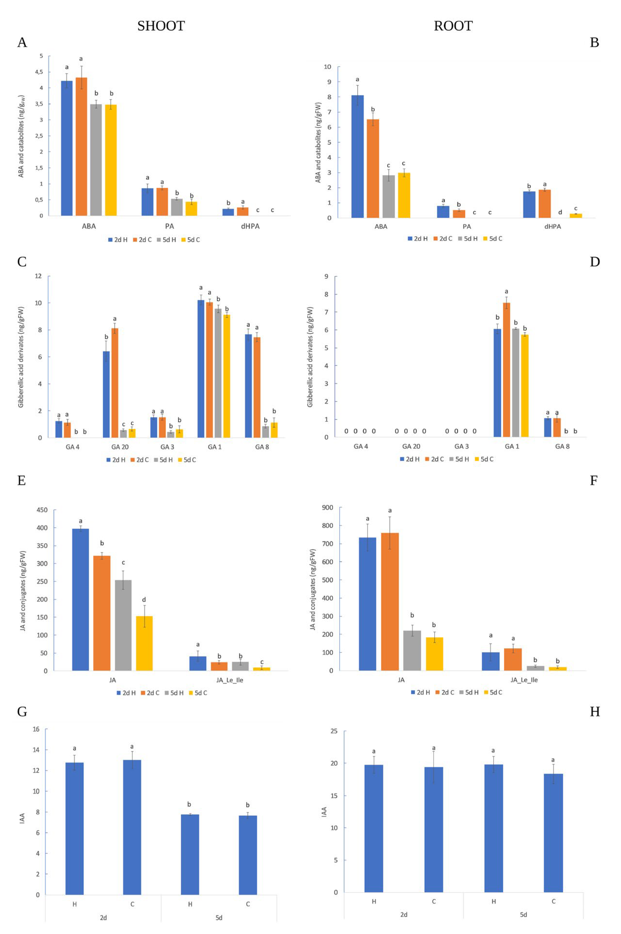
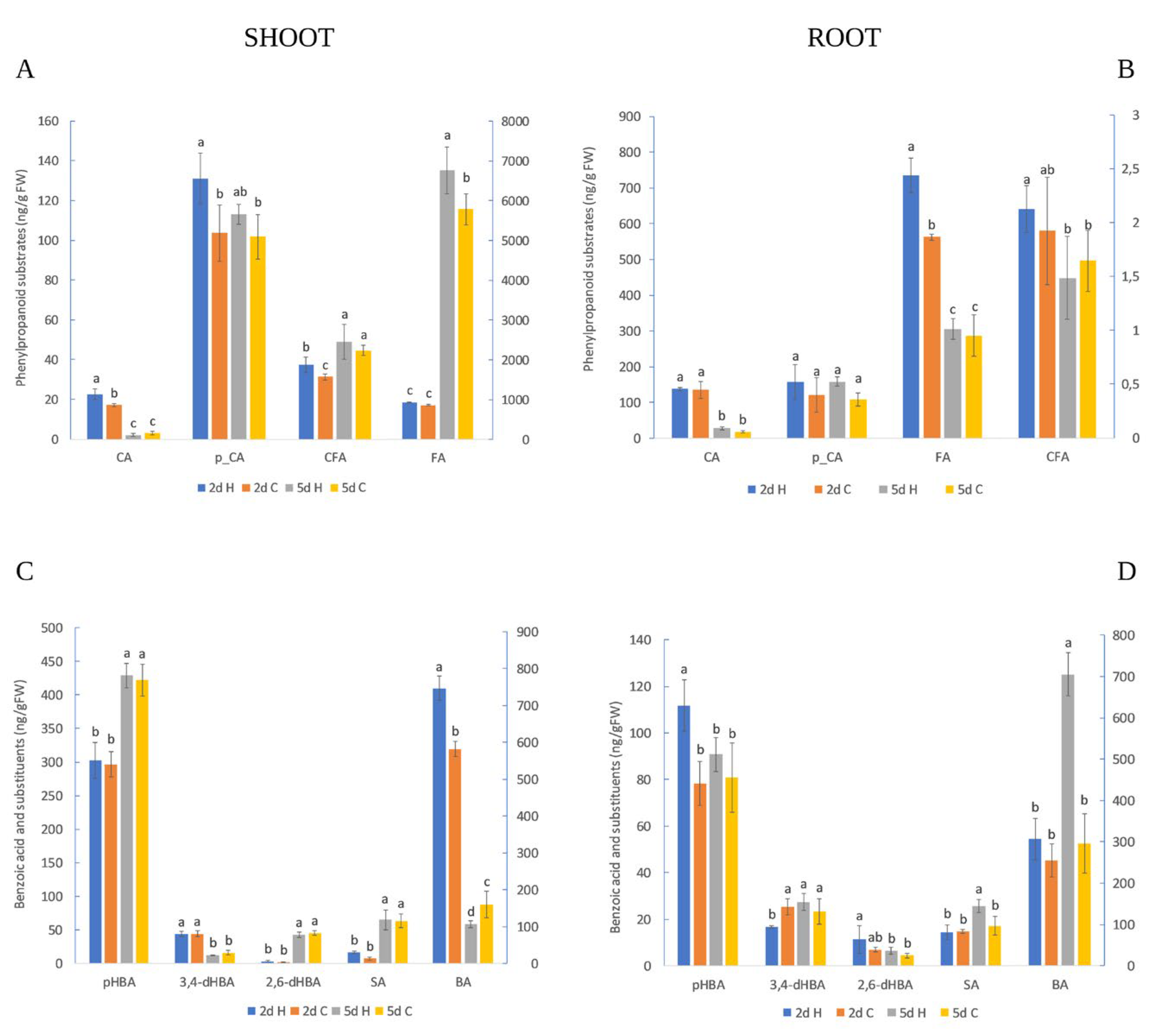
2.2. HVED Impact on Hormonal and Metabolite Profile

In the shoot, treatment had a significant impact on most of the phenylpropanoid substrates (except FA), JA and its conjugates and BA, as indicated by analysis of variance (Tables S2 and S3). On the other hand, time affected all variables except for p-CA acid, indicating that its reservoirs in shoots are time-independent. Finally, treatment and time interactions were noticed only for CA and BA, suggesting interconnections in the β-oxidation pathway of the BA metabolic route. In the root, treatment affected ABA, PA, dHPA, FA, GA1, BA and pHBA content, while variations in the time-dependent response were found for most of the compounds with the exception of p-CA, IAA and pHBA amount. The results of the statistical analysis are presented in the Supplementary Material (Tables S4 and S5). The significant effects for treatment and time points were observed for ABA, PA, FA, GA1, 3,4-dHBA and BA, respectively. Tables S6 and S7 present the numerical values of hormones and polyphenols in shoots and roots.
The accumulation of ABA and its catabolites in the shoot and root was higher in the earlier time point (2nd day) than on the 5th day in the control and HVED groups (Figure 3A,B). In addition, ABA metabolism differed in the root of the control and HVED groups. Namely, HVED treatment stimulated higher ABA and PA content production in 2-day-old roots compared to the control, while later (5th day) there were no differences between treatments (Figure 3B). The level of dHPA was higher under the control than after HVED application, in both the root (2nd and 5th day) and shoot (2nd day), referring to slower ABA decomposition under HVED. The GA distribution was tissue-specific and time-dependent as indicated via analysis of variance (Tables S2 and S4). In the shoot, bioactive GAs (GA4, GA3 and GA1) and their intermediates (GA20 and GA8) were detected except for GA4 which remained undetected on the 5th day of growth (Figure 3C). Similar to ABA metabolism, the production of all GA derivates in the shoot was higher on the 2nd day compared to the 5th day in both treatment groups. Unlike the shoot, the root contained only a few compounds (GA1 and GA8) involved in the early-13-hydroxylation pathway. The amount of GA1 was higher on the 2nd than on the 5th day in the control and HVED group, whilst its inactive form GA8 was detected only on the 2nd day (Figure 3D). Comparing GA accumulation within the one-time point, the 2-day-old control plants of shoots and roots showed a higher concentration of GA20 and GA1 than the HVED-treated plants (Figure 3C,D).
The biosynthesis of JA and JA_Le_Ile decreased over time in shoot and root, respectively (Figure 3E,F). Five-day-old seedlings had a lower amount of JA and JA-Le_Ile than two-day-old seedlings under both treatment groups (control and HVED). The HVED treatment triggered the overproduction of JA and JA_Ile derivates only in the shoot. Namely, the percentage of JA and JA_ILE derivates after the 2nd day was 23.5% and 68%, higher in the HVED group than in the control group, while later (5th day) their concentration increased by 66% and 174%, respectively. The control and HVED-exposed 2-day-old shoots contained higher IAA content than the 5-day-old shoots (Figure 3G), indicating IAA decomposition during the time. For the root, significant differences in IAA synthesis were not detected between days of growth or between treatments (Figure 3H).
The phenylpropanoid distribution in the root and shoot was more time-dependent than treatment-dependent (Figure 4A,B). The amount of CA was higher on the 2nd day than the 5th day of growth in both the root and shoot, independent of the treatment. Similarly to CA, the FA amounts were higher on the 2nd day in the root while an inverse response in FA amounts was obtained for the shoot; FA values were lower on the 2nd day compared to the 5th day’s values. HVED treatment provided a significant increase in CA, p-CA and CFA in the shoot and FA in the root on the 2nd day of growth. The differences disappeared on the 5th day, but became notable for FA whereby the amount was higher in the HVED group.
The results revealed a time-dependent accumulation of several BA compounds without treatment influence in the shoots (Figure 4C). In the shoot, the amount of 3,4-dHBA was higher on the 2nd day as compared to those data measured on the 5th days, independent of treatment, while inverse tendencies were observed for SA, pHBA and 2,6-dHBA (their amount was higher on the 5th day). The early effect of HVED on the shoot’s polyphenol response was notable only for BA whose levels increased compared to the control on the 2nd day. Later, HVED had an inhibitory effect on BA biosynthesis in shoots that showed a lower BA amount than the control. In roots, the HVED treatment immediately (2nd day) triggered the higher accumulation of pHBA but negatively affected 3,4-dHBA amounts in comparison to the control (Figure 4D). Unlike the control, the HVED group had a higher amount of SA and BA on the 5th day. Moreover, the same trend was detected during the time where the amount of BA and SA remained significantly higher in HVED-treated roots.

2.3. Comparative Statistical Analyses
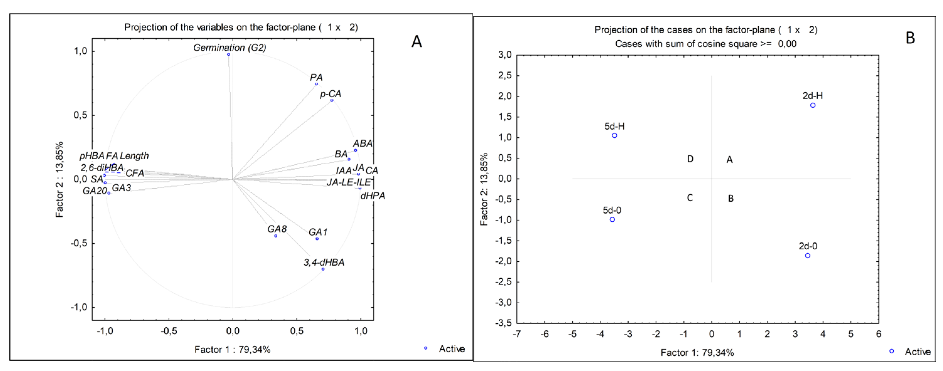
To reveal the connection between the effect of treatment during the times of germination (first three days), shoot and root growth, a comparative statistical analysis was performed (Table 1). The treatment was positively correlated with germination (G1-G3), shoot (S2-S5) and root (R2-R5) length during the time. A strong positive association was also notable for germination and growth. Comparative statistical analysis was also used to verify the interactions of treatments on growth parameters and changes in hormone and metabolite amounts in the time frame for shoot and root, separately (Table 2 and Table 3). In 2-year-old shoots, a correlation was found between treatments and the phenylpropanoids (except p-CA), BA (except pHBA and 3,4-dHBA), JA, germination percentage and shoot length (Table 2). During the time (5th day), treatments showed an association with the accumulation of JA and its conjugates, GA1, germination and shoot length, but a negative correlation with BA and 3,4 dHBA amount. On the 2nd day, a positive association was found between germination and shoot length (0.83 ***), CA (0.71 *), BA (0.75 *), p-CA (0.70 *) and SA (0.82 *) (Table 2). Moreover, shoot length was connected with CA (0.85 **), BA (0.91 **), p-CA (0.83 **), CFA (0.69 *), FA (0.95 ***) and JA (0.92 **), indicating improved activation of the shikimate acid pathway at an early time point. Most associations connected with shoot early metabolic changes were found for CA and BA. Thus, CA correlated with benzoic acid (0.95 ***), p-CA (0.70 *), CFA (0.64 *), FA (0.89 *), JA (0.99 ***) and JA_Le_Ile (0.73 *), respectively. A positive association was noticed for BA and p-CA (0.86 *), FA (0.94 **), JA (0.93 **) and the active form of JA (0.71 *), respectively. Over time, the number and nature of the relationship between metabolites and hormone amount were lower. Namely, on the 5th day, a negative association was noticed between p-CA and 2,6-dHBA (−0.76 *) and between BA and GA1 (−0.83 *). Moreover, a negative association was also noticed for FA and BA (−0.94 **) and between FA and GA20 (−0.89 *), respectively. A strong positive connection (p ≤ 0.001) was seen between BA and 3,4-dHBA, CFA and SA, FA and GA1, JA and JA_Le_Ile and finally between JA and PA.
Table 3 presents the correlation coefficients for root-related metabolites and hormones. In 2-day-old roots, a correlation existed between treatments and the amount of pHBA, FA and PA, ABA, germination percentage and root length, while the production of GA1 and 3,4-dHBA was downregulated by treatment. Later (5th day), a positive association was found in the amount of CA, BA, 2,6-dHBA, SA, GA1 and germination percentage, while the treatment was negatively correlated with dHPA. Correlation analyses revealed a relationship between germination and ABA content (0.99 ***) in the early stage of growth (2nd day), pHBA amount (0.78 *) and PA (0.71 *), while germination had an opposite effect on 3,4-dHBA synthesis (−0.74 *). ABA, FA, PA and pHBA amounts were connected with root length. The amount of IAA was correlated with FA (0.83*) and was negatively correlated with JA (−0.86 *) and dHPA (−0.89 *) suggesting a greater activation of metabolic routes connected with seed development. PA showed a negative correlation with 3,4-dHBA (−0.92 *) but a positive correlation with pHBA (0.72 *), FA (0.83 *) and ABA (0.83 *). Moreover, in 2-day-old roots, the dHBA amount correlated with JA content (0.90 *) and GA1 (0.99 ***), but it had the opposite relationship with p-CA amount (−0.90 *), IAA (−0.89 *) and FA (−0.98 **), respectively. On the other hand, JA levels were correlated with GA1 (0.90 *) and GA8 (0.86 *) but were negatively connected with IAA, p-CA and FA, indicating the crosstalk between JA signaling and gibberellin metabolism in the root. A positive linkage (0.88 *) was evident for CA and CFA while the amount of pHBA was in a negative association with 3,4-dHBA (−0.78 *) suggesting that the hydroxylation of benzoic acids depends on the availability of precursor molecules. Finally, the FA amount was negatively connected with GA1, JA and dHPA but positively connected with ABA and PA, indicating an early ABA regulatory role in polyphenol root induction. The germination percentage in 5-day-old roots correlated with GA1 growth but showed a negative connection with the dHPA level. The greatest number of associations was noticed for CA and BA in the root on the 5th day. Thus, CA was positively correlated with BA (0.78 *), SA (0.68 *) and p-CA (0.72 *) and was negatively correlated with dHPA (−0.89 **). Furthermore, BA levels were associated with SA (0.74 *) and 2,6-dHBA (0.76 *) while a negative connection was detected with dHBA (−0.81 *). The other positive connections (p ≤ 0.01) were noticed between IAA and 3,4-dHBA, p-HBA and p-CA, SA and 2,6-dHBA amount, root length and 3,4-dHBA, respectively. In contrast, negative associations were observed for dHPA and GA1 (−0.79 *), 2,6-dHBA (−0.71 *), CA (−0.89 **), BA (−0.81 **) and finally between JA and ABA amount (−0.83 *).

2.4. PCA Analyses

Figure 5A shows the percentage of the total variance and principal components (PCs) and their interactions with hormones and metabolites under control and HVED treatment for roots during the time. As the PCA analyses did not detect the explanatory variables for the shoot, the factor loadings are presented only in Table S8. For the root, the variable projection is presented for the first two components, contributing 93.19% of the total variance (Figure 5A). The cumulative contribution to the total variation in PC1 was 79.34% (Table S9). The PC1 was characterized by positive loadings of CA and BA, 3,4-dHBA, p-CA, dHPA, JA and its conjugates, ABA and IAA, respectively. Negative associations were evident for pHBA, SA, 2,6-dHBA, CF and FA, GA3, GA20 and root length. The PC2 was explained by negative loadings of 3,4-dHBA and positive loadings of PA and germination (2nd day), while the positive loading was obtained in PC3 for GA8. The projection of the corresponding plots reveals four clusters separated based on treatment and days of sampling (Figure 5B). The first cluster (A) belongs to the 2-day-old HVED roots (2d-H) segregated based on the higher amount of ABA, PA, FA and pHBA but the lower accumulation of dHPA and 3,4-dHBA compared to the 2-day-old control group (2d-0) (cluster B). The separation of 5-day-old roots into clusters C (5d-0) and D (5d-H) was the result of HVED treatment on root length, dHPA, SA and BA amount. Namely, the roots of HVED-treated seeds (cluster D) were longer and showed increased SA and BA levels and decreased dHPA compared to the control (cluster C). HVED-treated groups (A, D) were distinguished through specific time-frame responses. In group A, HVED promoted increased ABA, pHBA, dHBA, FA and PA accumulation, while group D had higher SA and BA concentrations under HVED. Finally, clusters B and C were apart due to a difference in the production of ABA and its catabolites, root length, GA8, JA and its derivates, CA, CFA and FA, whose production was upregulated earlier.

3. Discussion

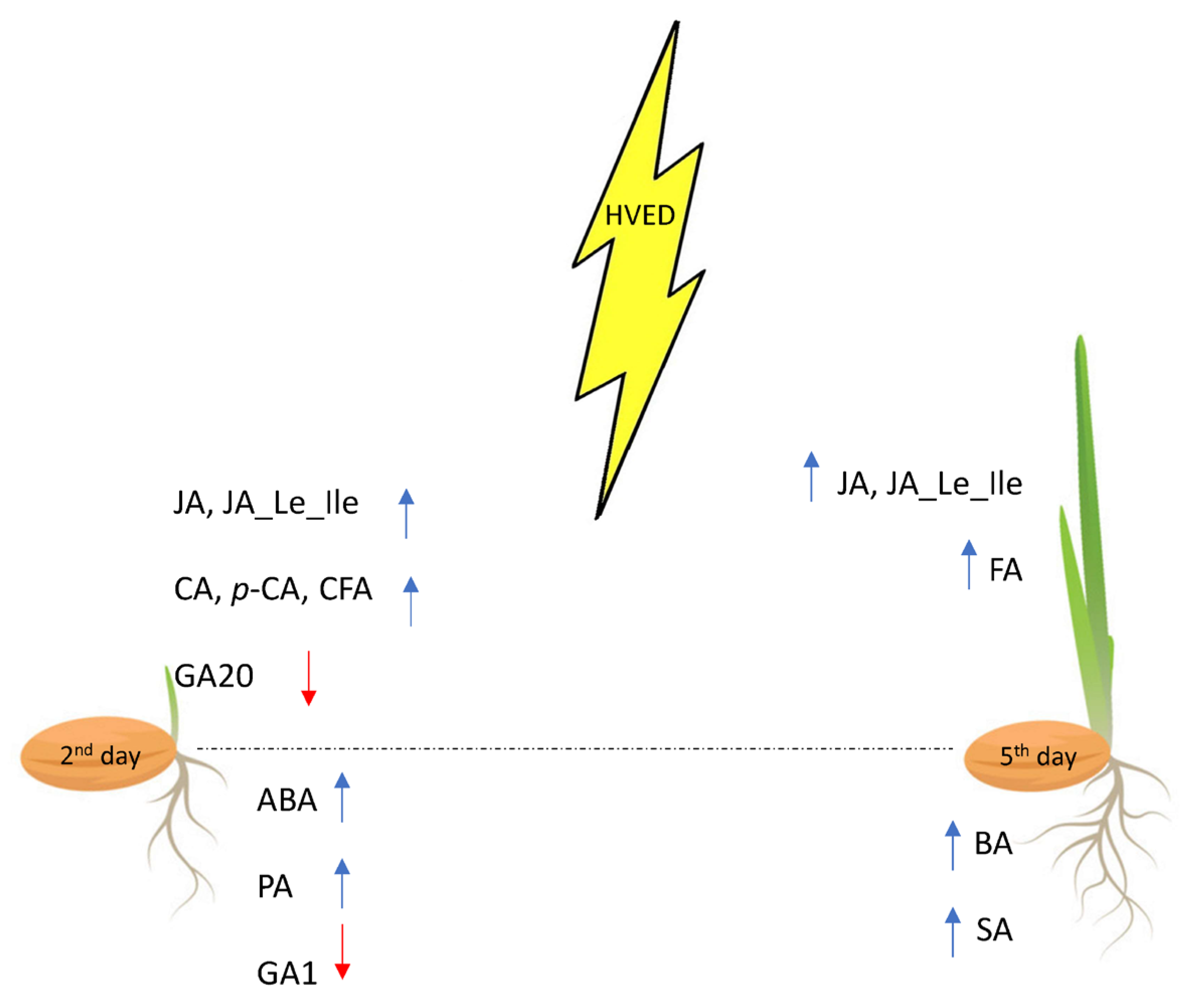
3.1. HVED Impact on Germination and Growth

In the present study, the metabolic background of the HVED-induced stimulation in germination and growth was studied. The stimulatory effect of HVED on seed germination was manifested in the shortening of the imbibition time, improving germination and root and shoot growth within the early (2–5 days) stages of growth (Figure 1 and Figure 2). Although the HVED-induced treatment improved seed germination, as has already been observed in wheat [12,43,44], Nasturtium seeds [45] and oilseed rape [46], the physiological and metabolic background have not been revealed yet. The stimulatory effect of HVED is based on the fact that this treatment weakens the seed surface layers, thus promoting seed invigoration and growth [47]. The exposure of seeds to HVED causes seed surface modification which shortens the imbibition time and increases wettability leading to faster oxygen diffusion into the embryo [15]. HVED treatment creates plasma-activated water (PAW) rich in ROS and RNS particles which can also affect germination [48]. Moreover, the PAW creates a higher amount of hydrogen peroxide (H2O2) and nitric oxides (NO), longer half-life molecules which may alter the phytohormone balance in the seeds and promote germination [49]. Recently, Mildaziene and Sera [50] and Priatama et al. [51] observed that the accumulation of ROS and RNS triggers changes in proteomic, metabolomic and transcriptomic levels and also affects phytohormone levels. The cavitation bubbles which occur during the HVED treatment create UV emission which could also be meritorious for germination dormancy disruption [9,44]. However, it should also be mentioned that the success of germination depended on the number of shots. Ji et al. [52] found that one or five energy shots had a beneficial effect on germination, while seeds treated with ten shots decreased growth and germination. These observations were found in spinach. In the present study, the energy input was 30 kV, suggesting that such a huge energy input still improved the germination of wheat seeds. In addition, HVED treatment induced several changes at the metabolic level too.
Correlation analysis reveals a positive association between germination and p-CA, CA, GA1, BA and SA in the shoot (2nd and 5th day) (Table 2). The positive crosstalk between GA and SA interaction in germination and early growth has been published for Arabidopsis thaliana [53]. Moreover, phenol compounds (SA, BA, p-CA and CA) were also important inducers of germination. Since all of these compounds originate from the Phe metabolic route, our results confirm the connection with complex hormone interaction. Curiously, germination in 2-day-old roots positively correlated with ABA, p-HBA and PA, while the 5-day-old root germination percentage was associated with GA1 indicating different regulation pathways of germination during the time (Table 3). These results support the observations of Pei et al. [54], who found that HVED treatment generates ROS and H2O2 which participate in ABA signaling. Moreover, it is also possible that the influence of H2O2 diminishes over time, which might be a reason for a decreased ABA amount in 5-day-old roots (Figure 3B). Correlation analysis displays a positive linkage between JA and shoot length (2nd day), indicating that growth promotion could be ascribed to the early activation of the JA regulation pathway. Over time, GA1 also contributed to shoot growth (p < 0.05), suggesting that gibberellins act through jasmonoyl-isoleucine in the coordination of growth. In the root, early growth was promoted by ABA, PA, FA and pHBA while, over time (5th day), length was associated with 3,4-dHBA (Table 3). This indicates that the ABA pathway and phenolic compounds have a similar action on early root formation. Moreover, a linkage between ABA and root growth could be also connected with ABA growth regulation under dark conditions since the seeds were kept for 48 h in the darkness before sampling. The ABA-induced growth occurs in the process of skotomorphogenesis where the cells of hypocotyl rapidly elongate [55]. In both 5-day-old shoot and root germination, the percentage was positively regulated by the GA1 amount (Table 2 and Table 3). According to Vishal and Kumar [56], the presence of GA-coding genes in the growing tissues suggests that active forms of GA can be produced in situ.
According to the results, different metabolic responses were found in the root and shoot. Thus, they are discussed here separately.

3.2. Root Hormonal and Polyphenol Response

The early root response revealed the antagonistic interaction in ABA/GA signaling in both the control and HVED groups, with slight alterations in the rate of ABA decomposition and GA1 formation. Namely, on the 2nd day, HVED increased ABA (p < 0.05, r = 0.88) and PA (p < 0.01, r = 0.87) content but inhibited the biosynthesis of the active form of gibberellins (GA1) (p < 0.05, r = −0.88) (Figure 3B,D and Table 3), suggesting that the ABA hormone suppresses GA biogenesis. Jacobsen et al. [57] stated that a low concentration of GAs could be the result of high ABA amounts due to changes in the conversion of their common precursor geranylgeranyl diphosphate (GGDP) to ent-kaurene. In contrast, the control displayed a higher GA1 level which might be connected with earlier activation of the 13-hydroxylation gibberellin pathway. In general, a lower ABA/GA ratio under optimal conditions participates in seed dormancy breakdown events [58]. In this study, the ABA/GA1 ratio in HVED roots was higher (1.33) compared to the control (0.86), indicating that HVED modifies the ABA pathway at the expense of GA routes. As the opposite, the GA3 concentration in spinach increased after non-thermal (NTP) treatment [52]. In another study, NTP application increased the expression of the LEA1, SnRK2 and P5CS drought-responsible genes and higher ABA generation in wheat seedlings [59]. Correlation analysis also showed a positive linkage of germination with ABA (p < 0.001, r = 0.99) and PA (p < 0.05, r = 0.71) (Table 3). A higher PA content and higher germination percentage on the 2nd day suggests that HVED in the root triggers the dormancy-breaking events compared to the control group earlier in which the ABA degradation was complete due to its final catabolite dHPA. As provided by PCA analysis, the 2-day-old control group (B cluster) was separated from the HVED group (A cluster) based on a higher dHPA amount (Figure 5A,B). Ferulic acid (FA), a phenylpropanoid substrate of the β-oxidative pathway, creates cross-connections between sugars, polymers and the lignin of the cell wall, increasing the mechanical strength especially important in the conditions of pathogen attack [60]. Our results showed a higher FA amount in 2-day-old-HVED roots which might be connected with an early root response to ROS and RNS in PAW medium and the stress response (Figure 4B). In the study of Cortese et al. [61], PAW caused an increase in cytosolic Ca2+ concentration in Arabidopsis. The role of Ca2+ signaling in the perception of ROS and RNS was described in [62,63]. In this context, it seems that HVED takes part in Ca2+-dependent signaling transduction. However, further investigations in this direction are required. Correlation analyses display the positive connections between FA content and root growth, and ABA and FA amount, respectively (Table 3). Yukiko et al. [64] and Marchiosi et al. [65] reported a dual role in growth regulation between hormones and polyphenols, indicating that some regulatory polyphenols inhibit growth by binding GAs, while others, through ABA signaling, promote it. Considering this, it seems that in early root growth, FA biosynthesis is ABA-dependent.
In 5-day-old roots, HVED promoted the accumulation of BA and SA compared to untreated roots. The separation of these two groups was presented via PCA analysis (Figure 5A,B). Correlation analysis also showed a positive linkage between BA and SA (p < 0.05, r = 0.74), suggesting that HVED for SA synthesis activates the phenylpropanoid rather than the chorismate pathway [66]. In wheat, plasma treatment increased the activity of Phe ammonia lyase (PAL), which catalyzes the deamination of Phe to CA [43,67]. Additionally, in tomatoes, PAW induced the tissue-specific expression of genes involved in JA and SA synthesis [37]. In the same study, shoots showed an induction of 12-oxophytodienoic acid reductase (opr 1) and allene oxidase synthase (aos) JA regulatory genes, while in PAW-treated roots their expression was suppressed. In contrast, the expression of phenylalanine ammonia-lyase (pal), the SA encoding gene, in PAW-treated root and shoot was higher. However, such a conclusion should be interpreted with caution. According to Widhalm and Dudareva [39], the generation of SA might be the result of the utilization of crosstalk intermediate compounds in chorismate and Phe pathways via feedback inhibition.
Correlation analysis reveals the difference between 2-day-old and 5-day-old roots in JA regulation and the type of metabolite expression. At the first time point, JA was positively regulated by dHPA (p < 0.05, r = 0.90), GA1 (p < 0.05, r = 0.90) and GA8 (p < 0.05, r = 0.86), while later (5th day) a negative correlation (p < 0.05, r = −0.83) existed between JA and ABA (Table 3). This leads to the conclusion that JA control of ABA/GA regulation in the root was time-dependent. In the root, HVED suppressed the production of 2,6 dHBA by 42.6% but increased SA by 77% and the BA amount by 130% with time (from the 2nd to the 5th day (Figure 4D)). The catabolism of SA includes hydroxylation reactions connected with the addition of the hydroxyl group (-OH) on three or five positions of the benzenoid ring creating sugar conjugates, 2,3-dHBA and 2,5-dHBA, respectively [68]. The LC-UV-MS dHBAs detection showed that 2,6-dHBA and 2,4-dHBA exhibited very similar retention times to 2,3-dHBA [68]. Our results reveal the positive connection between 2,6-dHBA and SA (p < 0.01, r = 0.83) (Table 3). According to this, a lower amount of 2,6-dHBA and higher SA and BA concentrations in 5-day-old roots suggest that SA catabolism is downregulated by HVED. In addition, an increased amount of SA and BA refers to the mobilization of the shikimate/chorismate regulatory network.

3.3. Shoot Hormonal and Polyphenol Response

In shoots, the early events (2 days) caused by HVED treatment were connected with reduced dHPA and GA20 amounts compared to the control (Figure 3A,C). The results show the synergism between ABA/GA derivates. As roots showed an antagonistic ABA/GA interaction, it can be concluded that HVED induces a tissue-specific response. In the root, ABA and GA operate together in the regulation of periclinal asymmetric cell divisions of endodermis during early root development (until 2 days) [69]. Li et al. [70] reported that in Arabidopsis root, a lower amount of active GA compounds was connected with higher activity of GA2ox2, a GA inactivator gene, resulting in a faster transition from cell division to cell expansion. This confirms the previous findings that the nature of ABA/GA regulation and the expression of ABA and GA coding genes are tissue-specific. On the other hand, in shoots, HVED stimulated the accumulation of the inactive prohormone JA and its active isoleucine conjugate JA_Le_Ile, revealing a positive interaction (p< 0.05, r = 0.82) (Table 2). HVED treatment caused mechanical changes on the seed coat which can trigger the traumatic signal followed by the accumulation of JAs. In Arabidopsis thaliana, mechanical injury triggered the formation of the AtPEP1, a polypeptide signal molecule, which binds to plasma membrane receptor PEPR1, resulting in the activation of JA signaling networks [71]. Decreased dHPA and GA20 and increased JA amounts in shoots indicate that HVED disturbs the counterbalance between the ABA and JA signaling pathways. According to Avramova [72], the type of JA and ABA crosstalk depends on the developmental stage or environment and each shift in hormonal routes causes metabolite redistribution. Metabolite analyses revealed a higher accumulation of cinnamic-acid-derived compounds (p-CA, CFA, BA and FA) in 2-day-old shoots after HVED exposure (Figure 4A,C). Additionally, correlation analysis confirmed a positive association between CA and FA (p< 0.01, r = 0.89), BA (p < 0.001, r = 0.95), p-CA (p < 0.05, r = 0.70), CFA (p < 0.05, r = 0.64) and FA (p < 0.05, r = 0.89), respectively (Table 2), indicating a higher induction of the phenylpropanoid pathway under HVED treatment. Similar results were obtained for Ocimum basilicum L. leaves in which cold plasma treatment caused a higher expression of phenylpropanoid coding genes cinnamate 4-hydroxylase (C4H), 4-coumarate coA ligase (4CL) and chavicol O-methyl transferase (CVOMT) [42]. In another study, plasma improved the phenylalanine lyase (PAL) activity of the root and shoot in Astragalus fridae [73]. Finally, NTP-treated rice seeds had a higher activity of enzymes related to phenylpropanoid synthesis (phenylalanine ammonia lyase, PAL) and the shikimic acid (shikimate dehydrogenase, SKDH) pathway [74].
A delayed shoot response (5th day) to HVED treatment was also characterized by higher FA production but lower BA levels (Figure 4A,C). It was reported that FA could be involved in growth promotion because of similar auxin-like properties [64]. Comparative analysis showed a negative linkage of FA with BA (p < 0.01, r= −0.94) and 3,4-dHBA (p < 0.05, r= −0.80), respectively (Table 2). Similarly, polyphenol content was increased in plasma-treated ginseng [75]. In addition, high levels of FA were found in the shoot after HVED treatment. Marcato et al. [76] observed that FA can absorb UV radiation through the creation of phenoxy radicals, resulting in cis-trans isomerization. Based on this, it is possible that FA might have a role in UV protection. However, further investigations are needed to prove the participation of UV in the HVED-induced accumulation of FA. In the shoot, HVED treatment stimulated JA and JA_Le_Ile production in both time points (Figure 3E). Correlation analysis for the shoot also displayed a tight association between JA metabolites on the 2nd day (p < 0.05, r = 0.82) and 5th day (p < 0.05, r = 0.94) (Table 2), confirming their role in the perception of the HVED-induced mechanical signal. The JA signaling pathway mediates plant photomorphogenesis and plays a part in the repairing of tissue damage caused by UVB [77,78]. Thus, we can assume that higher JA isoleucine accumulation could be ascribed to the developmental process or, what is more probable, the UV stress response. According to Ruan et al. [34], membrane damage activates membrane-related phospholipase which separates linoleic acid, a precursor of JA anabolism, from the phospholipid bilayer. Since the levels of JA metabolites remain elevated over time, it can be concluded that HVED in the shoot influences early and delayed JA signal transmission. Some reports even suggest the possible JA and JA-Ile resynthesis in uninjured distant tissue [79,80].
In summary, HVED treatment stimulated germination, shoot and root growth under optimal conditions. In roots, the HVED-responsible hormones were ABA and GA1, while JA and its conjugated derivate (JA_Le_Ile) were shoot-specific regulators (Figure 6). In both organs, HVED affected the phenylpropanoid pathway causing different metabolite distribution. The results show that the HVED treatment did not exert its effect through the stimulation of growth hormones (GA and IAA), but instead triggered a stress reaction, which was shown in the increase in the level of some stress hormones (e.g., JA and ABA). Altogether, the information presented here supports the idea that HVED modifies the crosstalk between the ABA, GA and JA signaling networks.

4. Materials and Methods

4.1. Plant Material and Treatment

Wheat (Triticum aestivum L.) cv. BC Opsesija was used in the experiments. The seeds were obtained from the BC Institute for Breeding and Production of Field Crops, Croatia. HVED treatment was applied as described previously in Marček et al. [12]. Briefly, non-sterilized seeds (30 g) were submerged in 800 mL dH2O and exposed to high voltage (30 kV) for 30 s and 30 Hz frequency with constant stirring. The electrical discharge was generated between the tip of the needle and the plate electrode at a distance of 1 cm. The control seeds were stirred in dH2O for 30 s without HVED application.
After treatment, the healthy, uniform seeds, without visible damage, were taken in plastic transparent pots (20 cm L × 15 cm W × 3 cm H) containing double-layer filter paper irrigated with 10 mL dH2O and covered to prevent evaporation. Seeds were left to germinate in the dark at room temperature (22 °C) and after 2 days they were exposed to fluorescent light illumination (80 mol m−2s−1) under 12 h light/dark photoperiod at 22 ± 2 °C for five days. Pots were watered (10 mL H2O) every second day. Each pot contained one hundred seeds and 5 pots were used for each treatment. The germination percentage was determined on 100 seeds per pot while for the shoot and root length measurements, ten plants per pot were used. Germination was evaluated visually when radicle and coleoptile were notable and expressed as germination percentages [81]. The germination was counted during the first five days whilst the shoot and root growth were recorded from the 2nd to the 5th day using a scaler ruler.

4.2. Hormone and Metabolite Measurements

For the hormone and metabolite determination, 200 mg shoot and root tissues were collected (200 mg per sample) from each pot from the 2nd to 5th day of germination. The 5 pots of each treatment provided the biological repetitions (n = 5). The samples were stored at −80 °C until analysis.
Samples were (200 mg) homogenized using a Qiagen TissueLyser II (Hilden, Germany) and extracted in 1 mL of 75% (v/v) methanol (UPLC gradient grade; VWR, Radnor, PA, USA) using a 1600 MiniG®-Automated Tissue Homogenizer and Cell Lyser (SPEX; Rickmansworth, UK) for 3 min at 1250 RPM. Twenty ng/mL [2H2]GA4 and [2H4]GA1 as internal standards were added to each sample. Then, samples were centrifuged at 16,500 g for 15 min at +4 °C and the extraction process was repeated. The two supernatants were pooled and partitioned against 1 mL diethyl-ether: petrolether = 4:1 (v/v%) (HPLC grade; VWR). After phase separation, the lower (methanol–water) phase was subjected to flow through type SPE clean-up using an ENVI-18 SPE tube (1 mL; Supelclean, Merck-Sigma Group; Schnelldorf, Germany). After clean-up, the final sample extract was evaporated under vacuum at 35 °C and reconstituted in 400 µL of 30% (v/v) methanol containing 0.1% (v/v) formic acid (MS grade; VWR). Then, the samples were filtered through a 0.22 µm disposable PTFE syringe filter and immediately submitted for UPLC-US-MS/MS analysis. The UPLC separation was performed using a Waters Acquity I class UPLC system (Milford, MA, USA) and a Waters Acquity HSS T3 column (1.8 μm, 100 mm × 2.1 mm) at 40 °C. The mobile phase was composed of a mixture of 0.1 v/v% formic acid (FA) (A) and acetonitrile containing 0.1 v/v % FA (B), according to Table S10. Samples of 1.5 μL were injected. Tandem mass spectrometric detection was performed on a Waters Xevo TQ-XS equipped with a UniSpray™ source (US) operated in timed multiple reaction monitoring (MRM) mode. The setting parameters are described in Table S10. Where possible, at least three MRM transitions were used for data acquisition and the transition having the highest S/N ratio was used for quantitation (Table S10). Calibration was carried out via external procedural calibration points and data processing was conducted using Waters MassLynx 4.2 and TargetLynx software.

4.3. Data Analysis

Statistical analyses were performed using Statistica 14.0.0.15 (TIBCO Software Inc., Palo Alto, CA, USA). The results in this study were expressed as the mean value ± standard deviation of 3–5 repetitions. To check the variability of the results and the interactions of treatment (T), day (D) and combined T × D, the factorial analysis of variance (ANOVA) was performed. Differences among the variables (T and D) were compared using Fisher’s least significant difference (LSD) test. To reveal the associations between variables, Spearman’s correlation coefficients were calculated in two time points (2nd and 5th day) for the root and shoot, separately. Principal component analysis (PCA) was applied to evaluate and discriminate the hormonal and metabolic responses of the shoot and root between different treatments during the time (2nd and 5th day). The data set for PCA consisted of 21 variables for the root and 22 variables for the shoot, respectively. Factor loadings were performed to assess the proportion of total variance with different principal components (PCs). The loadings showed the correlations between different PC and variables, whereas higher values (>0.71) were referred to as strong [82].

Supplementary Materials

The following are available online at https://www.mdpi.com/article/10.3390/plants12061235/s1: Table S1: Analysis variance of germination (from 1 to 5 days), shoot and root length (from 2 to 5 days) under control and high voltage electrical discharge (HVED) treatment. Mean squares followed by asterisks (*) are significantly different (p < 0.05). Analysis included five repetitions for germination, shoot and root length, Table S2: Analysis variance of hormone accumulation in shoot under control and high voltage electrical discharge (HVED) treatment for 2nd and 5th day. Mean squares followed by asterisks (*) are significantly different (p < 0.05). Analysis included three–five repetitions, Table S3: Analysis variance of polyphenol accumulation in shoot under control and high voltage electrical discharge (HVED) treatment for 2nd and 5th day. Mean squares followed by asterisks (*) are significantly different (p < 0.05). Analysis included three–five repetitions, Table S4: Analysis variance of hormone accumulation in root under control and high voltage electrical discharge (HVED) treatment for 2nd and 5th day. Mean squares followed by asterisks (*) are significantly different (p < 0.05). Analysis included five repetitions, Table S5: Analysis variance of polyphenol accumulation in root under control and high voltage electrical discharge (HVED) treatment for 2nd and 5th day. Mean squares followed by asterisks (*) are significantly different (p < 0.05). Analysis included three–five repetitions, Table S6: Amount of hormones in shoot and root under control (0) and high electrical voltage discharge (HVED) treatment (H) on the 2nd and 5th day. Values represent means ± standard deviation. Analysis included three–five repetitions, Table S7: Amount of polyphenols in shoot and root under control (0) and high electrical voltage discharge (HVED) treatment (H) on the 2nd and 5th day. Values represent means ± standard deviation. Analysis includes three–five repetitions, Table S8: Factor loadings of shoot hormones and polyphenols under control and high voltage electrical discharge (HVED) treatment, Table S9: Factor loadings of root hormones and polyphenols under control and high voltage electrical discharge (HVED) treatment, Table S10: Measuring conditions used for hormonal and metabolic analysis via UPLC-US-MS/MS.

Author Contributions

Conceptualization, T.M.; investigation, T.M., K.Á.H. and E.D.; writing—original draft, T.M. and E.D.; writing—review and editing, E.D. and T.J.; visualization, T.M. and E.D.; supervision, E.D. and T.J. All authors have read and agreed to the published version of the manuscript.

Funding

This work was supported by project No. TKP2021-NKTA-06 funded by the Ministry of Innovation and Technology from the National Research Development and Innovation Fund, in the frame of the Thematic Excellence Program 2021.

Data Availability Statement

Not applicable.

Acknowledgments

The authors thank Katarina Jukić, BC Institute for production and Field Crops, Croatia, for providing the seed material. In addition, we kindly thank the Agricultural Institute, Centre for Agricultural Research of the Hungarian Academy of Sciences, Martonvásár, Hungary, for analytical measurements.

Conflicts of Interest

The authors declare no conflict of interest.

Abbreviations

abscisic acid (ABA); auxin (IAA); benzoic acid (BA); caffeic acid (CFA); cinnamic acid (CA); 3,4-dihydroxybenzoic acid (3,4-dHBA); 2,6-dyhydroxybenzoic acid (2,6-dHBA); dihydrophaseic acid (dHPA); ferulic acid (FA); gibberellic acid derivates (GAs); high voltage electrical discharge (HVED); 4-hydroxybenzoic acid (p-HBA); jasmonic acid (JA); jasmonoyl-leucine-isoleucine (JA_Le_Ile); non-thermal plasma (NTP); p-coumaric acid (p-CA); phaseic acid (PA); plasma-activated water (PAW); salicylic acid (SA).

References

  1. Pretty, J. Agricultural sustainability: Concepts, principles and evidence. Philos. Trans. R. Soc. B Biol. Sci. 2008, 363, 447–465. [Google Scholar] [CrossRef] [PubMed] [Green Version]
  2. Baby, S.M.; Narayanaswamy, G.K.; Anand, A. Superoxide radical production and performance index of Photosystem II in leaves from magnetoprimed soybean seeds. Plant Signal. Behav. 2011, 6, 1635–1637. [Google Scholar] [CrossRef] [PubMed] [Green Version]
  3. Maffei, M.E. Magnetic field effects on plant growth, development, and evolution. Front. Plant Sci. 2014, 5, 445. [Google Scholar] [CrossRef] [PubMed] [Green Version]
  4. Marcu, D.; Damian, G.; Cosma, C.; Cristea, V. Gamma radiation effects on seed germination, growth and pigment content, and ESR study of induced free radicals in maize (Zea mays L.). J. Biol. Phys. 2013, 39, 625–634. [Google Scholar] [CrossRef] [Green Version]
  5. Qi, W.; Zhang, L.; Wang, L.; Xu, H.; Jin, Q.; Jiao, Z. Pretreatment with low-dose gamma irradiation enhances tolerance to the stress of cadmium and lead in Arabidopsis thaliana seedlings. Ecotoxicol. Environ. Saf. 2015, 115, 243–249. [Google Scholar] [CrossRef]
  6. Feng, J.; Wang, D.; Shao, C.; Zhang, L.; Tang, X. Effects of cold plasma treatment on alfalfa seed growth under simulated drought stress. Plasma Sci. Technol. 2018, 20, 035505. [Google Scholar] [CrossRef] [Green Version]
  7. Sadhu, S.; Thirumdas, R.; Deshmukh, R.R.; Annapure, U.S. Influence of cold plasma on the enzymatic activity in germinating mung beans (Vigna radiate). LWT Food Sci. Technol. 2017, 78, 97–104. [Google Scholar] [CrossRef]
  8. Park, Y.; Oh, K.S.; Oh, J.; Seok, D.C.; Kim, S.B.; Yoo, S.J.; Lee, M.-J. The biological effects of surface dielectric barrier discharge on seed germination and plant growth with barley. Plasma Process. Polym. 2016, 15, e1600056. [Google Scholar] [CrossRef]
  9. Boussetta, N.; Turk, M.; De Taeye, C.; Larondelle, Y.; Lanoisellé, J.; Vorobiev, E. Effect of high voltage electrical discharges, heating and ethanol concentration on the extraction of total polyphenols and lignans from flaxseed cake. Ind. Crop. Prod. 2013, 49, 690–696. [Google Scholar] [CrossRef]
  10. Thirumdas, R.; Kothakota, A.; Annapure, U.; Siliveru, K.; Blundell, R.; Gatt, R.; Valdramidis, V.P. Plasma activated water (PAW): Chemistry, physico-chemical properties, applications in food and agriculture. Trends Food Sci. Technol. 2018, 77, 21–31. [Google Scholar] [CrossRef]
  11. Jirešová, J.; Scholtz, V.; Julák, J.; Šerá, B. Comparison of the Effect of Plasma-Activated Water and Artificially Prepared Plasma-Activated Water on Wheat Grain Properties. Plants 2022, 11, 1471. [Google Scholar] [CrossRef] [PubMed]
  12. Marček, T.; Kovač, T.; Jukić, K.; Lončarić, A.; Ižaković, M. Application of High Voltage Electrical Discharge Treatment to Improve Wheat Germination and Early Growth under Drought and Salinity Conditions. Plants 2021, 10, 2137. [Google Scholar] [CrossRef]
  13. Bormashenko, E.; Grynyov, R.; Bormashenko, Y.; Drori, E. Cold Radiofrequency Plasma Treatment Modifies Wettability and Germination Speed of Plant Seeds. Sci. Rep. 2012, 2, 741. [Google Scholar] [CrossRef] [PubMed] [Green Version]
  14. Filatova, I.; Filatova, I.; Azharonok, V.; Kadyrov, M.; Beljavsky, V.; Gvozdov, A.; Shik, A.; Antonuk, A. The Effect of Plasma Treatment of Seeds of Some Grain and Legumes on Their Sowing Quality and Productivity Enhancement. Rom. J. Phys. 2011, 56, 139–143. [Google Scholar]
  15. Rathjen, J.R.; Strounina, E.V.; Mares, D.J. Water movement into dormant and non-dormant wheat (Triticum aestivum L.) grains. J. Exp. Bot. 2009, 60, 1619–1631. [Google Scholar] [CrossRef] [Green Version]
  16. Marthandan, V.; Geetha, R.; Kumutha, K.; Renganathan, V.; Karthikeyan, A.; Ramalingam, J. Seed Priming: A Feasible Strategy to Enhance Drought Tolerance in Crop Plants. Int. J. Mol. Sci. 2020, 21, 8258. [Google Scholar] [CrossRef]
  17. Qi, P.-F.; Jiang, Y.-F.; Guo, Z.-R.; Chen, Q.; Ouellet, T.; Zong, L.-J.; Wei, Z.-Z.; Wang, Y.; Zhang, Y.-Z.; Xu, B.-J.; et al. Transcriptional reference map of hormone responses in wheat spikes. BMC Genom. 2019, 20, 390. [Google Scholar] [CrossRef]
  18. Ali, A.S.; Elozeiri, A.A. Metabolic Processes During Seed Germination. In Seed Biology, 6th ed.; Jimenez-Lopez, J.C., Ed.; InTech: Rang-Du-Fliers, France, 2017; pp. 141–166. [Google Scholar]
  19. Nonogaki, H. Repression of Transcription Factors by MicroRNA during Seed Germination and Postgermination. Plant Signal. Behav. 2008, 3, 65–67. [Google Scholar] [CrossRef] [Green Version]
  20. Soliman, A.S. Plant Growth Hormones. In Cell Growth, 1st ed.; Vikas, B., Fasullo, M., Eds.; IntechOpen: Rang-Du-Fliers, France, 2000. [Google Scholar] [CrossRef]
  21. Hedden, P. The Current Status of Research on Gibberellin Biosynthesis. Plant Cell Physiol. 2020, 61, 1832–1849. [Google Scholar] [CrossRef]
  22. Salazar-Cerezo, S.; Martínez-Montiel, N.; García-Sánchez, J.; Pérez-Y-Terrón, R.; Martínez-Contreras, R.D. Gibberellin biosynthesis and metabolism: A convergent route for plants, fungi and bacteria. Microbiol. Res. 2018, 208, 85–98. [Google Scholar] [CrossRef]
  23. Cui, D.; Yin, Y.; Li, H.; Hu, X.; Zhuang, J.; Ma, R.; Jiao, Z. Comparative transcriptome analysis of atmospheric pressure cold plasma enhanced early seedling growth in Arabidopsis thaliana. Plasma Sci. Technol. 2021, 23, 085502. [Google Scholar] [CrossRef]
  24. Zaharia, L.I.; Walker-Simmon, M.K.; Rodríguez, C.N.; Abrams, S.R. Chemistry of Abscisic Acid, Abscisic Acid Catabolites and Analogs. J. Plant Growth Regul. 2005, 24, 274–284. [Google Scholar] [CrossRef]
  25. Attri, P.; Ishikawa, K.; Okumura, T.; Koga, K.; Shiratani, M.; Mildaziene, V. Impact of seed color and storage time on the radish seed germination and sprout growth in plasma agriculture. Sci. Rep. 2021, 11, 2539. [Google Scholar] [CrossRef] [PubMed]
  26. Suriyasak, C.; Hatanaka, K.; Tanaka, H.; Okumura, T.; Yamashita, D.; Attri, P.; Koga, K.; Shiratani, M.; Hamaoka, N.; Ishibashi, Y. Alterations of DNA Methylation Caused by Cold Plasma Treatment Restore Delayed Germination of Heat-Stressed Rice (Oryza sativa L.) Seeds. ACS Agric. Sci. Technol. 2021, 1, 5–10. [Google Scholar] [CrossRef]
  27. Belin, C.; Megies, C.; Hauserová, E.; Lopez-Molina, L. Abscisic Acid Represses Growth of the Arabidopsis Embryonic Axis after Germination by Enhancing Auxin Signaling. Plant Cell 2009, 21, 2253–2268. [Google Scholar] [CrossRef] [Green Version]
  28. Liu, X.D.; Zhang, H.; Zhao, Y.; Feng, Z.Y.; Li, Q.; Yang, H.-Q.; Luan, S.; Li, J.M.; He, Z.-H. Auxin controls seed dormancy through stimulation of abscisic acid signaling by inducing ARF-mediated ABI3 activation in Arabidopsis. Proc. Natl. Acad. Sci. USA 2013, 110, 15485–15490. [Google Scholar] [CrossRef] [Green Version]
  29. Shu, K.; Liu, X.-D.; Xie, Q.; He, Z.-H. Two Faces of One Seed: Hormonal Regulation of Dormancy and Germination. Mol. Plant 2016, 9, 34–45. [Google Scholar] [CrossRef] [Green Version]
  30. Wang, L.; Hua, D.P.; He, J.; Duan, Y.; Chen, Z.Z.; Hong, X.H.; Gong, Z.Z. Auxin Response Factor2 (ARF2) and Its Regulated Homeodomain Gene HB33 Mediate Abscisic Acid Response in Arabidopsis. PLoS Genet. 2011, 7, e1002172. [Google Scholar] [CrossRef] [Green Version]
  31. Watanabe, S.; Ono, R.; Hayashi, N.; Shiratani, M.; Tashiro, K.; Kuhara, S.; Inoue, A.; Yasuda, K.; Hagiwara, H. Growth enhancement and gene expression of Arabidopsis thaliana irradiated with active oxygen species. Jpn. J. Appl. Phys. 2016, 55, 07LG10. [Google Scholar] [CrossRef]
  32. Takahashi, K.; Hayashi, K.-I.; Kinoshita, T. Auxin Activates the Plasma Membrane H+-ATPase by Phosphorylation during Hypocotyl Elongation in Arabidopsis. Plant Physiol. 2012, 159, 632–641. [Google Scholar] [CrossRef] [Green Version]
  33. Wasternack, C. Action of jasmonates in plant stress responses and development—Applied aspects. Biotechnol. Adv. 2014, 32, 31–39. [Google Scholar] [CrossRef] [PubMed]
  34. Ruan, J.; Zhou, Y.; Zhou, M.; Yan, J.; Khurshid, M.; Weng, W.; Cheng, J.; Zhang, K. Jasmonic Acid Signaling Pathway in Plants. Int. J. Mol. Sci. 2019, 20, 2479. [Google Scholar] [CrossRef] [PubMed] [Green Version]
  35. Llanes, A.; Andrade, A.; Alemano, S.; Luna, V. Alterations of Endogenous Hormonal Levels in Plants under Drought and Salinity. Am. J. Plant Sci. 2016, 7, 1357–1371. [Google Scholar] [CrossRef] [Green Version]
  36. Wang, J.; Song, L.; Gong, X.; Xu, J.; Li, M. Functions of Jasmonic Acid in Plant Regulation and Response to Abiotic Stress. Int. J. Mol. Sci. 2020, 21, 1446. [Google Scholar] [CrossRef] [Green Version]
  37. Adhikari, B.; Adhikari, M.; Ghimire, B.; Park, G.; Choi, E.H. Cold Atmospheric Plasma-Activated Water Irrigation Induces Defense Hormone and Gene expression in Tomato seedlings. Sci. Rep. 2019, 9, 16080. [Google Scholar] [CrossRef] [PubMed] [Green Version]
  38. Koo, Y.M.; Heo, A.Y.; Choi, H.W. Salicylic Acid as a Safe Plant Protector and Growth Regulator. Plant Pathol. J. 2020, 36, 1. [Google Scholar] [CrossRef]
  39. Widhalm, J.R.; Dudareva, N. A Familiar Ring to It: Biosynthesis of Plant Benzoic Acids. Mol. Plant 2015, 8, 83–97. [Google Scholar] [CrossRef] [Green Version]
  40. Iriti, M.; Faoro, F. Bioactive Chemicals and Health Benefits of Grapevine Products. In Bioactive Foods in Promoting Health Fruits and Vegetables; Watson, R.R., Preedy, V.R., Eds.; Elsevier Inc.: Amsterdam, The Netherlands, 2010; pp. 581–620. [Google Scholar] [CrossRef]
  41. Dong, N.Q.; Lin, H.X. Contribution of phenylpropanoid metabolism to plant development and plant–environment interactions. J. Integr. Plant Biol. 2021, 63, 180–209. [Google Scholar] [CrossRef]
  42. Jasim, S.F.; Al-Zubaidi, A.; Abdulbaqi, N.J. The Effect of Cold Plasma on Gene Expression of Major Genes in the Biosynthesis of Phenylpropanoids and Essential Oil Contents in Ocimum basilicum L. Ann. Rom. Soc. Cell Biol. 2021, 25, 14996–15010. [Google Scholar]
  43. Los, A.; Ziuzina, D.; Boehm, D.; Cullen, P.J.; Bourke, P. Investigation of mechanisms involved in germination enhancement of wheat (Triticum aestivum L.) by cold plasma: Effects on seed surface chemistry and characteristics. Plasma Process. Polym. 2019, 16, 1800148. [Google Scholar] [CrossRef]
  44. Meng, Y.R.; Qu, G.Z.; Wang, T.C.; Sun, Q.H.; Liang, D.L.; Hu, S.B. Enhancement of Germination and Seedling Growth of Wheat Seed Using Dielectric Barrier Discharge Plasma with Various Gas Sources. Plasma Chem. Plasma Process. 2017, 37, 1105–1119. [Google Scholar] [CrossRef]
  45. Molina, R.; López-Santos, C.; Gómez-Ramírez, A.; Vílchez, A.; Espinós, J.P.; González-Elipe, A.R. Influence of irrigation conditions in the germination of plasma treated Nasturtium seeds. Sci. Rep. 2018, 8, 16442. [Google Scholar] [CrossRef] [PubMed] [Green Version]
  46. Ling, L.; Jiangang, L.; Minchong, S.; Chunlei, Z.; Yuanhua, D. Cold plasma treatment enhances oilseed rape seed germination under drought stress. Sci. Rep. 2015, 5, 13033. [Google Scholar] [CrossRef] [Green Version]
  47. De Sousa Araújo, S.; Paparella, S.; Dondi, D.; Bentivoglio, A.; Carbonera, D.; Balestrazzi, A. Physical Methods for Seed Invigoration: Advantages and Challenges in Seed Technology. Front. Plant Sci. 2016, 7, 646. [Google Scholar] [CrossRef] [Green Version]
  48. Lukacova, Z.; Svubova, R.; Selvekova, P.; Hensel, K. The Effect of Plasma Activated Water on Maize (Zea mays L.) under Arsenic Stress. Plants 2021, 10, 1899. [Google Scholar] [CrossRef] [PubMed]
  49. Mildaziene, V.; Ivankov, A.; Sera, B.; Baniulis, D. Biochemical and Physiological Plant Processes Affected by Seed Treatment with Non-Thermal Plasma. Plants 2022, 11, 856. [Google Scholar] [CrossRef]
  50. Mildaziene, V.; Sera, B. Effects of Non-Thermal Plasma Treatment on Plant Physiological and Biochemical Processes. Plants 2022, 11, 1018. [Google Scholar] [CrossRef] [PubMed]
  51. Priatama, R.A.; Pervitasari, A.N.; Park, S.; Park, S.J.; Lee, Y.K. Current Advancements in the Molecular Mechanism of Plasma Treatment for Seed Germination and Plant Growth. Int. J. Mol. Sci. 2022, 23, 4609. [Google Scholar] [CrossRef]
  52. Ji, S.-H.; Choi, K.-H.; Pengkit, A.; Im, J.S.; Kim, J.S.; Kim, Y.H.; Park, Y.; Hong, E.J.; Jung, S.K.; Choi, E.-H.; et al. Effects of high voltage nanosecond pulsed plasma and micro DBD plasma on seed germination, growth development and physiological activities in spinach. Arch. Biochem. Biophys. 2016, 605, 117–128. [Google Scholar] [CrossRef]
  53. Alonso-Ramírez, A.; Rodríguez, D.; Reyes, D.; Jiménez, J.A.; Nicolás, G.; López-Climent, M.; Gómez-Cadenas, A.; Nicolás, C. Cross-Talk between Gibberellins and Salicylic Acid in Early Stress Responses in Arabidopsis thaliana Seeds. Plant Signal. Behav. 2009, 4, 750–751. [Google Scholar] [CrossRef] [Green Version]
  54. Pei, Z.-M.; Murata, Y.; Gregor, B.; Sebastien, T.; Birgit, K.; Allen, G.J.; Grill, E.; Julian, S. Calcium Channels Activated by Hydrogen Peroxide Mediate Abscisic Acid Signalling in Guard Cells. Nature 2000, 406, 731–734. [Google Scholar] [CrossRef] [PubMed]
  55. Brookbank, B.P.; Patel, J.; Gazzarrini, S.; Nambara, E. Role of Basal ABA in Plant Growth and Development. Genes 2021, 12, 1936. [Google Scholar] [CrossRef] [PubMed]
  56. Vishal, B.; Kumar, P.P. Regulation of Seed Germination and Abiotic Stresses by Gibberellins and Abscisic Acid. Front. Plant Sci. 2018, 9, 838. [Google Scholar] [CrossRef] [PubMed] [Green Version]
  57. Jacobsen, J.V.; Pearce, D.W.; Poole, A.T.; Pharis, R.P.; Mander, L.N. Abscisic acid, phaseic acid and gibberellin contents associated with dormancy and germination in barley. Physiol. Plant. 2002, 115, 428–441. [Google Scholar] [CrossRef]
  58. Miransari, M.; Smith, D.L. Plant hormones and seed germination. Environ. Exp. Bot. 2014, 99, 110–121. [Google Scholar] [CrossRef]
  59. Guo, Q.; Wang, Y.; Zhang, H.; Qu, G.; Wang, T.; Sun, Q.; Liang, D. Alleviation of adverse effects of drought stress on wheat seed germination using atmospheric dielectric barrier discharge plasma treatment. Sci. Rep. 2017, 7, 16680. [Google Scholar] [CrossRef] [Green Version]
  60. Du, L.; Yu, P. Effect of barley variety and growth year on ferulic and para-coumaric acids, and their ratio in the seed and hull. Cereal Res. Commun. 2010, 38, 521–532. [Google Scholar] [CrossRef]
  61. Cortese, E.; Settimi, A.G.; Pettenuzzo, S.; Cappellin, L.; Galenda, A.; Famengo, A.; Dabalà, M.; Antoni, V.; Navazio, L. Plasma-Activated Water Triggers Rapid and Sustained Cytosolic Ca2+ Elevations in Arabidopsis thaliana. Plants 2021, 10, 2516. [Google Scholar] [CrossRef]
  62. Riveras, E.; Alvarez, J.M.; Vidal, E.A.; Oses, C.; Vega, A.; Gutiérrez, R.A. The Calcium Ion Is a Second Messenger in the Nitrate Signaling Pathway of Arabidopsis. Plant Physiol. 2015, 169, 1397–1404. [Google Scholar] [CrossRef] [Green Version]
  63. Wang, X.; Feng, C.; Tian, L.; Hou, C.; Tian, W.; Hu, B.; Zhang, Q.; Ren, Z.; Niu, Q.; Song, J.; et al. A transceptor–channel complex couples nitrate sensing to calcium signaling in Arabidopsis. Mol. Plant 2021, 14, 774–786. [Google Scholar] [CrossRef]
  64. Yukiko, I.; Yasuo, K.; Minoru, T. Effects of Phenolic Compounds on Seed Germination of Shirakamba Birch, Betula platyphylla var. japonica. Eurasian J. For. Res. 2001, 2, 17–25. [Google Scholar]
  65. Marchiosi, R.; Dos Santos, W.D.; Constantin, R.P.; De Lima, R.B.; Soares, A.R.; Finger-Teixeira, A.; Mota, T.R.; de Oliveira, D.M.; Foletto-Felipe, M.D.P.; Abrahão, J.; et al. Biosynthesis and metabolic actions of simple phenolic acids in plants. Phytochem. Rev. 2020, 19, 865–906. [Google Scholar] [CrossRef]
  66. Chen, Z.; Zheng, Z.; Huang, J.; Lai, Z.; Fan, B. Biosynthesis of Salicylic Acid 494 Plant Signaling. Plant Signal. Behav. 2009, 4, 6. [Google Scholar] [CrossRef] [PubMed]
  67. Hasan, M.; Sohan, S.R.; Sajib, S.A.; Hossain, F.; Miah, M.; Maruf, M.H.; Khalid-Bin-Ferdaus, K.M.; Kabir, A.H.; Talukder, M.R.; Rashid, M.; et al. The Effect of Low-Pressure Dielectric Barrier Discharge (LPDBD) Plasma in Boosting Germination, Growth, and Nutritional Properties in Wheat. Plasma Chem. Plasma Process. 2021, 42, 339–362. [Google Scholar] [CrossRef]
  68. Berim, A.; Gang, D.R. Accumulation of Salicylic Acid and Related Metabolites in Selaginella moellendorffii. Plants 2022, 11, 461. [Google Scholar] [CrossRef]
  69. Lee, S.A.; Jang, S.; Yoon, E.K.; Heo, J.-O.; Chang, K.S.; Choi, J.W.; Dhar, S.; Kim, G.; Choe, J.-E.; Heo, J.B.; et al. Interplay between ABA and GA Modulates the Timing of Asymmetric Cell Divisions in the Arabidopsis Root Ground Tissue. Mol. Plant 2016, 9, 870–884. [Google Scholar] [CrossRef] [PubMed] [Green Version]
  70. Li, H.; Torres-Garcia, J.; Latrasse, D.; Benhamed, M.; Schilderink, S.; Zhou, W.; Kulikova, O.; Hirt, H.; Bisseling, T. Plant-Specific Histone Deacetylases HDT1/2 Regulate GIBBERELLIN 2-OXIDASE2 Expression to Control Arabidopsis Root Meristem Cell Number. Plant Cell 2017, 29, 2183–2196. [Google Scholar] [CrossRef] [PubMed] [Green Version]
  71. Yamaguchi, Y.; Huffaker, A.; Bryan, A.C.; Tax, F.E.; Ryan, C.A. PEPR2 Is a Second Receptor for the Pep1 and Pep2 Peptides and Contributes to Defense Responses in Arabidopsis. Plant Cell 2010, 22, 508–522. [Google Scholar] [CrossRef] [Green Version]
  72. Avramova, Z. The jasmonic acid-signalling and abscisic acid-signalling pathways cross talk during one, but not repeated, dehydration stress: A non-specific ‘panicky’ or a meaningful response? Plant Cell Environ. 2017, 40, 1704–1710. [Google Scholar] [CrossRef]
  73. Moghanloo, M.; Iranbakhsh, A.; Ebadi, M.; Ardebili, Z.O. Differential physiology and expression of phenylalanine ammonia lyase (PAL) and universal stress protein (USP) in the endangered species Astragalus fridae following seed priming with cold plasma and manipulation of culture medium with silica nanoparticles. 3 Biotech 2019, 9, 288. [Google Scholar] [CrossRef]
  74. Sheteiwy, M.S.; An, J.; Yin, M.; Jia, X.; Guan, Y.; He, F.; Hu, J. Cold plasma treatment and exogenous salicylic acid priming enhances salinity tolerance of Oryza sativa seedlings. Protoplasma 2018, 256, 79–99. [Google Scholar] [CrossRef] [PubMed]
  75. Ji, S.H.; Yoo, S.; Choi, E.H.; Kim, S.B.; Oh, J. Biochemical and molecular characterization of enhanced growth of Panax ginseng C. A. Meyer treated with atmospheric pressure plasma. J. Phys. D Appl. Phys. 2020, 53, 494001. [Google Scholar] [CrossRef]
  76. Marcato, D.C.; Spagnol, C.M.; Salgado, H.R.N.; Isaac, V.L.B.; Corrêa, M.A. New and potential properties, characteristics, and analytical methods of ferulic acid: A review. Braz. J. Pharm. Sci. 2022, 58, e18747. [Google Scholar] [CrossRef]
  77. Gupta, N.; Prasad, V.B.R.; Chattopadhyay, S. LeMYC2 acts as a negative regulator of blue light mediated photomorphogenic growth, and promotes the growth of adult tomato plants. BMC Plant Biol. 2014, 14, 38. [Google Scholar] [CrossRef] [PubMed] [Green Version]
  78. Mewis, I.; Schreiner, M.; Nguyen, C.N.; Krumbein, A.; Ulrichs, C.; Lohse, M.; Zrenner, R. UV-B Irradiation Changes Specifically the Secondary Metabolite Profile in Broccoli Sprouts: Induced Signaling Overlaps with Defense Response to Biotic Stressors. Plant Cell Physiol. 2012, 53, 1546–1560. [Google Scholar] [CrossRef] [PubMed] [Green Version]
  79. Koo, A.J.; Gao, X.; Jones, A.D.; Howe, G.A. A rapid wound signal activates the systemic synthesis of bioactive jasmonates in Arabidopsis. Plant J. 2009, 59, 974–986. [Google Scholar] [CrossRef]
  80. Larrieu, A.; Vernoux, T. Q&A: How does jasmonate signaling enable plants to adapt and survive? BMC Biol. 2016, 14, 79. [Google Scholar] [CrossRef]
  81. Thabet, S.G.; Moursi, Y.S.; Karam, M.A.; Graner, A.; Alqudah, A.M. Genetic basis of drought tolerance during seed germination in barley. PLoS ONE 2018, 13, e0206682. [Google Scholar] [CrossRef]
  82. Pituch, K.A.; Stevens, J.P. Applied Multivariate Statistics for the Social Sciences, 6th ed.; Riegert, D., Ed.; Taylor and Francis: New York, NY, USA, 2016. [Google Scholar]
Figure 1. Germination percentage under control and high voltage electrical discharge (HVED) treatment across five days. Values are means of five repetitions ± S.D. The different letters denote significances among the means between days and treatments at p < 0.05 using the LSD post hoc test.
Plants 12 01235 g001
Figure 2. Shoot (A) and root (B) length under control C and high voltage electrical discharge (HVED) treatment for the 2nd and 5th day. Values are means of five repetitions ± S.D. The different letters denote significances among means between days and treatments at p < 0.05 using the LSD post hoc test. C- denotes control group.
Plants 12 01235 g002
Figure 3. Amount of hormones in shoot (A,C,E,G) and root (B,D,F,H) under control (C) and high electrical voltage discharge (HVED) treatment (H) on the 2nd and 5th day, respectively. Abscisic acid (ABA); phaseic acid (PA); dihydrophaseic acid (dHPA); gibberellic acid (GA) derivates; jasmonic acid (JA); jasmonoyl-leucine-isoleucine (JA_Le_Ile) and auxin (IAA). Values are means of 3–5 repetitions ± S.D. The different letters denote significances among means between days and treatments at p < 0.05 using the LSD post hoc test.
Plants 12 01235 g003
Figure 4. Amount of polyphenols in shoot (A,C) and root (B,D) under control (C) and high electrical voltage discharge (HVED) treatment (H) on 2nd and 5th day, respectively. In 4A, the left scale shows the amount of CA, p-CA and CFA, and the right scale shows FA. In 4B, the left scale shows CA, p-CA and FA, and the right scale presents CFA. Cinnamic acid (CA); p-coumaric acid (p-CA); ferulic acid (FA); caffeic acid (CFA); p-hydroxybenzoic acid (p-HBA); 3,4-dihydroxybenzoic acid (3,4-dHBA); 2,6-dyhydroxybenzoic acid (2,6-dHBA); salicylic acid (SA) and benzoic acid (BA). Values are means of 3–5 repetitions ± S.D. The different letters denote significances among means between days and treatments at p < 0.05 using the LSD post hoc test.
Plants 12 01235 g004
Figure 5. Principal component analysis of data sets of hormone and polyphenol accumulation in the root under control (0) and high voltage electrical discharge (HVED) treatment on the 2nd and 5th day. Factor loading projection (A) and scores (B) of two principal components. The capital letters denote groups. Abbreviations: abscisic acid (ABA); auxin (IAA); benzoic acid (BA); caffeic acid (CFA); cinnamic acid (CA); 3,4- dihydroxybenzoic acid (3,4-dHBA); 2,6-dihydroxybenzoic acid (2,6-dHBA); dihydrophaseic acid (dHPA); ferulic acid (FA); gibberellic acid derivates (GAs); high voltage electrical discharge (HVED); 4-hydroxybenzoic acid (p-HBA); jasmonic acid (JA); jasmonoyl-leucine-isoleucine (JA_Le_Ile); p-coumaric acid (p-CA); phaseic acid (PA); salicylic acid (SA).
Plants 12 01235 g005
Figure 6. Time-dependent HVED impact on hormonal and phenylpropanoid distribution in the root and shoot. Blue arrows denote an increase, and red arrows denote a decrease.
Plants 12 01235 g006
Table 1. Spearman’s correlation coefficients among germination (G), shoot (S) and root (R) length under control and high voltage electrical discharge (HVED) treatment (*, **, ***: significant at 0.05, 0.01 and 0.001 level). Numbers beside capital letters represent the treatment day.
VariableTreatmentS2S3S4S5R2R3R4R5G1G2G3
Treatment
S20.91 ***
S30.90 ***0.82 ***
S40.89 ***0.86 ***0.82 ***
S50.89 ***0.75 ***0.80 ***0.68 *
R20.90 ***0.83 ***0.76 ***0.85 ***0.74 ***
R30.89 ***0.84 ***0.70 ***0.77 ***0.69 ***0.82 ***
R40.88 ***0.81 ***0.75 ***0.78 ***0.66 ***0.83 ***0.86 ***
R50.77 ***0.78 ***0.59 *0.57 *0.68 ***0.75 ***0.78 ***0.74 ***
G10.88 ***0.78 ***0.84 ***0.81 ***0.77 ***0.80 ***0.74 ***0.75 ***0.65 *
G20.81 ***0.70 ***0.78 ***0.78 ***0.62 *0.72 ***0.74 ***0.77 ***0.58 *0.85 ***
G30.80 ***0.70 ***0.79 ***0.77 ***0.64 *0.73 ***0.71 ***0.75 ***0.53 **0.86 ***0.97 ***
Table 2. Spearman’s correlation coefficients among germination, shoot length, hormones and their derivates under control and high voltage electrical discharge (HVED) treatment after the 2nd and 5th day of growth (*, **, ***: significant at 0.05, 0.01 and 0.001 level).
5 DAY
VariableTreatmentCAIAABApHBASA3,4-dHBA2,6-dHBAp-CACFAFAJAABAPAdHPAJA-Le-IleGA 4GA 20GA 3GA 1GA 8G% ShL
2 DAYTreatment −0.58 ns0.06 ns−0.87 *0.280.17 ns−0.73 *−0.39 ns0.51 ns0.28 ns0.68 ns0.88 *−0.06 ns0.51 nsn.d.0.85 **n.d.−0.28 ns−0.51 ns0.88 *−0.28 ns0.80 ***0.88 ***
CA0.87 ** −0.14 ns0.36 ns0.57 ns0.21 ns0.43 ns−0.21 ns0.46 ns0.11 ns−0.20 ns−0.60 ns−0.05 ns−0.25 ns−0.57 ns0.04 ns0.32 ns−0.37 ns0.39 ns−0.44 ns−0.47 ns
IAA−0.25 ns−0.27 ns −0.61 ns−0.33 ns0.02 ns−0.19 ns0.21 ns−0.12 ns−0.26 ns0.71 ns0.89 *0.20 ns0.40 ns−0.29 ns−0.26 ns−0.21 ns−0.03 ns0.05 ns−0.14 ns0.01 ns
BA0.87 **0.95 ***0.00 ns −0.32 ns−0.54 ns0.93 **0.21 ns−0.46 ns−0.46 ns−0.94 **−0.77 ns0.23 ns−0.54 ns−0.61 ns0.57 ns0.57 ns−0.83 *0.43 ns−0.65 ns−0.67 ns
pHBA0.14 ns0.10 ns−0.14 ns0.04 ns 0.45 ns−0.45 ns−0.45 ns0.74 *0.43 ns0.60 ns−0.03 ns−0.61 ns0.00 ns0.48 ns−0.45 ns−0.14 ns0.37 ns−0.36 ns0.17 ns0.06 ns
SA0.87 **0.69 ns−0.22 ns0.60 ns0.23 ns −0.64 ns0.26 ns0.24 ns0.90 **0.71 ns−0.03 ns−0.60 ns−0.52 ns−0.02 ns−0.69 ns−0.48 ns0.20 ns−0.62 ns0.10 ns0.08 ns
3,4-dHBA−0.03 ns0.22 ns−0.70 *0.12 ns0.18 ns−0.19 ns −0.07 ns−0.38 ns−0.55 ns−0.8 *−0.71 ns0.42 ns−0.02 ns−0.55 ns0.71 *0.55 ns−0.60 ns0.71 *−0.45 ns−0.50 ns
2,6-dHBA0.76 *0.67 ns0.02 ns0.66 ns0.05 ns0.71 ns−0.31 ns −0.76 *0.14 ns−0.09 ns−0.49 ns−0.28 ns−0.55 ns−0.52 ns−0.14 ns0.17 ns−0.49 ns−0.45 ns−0.41 ns−0.26 ns
p-CA0.69 *0.70 *0.29 ns0.86 *0.50 ns0.71 ns−0.10 ns0.52 ns 0.31 ns0.43 ns0.26 ns−0.01 ns0.36 ns0.45 ns−0.10 ns−0.12 ns0.49 ns0.17 ns0.49 ns0.44 ns
CFA0.66 *0.64 *−0.21 ns0.57 ns0.78 **0.71 *0.13 ns0.60 ns0.70 * 0.54 ns−0.09 ns−0.54 ns−0.48 ns0.10 ns−0.40 ns−0.33 ns0.49 ns−0.48 ns0.38 ns0.34 ns
FA0.87 *0.89 *−0.11 ns0.94 **0.14 ns0.60 ns0.00 ns0.49 ns0.94 *0.57 ns 0.60 ns−0.54 ns0.54 ns0.54 ns−0.89 *−0.37 ns0.90 *−0.66 ns0.62 ns0.60 ns
JA0.87 *0.99 ***−0.11 ns0.93 **0.20 ns0.66 ns0.36 ns0.70 ns0.77 ns0.82 *0.90 * 0.09 ns0.94 **0.94 *−0.60 ns−0.77 ns0.70 ns−0.26 ns0.62 ns0.54 ns
ABA0.00 ns0.30 ns−0.13 ns0.14 ns0.33 ns0.21 ns0.10 ns0.36 ns0.05 ns0.42 ns0.20 ns0.43 ns 0.29 ns−0.22 ns0.59 ns0.14 ns−0.26 ns0.78 *0.00 ns0.09 ns
PA−0.24 ns−0.08 ns0.25 ns−0.02 ns−0.04 ns0.05 ns0.14 ns0.05 ns−0.05 ns−0.02 ns−0.36 ns0.14 ns0.20 ns 0.43 ns0.24 ns0.05 ns0.71 ns0.38 ns0.49 ns0.50 ns
dHPA−0.35 ns−0.43 ns0.37 ns−0.39 ns−0.17 ns−0.32 ns−0.65 ns−0.12 ns−0.10 ns−0.30 ns−0.14 ns−0.54 ns−0.33 ns−0.62 ns n.d.n.d.
JA-Le_Ile0.59 ns0.73 *−0.35 ns0.71 *−0.18 ns0.74 *0.03 ns0.86 **0.37 ns0.28 ns0.54 ns0.82 *0.33 ns−0.09 ns−0.02 ns −0.29 ns−0.43 ns0.77 ns−0.36 ns0.68 ns0.56 ns
GA 40.38 ns0.13 ns−0.52 ns−0.07 ns−0.35 ns0.17 ns−0.01 ns0.19 ns−0.35 ns−0.09 ns−0.07 ns−0.18 ns−0.33 ns−0.55 ns0.17 ns0.19 ns n.d.
GA 20−0.48 ns−0.47 ns0.46 ns−0.71 ns−0.35 ns−0.36 ns−0.38 ns−0.02 ns−0.22 ns−0.48 ns−0.43 ns−0.71 ns−0.54 ns0.36 ns0.34 ns−0.06 ns−0.21 ns 0.76 *0.03 ns0.81 *0.10 ns0.14 ns
GA 30.10 ns0.02 ns−0.29 ns−0.05 ns0.35 ns−0.02 ns−0.04 ns0.29 ns0.13 ns0.16 ns0.32 ns−0.07 ns0.30 ns−0.42 ns0.10 ns0.18 ns−0.02 ns−0.13 ns −0.37 ns0.48 ns−0.22 ns−0.12 ns
GA 10.30 ns0.37 ns−0.40 ns0.67 ns0.22 ns−0.45 ns0.89 **−0.30 ns0.37 ns0.11 ns0.67 ns0.63 ns−0.41 ns−0.19 ns−0.48 ns−0.37 ns0.07 ns−0.39 ns−0.11 ns −0.09 ns0.93 **0.84 *
GA 80.24 ns0.33 ns−0.13 ns0.17 ns0.81 **0.02 ns0.35 ns0.43 ns0.60 ns0.76 *0.32 ns0.43 ns0.38 ns0.10 ns−0.45 ns−0.08 ns−0.38 ns−0.34 ns0.38 ns0.48 ns −0.01 ns0.01 ns
G% 0.81 ***0.71 *−0.17 ns0.75 *0.20 ns0.82 *−0.08 ns0.53 ns0.70 *0.61 ns0.73 ns0.65 ns−0.14 ns−0.23 ns−0.34 ns0.41 ns0.32 ns−0.64 ns−0.06 ns0.32 ns0.09 ns 0.77 **
ShL0.92 ***0.85 **−0.15 ns0.91 **0.32 ns0.70 ns0.01 ns0.60 ns0.83 **0.69 *0.95 ***0.92 **0.19 ns−0.34 ns−0.27 ns0.47 ns0.13 ns−0.65 ns0.24 ns0.39 ns0.40 ns0.83 ***
Plants 12 01235 i001
Abbreviations: CA (cinnamic a.); p-CA (p-coumaric a.); CFA (caffeic a.); FA (ferulic a.); PA (phaseic a.); ABA (abscisic acid); PA (phaseic acid); dHPA (dihydrophaseic acid); JA-Le_Ile (jasmonoyl-leucine-isoleucine); IAA (auxin); pHBA (p-hydroxybenzoic acid); 3,4-dHBA (3,4-dihydroxybenzoic acid); 2,6-dHBA (2,6-dihydroxybenzoic acid); SA (salicylic acid); BA (benzoic acid); GA (gibberelins 1,3,4,8,20); JA (jasmonic acid); G% (germination percentage); ShL (shoot length); n.d. (not detected).
Table 3. Spearman’s correlation coefficients among germination, root length, hormones and their derivates under control and high voltage electrical discharge (HVED) treatment after the 2nd and 5th day of growth (*, **, ***: significant at 0.05, 0.01 and 0.001 level).
5 DAY
VariableTreatmentCAIAABApHBASA3,4-dHBA2,6-dHBAp-CACFAFAJAABAPAdHPAJA-Le_IleGA 1GA 8G%RhL
2 DAYTreatment 0.87 **0.52 ns0.87 **0.52 ns0.69 *0.43 ns0.78 *0.61−0.17 ns0.22 ns0.65 ns−0.26 nsn.d.−0.90 ***0.43 ns0.87 *n.d.0.81 **0.18 ns
CA0.10 ns 0.41 ns0.78 *0.53 ns0.68 *0.30 ns0.64 ns0.72 *−0.04 ns0.36 ns0.63 ns−0.46 ns−0.89 **0.09 ns0.56 ns0.62 ns−0.08 ns
IAA0.09 ns−0.32 ns 0.60 ns0.07 ns0.47 ns0.90 **0.48 ns0.22 ns−0.12 ns0.29 ns0.33 ns−0.30 ns−0.52 ns0.35 ns0.25 ns0.47 ns0.62 ns
BA0.58 ns0.40 ns0.50 ns 0.62 ns0.74 *0.36 ns0.76 *0.60 ns−0.24 ns0.25 ns0.11 ns0.12 ns−0.81 *0.31 ns0.75 ns0.69 ns0.00 ns
pHBA0.87 **0.26 ns−0.22 ns0.36 ns 0.30 ns−0.14 ns0.27 ns0.83 **0.18 ns0.57 ns0.24 ns−0.28 ns−0.30 ns−0.08 ns0.71 ns0.50 ns−0.52 ns
SA0.17 ns−0.67 ns0.11 ns0.58 ns0.20 ns 0.55 ns0.83 **0.50 ns0.00 ns0.40 ns0.26 ns0.03 ns−0.64 ns0.23 ns0.21 ns0.45 ns0.28 ns
3,4-dHBA−0.88 **−0.41 ns−0.01 ns−0.31 ns−0.78 *0.04 ns 0.54 ns0.08 ns−0.13 ns0.14 ns0.40 ns−0.28 ns−0.40 ns0.44 ns0.14 ns0.41 ns0.81 **
2,6-dHBA0.26 ns0.03 ns0.42 ns0.64 ns0.08 ns0.30 ns−0.20 ns 0.47 ns−0.28 ns0.14 ns0.38 ns0.02 ns−0.71 *0.58 ns0.64 ns0.57 ns0.44 ns
p-CA0.49 ns0.40 ns0.77 ns0.54 ns0.31 ns−0.40 ns−0.70 ns0.31 ns 0.42 ns0.38 ns0.55 ns−0.52 ns−0.41 ns0.15 ns0.54 ns0.47 ns−0.22 ns
CFA0.35 ns0.88 *−0.43 ns−0.05 ns0.54 ns−0.46 ns−0.59 ns−0.11 ns0.37 ns −0.33 ns0.14 ns−0.30 ns0.37 ns0.02 ns−0.29 ns−0.10 ns−0.21 ns
FA0.88 *−0.30 ns0.83 *0.49 ns0.66 ns0.20 ns−0.60 ns0.60 ns0.8 *0.06 ns −0.11 ns−0.05 ns−0.43 ns−0.62 ns−0.25 ns0.01 ns−0.23 ns
JA0.00 ns0.20 ns−0.86 *−0.43 ns0.21 ns0.43 ns−0.31 ns−0.36 ns−0.89 *0.36 ns−0.90 * −0.83 *−0.46 ns0.48 ns0.37 ns0.70 ns0.06 ns
ABA0.88 *0.50 ns0.09 ns0.70 ns0.77 ns0.10 ns−0.80 ns0.71 ns0.20 ns0.37 ns0.8 *0.14 ns 0.18 ns−0.05 ns−0.07 ns−0.28 ns−0.05 ns
PA0.87 **0.26 ns0.02 ns0.32 ns0.72 *0.30 ns−0.92 **0.28 ns0.31 ns0.47 ns0.83 *0.36 ns0.83 * n.d.n.d.
dHPA−0.49 ns0.40 ns−0.89 *−0.60 ns−0.20 ns0.20 ns0.52 ns−0.37 ns−0.90 *0.20 ns−0.98 **0.90 *−0.20 ns−0.37 ns −0.22 ns−0.79 *−0.68 *−0.08 ns
JA-LE_Ile−0.22 ns−0.26 ns0.12 ns0.22 ns−0.25 ns0.61 ns0.57 ns0.51 ns−0.55 ns−0.47 ns0.350.05 ns0.03 ns−0.04 ns0.38 ns 0.68 ns0.53 ns0.56 ns
GA 1−0.88 *0.50 ns−0.60 ns−0.20 ns−0.66 ns0.00 ns0.60 ns−0.60 ns−0.8 *−0.06 ns−0.83 *0.90 *−0.60 ns−0.66 ns0.99 ***0.03 ns 0.89**−0.09 ns
GA 80.26 ns0.32 ns−0.56 ns−0.07 ns0.38 ns0.31 ns−0.36 ns0.27 ns−0.60 ns0.45 ns−0.14 ns0.86 *0.31 ns0.57 ns0.70 ns0.36 ns0.20 ns n.d.
G%0.8 **0.31 ns−0.01 ns0.64 ns0.78 *−0.01 ns−0.74 *0.42 ns0.27 ns0.33 ns0.74 ns−0.04 ns0.99 ***0.71 *−0.21 ns−0.11 ns−0.74 ns0.25 ns 0.30 ns
RhL0.90 ***0.19 ns−0.20 ns0.16 ns0.82 **−0.16 ns−0.85 **−0.04 ns0.38 ns0.57 ns0.79 *0.24 ns0.79 *0.85 **−0.26 ns−0.45 ns−0.88 *0.33 ns0.69 *
Plants 12 01235 i002
Abbreviations: CA (cinnamic a.); p-CA (p-coumaric a.); CFA (caffeic a.); FA (ferulic a.); PA (phaseic a.); ABA (abscisic acid); PA (phaseic acid); dHPA (dihydrophaseic acid); JA-Le_Ile (jasmonoyl-leucine-isoleucine); IAA (auxin); pHBA (p-hydroxybenzoic acid); 3,4-dHBA (3,4-dihydroxybenzoic acid); 2,6-dHBA (2,6-dihydroxybenzoic acid); SA (salicylic acid); BA (benzoic acid); GA (gibberelins 1,3,4,8,20); JA (jasmonic acid); G% (germination percentage); RhL (root length); not detected (n.d.).
Disclaimer/Publisher’s Note: The statements, opinions and data contained in all publications are solely those of the individual author(s) and contributor(s) and not of MDPI and/or the editor(s). MDPI and/or the editor(s) disclaim responsibility for any injury to people or property resulting from any ideas, methods, instructions or products referred to in the content.

