
Hepatoprotective Effect of Opuntia robusta Fruit Biocomponents in a Rat Model of Thioacetamide-Induced Liver Fibrosis

 ,
,  , ,
, ,  and
and
Abstract
:1. Introduction
2. Results and Discussion
2.1. Nutraceutical Characterization
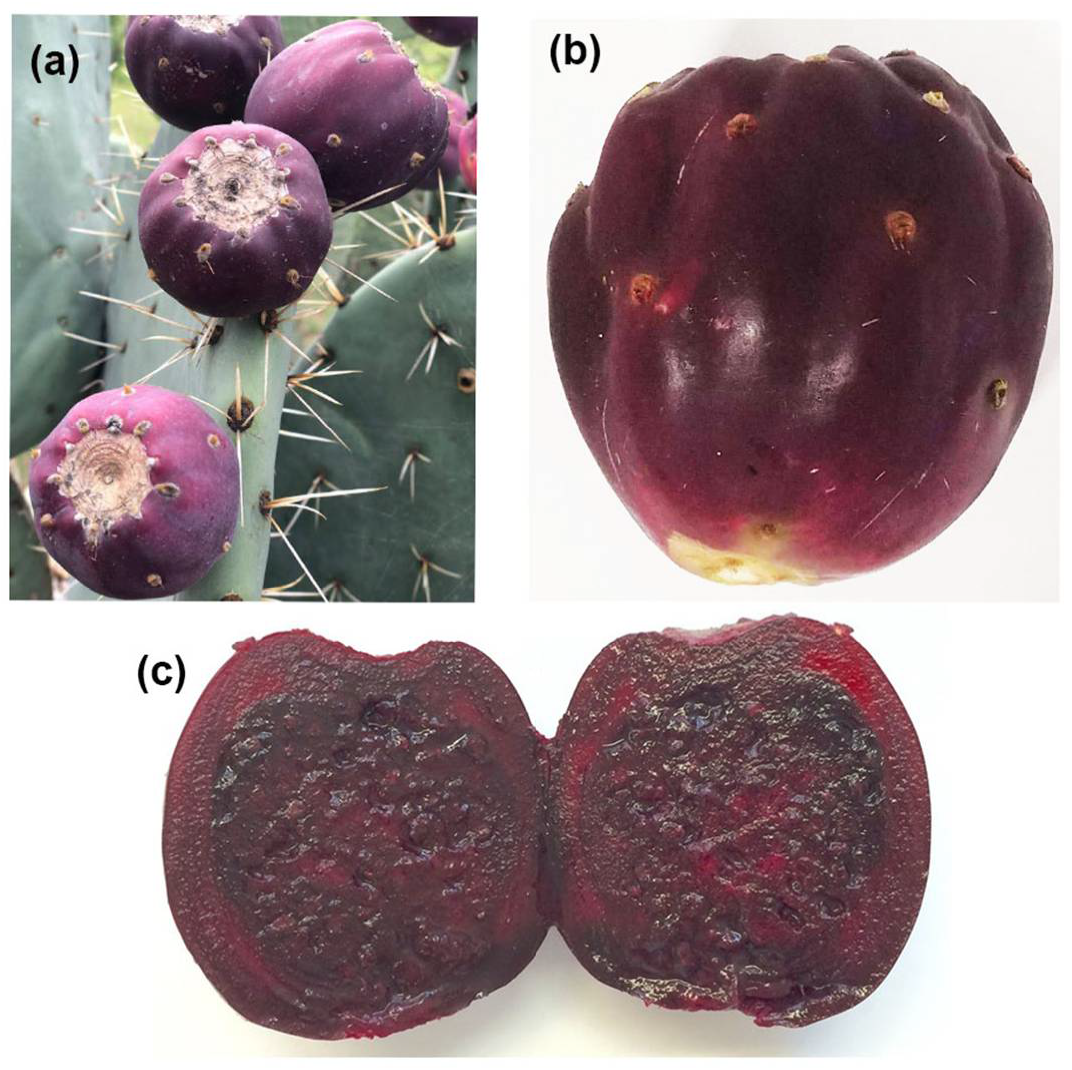
2.1.1. Fruit Material
2.1.2. Determination of Moisture Content
2.1.3. Bromatological Analysis of OrP
2.1.4. Betacyanins
2.1.5. Total Soluble Phenols
2.1.6. Total Soluble Flavonoids
2.1.7. Antioxidant Capacity
DPPH (2,2-Diphenyl-1-picrylhydrazyl)
ABTS•+ (2,2′-Azino-bis(3-ethylbenzothiazoline-6-sulfonic Acid) Diammonium Salt)
FRAP (Ferric Reducing Antioxidant Power)
AAPH (2,2-Azobis(2-amidinopropane) Dihydrochloride)
Capacity to Scavenge H2O2
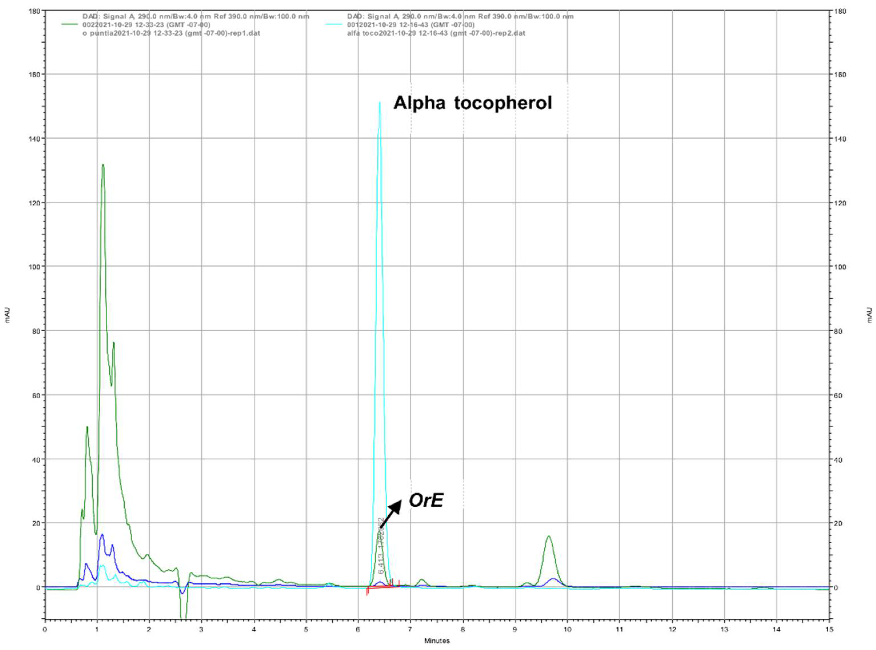
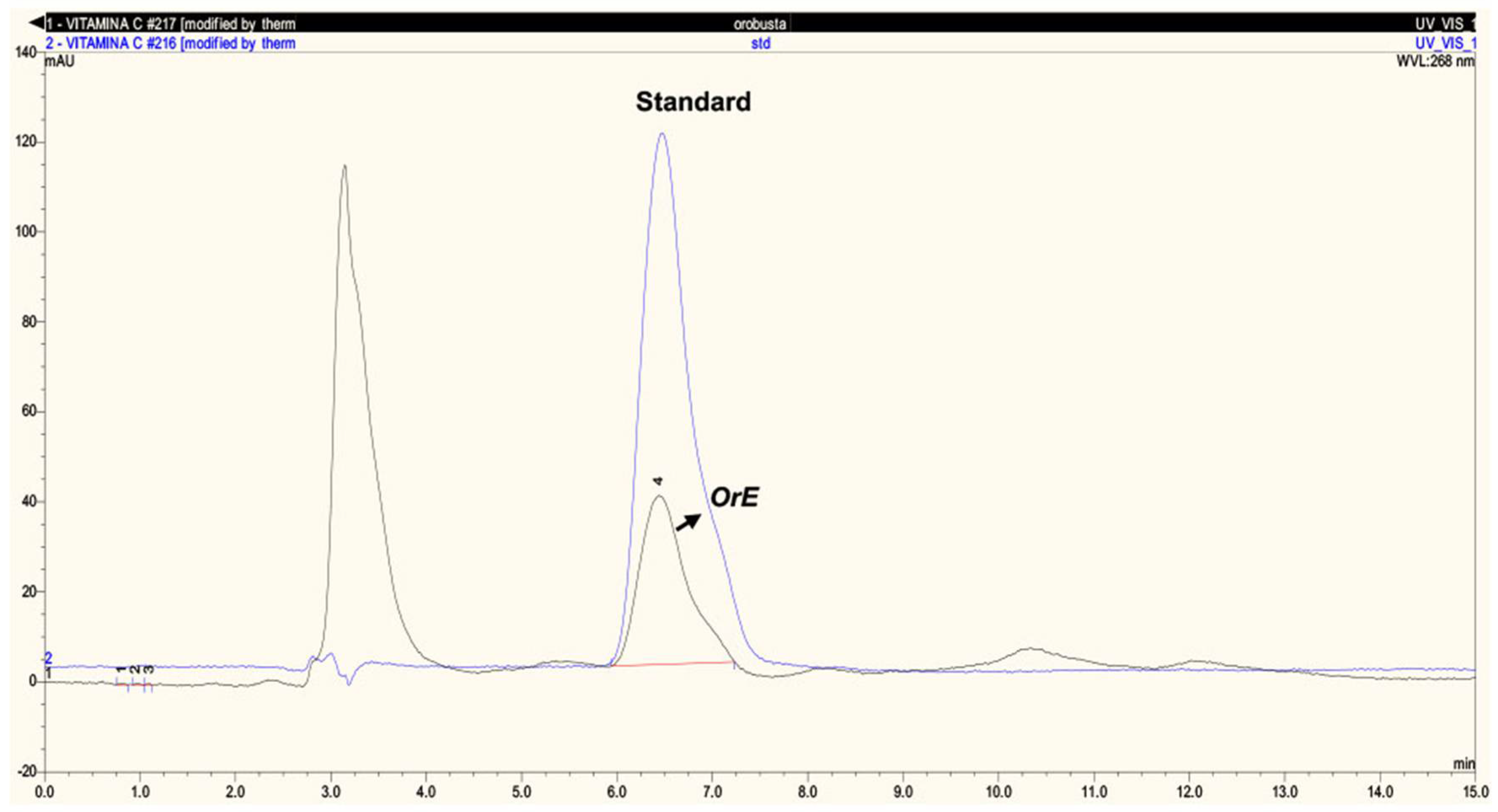
Vitamins E and C
2.2. Animal Studies with Thioacetamide
2.2.1. Animals and Treatments
2.2.2. Liver Damage Markers
2.2.3. Oxidative Stress Markers
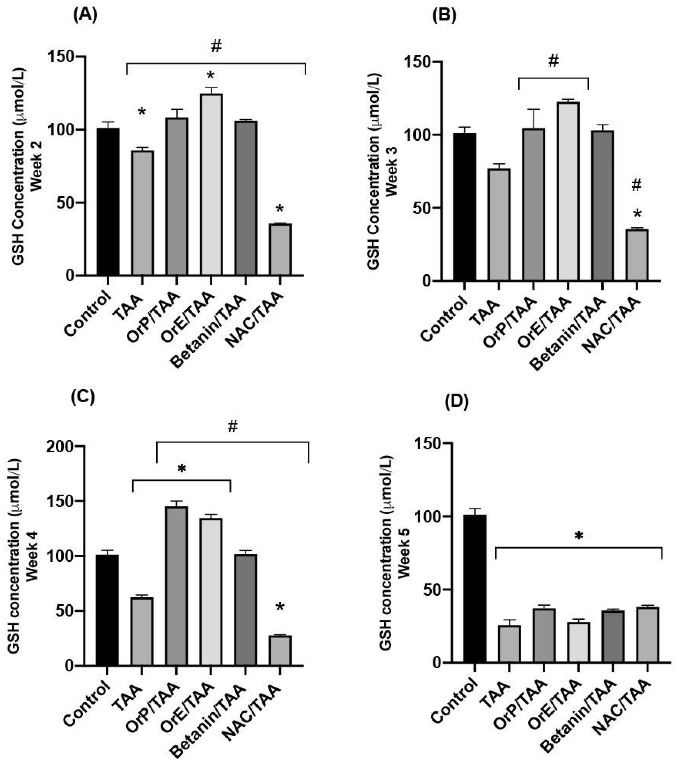
GSH (Reduced Glutathione)
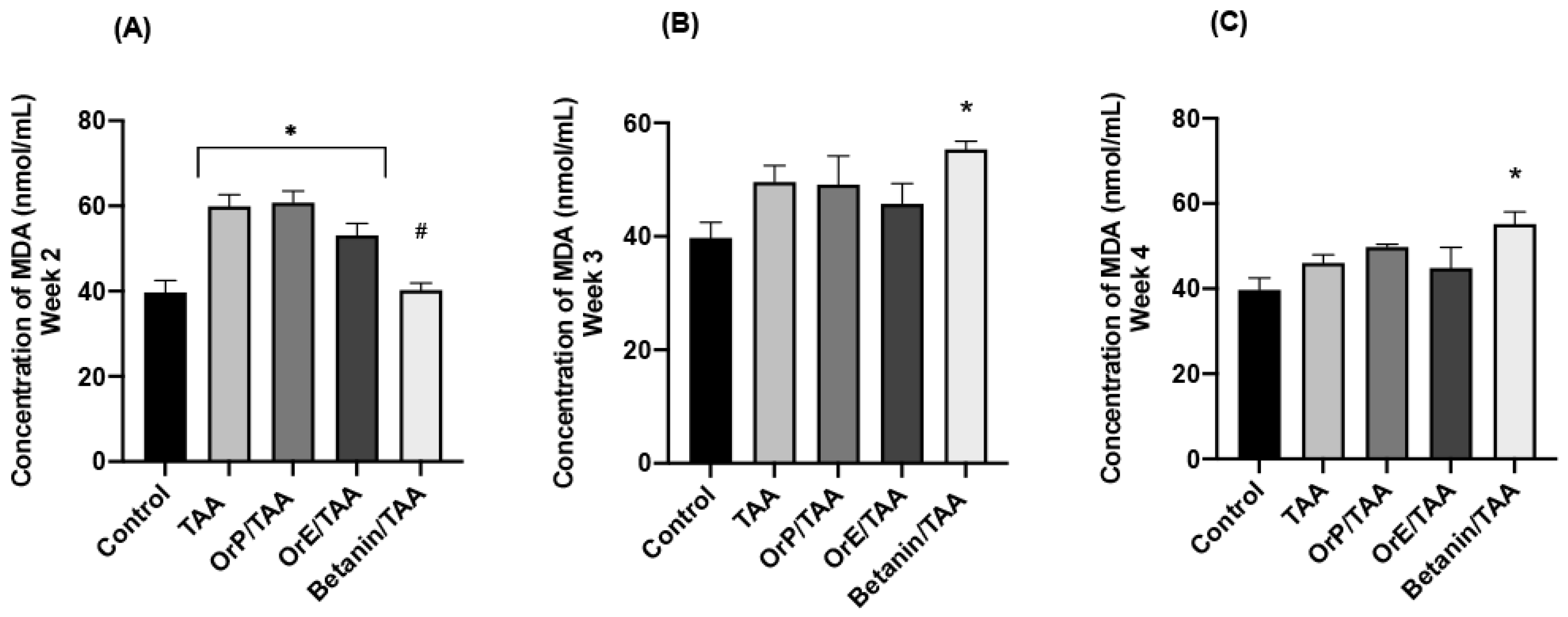
MDA (Malondialdehyde)
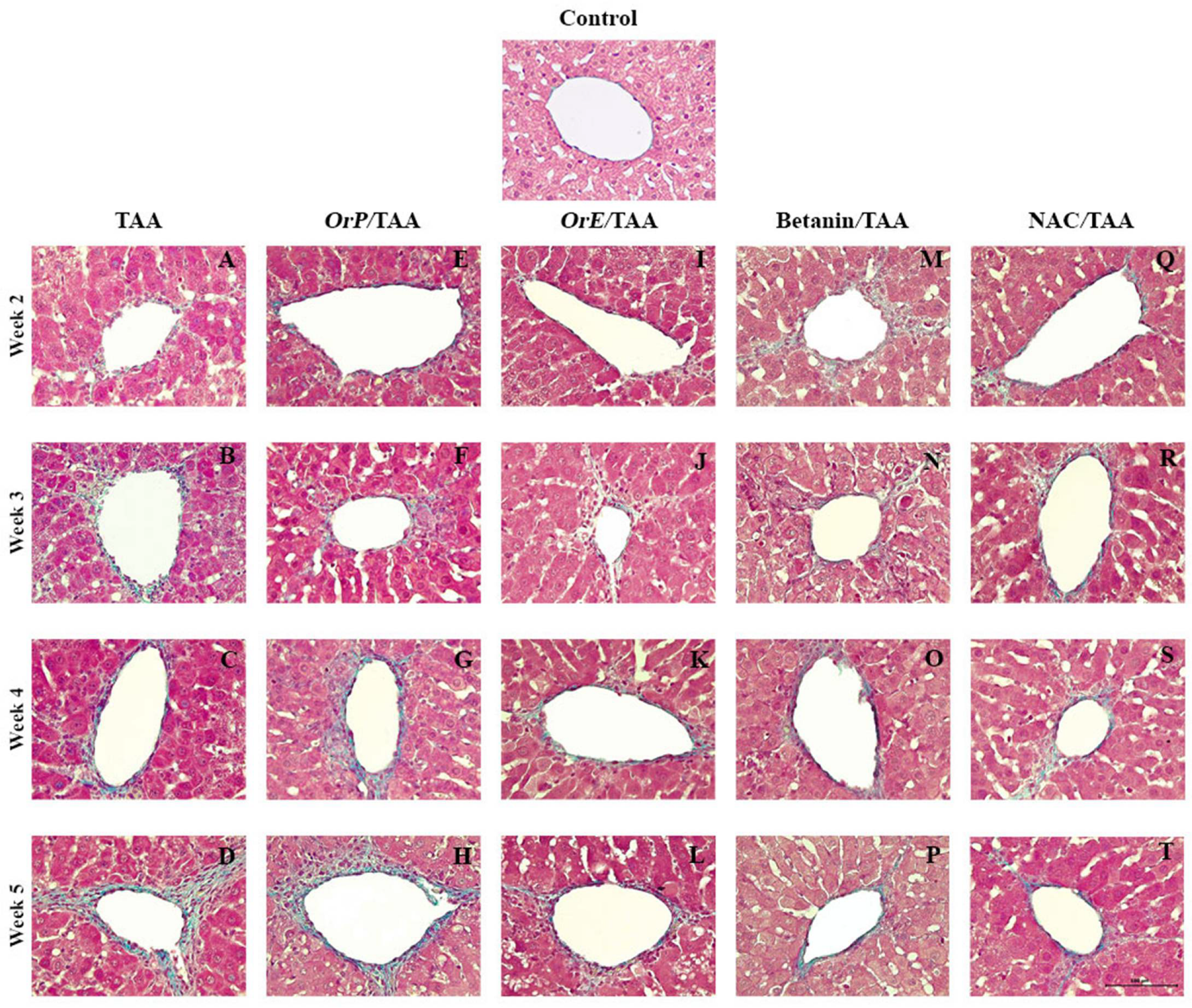
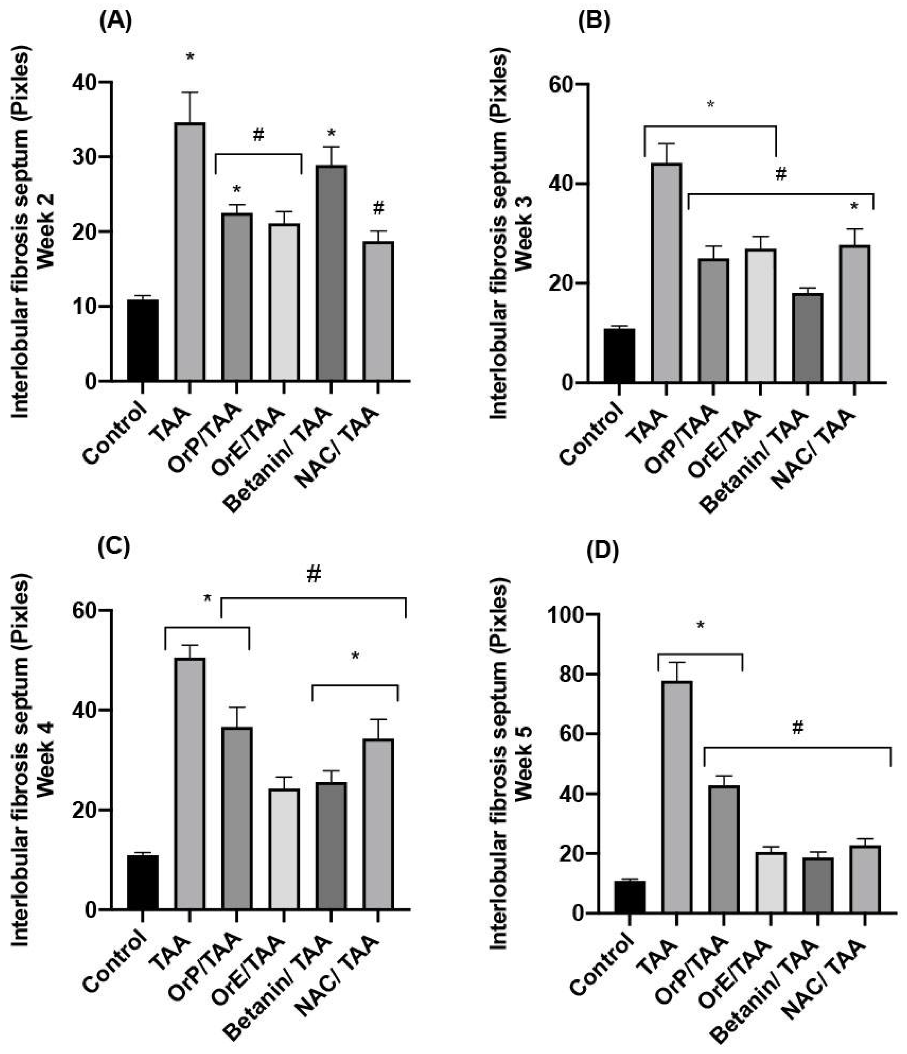
2.2.4. Histopathological Study
3. Materials and Methods
3.1. Chemicals and Reagents
3.2. Nutraceutical Characterization of Opuntia Robusta Fruit
3.2.1. Fruit Material
3.2.2. Determination of Moisture Content of OrP
3.2.3. Bromatological Analysis of OrP
3.2.4. Betacyanins
3.2.5. Total Soluble Phenols
3.2.6. Total Soluble Flavonoids
3.2.7. Antioxidant Capacity
Methanolic Extract
DPPH (2,2-Diphenyl-1-picrylhydrazyl)
ABTS•+ (2,2′-Azino-bis (3-ethylbenzothiazoline-6-sulfonic Acid) Diammonium Salt)
FRAP (Ferric Reducing Antioxidant Power)
AAPH (2,2-Azobis(2-amidinopropane) Dihydrochloride)
Capacity to Scavenge Hydrogen Peroxide (H2O2)
Determination of Vitamins C and E by High Performance Liquid Chromatography-UV (HPLC-UV)
3.3. Experimental Groups
3.3.1. Animals and Treatments
3.3.2. Biomarkers of Liver Damage
3.3.3. Oxidative Stress Biomarkers
GSH (Reduced Glutathione)
MDA (Malondialdehyde)
3.3.4. Histopathological Study
3.3.5. Measurements of Interlobular Fibrotic Septa
3.3.6. Statistical Analysis
4. Conclusions
Author Contributions
Funding
Institutional Review Board Statement
Informed Consent Statement
Data Availability Statement
Acknowledgments
Conflicts of Interest
References
- Xu, J.; Wang, X.; Su, G.; Yue, J.; Sun, Y.; Cao, J.; Zhao, Y. The antioxidant and anti-hepatic fibrosis activities of acorns (Quercus liaotungensis) and their natural galloyl triterpenes. J. Funct. Foods 2018, 46, 567–578. [Google Scholar] [CrossRef]
- Berumen, J.; Baglieri, J.; Kisseleva, T.; Mekeel, K. Liver fibrosis: Pathophysiology and clinical implications. Wiley Interdiscip. Rev. Syst. Biol. Med. 2020, 13, e1499. [Google Scholar] [CrossRef] [PubMed]
- Shi, L.; Zhang, X.; Liu, X.; Jiang, Y.; Deng, Y.; Liu, J. Cranberry (Vacinium macrocarpon) phytochemicals inhibit hepatic stellate cell activation and liver fibrosis. Food Biosci. 2021, 42, 101176. [Google Scholar] [CrossRef]
- Yuan, L.; Kaplowitz, N. Mechanisms of Drug-induced Liver Injury. Clin. Liver Dis. 2013, 17, 507–518. [Google Scholar] [CrossRef] [PubMed] [Green Version]
- Novo, E.; Busletta, C.; Bonzo, L.; Povero, D.; Paternostro, C.; Mareschi, K.; Ferrero, I.; David, E.; Bertolani, C.; Caligiuri, A.; et al. Intracellular reactive oxygen species are required for directional migration of resident and bone marrow-derived hepatic pro-fibrogenic cells. J. Hepatol. 2010, 54, 964–974. [Google Scholar] [CrossRef]
- Reyes-Agüero, J.A.; Rivera, J.R.; Flores, J. Variación morfológica de “Opuntia” (“Cactaceae”) en relación con su domesticación en la altiplanicie meridional de México. Intercienc. Rev. Cienc. Tecnol. Am. 2005, 30, 476–484. [Google Scholar]
- Castellanos-Santiago, E.; Yahia, E.M. Identification and Quantification of Betalains from the Fruits of 10 Mexican Prickly Pear Cultivars by High-Performance Liquid Chromatography and Electrospray Ionization Mass Spectrometry. J. Agric. Food Chem. 2008, 56, 5758–5764. [Google Scholar] [CrossRef]
- Patel, S. Reviewing the prospect of Opuntia pears as low cost functional foods. Rev. Environ. Sci. Bio Technol. 2012, 12, 223–234. [Google Scholar] [CrossRef]
- Patil, K.; Dagadkhair, A. Physicochemical characteristics and antioxidant potential of Opuntia fruit: A review Kartika V Patil and Amol C Dagadkhair. Seeds 2019, 2, 10. [Google Scholar]
- Ali, R. Antioxidant and Anticancer Activities of Different Constituents Extracted from Egyptian Prickly Pear Cactus (Opuntia Ficus-Indica) Peel Faten, M. Abou-Elella and Rehab Farouk Mohammed Ali* Department of. Biochem. Anal. Biochem. 2014, 3, 1–8. [Google Scholar] [CrossRef] [Green Version]
- De Wit, M.; du Toit, A.; Osthoff, G.; Hugo, A. Antioxidant Content, Capacity and Retention in Fresh and Processed Cactus Pear (Opuntia ficus-indica and O. robusta) Fruit Peels from Different Fruit-Colored Cultivars. Front. Sustain. Food Syst. 2020, 4, 133. [Google Scholar] [CrossRef]
- González-Ponce, H.A.; Martínez-Saldaña, M.C.; Rincón-Sánchez, A.R.; Sumaya-Martínez, M.T.; Buist-Homan, M.; Faber, K.N.; Moshage, H.; Jaramillo-Juárez, F. Hepatoprotective Effect of Opuntia robusta and Opuntia streptacantha Fruits against Acetaminophen-Induced Acute Liver Damage. Nutrients 2016, 8, 607. [Google Scholar] [CrossRef] [PubMed] [Green Version]
- Bankova, V. Chemical diversity of propolis and the problem of standardization. J. Ethnopharmacol. 2005, 100, 114–117. [Google Scholar] [CrossRef] [PubMed]
- Jiménez-Aguilar, D.M.; López-Martínez, J.M.; Hernández-Brenes, C.; Gutiérrez-Uribe, J.A.; Welti-Chanes, J. Dietary fiber, phytochemical composition and antioxidant activity of Mexican commercial varieties of cactus pear. J. Food Compos. Anal. 2015, 41, 66–73. [Google Scholar] [CrossRef]
- Chavez-Santoscoy, A.; Gutiérrez-Uribe, J.; Serna-Saldivar, S. Phenolic Composition, Antioxidant Capacity and In Vitro Cancer Cell Cytotoxicity of Nine Prickly Pear (Opuntia spp.) Juices. Plant Foods Hum. Nutr. 2009, 64, 146–152. [Google Scholar] [CrossRef]
- Chang, S.F.; Hsieh, C.L.; Yen, G.C. The protective effect of Opuntia dillenii Haw fruit against low-density lipoprotein peroxidation and its active compounds. Food Chem. 2008, 106, 569–575. [Google Scholar] [CrossRef]
- Budinsky, A.; Wolfram, R.; Oguogho, A.; Efthimiou, Y.; Stamatopoulos, Y.; Sinzinger, H. Regular ingestion of opuntia robusta lowers oxidation injury. Prostaglandins Leukot. Essent. Fat. Acids (PLEFA) 2001, 65, 45–50. [Google Scholar] [CrossRef] [Green Version]
- González-Ponce, H.A.; Martínez-Saldaña, M.C.; Tepper, P.G.; Quax, W.J.; Buist-Homan, M.; Faber, K.N.; Moshage, H. Betacyanins, major components in Opuntia red-purple fruits, protect against acetaminophen-induced acute liver failure. Food Res. Int. 2020, 137, 109461. [Google Scholar] [CrossRef]
- Villa-Jaimes, G.S.; Aguilar-Mora, F.A.; González-Ponce, H.A.; Avelar-González, F.J.; Martínez Saldaña, M.C.M.; Buist-Homan, M.; Moshage, H. Biocomponents from Opuntia robusta and Opuntia streptacantha fruits protect against diclofenac-induced acute liver damage in vivo and in vitro. J. Funct. Foods 2022, 89, 104960. [Google Scholar] [CrossRef]
- Piga, A. Cactus Pear: A Fruit of nutraceutical and functional importance. J. Prof. Assoc. Cactus Dev. 2004, 6, 9–22. [Google Scholar]
- AOAC. AOAC International. Official Method 920.151 Solids (Total) in Fruits and Fruit Products, 17th ed.; AOAC: Gaithersburg MD, USA, 2000. [Google Scholar]
- Torres-Bojórquez, A.E.; García, O.; Miranda-Lopez, R.; Anaberta, C.M. Evaluation of antioxidant capacity, physicochemical characteristics and sensory profile of Opuntia robusta and O. ficus-indica. Arch. Latinoam. Nutr. 2017, 67, 291–299. [Google Scholar]
- Chaouch, M.; Hafsa, J.; Rihouey, C.; le Cerf, D.; Majdoub, H. Effect of extraction conditions on the antioxidant and antiglycation capacity of carbohydrates from Opuntia robusta cladodes. Int. J. Food Sci. Technol. 2016, 51, 929–937. [Google Scholar] [CrossRef]
- Van Soest, P.J. Nutritional Ecology of the Ruminant, 2nd ed.; Conrnell University: Ithaca, NY, USA, 1994. [Google Scholar]
- González Ponce, H.; Rincón-Sánchez, A.; Jaramillo-Juárez, F.; Moshage, H. Natural Dietary Pigments: Potential Mediators against Hepatic Damage Induced by Over-The-Counter Non-Steroidal Anti-Inflammatory and Analgesic Drugs. Nutrients 2018, 10, 117. [Google Scholar] [CrossRef] [PubMed] [Green Version]
- Buettner, G. The pecking order of free radicals and antioxidants: Lipid peroxidation, a -tocopherol, and ascorbate. Arch. Biochem. Biophys. 1993, 300, 535–543. [Google Scholar] [CrossRef] [PubMed]
- Livrea, M.A.; Tesoriere, L. Lipoperoxyl Radical Scavenging and Antioxidative Effects of Red Beet Pigments BT—Red Beet Biotechnology. In Food and Pharmaceutical Applications; Neelwarne, B., Ed.; Springer: Boston, MA, USA, 2012; pp. 105–124. [Google Scholar] [CrossRef] [Green Version]
- Krajka-Kuźniak, V.; Paluszczak, J.; Szaefer, H.; Baer-Dubowska, W. Betanin, a beetroot component, induces nuclear factor erythroid-2-related factor 2-mediated expression of detoxifying/antioxidant enzymes in human liver cell lines. Br. J. Nutr. 2013, 110, 2138–2149. [Google Scholar] [CrossRef] [Green Version]
- Marañón-Ruiz, V.F.; Rizo de la Torre L del, C.; Chiu-Zarate, R. Caracterización de las propiedades ópticas de Betacianinas y Betaxantinas por espectroscopía Uv-Vis y barrido en Z. Superf. Vacío 2011, 24, 113–120. [Google Scholar]
- Fernández-López, J.A.; Almela, L.; Obón, J.M.; Castellar, R. Determination of Antioxidant Constituents in Cactus Pear Fruits. Plant Foods Hum. Nutr. 2010, 65, 253–259. [Google Scholar] [CrossRef]
- Macheix, J.J.; Fleuriet, A.; Billot, J. Fruit Phenolics, 1st ed.; CRC Press: Boca Ratón, FL, USA, 1990; p. 390. [Google Scholar]
- Galati, E.M.; Tripodo, M.M.; Trovato, A.; Miceli, N.; Monforte, M.T. Biological effect of Opuntia ficus indica (L.) Mill. (Cactaceae) waste matter: Note I: Diuretic activity. J. Ethnopharmacol. 2002, 79, 17–21. [Google Scholar] [CrossRef]
- Cazzola, R.; Cestaro, B. Red wine polyphenols protect n−3 more than n−6 polyunsaturated fatty acid from lipid peroxidation. Food Res. Int. 2011, 44, 3065–3071. [Google Scholar] [CrossRef]
- Hwang, Y.P.; Choi, J.H.; Han, E.H.; Kim, H.G.; Wee, J.-H.; Jung, K.O.; Jung, K.H.; Kwon, K.-I.; Jeong, T.C.; Chung, Y.C.; et al. Purple sweet potato anthocyanins attenuate hepatic lipid accumulation through activating adenosine monophosphate–activated protein kinase in human HepG2 cells and obese mice. Nutr. Res. 2011, 31, 896–906. [Google Scholar] [CrossRef]
- Toufektsian, M.C.; Salen, P.; Laporte, F.; Tonelli, C.; de Lorgeril, M. Dietary Flavonoids Increase Plasma Very Long-Chain (n-3) Fatty Acids in Rats. J. Nutr. 2011, 141, 37–41. [Google Scholar] [CrossRef] [PubMed] [Green Version]
- Villasante, A.; Patro, B.; Chew, B.; Becerra, M.; Wacyk, J.; Overturf, K.; Powell, M.S.; Hardy, R.W. Dietary Intake of Purple Corn Extract Reduces Fat Body Content and Improves Antioxidant Capacity and n-3 Polyunsaturated Fatty Acid Profile in Plasma of Rainbow Trout, Oncorhynchus mykiss. J. World Aquac. Soc. 2015, 46, 381–394. [Google Scholar] [CrossRef]
- Pérez-Torres, I.; Zúñiga Muñoz, A.; Beltrán-Rodríguez, U.; Díaz-Díaz, E.; Martínez-Memije, R.; Guarner Lans, V. Modification of the liver fatty acids by Hibiscus sabdariffa Linnaeus (Malvaceae) infusion, its possible effect on vascular reactivity in a metabolic syndrome model. Clin. Exp. Hypertens. 2014, 36, 123–131. [Google Scholar] [CrossRef] [PubMed]
- Miler, M.; Živanović, J.; Ajdžanović, V.; Oreščanin-Dušić, Z.; Milenković, D.; Konić-Ristić, A.; Blagojević, D.; Milošević, V.; Šošić-Jurjević, B. Citrus flavanones naringenin and hesperetin improve antioxidant status and membrane lipid compositions in the liver of old-aged Wistar rats. Exp. Gerontol. 2016, 84, 49–60. [Google Scholar] [CrossRef]
- Vargas Mendoza, N. Efecto Hepatoprotector y Antioxidante del Extracto y los Principios Activos de Geranium shiedeanum. Ph.D. Thesis, San Agustín Tlaxiaca, Hgo, Mexico, 2012. Available online: http://dgsa.uaeh.edu.mx:8080/bibliotecadigital/handle/231104/1803 (accessed on 5 July 2022).
- Esatbeyoglu, T.; Wagner, A.E.; Motafakkerazad, R.; Nakajima, Y.; Matsugo, S.; Rimbach, G. Free radical scavenging and antioxidant activity of betanin: Electron spin resonance spectroscopy studies and studies in cultured cells. Food Chem. Toxicol. 2014, 73, 119–126. [Google Scholar] [CrossRef]
- Sumaya-Martínez, M.T.; Cruz-Jaime, S.; Madrigal-Santillán, E.; García-Paredes, J.D.; Cariño-Cortés, R.; Cruz-Cansino, N.; Valadez-Vega, C.; Martinez-Cardenas, L.; Alanís-García, E. Betalain, Acid ascorbic, phenolic contents and antioxidant properties of purple, red, yellow and white cactus pears. Int. J. Mol. Sci. 2011, 2, 6452–6468. [Google Scholar] [CrossRef] [Green Version]
- Butera, D.; Tesoriere, L.; Di Gaudio, F.; Bongiorno, A.; Allegra, M.; Pintaudi, A.M.; Kohen, R.; Livrea, M.A. Antioxidant Activities of Sicilian Prickly Pear (Opuntia ficus indica) Fruit Extracts and Reducing Properties of Its Betalains: Betanin and Indicaxanthin. J. Agric. Food Chem. 2002, 50, 6895–6901. [Google Scholar] [CrossRef] [Green Version]
- Amjadi, S.; Mesgari Abbasi, M.; Shokouhi, B.; Ghorbani, M.; Hamishehkar, H. Enhancement of therapeutic efficacy of betanin for diabetes treatment by liposomal nanocarriers. J. Funct. Foods 2019, 59, 119–128. [Google Scholar] [CrossRef]
- Prior, R.L.; Wu, X.; Schaich, K. Standardized Methods for the Determination of Antioxidant Capacity and Phenolics in Foods and Dietary Supplements. J. Agric. Food Chem. 2005, 53, 4290–4302. [Google Scholar] [CrossRef]
- Roginsky, V.; Lissi, E.A. Review of methods to determine chain-breaking antioxidant activity in food. Food Chem. 2005, 92, 235–254. [Google Scholar] [CrossRef]
- Ou, B.; Huang, D.; Hampsch-Woodill, M.; Flanagan, J.A.; Deemer, E.K. Analysis of Antioxidant Activities of Common Vegetables Employing Oxygen Radical Absorbance Capacity (ORAC) and Ferric Reducing Antioxidant Power (FRAP) Assays: A Comparative Study. J. Agric. Food Chem. 2002, 50, 3122–3128. [Google Scholar] [CrossRef]
- Chisté, R.; Freitas, M.; Mercadante, A.; Fernandes, E. Carotenoids are Effective Inhibitors of in vitro Hemolysis of Human Erythrocytes, as Determined by a Practical and Optimized Cellular Antioxidant Assay. J. Food Sci. 2014, 79, H1841–H1847. [Google Scholar] [CrossRef] [PubMed]
- Kanner, J.; Harel, S.; Granit, R. BetalainsA New Class of Dietary Cationized Antioxidants. J. Agric. Food Chem. 2001, 49, 5178–5185. [Google Scholar] [CrossRef] [PubMed]
- Sakihama, Y.; Maeda, M.; Hashimoto, M.; Tahara, S.; Hashidoko, Y. Beetroot betalain inhibits peroxynitrite-mediated tyrosine nitration and DNA strand damage. Free. Radic. Res. 2011, 46, 93–99. [Google Scholar] [CrossRef]
- Keser, S.; Celik, S.; Türkoğlu, S.; Yilmaz, O.; Turkoglu, I. Hydrogen Peroxide Radical Scavenging and Total Antioxidant Activity of Hawthorn. Chem. J. 2012, 2, 9–12. [Google Scholar]
- Valko, M.; Leibfritz, D.; Moncol, J.; Cronin, M.T.D.; Mazur, M.; Telser, J. Free radicals and antioxidants in normal physiological functions and human disease. Int. J. Biochem. Cell Biol. 2007, 39, 44–84. [Google Scholar] [CrossRef]
- Musyarofah, N.; Susanto, S.; Aziz, S.A.; Suketi, K.; Dadang, D. The diversity of ‘kristal’ guava (Psidium guajava) fruit quality in response to different altitudes and cultural practices. Biodiversitas 2020, 21, 3310–3316. [Google Scholar] [CrossRef]
- Alkaladi, A. Vitamins E and C ameliorate the oxidative stresses induced by Zinc oxide nanoparticles on liver and gills of Oreochromis niloticus. Saudi J. Biol. Sci. 2018, 26, 357–362. [Google Scholar] [CrossRef]
- El-Aal, A.A.; El-Ghffar, E.A.A.; Ghali, A.A.; Zughbur, M.R.; Sirdah, M.M. The effect of vitamin C and/or E supplementations on type 2 diabetic adult males under metformin treatment: A single-blinded randomized controlled clinical trial. Diabetes Metab. Syndr. Clin. Res. Rev. 2018, 12, 483–489. [Google Scholar] [CrossRef]
- Sumida, Y.; Niki, E.; Naito, Y.; Yoshikawa, T. Involvement of free radicals and oxidative stress in NAFLD/NASH. Free Radic. Res. 2013, 47, 869–880. [Google Scholar] [CrossRef]
- Al-Mehdar, A.A.; El-Denshary, E.S.; Addel-wahhab, M.A. Alpha Lipoic Acid and Alpha-Tocopherol Counteract the Oxidative Stress and Liver Damage in Rats Sub-Chronically Treated with Khat (Catha edulis) Extract. Glob. J. Pharmacol. 2012, 6, 94–105. [Google Scholar]
- Abhilash, P.A.; Harikrishnan, R.; Indira, M. Ascorbic acid suppresses endotoxemia and NF-κB signaling cascade in alcoholic liver fibrosis in guinea pigs: A mechanistic approach. Toxicol. Appl. Pharmacol. 2014, 274, 215–224. [Google Scholar] [CrossRef] [PubMed]
- Gliszczyńska-Świgło, A.; Szymusiak, H.; Malinowska, P. Betanin, the main pigment of red beet: Molecular origin of its exceptionally high free radical-scavenging activity. Food Addit. Contam. 2006, 23, 1079–1087. [Google Scholar] [CrossRef] [PubMed] [Green Version]
- Tada, M.; Kohno, M.; Niwano, Y. Scavenging or Quenching Effect of Melanin on Superoxide Anion and Singlet Oxygen. J. Clin. Biochem. Nutr. 2010, 46, 224–228. [Google Scholar] [CrossRef] [Green Version]
- Brahmi, D.; Bouaziz, C.; Ayed, Y.; ben Mansour, H.; Zourgui, L.; Bacha, H. Chemopreventive effect of cactus Opuntia ficus indica on oxidative stress and genotoxicity of aflatoxin B1. Nutr. Metab. 2011, 8, 73. [Google Scholar] [CrossRef] [PubMed] [Green Version]
- Xie, Y.; Wang, G.; Wang, H.; Yao, X.; Jiang, S.; Kang, A.; Zhou, F.; Xie, T.; Hao, H. Cytochrome P450 Dysregulations in Thioacetamide-Induced Liver Cirrhosis in Rats and the Counteracting Effects of Hepatoprotective Agents. Drug Metab. Dispos. 2012, 40, 796–802. [Google Scholar] [CrossRef] [Green Version]
- Ramos-Tovar, E.; Casas-Grajales, S.; Hernández-Aquino, E.; Flores-Beltrán, R.E.; Galindo-Gómez, S.; Vera-Aguilar, E.; Diaz-Ruiz, A.; Montes, S.; Camacho, J.; Tsutsumi, V.; et al. Cirrhosis induced by thioacetamide is prevented by stevia. Molecular mechanisms. J. Funct. Foods 2019, 52, 552–564. [Google Scholar] [CrossRef]
- El-Latif El-Ghazaly, M.A.; Rashed, E.R.; Shafey, G.M.; Zaki, H.F.; Attia, A.S. Amelioration of thioacetamide-induced hepatic encephalopathy in rats by low-dose gamma irradiation. Environ. Sci. Pollut. Res. 2020, 27, 334–343. [Google Scholar] [CrossRef]
- Gulbahar, O.; Karasu, Z.; Ersoz, G.; Akarca, U.; Musoglu, A. Treatment of nonalcoholic steatohepatitis with N-acetylcysteine (Abstract). Gastroenterology 2000, 118, A1444. [Google Scholar] [CrossRef]
- Bashandy, S.A.E.; el Awdan, S.A.; Mohamed, S.M.; Omara, E.A.A. Allium porrum and Bauhinia Variegata Mitigate Acute Liver Failure and Nephrotoxicity Induced by Thioacetamide in Male Rats. Indian J. Clin. Biochem. 2020, 35, 147–157. [Google Scholar] [CrossRef]
- Abul, H.; Mathew, T.C.; Dashti, H.M.; Al-Bader, A. Level of Superoxide Dismutase, Glutathione Peroxidase and Uric Acid in Thioacetamide-Induced Cirrhotic Rats. Anat. Histol. Embryol. 2002, 31, 66–71. [Google Scholar] [CrossRef] [PubMed]
- Tesoriere, L.; Allegra, M.; Gentile, C.; Livrea, M. Betacyanins as phenol antioxidants. Chemistry and mechanistic aspects of the lipoperoxyl radical-scavenging activity in solution and liposomes. Free Radic. Res. 2009, 43, 706–717. [Google Scholar] [CrossRef] [PubMed]
- Dwivedi, D.; Jena, G. Glibenclamide protects against thioacetamide-induced hepatic damage in Wistar rat: Investigation on NLRP3, MMP-2, and stellate cell activation. Naunyn-Schmiedeberg’s Arch. Pharmacol. 2018, 391, 1257–1274. [Google Scholar] [CrossRef] [PubMed]
- Yormaz, S.; Bulbuloglu, E.; Kurutas, E.B.; Ciralik, H.; Yuzbasioglu, M.F.; Yildiz, H.; Coskuner, I.; Silay, E.; Kantarceken, B.; Goksu, M.; et al. The comparison of the effects of hepatic regeneration after partial hepatectomy, silybum marinaum, propofol, N-acetylcysteine and vitamin E on liver. Bratisl. Lekárske Listy 2012, 113, 145–151. [Google Scholar] [CrossRef] [Green Version]
- AOAC. AOAC International. Official Method 954.02, Fat Crude or Ether Extract in Pet Food; AOAC: Gaithersburg MD, USA, 1977. [Google Scholar]
- Takoudjou Miafo, A.P.; Koubala, B.B.; Muralikrishna, G.; Kansci, G.; Fokou, E. Non-starch polysaccharides derived from sorghum grains, bran, spent grain and evaluation of their antioxidant properties with respect to their bound phenolic acids. Bioact. Carbohydr. Dietary Fibre 2022, 28, 100314. [Google Scholar] [CrossRef]
- Ilyas, M.; Khan, W.A.; Ali, T.; Ahmad, N.; Khan, Z.; Fazal, H.; Zaman, N.; Ualiyeva, D.; Ali, M.; Amissah, O.B.; et al. Cold Stress-induced Seed Germination and Biosynthesis of Polyphenolics Content in Medicinally Important Brassica rapa. Phytomedicine Plus 2022, 2, 100185. [Google Scholar] [CrossRef]
- Lasano, N.F.; Hamid, A.H.; Karim, R.; Dek, M.S.P.; Shukri, R.; Shazini Ramli, N. Nutritional Composition, Anti-Diabetic Properties and Identification of Active Compounds Using UHPLC-ESI-Orbitrap-MS/MS in Mangifera odorata L. Peel Seed Kernel. Mol. 2019, 24, 320. [Google Scholar] [CrossRef] [Green Version]
- Ruslan, K.; Happyniar, S.; Fidrianny, I. Antioxidant potential of two varieties of Sesamum indicum L. collected from Indonesia. J. Taibah Univ. Med. Sci. 2018, 13, 211–218. [Google Scholar] [CrossRef]
- Xu, P.; Qian, Y.; Wang, R.; Chen, Z.; Wang, T. Entrapping curcumin in the hydrophobic reservoir of rice proteins toward stable antioxidant nanoparticles. Food Chem. 2022, 387, 132906. [Google Scholar] [CrossRef]
- Khudyakov, D.; Sosnin, M.; Shorstkii, I.; Okpala, C.O.R. Cold filamentary microplasma pretreatment combined with infrared dryer: Effects on drying efficiency and quality attributes of apple slices. J. Food Eng. 2022, 329, 111049. [Google Scholar] [CrossRef]
- Vinjamuri, S.; Shanker, D.; Ramesh, R.S.; Nagarajan, S. In vitro evaluation of hemolytic activity and cell viability assay of hexanoic extracts of Bridelia ferruginea Benth. World J. Pharm. Pharm. Sci. (WJPPS) 2015, 4, 1263–1268. [Google Scholar]
- Khan, S.; Rehman, M.; Muhammed, K.; Khan, M.; Haq, I.; Khan, M. In vitro and in vivo antioxidant therapeutic evaluation of Dodonaea viscosa. Biorxiv 2022. [Google Scholar] [CrossRef]
- Singh, G.; Mohanty, B.P.; Saini, G.S.S. Structure, spectra and antioxidant action of ascorbic acid studied by density functional theory, Raman spectroscopic and nuclear magnetic resonance techniques. Spectrochim. Acta Part A Mol. Biomol. Spectrosc. 2016, 155, 61–74. [Google Scholar] [CrossRef] [PubMed]
- Limbach, J.R.; Espinosa, C.D.; Perez-Calvo, E.; Stein, H.H. Effect of dietary crude protein level on growth performance, blood characteristics, and indicators of intestinal health in weanling pigs. J. Anim. Sci. 2021, 99, skab166. [Google Scholar] [CrossRef] [PubMed]
- El-Baz, F.K.; Salama, A.A.A.; Hussein, R.A. Dunaliella salina microalgae oppose thioacetamide-induced hepatic fibrosis in rats. Toxicol. Rep. 2020, 7, 36–45. [Google Scholar] [CrossRef]
- Han, J.; Ma, D.; Zhang, M.; Yang, X.; Tan, D. Natural Antioxidant Betanin Protects Rats from Paraquat-Induced Acute Lung Injury Interstitial Pneumonia. BioMed Res. Int. 2015, 2015, 608174. [Google Scholar] [CrossRef] [Green Version]
- Prophet, E.B. Methods in Histotechnology, 1st ed.; Prophet Edna, Ed.; The Libraries: Washington, DC, USA, 1992; Volume I, pp. 123–132. [Google Scholar]


| Component (%) | DMB | WMB |
|---|---|---|
| Dry matter | 100 | 37.99 |
| Crude protein | 1.18 | 0.45 |
| Crude fiber | 2.84 | 1.08 |
| Crude fat | 0.76 | 0.29 |
| Ashes | 6.75 | 2.56 |
| Nitrogen free extract | 88.47 | 33.61 |
| Betacyanins | Total Phenols | Flavonoids |
|---|---|---|
| 436.5 ± 57 mg of betacyanin equivalents/L | 1118.0 mg GAE/100 g, dmb | 793 mg CAE/100 g, dmb |
| DPPH | ABTS•+ | FRAP | AAPH | Capacity to Scavenge H2O2 |
|---|---|---|---|---|
| 2.27 mmol TE/L | 62.2 ± 5.0 μmol TE/g, db | 80.2± 11.7 μmol TE/g, db | 247.9 ± 15.6 μmol TE/g, db | 15 ± 0.8% at. 100 µg/mL |
Publisher’s Note: MDPI stays neutral with regard to jurisdictional claims in published maps and institutional affiliations. |
© 2022 by the authors. Licensee MDPI, Basel, Switzerland. This article is an open access article distributed under the terms and conditions of the Creative Commons Attribution (CC BY) license (https://creativecommons.org/licenses/by/4.0/).
Share and Cite
Pulido-Hornedo, N.A.; Ventura-Juárez, J.; Guevara-Lara, F.; González-Ponce, H.A.; Sánchez-Alemán, E.; Buist-Homan, M.; Moshage, H.; Martínez-Saldaña, M.C. Hepatoprotective Effect of Opuntia robusta Fruit Biocomponents in a Rat Model of Thioacetamide-Induced Liver Fibrosis. Plants 2022, 11, 2039. https://doi.org/10.3390/plants11152039
Pulido-Hornedo NA, Ventura-Juárez J, Guevara-Lara F, González-Ponce HA, Sánchez-Alemán E, Buist-Homan M, Moshage H, Martínez-Saldaña MC. Hepatoprotective Effect of Opuntia robusta Fruit Biocomponents in a Rat Model of Thioacetamide-Induced Liver Fibrosis. Plants. 2022; 11(15):2039. https://doi.org/10.3390/plants11152039
Chicago/Turabian StylePulido-Hornedo, Nayeli Amalinalli, Javier Ventura-Juárez, Fidel Guevara-Lara, Herson Antonio González-Ponce, Esperanza Sánchez-Alemán, Manon Buist-Homan, Han Moshage, and Ma. Consolación Martínez-Saldaña. 2022. "Hepatoprotective Effect of Opuntia robusta Fruit Biocomponents in a Rat Model of Thioacetamide-Induced Liver Fibrosis" Plants 11, no. 15: 2039. https://doi.org/10.3390/plants11152039
APA StylePulido-Hornedo, N. A., Ventura-Juárez, J., Guevara-Lara, F., González-Ponce, H. A., Sánchez-Alemán, E., Buist-Homan, M., Moshage, H., & Martínez-Saldaña, M. C. (2022). Hepatoprotective Effect of Opuntia robusta Fruit Biocomponents in a Rat Model of Thioacetamide-Induced Liver Fibrosis. Plants, 11(15), 2039. https://doi.org/10.3390/plants11152039