Abstract
Pulmonary hypertension (PH) is a rare disease, which still carries a poor prognosis. PH is characterized by a pressure overload on the right ventricle (RV), which develops hypertrophy, followed by a progressive failure. Accordingly, recent evidence showed that RV function has an important prognostic role in patients with PH. Echocardiography, cardiac magnetic resonance (CMR), computed tomography, and nuclear imaging allow a non-invasive evaluation of the RV size and function, but only the first two are routinely used in the clinical arena. Some conventional echocardiographic parameters, such as TAPSE (tricuspid anular plane systolic excursion), have demonstrated prognostic value in patients with PH. Moreover, there are some new advanced echo techniques, which can provide a more detailed assessment of RV function. Three-dimensional (3D) echocardiography allows measurement of RV volumes and ejection fraction, and two-dimensional (2D) speckle tracking (STE), allows assessment of RV myocardial mechanics. CMR provides accurate measurement of RV volumes, ejection fraction, and mass and allows the characterization of the RV wall composition by identifying the presence of fibrosis by late gadolinium enhancement. Although CMR seems to hold promise for both initial assessment and follow-up of patients with PH, its main role has been restricted to diagnostic work-up only.
1. Introduction
Pulmonary hypertension (PH) is a rare disease which carries a poor prognosis [1,2]. Recent evidence showed that right ventricular (RV) function has an important prognostic role in patients with PH [3,4,5,6,7,8,9,10], the onset of RV failure being associated with increased mortality, independent from pulmonary vascular resistance values and the etiology of PH [1,11,12,13,14]. Therefore, accurate assessment of RV function appears to be critical in PH patients’ initial evaluation and follow-up.
In this scenario non-invasive imaging techniques, such as cardiac magnetic resonance (CMR) and echocardiography [15], have been shown to be able to provide quantitative assessment of RV morphology, size, and function, covering a major role in diagnosis and follow-up of patients with PH.
This review paper will summarize the current knowledge about the RV assessment by conventional and novel non-invasive imaging modalities in the setting of PH.
2. The Right Ventricle
Compared to its left counterpart, the RV shows a complex pyramidal shape, and can be divided in three main parts: the inlet, the outlet, and the apical, trabeculated part. The RV free wall myocardium is composed mainly by circumferential fibers in the superficial layer and longitudinal fibers in the subendocardial layer [16]. Usually, the circumferential fiber layer is less developed than the longitudinal one. This myocardial fiber architecture explains why, in healthy subjects, RV pump function is determined mainly by longitudinal shortening, rather than by transversal displacement [17,18].
The RV contraction follows a peristaltic pattern that starts from the inlet part and ends to the outlet one 25–50 msec later [19,20]. Several distinct events contribute to overall RV pump function: (i) inward movement of the RV free wall, which produces a bellows effect; (ii) contraction of the longitudinal flbers, which draws the tricuspid annulus toward the RV apex; (iii) infundibular contraction; and (iv) contraction of the left ventricle, which assists RV contraction via mechanical transduction across the shared interventricular septum, as well as via circumferentially oriented superflcial myoflbers that are contiguous between the ventricles. Therefore, the RV presents regional contraction differences that contribute in distinct magnitude and timing to its global systolic function [21,22].
In patients with RV pressure overload, with consequent RV hypertrophy, the hypertrophied fibers change their spatial orientation and become more circumferential. As a consequence, in PH patients, circumferential and radial shortening increase their contribution to the RV pump function compared to normal subjects [23,24].
3. Echocardiography
Conventional echocardiography provides several parameters to measure RV function, such as RV fractional area change, tricuspid anulus plane systolic escursion (TAPSE), lateral wall S wave velocity by Tissue Doppler, myocardial performance index, and longitudinal strain. Among them only TAPSE has shown to be a strong predictor of survival [25].
However, TAPSE reflects only the longitudinal shortening of the RV which is only one part of the whole complex mechanics. Longitudinal shortening is the predominant component of RV pump function in healthy subjects [26]. However, in presence of RV pressure overload, as it occurs in patients with PH, RV ejection fraction is more dependent on radial (transversal) displacement than to longitudinal shortening [27]. Particularly, Kind et al. [27] reported that transversal displacement of the RV free wall at mid-level was a more powerful predictor of lower RV ejection fraction than longitudinal shortening. This different RV mechanics between healthy subjects and PH patients can be explained by changes in RV geometry and myocardial fiber orientation. When the RV contracts against high pressure, it dilates predominantly in cross-section changing the shape of the transverse section from crescentic to more circular. Accordingly, Pettersen et al. [24] reported that, in PH patients, the mid RV free wall shortening occurs predominantly in the circumferential than in the longitudinal direction, possibly due to re-arrangement of myocardial fiber architecture in the RV free wall. Accordingly, Mauritz et al. [28] confirmed that the progression of RV function decline in PH patients is mainly related to the loss of RV transversal displacement, showing also a good correlation between prognosis and extent of residual transverse displacement. Moreover, they showed that the decline in transversal displacement is mostly due to a progressive leftward displacement of interventricular septum rather than to a further decrease of RV free wall transverse displacement [28]. They hypothesized that RV dysfunction in PH patients may start with a reduction of TAPSE, that continues to decrease until a lower limit is reached. Progression of the disease induces a further deterioration of the RV function through a loss of transversal shortening. As for longitudinal shortening, a lower limit also is reached for the RV free wall transversal displacement; thus, increased leftward septal bowing is the main explanation for a further decline in RV transverse shortening in progressive PH [28].
Conventional echocardiographic measurements of RV function do not always reflect true RV pump function due to the complex geometry of the RV and its extreme dependence on loading conditions [29]. Accordingly, new echocardiographic techniques, such as 2D speckle tracking echocardiography (STE) and 3D echocardiography, have been employed to provide a more comprehensive assessment of RV function. Indeed, these new echocardiographic techniques overcome the main limitations of both two-dimensional echo (geometric assumptions about RV geometry) and M-mode and Doppler related techniques (external reference, angle-dependency) and provide a more comprehensive assessment of RV function, which is not limited to the assessment of the longitudinal excursion anymore.
3.1. Right Ventricular Myocardial Deformation by Speckle Tracking Echocardiography
Deformation imaging by STE is a new echo technique that has been developed to assess left ventricular myocardial deformation. Nevertheless, recent studies have demonstrated its feasibility in measuring RV strain (Figure 1) to detect RV myocardial function changes in several pathological conditions [30,31,32,33,34,35,36,37,38]. Compared to Tissue Doppler Imaging, STE is an angle-indipendent technique based on internal reference, thus, avoiding limitations related to translational cardiac motion. Preliminary researches showed good correlations with the other standard echo parameters of RV function, in particular with TAPSE [39]. Among non-volumetric echo parameters, RV free-wall strain by STE demonstrated the closest correlation with RV ejection fraction measured by CMR [40].
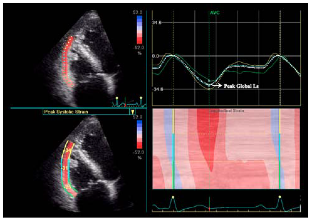
Figure 1.
Right ventricular longitudinal strain by two-dimensional speckle tracking in a healthy subject. Upper right: longitudinal strain dotted curve showing the main values. Ls = longitudinal strain.
RV longitudinal strain assessed by STE strain imaging is significantly impaired in patients with PH (Figure 2) and it is inversely correlated with systolic pulmonary artery pressure and RV dimensions [29,34,39,41,42].
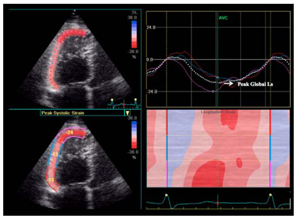
Figure 2.
Right ventricular longitudinal strain impairment in pulmonary hypertension patient. Ls = longitudinal strain.
RV longitudinal strain values demonstrated a significant prognostic role in PH which was incremental to clinical status, overcoming the other echocardiographic parameters [38,43,44]. RV longitudinal strain has been reported to be a predictor of cardiovascular events [43], all-cause mortality and complications [44]. Moreover, Fine et al. [44] demonstrated that abnormal RV strain is predictive of reduced survival, stratifying patients prognosis.
However, it is important to underline that the prognostic power of RV longitudinal strain has been demonstrated in patients with pre-capillary PH, but not in patients with post-capillary PH (group 2 according to Dana Point classification) [45]. The exclusion of group 2 from these studies is justified by the different hemodynamic pattern that characterize this pathological condition [44]. Highlighted in a recent study [46], pulmonary vascular resistance and compliance demonstrated a highly predictable inverse relationship with RV loading, which is similar across different forms of pre-capillary PH. In presence of post-capillary PH, as occurs in group 2, this relationship is altered and elevated capillary wedge pressure lowers pulmonary vascular compliance for any given resistance, augmenting RV pulsatile afterload. Accordingly, Haeck et al. [38] demonstrated, in a little cohort of group 2 PH patients, that in these patients RV longitudinal strain doesn’t confirm its prognostic value.
RV function is heavily load dependent and myocardial deformation is influenced by loading conditions, too. Accordingly, it has been demonstrated that in patients with RV pressure overload, both with and without loss of myocardial contractility, RV strain was significantly lower than in normal subjects [47]. These data show that RV strain can be decreased in presence of both increased afterload and normal contractility, and in presence of increased afterload and myocardial damage with decreased contractility [47], underlying the fact that the diagnostic significance of RV strain values must always be related to RV loading conditions [48,49]. However, the main determinant of RV strain impairment seems to be myocardial contractility. Indeed PH patients with RV dysfunction showed lower RV strain values in comparison to PH patients with preserved RV function [47]. Further evidences are needed to support the hypothesis that, considering its load-dependency, a mild reduction of RV strain alone should not be considered a marker of RV myocardial dysfunction in the PH setting, but a marked reduction of RV strain could be, underlying the need of cut-off values identification.
Not only the absolute value of RV strain is predictive of prognosis in PH patients, but also its variation during follow-up demonstrated a prognostic role. Patients with an improvement >5% of RV strain during follow-up demonstrated better survival respect to patients without [50].
Recently, Atsumi et al. [51] studied, for the first time, the application of 3D-STE to assess RV myocardial deformation. They have been able to demonstrate a good feasibility (around 80%) of the technique (feasibility close to that of 2D-STE). In comparison to 2D-STE, 3D-STE allowed to assess not only the limited part of the RV free-wall encompassed in the 2DE four-chamber view but the whole RV free wall, and to confirm the significant heterogeneity of RV contraction. Moreover, 3D-STE explores simultaneously the longitudinal and circumferential deformation of the myocardium, that are expression of the function of myocardial fibers positioned in different layers of the RV wall. The Authors found that, in presence of conditions affecting the RV, such as arrrhytmogenic RV cardiomyopathy and PH, longitudinal function was significantly reduced, whereas circumferential function was relatively preserved [51]. These findings confirm previous data showing that the motion of the base towards the apex is the first function to be impaired [28]. The importance of assessing RV radial function has been demonstrated by Di Salvo et al. [52], who found a strong correlation between the extent of radial function and exercise tolerance in RV pressure overload condition, such as in patients with transposition of great arteries who underwent atrial-switch repair.
Despite all these data, the use of STE to assess RV myocardial function is not widespread because the feasibility of STE is suboptimal in the thin RV wall (better in hypertrophied RV as seen in patients with PH). Reference values for different age groups, body size, and gender have not been established yet, and standardization among the different software packages available on the market is still a pending issue [17].
3.2. Three-Dimensional Echocardiography
Three-dimensional echocardiography (Figure 3; Videos 1–2) has been demonstrated to be feasible, accurate and reproducible in measuring RV volumes and ejection fraction in adults [53,54,55,56,57,58,59,60,61], as well as in children [62,63]. Recent studies demonstrated a close correlation between 3D-echocardiography and CMR in measuring RV volumes and ejection fraction [61,64,65] (Table 1). However, 3D echocardiography appeared to underestimate RV volumes in comparison to CMR [65]. Anyway this underestimation is systematic [58], confirming the good agreement between the two techniques and the accuracy of 3D echo. 3D echocardiography confirmed the correlation between RV volumes and gender, age, and body size, previously demonstrated by CMR [58] (Figure 4).
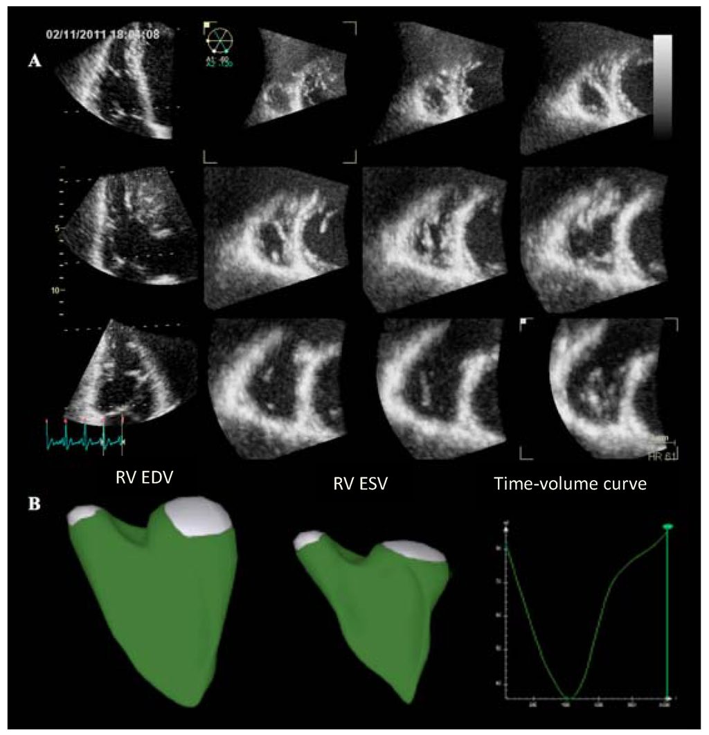
Figure 3.
Three-dimensional echocardiographic visualization of the right ventricle in a healthy subject. (A) Multislice view showing three apical views and corresponding short axis. (B) Right ventricular beutel in end-diastole and end-systole and the corresponding time-volume curve. EDV = end-diastolic volume; ESV = end-systolic volume; RV = right ventricle.

Table 1.
Differences between right ventricular volumes assessed by three-dimensional echocardiography and cardiac magnetic resonance (Modified from: Badano et al. [61]).
| Author | Three-dimensional echocardiography | ||
|---|---|---|---|
| End-diastolic volume (mL) | End-systolic volume (mL) | Ejection fraction (%) | |
| Grapsa et al. | −4 (−11, 4) | 0 (−6, 6) | −1 (−3, 0) |
| Sugeng et al. | −14 (−28, 0) | −9 (−19, 1) | −2 (−4, 0) |
| van der Zwaan et al. | −34 (−43, −25) | −11 (−19, 3) | −4 (−6, −2) |
| Leibundgut et al. [66] | −10 (−15, −6) | −5 (−8, −1) | 0 (−2, 1) |
| Shimada et al. [67] | −14 (−18, −10) | −6 (−8, −3) | −1 (−2, 0) |
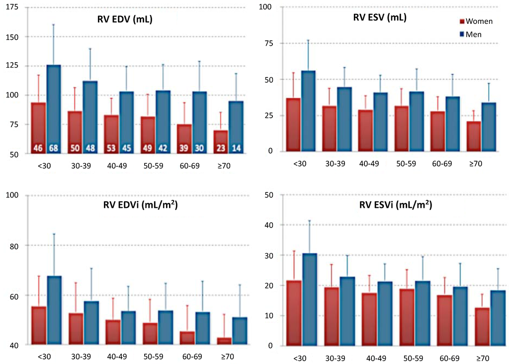
Figure 4.
Right ventricular volumes varies with gender and age, unrespect to body size. (Modified from Maffesanti et al. [58]). EDV = end-diastolic volume; ESV = end-systolic volume; RV = right ventricle.
Accordingly, Morikawa et al. [68] proved a strong correlation between ≥3D echocardiography and CMR measurements of the RV volumes and ejection fraction in patients with PH. Similar results were reported in a population of patients with congenital heart disease [69].
The presence of synus rhytm and the patient’s compliance for breath-holding are necessary requirements for obtaining a good quality 3D echo assessment with the multi-beat technique. However, recently Zhang et al. [70] demonstrated that the 3D-echocardiography assessment of RV geometry by a single-beat acquisition is as accurate as with multi-beat acquisition when compared with CMR. Nevertheless, the feasibility of obtaining a good quality full volume 3D data set acquisition which includes the RV anterior and apical lateral segments as well as the RV outflow tract, in patients with poor imaging windows and/or dilated RV, remains the main limitation of the technique. Accuracy tends to decrease with increasing RV size, limiting its application in patients with more dilated RV [69].
Using 3D echocardiography, it has been confirmed that in normal people the three compartments of the right ventricle provides a different contribution in RV systolic contraction, both in timing as in strength [71,72]. Inflow and outflow tracts are the most active RV compartments contributing to its pump function, whereas the apex contributes less, following a chronological order in contraction, that reflects the peristaltic pump function of the RV [71]. In pts with PH, the relative contribution of the three compartments to RV pump function seems to remain unchanged, with the loss of the timing differences. The three compartments contract simultaneously loosing the peristaltic function and making the RV behave as a single chamber. This change is coupled by changes of the RV shape, from a triangular to a cilindrical one [71].
These findings underline once more the importance of a comprehensive assessment of RV function, that could not be adequately reflected by assessing a single parameter, such as TAPSE. Indeed, the latter is representative only of the inflow tract displacement, with the consequently risk to underestimate the whole RV pump function.
Three-dimensional echocardiography demonstrated to be superior in comparison to conventional echocardiography in identifying RV dysfunction in patients with PH [73] (Figure 5; video 3). Recently, RV function assessed by 3D echocardiography has been reported to be prognostically important in patients with acute pulmonary embolism [74].
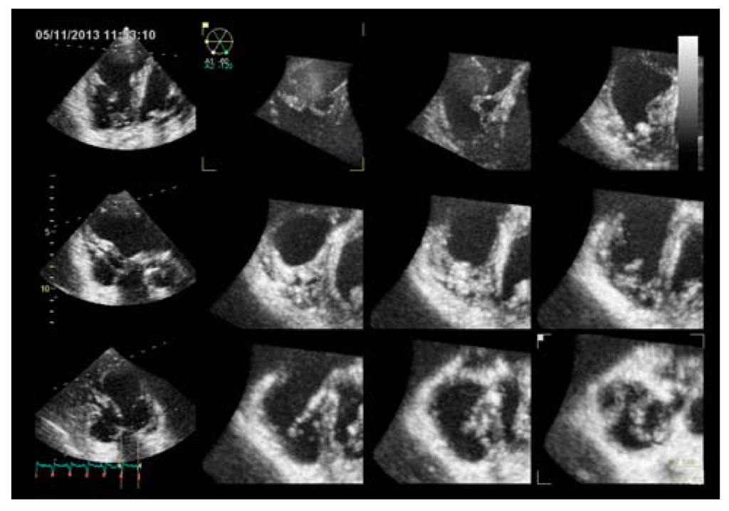
Figure 5.
Three-dimensional echocardiography of the right ventricle in a pulmonary hypertension patient, showing three apical views and corresponding short axis.
4. Cardiac Magnetic Resonance
CMR is used to define the anatomy, to assess biventricular function and haemodynamics, and to measure blood-flow by using phase contrast technique. Compared to echocardiography, CMR has higher spatial resolution, lower temporal resolution and does not suffer from the limitations about the acoustic window [75], allowing calculation of RV volumes and ejection fraction using the Simpson’s approach. CMR-derived RV volumes have shown good correlation with in vivo standards [76]. Moreover, this technique demonstrated good accuracy, when compared to the directly measured cardiac mass in calf hearts [77], with excellent intra- and inter-observer variability [78] and good interstudy reproducibility [79] in RV volumes, mass, and function quantification. At this time, CMR is regarded as the reference standard for the assessment of RV volumes (Figure 6) and ejection fraction [17,80]. Recent data obtained by CMR have demonstrated that normal values of RV systolic and diastolic parameters vary significantly by gender, body size, and age [81,82].
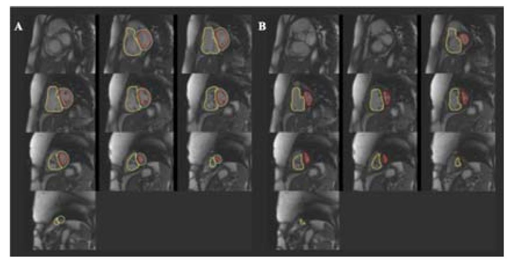
Figure 6.
Right ventricular volumes obtained with cardiac magnetic resonance in a patient affected by pulmonary hypertension. (A) End-diastolic volume; (B) End-systolic volume.
In PH, RV end-diastolic volume [3] and ejection fraction [14] measured by CMR have demonstrated prognostic power. In particular, RV end-diastolic volume index ≤84 mL/m2, left ventricular end-diastolic volume index ≥40 mL/m2, and a stroke volume index ≥25 mL/m2 were associated with better survival in patients with idiopathic PH [3]. RV ejection fraction has also been assessed by CMR in PH patients and a value <35% predicts increased mortality [14]. Moreover, the RV mass demonstrated a diagnostic and prognostic role in PH patients, too [3,83,84]. An open issue about quantification of RV mass by CMR is whether or not the trabeculae and papillary muscles should be included in RV mass measurements. Previous studies have variably included and excluded them [79,85,86,87,88,89,90]. The reason why this issue is controversial is that RV is highly trabeculated in normal subjects and even more in PH patients, but we do not know if hypertrophy involves all the mass components in the same extent. Recently, van de Veerdonk et al. [91] demonstrated that in PH population trabeculae and papillary muscles give a large contribution (about 35%) to the RV mass. Moreover, the trabeculae and papillary muscle mass seem to be related to changes in pulmonary pressure, closer than the RV free wall mass. According to van de Veerdonk [91], Driessen et al. [92] have recently showed that inclusion or exclusion of trabeculae has an important impact on the RV volumes and mass measurements, particularly in patients with overloaded RVs. Then, these findings highlights two main aspects: (i) in presence of RV pressure overload RV hypertrophy is more prominent in the trabeculae and papillary components, than in the compact free wall; consequently, (ii) exclusion or inclusion of trabeculae and papillary muscles measurements significantly affect RV volumes and mass measurements.
The presence of late gadoliniun enhancement (LGE) pattern in the RV myocardium of patients with PH has been first described by McCann and colleagues in 2005 [93]. LGE imaging shows a characteristic retention of gadolinium at the septal insertion (junctional pattern) [94,95,96], without any retention in the free wall [94] (Figure 7). Looking for LGE deposition in RV free wall of normal subjects or in pathologies like arrhytmogenic RV cardiomyopathy is difficult, because of partial volume effects due to the fact that the RV wall is particularly thin. Conversely, PH patients show thick RV walls, which does not pose any difficulty in nulling the RV myocardium. Further, LGE junctional pattern is not specific for PH, but it has been found in other pathologies like hypertrophic cardiomyopathy [97,98]. The etiology of this gadolinium retention remains to be clarified. The first hypothesis was the presence of fibrosis. Bradlow et al. [99] examined the heart of a patient with idiopathic PH and LGE junctional pattern at autopsy. They found increased collagen and fat between fiber bundles (plexiform fibrosis) consistent with myocardial disarray, but no pathological fibrosis. Myocardial disarray is common in healthy people at the interventricular junction site, but it appears particularly exaggerated in PH patients. A recent prospective observational study [100] demonstrated a moderate correlation among the presence of LGE junctional pattern and paradoxical septal motion, assessed by echocardiography, associated to a weaker correlation with RV function parameters. This data suggest that the abnormal interventricular septal motion, rather than the elevated RV pressure and the consequent remodeling may be the mechanism underlying the appearance of LGE junctional pattern. The reported occurrence of LGE junctional pattern in PH is not univocal, it ranges from 69% [101] to 100% [94]. The amount of the LGE junctional pattern has been demonstrated to be moderately related to the amount of RV dysfunction measured as ejection fraction, stroke volume and end-systolic volume [94] and it appears to predict RV remodeling in response to increased afterload [102]. Moreover, patients with the junctional pattern of the LGE are significantly more likely to experience a worse clinical outcome than those without this marker. LGE junctional pattern has been reported to be related to worse outcome in term of death, need of lung transplantation, initiation of prostacyclin therapy, and decompensated RV heart failure, stratifying prognosis [101].
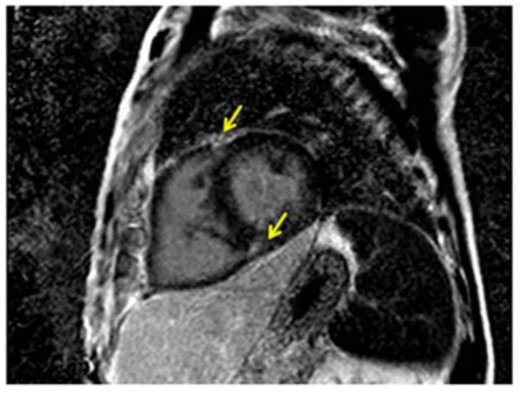
Figure 7.
CMR imaging of a PH patient showing the typical septal junctional pattern of late gadolinium enhancement (yellow arrows).
Swift et al. [66] showed that changes in RV morphology assessed by CMR are able to identify PH patients quite accurately. Particularly, presence of LGE junctional pattern, RV mass index, retrograde flow and pulmonary artery relative area change predicted the presence of PH with a positive predictive value of 98%, 97%, 95%, and 94%, respectively [66]. More recently, the same research group demonstrated that CMR imaging, in addition to provide detailed functional and morphological information about the RV, can accurately estimate mean pulmonary artery pressure in patients with PH and calculate pulmonary vascular resistance by estimating all major pulmonary hemodynamic metrics measured at right heart catheterization (RHC) [67]. If these findings would be confirmed in future studies and CMR will prove to be also accurate in estimating the changes in PVR/PAP over time, the follow-up of PH patients could be managed with CMR, reducing the need of serial RHC studies.
Although CMR seems to hold promise for initial assessment and follow-up of patients with PH, data are currently limited. In addition, CMR is costly, is not widely available for serial follow-up of patients with PH and cannot be performed in patients with several conditions (severe renal disease, implanted methallic devices, claustrophobia, irregular heart rhythm). Therefore, at present, its main role currently consists in providing baseline assessment in patients in whom echo cannot be performed or it is not exhaustive [103].
5. Multi Detector Cardiac Tomography
Several authors have validated the use of multi detector cardiac tomography (MDCT), in comparison with echocardiography, scintigraphic techniques, and CMR, for RV function assessment [104,105,106]. Although MDCT cannot be regarded as a first-line imaging technique because of the radiation dose and iodinated contrast agent injection involved, MDCT evaluation of RV function is accurate and provides information on the adjacent lung parenchyma [107]. Recent improvements in temporal and spatial resolution of the technique have allowed to obtain useful information about the heart [107]. However, the use of MDCT in the setting of PH has been mainly validated for the detection of acute pulmonary embolism and for the initial evaluation of the patient with PH in order to assess the presence of chronic pulmonary embolism or lung parenchima disease. Reference values for RV volumes and ejection fraction measured by MDCT have been recently published [108]. Compared with CMR, MDCT has lower temporal resolution and tends to overestimate end-systolic and end-diastolic volumes [109].
6. Nuclear Imaging
Radionuclide techniques have historically been the first imaging modalities used for assessing RV size and function [110], although they have widely been replaced by CMR and echocardiography. First pass planar equilibrium radionuclide angiocardiography (FPRNA) is currently the technique of choice because it allows temporal separation of structures that are spatially superimposed [110,111]. However it has many limitations. Gated blood-pool single photon emission computed tomography (SPECT) offers adequate 3D resolution of the cardiac chambers, without the need for multiple acquisition views; then it is the currently recommended nuclear modality for quantifying RV function [110,111,112]. Unfortunately, the absence of completely automated and clinically validated processing software limits the utilization of this promising technique for RV evaluation. Recently, Anderson and colleagues [113] demonstrated significant correlations of RV volumes and function evaluated by gated blood-pool SPECT with CMR. Further studies for validation of automatic measurement algorithms are still pending. Radionuclide techniques are of additional particular interest for assessing myocardial metabolism and perfusion. Experimental studies using SPECT have shown that acute or chronic RV pressure overload leads to a myocardial metabolic shift from fatty acid to glucose [114].
In the setting of RV evaluation in PH patients, nuclear imaging and MDCT do not have a major role. Their principal applications are in patients with controindications to CMR and/or inconclusive echocardiographic exams. Conversely, CMR and echocardiography, the latter with the new techniques STE and 3D echocardiography, are promising tools. They need to be tested in multicenter longitudinal studies to have their prognostic power recognized for clinical practice [103]. In Table 2, advantages and disadvantages of the various imaging modalities are briefly summarized.

Table 2.
Advantages and disadvantages of the different imaging modalities in right ventricle (RV) evaluation in the pulmonary hypertension (PH) setting.
| Advantages | Disadvantages | |
|---|---|---|
| Echocardiography | widely available; easily repeatable; independent from cardiac rhythm; complete and accurate RV function assessment by 3D echo and STE; no exposure to radiation | operator-dependent; acoustic window-dependent |
| Cardiac Magnetic Resonance | I independent from acoustic window; no exposure to radiation; quantification of RV mass; evaluation of myocardial tissue by Late Gadolinium Enhancement | presence of contraindications such as arrhytmias, claustrophobia, end-stage renal disease, implanted methallic devices |
| Multi Detector Computed Tomography | independent from acoustic window; provides information on the adjacent lung parenchyma | exposure to radiation; use of iodinated contrast agent injection |
| Nuclear Imaging | independent from acoustic window; opportunity to assess myocardial perfusion and metabolism. | exposure to radiation; lack of validated automatic measurements algorithms |
7. Conclusions
Right ventricular function has been recognized as the main predictor of prognosis in patients with pulmonary hypertension. According to current guidelines, prognostic parameters are obtained from right heart catheterization, or from assessment of functional capacity either by WHO functional class or six-minute walking test. In this scenario, cardiac magnetic resonance and echocardiography have demonstrated to be able to assess right ventricular function in an accurate and reproducible manner, both using with conventional and new echocardiographic techniques. However, these new parameters still need further validation to be introduced in the clinical routine.
Abbreviations
| 2D | two-dimensional |
| 3D | three-dimensional |
| CMR | cardiac magnetic resonance |
| FPRNA | first pass planar equilibrium radionuclide angiocardiography |
| LGE | late gadolinium enhancement |
| MDCT | multi detector cardiac tomography |
| PH | pulmonary hypertension |
| RHC | right heart catheterization |
| RV | right ventricle/ventricular |
| SPECT | single photon emission computed tomography |
| STE | speckle tracking echocardiography |
| TAPSE | tricuspid annular plane systolic excursion |
Author Contributions
All authors contributed to the submitted work. Particularly, DP and LPB have written the review; DM and UC revised the echocardiographic section; FT and MPM revised the cardiac magnetic resonance section; GR and SI revised the multi detector cardiac tomography and nuclear imaging section.
Conflicts of Interest
The authors declare no conflict of interest.
References
- D’Alonzo, G.E.; Barst, R.J.; Ayres, S.M.; Bergofsky, E.H.; Brundage, B.H.; Detre, K.M.; Fishman, A.P.; Goldring, R.M.; Groves, B.M.; Kernis, J.T.; et al. Survival in patients with primary pulmonary hypertension. Results from a national prospective registry. Ann. Intern. Med. 1991, 115, 343–349. [Google Scholar]
- Humbert, M.; Sitbon, O.; Yaïci, A.; Montani, D.; O’Callaghan, D.S.; Jaïs, X.; Parent, F.; Savale, L.; Natali, D.; Günther, S.; et al. Survival in incident and prevalent cohorts of patients with pulmonary arterial hypertension. Eur. Respir. J. 2010, 36, 549–555. [Google Scholar]
- van Wolferen, S.A.; Marcus, J.T.; Boonstra, A.; Marques, K.M.J.; Spreeuwenberg, M.D.; Postmus, P.E.; Bronzwaer, J.G.F.; Vonk-Noordegraaf, A. Prognostic value of right ventricular mass, volume, and function in idiopathic pulmonary arterial hypertension. Eur. Heart J. 2007, 28, 1250–1257. [Google Scholar]
- Zafrir, N.; Zingerman, B.; Solodky, A.; Ben-Dayan, D.; Sagie, A.; Sulkes, J.; Mats, I.; Kramer, M.R. Use of noninvasive tools in primary pulmonary hypertension to assess the correlation of right ventricular function with functional capacity and to predict outcome. Int. J. Cardiovasc. Imaging 2007, 23, 209–215. [Google Scholar] [CrossRef] [PubMed]
- Kawut, S.M.; Horn, E.M.; Berekashvili, K.K.; Garofano, R.P.; Goldsmith, R.L.; Widlitz, A.C.; Rosenzweig, E.B.; Kerstein, D.; Barst, R.J. New predictors of outcome in idiopathic pulmonary arterial hypertension. Am. J. Cardiol. 2005, 95, 199–203. [Google Scholar] [CrossRef] [PubMed]
- Voelkel, N.F.; Quaife, R.A.; Leinwand, L.A.; Barst, R.J.; McGoon, M.D.; Meldrum, D.R.; Dupuis, J.; Long, C.S.; Rubin, L.J.; Smart, F.W.; et al. Report of a National Heart, Lung, and Blood Institute working group on cellular and molecular mechanisms of right heart failure. Circulation 2006, 114, 1883–1891. [Google Scholar] [CrossRef] [PubMed]
- Raymond, R.J.; Hinderliter, A.L.; Willis, P.W.; Ralph, D.; Caldwell, E.J.; Williams, W.; Ettinger, N.A.; Hill, N.S.; Summer, W.R.; de Boisblanc, B.; et al. Echocardiographic predictors of adverse outcomes in primary pulmonary hypertension. J. Am. Coll. Cardiol. 2002, 39, 1214–1219. [Google Scholar] [CrossRef] [PubMed]
- Ghio, S.; Klersy, C.; Magrini, G.; D'Armini, A.M.; Scelsi, L.; Raineri, C.; Pasotti, M.; Serio, A.; Campana, C.; Vigano, M. Prognostic relevance of the echocardiographic assessment of right ventricular function in patients with idiopathic pulmonary arterial hypertension. Int. J. Cardiol. 2010, 140, 272–278. [Google Scholar] [CrossRef] [PubMed]
- Badesch, D.B.; Champion, H.C.; Sanchez, M.A.; Hoeper, M.M.; Loyd, J.E.; Manes, A.; McGoon, M.; Naeije, R.; Olschewski, H.; Oudiz, R.J.; et al. Diagnosis and assessment of pulmonary arterial hypertension. J. Am. Coll. Cardiol. 2009, 54 (Suppl. 1), S55–S66. [Google Scholar]
- Thenappan, T.; Shah, S.J.; Rich, S.; Tian, L.; Archer, S.L.; Gomberg-Maitland, M. Survival in pulmonary arterial hypertension: A reappraisal of the NIH risk stratification equation. Eur. Respir. J. 2010, 35, 1079–1087. [Google Scholar] [CrossRef] [PubMed]
- Farber, H.W.; Loscalzo, J. Pulmonary arterial hypertension. N. Engl. J. Med. 2004, 351, 1655–1665. [Google Scholar] [CrossRef] [PubMed]
- McLaughlin, V.V.; Presberg, K.W.; Doyle, R.L.; Abman, S.H.; McCrory, D.C.; Fortin, T.; Ahearn, G. Prognosis of pulmonary arterial hypertension: Accp evidence-based clinical practice guidelines. Chest 2004, 126, 78S–92S. [Google Scholar] [CrossRef] [PubMed]
- Burgess, M.I.; Mogulkoc, N.; Bright-Thomas, R.J.; Bishop, P.; Egan, J.J.; Ray, S.G. Comparison of echocardiographic markers of right ventricular function in determining prognosis in chronic pulmonary disease. J. Am. Soc. Echocardiogr. 2002, 15, 633–639. [Google Scholar] [CrossRef] [PubMed]
- van de Veerdonk, M.C.; Kind, T.; Marcus, J.T.; Mauritz, G.-J.; Heymans, M.W.; Bogaard, H.-J.; Boonstra, A.; Marques, K.M.J.; Westerhof, N.; Vonk-Noordegraaf, A. Progressive right ventricular dysfunction in patients with pulmonary arterial hypertension responding to therapy. J. Am. Coll. Cardiol. 2011, 58, 2511–2519. [Google Scholar]
- Badano, L.P.; Ginghina, C.; Easaw, J.; Muraru, D.; Grillo, M.T.; Lancellotti, P.; Pinamonti, B.; Coghlan, G.; Perazzolo Marra, M.; Popescu, B.A.; et al. Right ventricle in pulmonary hypertension: haemodynamics, structural changes, imaging, and proposal of a study protocol aimed to assess remodelling and treatment effects. Eur. J. Echoc. 2010, 11, 27–37. [Google Scholar] [CrossRef]
- Ho, S.Y.; Nihoyannopoulos, P. Anatomy, echocardiography, and normal right ventricular dimensions. Heart 2006, 92 (Suppl. 1), i2–i13. [Google Scholar]
- Valsangiacomo Buechel, E.R.; Mertens, L.M. Imaging the right heart: The use of integrated multimodality imaging. Eur. Heart J. 2012, 33, 949–960. [Google Scholar]
- Kukulski, T.; Hübbert, L.; Arnold, M.; Wranne, B.; Hatle, L.; Sutherland, G.R. Normal regional right ventricular function and its change with age: A Doppler myocardial imaging study. J. Am. Soc. Echocardiogr. 2000, 13, 194–204. [Google Scholar] [CrossRef] [PubMed]
- Haber, I.; Metaxas, D.N.; Geva, T.; Axel, L. Three-dimensional systolic kinematics of the right ventricle. Am. J. Physiol. Heart Circ. Physiol. 2005, 289, H1826–H1833. [Google Scholar] [CrossRef] [PubMed]
- Selton-Suty, C.; Juilliere, Y. Non-invasive investigations of the right heart: How and why? Arch. Cardiovasc. Dis. 2009, 102, 219–232. [Google Scholar]
- Geva, T.; Powell, A.J.; Crawford, E.C.; Chung, T.; Colan, S.D. Evaluation of regional differences in right ventricular systolic function by acoustic quantification echocardiography and cine magnetic resonance imaging. Circulation 1998, 98, 339–345. [Google Scholar] [CrossRef] [PubMed]
- McConnell, M.V.; Solomon, S.D.; Rayan, M.E.; Come, P.C.; Goldhaber, M.S.Z.; Lee, R.T. Regional right ventricular dysfunction detected by echocardiography in acute pulmonary embolism. Am. J. Cardiol. 1996, 78, 469–473. [Google Scholar] [CrossRef] [PubMed]
- Sanchez-Quintana, D.; Anderson, R.H.; Ho, S.Y. Ventricular myoarchitecture in tetralogy of Fallot. Heart 1996, 76, 280–286. [Google Scholar]
- Pettersen, E.; Helle-Valle, T.; Edvardsen, T.; Lindberg, H.; Smith, H.J.; Smevik, B.; Smiseth, O.A.; Andersen, K. Contraction pattern of the systemic right ventricle shift from longitudinal to circumferential shortening and absent global ventricular torsion. J. Am. Coll. Cardiol. 2007, 49, 2450–2456. [Google Scholar] [CrossRef] [PubMed]
- Forfia, P.R.; Fisher, M.R.; Mathai, S.C.; Housten-Harris, T.; Hemnes, A.R.; Borlaug, B.A.; Chamera, E.; Corretti, M.C.; Champion, H.C.; Abraham, T.P.; et al. Tricuspid annular displacement predicts survival in pulmonary hypertension. Am. J. Respir. Crit. Care Med. 2006, 174, 1034–1041. [Google Scholar] [CrossRef] [PubMed]
- Rushmer, R.F.; Crystal, D.K.; Wagner, C. The functional anatomy of ventricular contraction. Circ. Res. 1953, 1, 162–170. [Google Scholar] [CrossRef] [PubMed]
- Kind, T.; Mauritz, G.J.; Marcus, J.T.; van de Veerdonk, M.; Westerhof, N.; Vonk-Noordegraaf, A. Right ventricular ejection fraction is better reflected by transverse rather than longitudinal wall motion in pulmonary hypertension. J. Cardiovasc. Magn. Res. 2010, 12. [Google Scholar] [CrossRef]
- Mauritz, G.-J.; Kind, T.; Marcus, J.T.; Bogaard, H.-J.; van de Veerdonk, M.; Postmus, P.E.; Boonstra, A.; Westerhof, N.; Vonk-Noordegraaf, A. Progressive changes in right ventricular geometric shortening and long-term survival in pulmonary arterial hypertension. Chest 2012, 141, 935–943. [Google Scholar] [CrossRef] [PubMed]
- Giusca, S.; Dambrauskaite, V.; Scheurwegs, C.; D’hooge, J.; Claus, P.; Herbots, L.; Magro, M.; Rademakers, F.; Meyns, B.; Delcroix, M.; et al. Deformation imaging describes right ventricular function better than longitudinal displacement of the tricuspid ring. Heart 2010, 96, 281–288. [Google Scholar] [CrossRef] [PubMed]
- Teske, A.J.; De Boeck, B.W.; Olimulder, M.; Prakken, N.H.; Doevendans, P.A.; Cramer, M.J. Echocardiographic assessment of regional right ventricular function: A head-to-head comparison between 2-dimensional and tissue Doppler-derived strain analysis. J. Am. Soc. Echocardiogr. 2008, 21, 275–283. [Google Scholar] [CrossRef] [PubMed]
- Scherptong, R.W.; Mollema, S.A.; Blom, N.A.; Kroft, L.J.; de Roos, A.; Vliegen, H.W.; van der Wall, E.E.; Bax, J.J.; Holman, E.R. Right ventricular peak systolic longitudinal strain is a sensitive marker for right ventricular deterioration in adult patients with tetralogy of Fallot. Int. J. Cardiovasc. Imaging 2009, 25, 669–676. [Google Scholar] [CrossRef] [PubMed]
- Dragulescu, A.; Mertens, L.L. Developments in echocardiographic techniques for the evaluation of ventricular function in children. Arch. Cardiovasc. Dis. 2010, 103, 603–614. [Google Scholar] [CrossRef] [PubMed]
- Mertens, L.L.; Friedberg, M.K. Imaging the right ventricle—Current state of the art. Nat. Rev. Cardiol. 2010, 7, 551–563. [Google Scholar] [CrossRef] [PubMed]
- Pirat, B.; McCulloch, M.L.; Zoghbi, W.A. Evaluation of global and regional right ventricular systolic function in patients with pulmonary hypertension using a novel speckle tracking method. Am. J. Cardiol. 2006, 98, 699–704. [Google Scholar] [CrossRef] [PubMed]
- Fukuda, Y.; Tanaka, H.; Sugiyama, D.; Ryo, K.; Onishi, T.; Fukuya, H.; Nogami, M.; Ohno, Y.; Emoto, N.; Kawai, H.; et al. Utility of right ventricular free wall speckle-tracking strain for evaluation of right ventricular performance in patients with pulmonary hypertension. J. Am. Soc. Echocardiogr. 2011, 24, 1101–1108. [Google Scholar] [CrossRef] [PubMed]
- Sachdev, A.; Villarraga, H.R.; Frantz, R.P.; McGoon, M.D.; Hsiao, J.F.; Maalouf, J.F.; Ammash, N.M.; McCully, R.B.; Miller, F.A.; Pellikka, P.A.; et al. Right ventricular strain for prediction of survival in patients with pulmonary arterial hypertension. Chest 2011, 139, 1299–1309. [Google Scholar] [CrossRef] [PubMed]
- Haeck, M.L.; Scherptong, R.W.; Antoni, M.L.; Marsan, N.A.; Vliegen, H.W.; Holman, E.R.; Schalij, M.J.; Bax, J.J.; Delgado, V. Right ventricular longitudinal peak systolic strain measurements from the subcostal view in patients with suspected pulmonary hypertension: A feasibility study. J. Am. Soc. Echocardiogr. 2012, 25, 674–681. [Google Scholar] [CrossRef] [PubMed]
- Haeck, M.L.; Scherptong, R.W.; Ajmone Marsan, N.; Holman, E.R.; Schalij, M.J.; Bax, J.J.; Vliegen, H.W.; Delgado, V. Prognostic value of right ventricular longitudinal peak systolic strain in patients with pulmonary hypertension. Circ. Cardiovasc. Imaging 2012, 5, 628–636. [Google Scholar] [CrossRef] [PubMed]
- Meris, A.; Faletra, F.; Conca, C.; Klersy, C.; Regoli, F.; Klimusina, J.; Penco, M.; Pasotti, E.; Pedrazzini, G.B.; Moccetti, T.; et al. Timing and Magnitude of Regional Right Ventricular Function: A Speckle Tracking-Derived Strain Study of Normal Subjects and Patients with Right Ventricular Dysfunction. J. Am. Soc. Echocardiogr. 2010, 23, 823–831. [Google Scholar] [CrossRef] [PubMed]
- Leong, D.P.; Grover, S.; Molaee, P.; Chakrabarty, A.; Shirazi, M.; Cheng, Y.H.; Penhall, A.; Perry, R.; Greville, H.; Joseph, M.X.; et al. Nonvolumetric Echocardiographic Indices of Right Ventricular Systolic Function: Validation with Cardiovascular Magnetic Resonance and Relationship with Functional Capacity. Echocardiography 2012, 29, 455–463. [Google Scholar] [CrossRef] [PubMed]
- Puwanant, S.; Park, M.; Popovic, Z.B.; Tang, W.H.; Farha, S.; George, D.; Sharp, J.; Puntawangkoon, J.; Loyd, J.E.; Erzurum, S.C.; et al. Ventricular geometry, strain, and rotational mechanics in pulmonary hypertension. Circulation 2010, 121, 259–266. [Google Scholar] [CrossRef] [PubMed]
- Dambrauskaite, V.; Delcroix, M.; Claus, P.; Herbots, L.; D’Hooge, J.; Bijnens, B.; Rademakers, F.; Sutherland, G.R. Regional right ventricular dysfunction in chronic pulmonary hypertension. J. Am. Soc. Echocardiogr. 2007, 20, 1172–1180. [Google Scholar] [CrossRef] [PubMed]
- Motoji, Y.; Tanaka, H.; Fukuda, Y.; Ryo, K.; Emoto, N.; Kawai, H.; Hirata, K. Efficacy of right ventricular free-wall longitudinal speckle-tracking strain for predicting long-term outcome in patients with pulmonary hypertension. Circ. J. 2013, 77, 756–763. [Google Scholar] [CrossRef] [PubMed]
- Fine, N.M.; Chen, L.; Bastiansen, P.M.; Frantz, R.P.; Pellikka, P.; Oh, J.K.; Kane, G.C. Outcome prediction by quantitative right ventricular function assessment in 575 subjects evaluated for pulmonary hypertension. Circ. Cardiovasc. Imag. 2013, 6, 711–721. [Google Scholar] [CrossRef]
- Galiè, N.; Hoeper, M.M.; Humbert, M.; Torbicki, A.; Vachiery, J.-L.; Barbera, J.A.; Beghetti, M.; Corris, P.; Gaine, S.; Gibbs, J.S.; et al. Guidelines for the diagnosis and treatment of pulmonary hypertension. Eur. Heart J. 2009, 30, 2493–2537. [Google Scholar]
- Tedford, R.J.; Hassoun, P.M.; Mathai, S.C.; Girgis, R.E.; Russell, S.D.; Thiemann, D.R.; Cingolani, O.H.; Mudd, J.O.; Borlaug, B.A.; Redfield, M.M.; et al. Pulmonary capillary wedge pressure augments right ventricular pulsatile loading. Circulation 2012, 125, 289–297. [Google Scholar] [CrossRef] [PubMed]
- Simon, M.A.; Rajagopalan, N.; Mathier, M.A.; Shroff, S.G.; Pinsky, M.R.; López-Candales, A. Tissue Doppler imaging of right ventricular decompensation in pulmonary hypertension. Congest. Heart Fail. 2009, 15, 271–276. [Google Scholar] [CrossRef] [PubMed]
- Reichek, N. Right ventricular strain in pulmonary hypertension. Flavor du jour enduring prognostic index? Circul. Cardiovasc. Imag. 2013, 6, 611–613. [Google Scholar]
- La Gerche, A.; Ruxandra, J.; Voigt, J.-U. Right ventricular function by strain echocardiography. Curr. Opin. Cardiol. 2010, 25, 430–436. [Google Scholar]
- Hardegree, E.L.; Sachdev, A.; Villarraga, H.R.; Frantz, R.P.; McCoon, M.D.; Kushwaha, S.S.; Hsiao, J.-F.; McCully, R.B.; Oh, J.K.; Pellikka, P.A.; et al. Role of serial quantitative assessment of right ventricular function by strain in pulmonary arterial hypertension. Am. J. Cardiol. 2013, 111, 143–148. [Google Scholar] [CrossRef] [PubMed]
- Atsumi, A.; Ishizu, T.; Kameda, Y.; Yamamoto, M.; Harimura, Y.; Machino-Ohtsuka, T.; Kawamura, R.; Enomoto, M.; Seo, Y.; Aonuma, K. Application of 3-dimensional speckle tracking imaging to the assessment of right ventricular regional deformation. Circul. J. 2013, 77, 1760–1768. [Google Scholar] [CrossRef]
- Di Salvo, G.; Pacileo, G.; Rea, A.; Limongelli, G.; Baldini, L.; D’Andrea, A.; D’Alto, M.; Sarubbi, B.; Russo, M.G.; Calabrò, R. Transverse strain predicts exercise capacity in systemic right ventricle patients. Int. J. Cardiol. 2010, 145, 193–196. [Google Scholar]
- Schindera, S.T.; Mehwald, P.S.; Sahn, D.J.; Kececioglu, D. Accuracy of real time three-dimensional echocardiography for quantifying right ventricular volume. J. Ultrasound Med. 2002, 21, 1069–1075. [Google Scholar] [PubMed]
- Nesser, H.J.; Tkalec, W.; Patel, A.R.; Masani, N.D.; Niel, J.; Markt, B.; Pandian, N.G. Quantitation of right ventricular volumes and ejection fraction by three-dimensional echocardiography in patients: Comparison with magnetic resonance imaging and radionuclide ventriculography. Echocardiography 2006, 23, 666–680. [Google Scholar] [CrossRef] [PubMed]
- Chen, G.; Sun, K.; Huang, G. In vitro validation of right ventricular volume and mass measurement by real- time three dimensional echocardiography. Echocardiography 2006, 23, 395–399. [Google Scholar] [CrossRef] [PubMed]
- Angelini, E.D.; Homma, S.; Pearson, G.; Holmes, J.W.; Laine, A.F. Segmentation of real time three dimensional ultrasound for quantification of ventricular function: A clinical study on right and left ventricles. Ultrasound Med. Biol. 2005, 31, 1143–1158. [Google Scholar] [CrossRef] [PubMed]
- Jenkins, C.; Chan, J.; Bricknell, K.; Strudwick, M.; Marwick, T.H. Reproducibility of right ventricular volumes and ejection fraction using real-time three-dimensional echocardiography: comparison with cardiac MRI. Chest 2007, 131, 1844–1851. [Google Scholar] [CrossRef] [PubMed]
- Maffesanti, F.; Muraru, D.; Esposito, R.; Gripari, P.; Ermacora, D.; Santoro, C.; Tamborini, G.; Galderisi, M.; Pepi, M.; Badano, L.P. Age-, body size-, and sex-specific reference values for right ventricular volumes and ejection fraction by three-dimensional echocardiography. Circ. Cardiovasc. Imag 2013, 6, 700–710. [Google Scholar] [CrossRef]
- Chua, S.; Levine, R.A.; Yosefy, C.; Handschumacher, M.D.; Chu, J.; Qureshi, A.; Neary, J.; Ton-Nu, T.-T.; Fu, M.; Jen Wu, C.; et al. Assessment of right ventricular function by real-time three-dimensional echocardiography improves accuracy and decreases interobserver variability compared with conventional two-dimensional views. Eur. J. Echocardiogr. 2009, 10, 619–624. [Google Scholar] [CrossRef] [PubMed]
- Niemann, P.S.; Pinho, L.; Balbach, T.; Galuschky, C.; Blankenhagen, M.; Silberbach, M.; Broberg, C.; Jerosch-Herold, M.; Sahn, D.J. Anatomically oriented right ventricular volume measurements with dynamic three-dimensional echocardiography validated by 3-Tesla magnetic resonance imaging. J. Am. Coll. Cardiol. 2007, 50, 1668–1676. [Google Scholar] [CrossRef] [PubMed]
- Badano, L.P.; Boccalini, F.; Muraru, D.; Dal Bianco, L.; Peluso, D.; Bellu, R.; Zoppellaro, G.; Iliceto, S. Current Clinical Applications of Transthoracic Three-Dimensional Echocardiography. J. Cardiovasc. Ultrasound 2012, 20, 1–22. [Google Scholar] [CrossRef] [PubMed]
- Lu, X.; Nadvoretskiy, B.L.; Stolpen, A.; Ayres, N.; Pignatelli, R.H.; Kovalchin, J.P.; Grenier, M.; Klas, B.; Ge, S. Accuracy and reproducibility of real-time three-dimensional echocardiography for assessment of right ventricular volumes and ejection fraction in children. J. Am. Soc. Echocardiogr. 2008, 21, 84–89. [Google Scholar] [CrossRef] [PubMed]
- Grison, A.; Maschietto, N.; Reffo, E.; Stellin, G.; Padalino, M.; Vida, V.; Milanesi, O. Three-dimensional echocardiographic evaluation of right ventricular volume and function in pediatric patients: validation of the technique. J. Am. Soc. Echocardiogr. 2007, 20, 921–929. [Google Scholar] [CrossRef] [PubMed]
- Leibundgut, G.; Rohner, A.; Grize, L.; Bernheim, A.; Kessel-Schaefer, A.; Bremerich, J.; Zellweger, M.; Buser, P.; Handke, M. Dynamic assessment of right ventricular volumes and function by real-time three-dimensional echocardiography: A comparison study with magnetic resonance imaging in 100 adult patients. J. Am. Soc. Echocardiogr. 2010, 23, 116–126. [Google Scholar] [CrossRef] [PubMed]
- Shimada, Y.J.; Shiota, M.; Siegel, R.J.; Shiota, T. Accuracy of right ventricular volumes and function determined by three-dimensional echocardiography in comparison with magnetic resonance imaging: A meta-analysis study. J. Am. Soc. Echocardiogr. 2010, 23, 943–995. [Google Scholar] [CrossRef] [PubMed]
- Swift, A.J.; Rajaram, S.; Condliffe, R.; Capener, D.; Hurdman, J.; Elliot, C.A.; Wild, J.M.; Kiely, D.G. Diagnostic accuracy of cardiovascular magnetic resonance imaging of right ventricular morphology and function in the assessment of suspected pulmonary hypertension results from the ASPIRE registry. J. Cardiovasc. Magn. Res. 2012, 14. [Google Scholar] [CrossRef]
- Swift, A.J.; Rajaram, S.; Hurdman, J.; Hill, C.; Davies, C.; Sproson, T.W.; Morton, A.C.; Capener, D.; Elliot, C.; Condliffe, R.; et al. Noninvasive Estimation of PA Pressure, Flow, and Resistance With CMR Imaging. Derivation and Prospective Validation Study From the ASPIRE Registry. JACC Cardiovasc. Imag. 2013, 6, 1036–1047. [Google Scholar]
- Morikawa, T.; Murata, M.; Okuda, S.; Tsuruta, H.; Iwanaga, S.; Murata, M.; Satoh, T.; Ogawa, S.; Fukuda, K. Quantitative Analysis of Right Ventricular Function in Patients With Pulmonary Hypertension Using Three-Dimensional Echocardiography and a Two-Dimensional Summation Method Compared to Magnetic Resonance Imaging. Am. J. Cardiol. 2011, 107, 484–489. [Google Scholar] [CrossRef] [PubMed]
- Grewal, J.; Majdalany, D.; Syed, I.; Pellikka, P.; Warnes, C.A. Three-dimensional echocardiographic assessment of right ventricular volume and function in adult patients with congenital heart disease: Comparison with magnetic resonance imaging. J. Am. Soc. Echocardiogr. 2010, 23, 127–133. [Google Scholar] [CrossRef] [PubMed]
- Zhang, Q.B.; Sun, J.P.; Gao, R.F.; Lee, A.P.-W.; Feng, Y.L.; Liu, X.R.; Sheng, W.; Liu, F.; Yang, X.S.; Fang, F.; et al. Feasibility of single-beat full-volume capture real-time three-dimensional echocardiography for quantification of right ventricular volume: validation by cardiac magnetic resonance imaging. Int. J. Cardiol. 2013, 168, 3991–3995. [Google Scholar] [CrossRef] [PubMed]
- Calcutteea, A.; Chung, R.; Lindqvist, P.; Hodson, M.; Henein, M.Y. Differential right ventricular regional function and the effect of pulmonary hypertension: Three-dimensional echo study. Heart 2011, 97, 1004–1011. [Google Scholar] [CrossRef] [PubMed]
- Kong, D.; Shu, X.; Pan, C.; Cheng, L.; Dong, L.; Yao, H.; Zhou, D. Evaluation of right ventricular regional volume and systolic function in patients with pulmonary arterial hypertension using three-dimensional echocardiography. Echocardiography 2012, 29, 706–712. [Google Scholar] [CrossRef] [PubMed]
- Di Bello, V.; Conte, L.; Delle Donne, M.G.; Giannini, C.; Barletta, V.; Fabiani, I.; Palagi, C.; Nardi, C.; Dini, F.L.; Marconi, L.; et al. Advantages of real time three-dimensional echocardiography in the assessment of right ventricular volumes and function in patients with pulmonary hypertension compared with conventional two-dimensional echocardiography. Echocardiography 2013, 30, 820–828. [Google Scholar]
- Vitarelli, A.; Barillà, F.; Capotosto, L.; D’Angeli, I.; Truscelli, G.; De Maio, M.; Ashurov, R. Right ventricular function in acute pulmonary embolism: A combined assesment by three-dimensional and speckle tracking echocardiography. J. Am. Soc. Echocardiogr. 2014, 27, 329–338. [Google Scholar] [CrossRef] [PubMed]
- Bradlow, W.M.; Gibbs, J.S.R.; Mohiaddin, RH. Cardiovascular magnetic resonance in pulmonary hypertension. J. Cardiovasc. Magn. Res. 2012, 14. [Google Scholar] [CrossRef]
- Mogelvang, J.; Stokholm, K.H.; Stubgaard, M. Assessment of right ventricular volumes by magnetic resonance imaging and by radionuclide angiography. Am. J. Noninvasive Cardiol. 1991, 5, 321. [Google Scholar]
- Katz, J.; Whang, J.; Boxt, L.M.; Barst, R.J. Estimation of right ventricular mass in normal subjects and in patients with primary pulmonary hypertension by nuclear magnetic resonance imaging. J. Am. Coll. Cardiol. 1993, 21, 1475–1481. [Google Scholar] [CrossRef] [PubMed]
- Alfakih, K.; Plein, S.; Thiele, H.; Jones, T.; Ridgway, J.P.; Sivananthan, M.U. Normal human left and right ven- tricular dimensions for MRI as assessed by turbo gradient echo and steady-state free precession im- aging sequences. J. Magn. Reson. Imaging 2003, 17, 323–329. [Google Scholar] [CrossRef] [PubMed]
- Grothues, F.; Moon, J.C.; Bellenger, N.G.; Smith, G.S.; Klein, H.U.; Pennell, D.J. Interstudy reproducibility of right ventricular volumes, function, and mass with cardiovascular magnetic resonance. Am. Heart J. 2004, 147, 218–223. [Google Scholar] [CrossRef] [PubMed]
- Kilner, P.J.; Geva, T.; Kaemmerer, H.; Trindade, P.T.; Schwitter, J.; Webb, G.D. Recommendations for cardiovascular magnetic resonance in adults with congenital heart disease from the respective working groups of the European Society of Cardiology. Eur. Heart J. 2010, 31, 794–805. [Google Scholar] [CrossRef] [PubMed]
- Maceira, A.; Prasad, S.; Khan, M.; Pennell, D. Reference right ventricular systolic and diastolic function normalized to age, gender and body surface area from steady-state free precession cardiovascular magnetic resonance. Eur. Heart J. 2006, 27, 2879–2888. [Google Scholar] [CrossRef] [PubMed]
- Buechel, E.; Kaiser, T.; Jackson, C.; Schmitz, A.; Kellenberger, C. Normal right- and left ventricular volumes and myocardial mass in children measured by steady state free precession cardiovascular magnetic resonance. J. Cardiovasc. Magn. Reson. 2009, 11. [Google Scholar] [CrossRef]
- Saba, T.S.; Foster, J.; Cockburn, M.; Cowan, M.; Peacock, A.J. Ventricular mass index using magnetic resonance imaging accurately estimates pulmonary artery pressure. Eur. Respir. J. 2002, 20, 1519–1524. [Google Scholar] [CrossRef] [PubMed]
- Hagger, D.; Condliffe, R.; Woodhouse, N.; Elliot, C.A.; Armstrong, I.J.; Davies, C.; Hill, C.; Akil, M.; Wild, J.M.; Kiely, D.G. Ventricular mass index correlates with pulmonary artery pressure and predicts survival in suspected systemic sclerosis-associated pulmonary arterial hypertension. Rheumatology 2009, 48, 1137–1142. [Google Scholar] [CrossRef] [PubMed]
- Hudsmith, L.E.; Petersen, S.E.; Francis, J.M.; Robson, M.D.; Neubauer, S. Normal human left and right ventricular and left atrial dimensions using steady state free precession magnetic resonance imaging. J. Cardiovasc. Magn. Reson. 2005, 7, 775–782. [Google Scholar] [CrossRef] [PubMed]
- Lorenz, C.H.; Walker, E.S.; Morgan, V.L.; Klein, S.S.; Graham, T.P., Jr. Normal human right and left ventricular mass, systolic function, and gender differences by cine magnetic resonance imaging. J. Cardiovasc. Magn. Reson. 1999, 1, 7–21. [Google Scholar] [CrossRef] [PubMed]
- Hardziyenka, M.; Campian, M.E.; Reesink, H.J.; Surie, S.; Bouma, B.J.; Groenink, M.; Klemens, C.A.; Beekman, L.; Remme, C.A.; Bresser, P.; et al. Right ventricular failure following chronic pressure overload is associated with reduction in left ventricular mass evidence for atrophic remodeling. J. Am. Coll. Cardiol. 2011, 57, 921–928. [Google Scholar] [CrossRef] [PubMed]
- Dibble, C.T.; Lima, J.A.; Bluemke, D.A.; Chirinos, J.A.; Chahal, H.; Bristow, M.R.; Kronmal, R.A.; Barr, R.G.; Ferrari, V.A.; Propert, K.J.; et al. Regional left ventricular systolic function and the right ventricle: The multi-ethnic study. Chest 2011, 140, 310–316. [Google Scholar] [CrossRef] [PubMed]
- Sandstede, J.; Lipke, C.; Beer, M.; Hofmann, S.; Pabst, T.; Kenn, W.; Neubauer, S.; Hahn, D. Age- and gender-specific differences in left and right ventricular cardiac function and mass determined by cine mag- netic resonance imaging. Eur. Radiol. 2000, 10, 438–442. [Google Scholar] [CrossRef] [PubMed]
- Doherty, N.E., III.; Fujita, N.; Caputo, G.R.; Higgins, C.B. Measurement of right ventricular mass in normal and dilated car- diomyopathic ventricles using cine magnetic resonance imaging. Am. J. Cardiol. 1992, 69, 1223–1228. [Google Scholar] [CrossRef] [PubMed]
- van de Veerdonk, M.C.; Dusoswa, S.A.; Marcus, J.T.; Bogaard, H.J.; Spruijt, O.; Kind, T.; Westerhof, N.; Vonk-Noordegraaf, A. The importance of trabecular hypertrophy in right ventricular adaptation to chronic pressure overload. Int. J. Cardiovasc. Imaging 2014, 30, 357–365. [Google Scholar]
- Driessen, M.M.; Baggen, V.J.; Freling, H.G.; Pieper, P.G.; van Dijk, A.P.; Doevendans, P.A.; Snijder, R.J.; Post, M.C.; Meijboom, F.J.; Sieswerda, G.T.; et al. Pressure overloaded right ventricles: A multicenter study on the importance of trabeculae in RV function measured by CMR. Int. J. Cardiovasc. Imag. 2014, 30, 599–608. [Google Scholar] [CrossRef]
- McCann, G.P.; Beek, A.M.; Vonk-Noordegraaf, A.; van Rossum, A.C. Delayed contrast-enhanced magnetic resonance imaging in pulmonary arterial hypertension. Circulation 2005, 112. [Google Scholar] [CrossRef]
- McCann, G.P.; Gan, C.T.; Beek, A.M.; Niessen, H.W.; Vonk Noordegraaf, A.; van Rossum, A.C. Extent of MRI delayed enhancement of myocardial mass is related to right ventricular dysfunction in pulmonary artery hypertension. Am. J. Roentgenol. 2007, 188, 349–355. [Google Scholar] [CrossRef]
- Blyth, K.G.; Groenning, B.A.; Martin, T.N.; Foster, J.E.; Mark, P.B.; Dargie, H.J.; Peacock, A.J. Contrast enhanced cardiovascular magnetic resonance imaging in patients with pulmonary hypertension. Eur. Heart J. 2005, 26, 1993–1999. [Google Scholar] [CrossRef] [PubMed]
- Sanz, J.; Dellegrottaglie, S.; Kariisa, M.; Sulica, R.; Poon, M.; O’Donnell, T.P.; Mehta, D.; Fuster, V.; Rajagopalan, S. Prevalence and correlates of septal delayed contrast enhancement in patients with pulmonary hypertension. Am. J. Cardiol. 2007, 100, 731–735. [Google Scholar] [CrossRef] [PubMed]
- Choudhury, L.; Mahrholdt, H.; Wagner, A.; Choi, K.M.; Elliott, M.D.; Klocke, F.J.; Bonow, R.O.; Judd, R.M.; Kim, R.J. Myocardial scarring in asymptomatic or mildly symptomatic patients with hypertrophic cardiomyopathy. J. Am. Coll. Cardiol. 2002, 40, 2156–2164. [Google Scholar] [CrossRef] [PubMed]
- Moon, J.C.C.; McKenna, W.J.; McCrohon, J.A.; Elliott, P.M.; Smith, G.C.; Pennell, D.J. Toward clinical risk assessment in hypertrophic cardiomyopathy with gadolinium cardiovascular magnetic resonance. J. Am. Coll. Cardiol. 2003, 41, 1561–1567. [Google Scholar] [CrossRef] [PubMed]
- Bradlow, W.M.; Assomull, R.; Kilner, P.J.; Gibbs, S.R.; Sheppard, M.N.; Mohiaddin, R.H. Understanding late gadolinium enhancement in pulmonary hypertension. Circ. Cardiovasc. Imaging 2010, 3, 501–503. [Google Scholar] [CrossRef] [PubMed]
- Sato, T.; Tsujino, I.; Ohira, H.; Oyama-Manabe, N.; Ito, Y.M.; Noguchi, T.; Yamada, A.; Ikeda, D.; Watanabe, T.; Nishimura, M. Paradoxical Interventricular Septal Motion as a Major Determinant of Late Gadolinium Enhancement in Ventricular Insertion Points in Pulmonary Hypertension. Plos One 2013, 8. [Google Scholar] [CrossRef]
- Freed, B.H.; Gomberg-Maitland, M.; Chandra, S.; Mor-Avi, V.; Rich, S.; Archer, S.L.; Jamison, E.B., Jr.; Lang, R.M.; Patel, A.R. Late gadolinium enhancement cardiovascular magnetic resonance predicts clinical worsening in patients with pulmonary hypertension. J. Cardiovasc. Magn. Res. 2012, 14. [Google Scholar] [CrossRef]
- Shehata, M.L.; Lossnitzer, D.; Skrok, J.; Boyce, D.; Lechtzin, N.; Mathai, S.C.; Girgis, R.E.; Osman, N.; Lima, J.A.; Bluemke, D.A.; et al. Myocardial delayed enhancement in pulmonary hypertension: pulmonary hemodynamics, right ventricular function, and remodeling. Am. J. Roentgenol. 2011, 196, 87–94. [Google Scholar] [CrossRef]
- McLaughlin, V.V.; Gaine, S.P.; Howard, L.S.; Leuchte, H.H.; Mathier, M.A.; Mehta, S.; Palazzini, M.; Park, M.H.; Tapson, V.F.; Sitbon, O. Treatment goals of pulmonary hypertension. J. Am. Coll. Cardiol. 2013, 62, D73–D81. [Google Scholar] [CrossRef] [PubMed]
- Coche, E.; Vlassenbroek, A.; Roelants, V.; D'Hoore, W.; Verschuren, F.; Goncette, L.; Maldague, B. Evaluation of biventricular ejection fraction with ECG-gated 16-slice CT: Preliminary findings in acute pulmonary embolism in comparison with radionuclide ventriculography. Eur. Radiol. 2005, 15, 1432–1444. [Google Scholar] [CrossRef] [PubMed]
- Doğan, H.; Kroft, L.J.; Bax, J.J.; Schuijf, J.D.; van der Geest, R.J.; Doornbos, J.; de Roos, A. MDCT assessment of right ventricular systolic function. Am. J. Roentgenol. 2006, 186, S366–S370. [Google Scholar]
- Guo, Y.K.; Gao, H.L.; Zhang, X.C.; Wang, Q.L.; Yang, Z.G.; Ma, E.S. Accuracy and reproducibility of as- sessing right ventricular function with 64-section multi-detector row CT: Comparison with magnetic resonance imaging. Int. J. Cardiol. 2010, 139, 254–262. [Google Scholar] [CrossRef] [PubMed]
- Dupont, M.V.M.; Dra ̈gean, C.A.; Coche, E.E. Right ventricle function assessment by MDCT. Am. J. Roentgenol. 2011, 196, 77–86. [Google Scholar] [CrossRef]
- Lin, F.Y.; Devereux, R.B.; Roman, M.J.; Meng, J.; Jow, V.M.; Jacobs, A.; Weinsaft, J.W.; Shaw, L.J.; Berman, D.S.; Callister, T.Q.; Min, J.K. Cardiac chamber volumes, function, and mass as determined by 64-multidetector row computed tomography: mean values among healthy adults free of hypertension and obesity. JACC Cardiovasc. Imaging 2008, 1, 782–786. [Google Scholar] [CrossRef] [PubMed]
- Plumhans, C.D.; Muehlenbruch, G.; Rapaee, A.; Sim, K.-H.; Seyfarth, T.; Gaenther, R.W.; Mahnken, A.H. Assessment of global right ventricular function on 64-MDCT compared with MRI. Am. J. Roentgenol. 2008, 190, 1358–1361. [Google Scholar] [CrossRef]
- Rich, J.D.; Ward, R.P. Right-ventricular function by nuclear cardiology. Curr. Opin. Cardiol. 2010, 25, 445–450. [Google Scholar] [CrossRef] [PubMed]
- Ramani, G.V.; Gurm, G.; Dilsizian, V.; Park, M.H. Noninvasive assessment of right ventric- ular function: Will there be resurgence in radionuclide imaging techniques? Curr. Cardiol. Rep. Mar. 2010, 12, 162–169. [Google Scholar] [CrossRef]
- Daou, D.V.K.S.; Coaquila, C.; Lebtahi, R.; Fourme, T.; Sitbon, O.; Parent, F.; Slama, M.; Le Guludec, D.; Simonneau, G. Automatic quantification of right ventricular function with gated blood pool SPECT. J. Nucl. Cardiol. 2004, 11, 293–304. [Google Scholar] [CrossRef] [PubMed]
- Anderson, K.; Prylutska, H.; Ducharme, A.; Finnerty, V.; Grègoire, J.; Marcotte, F.; Harel, F. Evaluation of the right ventricle: Comparison of gated blood-pool single photon electron computed tomography and echocardiography with cardiac magnetic resonance. Int. J. Cardiol. 2014, 171, 1–8. [Google Scholar] [CrossRef] [PubMed]
- Nagaya, N.; Goto, Y.; Satoh, T.; Uematsu, S.; Hamada, S.; Kuribayashi, S.; Okano, Y.; Kyotani, S.; Shimotsu, Y.; Fukuchi, K.; et al. Impaired regional fatty acid uptake and systolic dysfunction in hypertrophied right ven-tricle. J. Nucl. Med. 1998, 39, 1676–1680. [Google Scholar] [PubMed]
© 2014 by the authors; licensee MDPI, Basel, Switzerland. This article is an open access article distributed under the terms and conditions of the Creative Commons Attribution license (http://creativecommons.org/licenses/by/3.0/).