Article

Phytochemical Screening and Antibacterial Activity of Jatropha multifida L. (Coral Bush) Leaf Extract-Based Soap Against Staphylococcus aureus and Escherichia coli


by
Trixie Joy B. Gano
1 and
Ariel M. Alcones
2,*
1
Research and Development, Apayao State College, Malama, CAR, Conner 3807, Apayao, Philippines
2
Industrial Technology Program, Apayao State College, Malama, CAR, Conner 3807, Apayao, Philippines
*
Author to whom correspondence should be addressed.
Cosmetics 2026, 13(4), 193; https://doi.org/10.3390/cosmetics13040193
Submission received: 1 June 2026 / Revised: 12 July 2026 / Accepted: 21 July 2026 / Published: 29 July 2026
(This article belongs to the Section Cosmetic Formulations)

Abstract

The development of bio-based hygiene products offers a sustainable approach to managing infectious topical pathogens while reducing dependence on synthetic antimicrobials. This study evaluated the antibacterial efficacy of soap formulated with Jatropha multifida leaf ethanolic extract (25%, 50%, and 75% v/v) against Staphylococcus aureus and Escherichia coli. Antibacterial activity was determined using the Kirby–Bauer disk diffusion method alongside positive and negative controls. All extract-infused soap formulations exhibited distinct, measurable zones of inhibition (ZOI) ranging from 16.33 mm to 17.67 mm for S. aureus and 16.33 mm to 18.00 mm for E. coli, consistently achieving an “Active” qualitative classification. A one-way analysis of variance (ANOVA) demonstrated highly significant differences across the entire dataset (p < 0.001). However, Tukey’s Honestly Significant Difference (HSD) post hoc test revealed no statistically significant differences in antibacterial performance among the 25%, 50%, and 75% concentrations (p > 0.05). This indicates a performance plateau caused by agar diffusion limits or micellar entrapment within the soap base. J. multifida maintains antibacterial integrity within a soap matrix, offering a viable plant-based antiseptic alternative against S. aureus and E. coli. Formulating at a 25% concentration represents the optimal commercial choice, maximizing antimicrobial performance while minimizing raw material costs.

1. Introduction

Microbial infections present a critical challenge to global public health, with Staphylococcus aureus and Escherichia coli identified as primary drivers of sepsis-related mortality [1]. This burden is exacerbated by rising bacterial resistance to conventional antibiotics, which has intensified the search for novel antimicrobial compounds derived from natural matrices [2,3]. Within this context, medicinal plants serve as a vital repository of pharmacologically active natural products capable of treating systemic ailments [4]. Furthermore, these botanical resources function as effective antimicrobial agents for integration into natural hygiene products [5]. Ultimately, prioritizing rigorous sanitation practices across diverse sectors remains a fundamental intervention to mitigate the global transmission and burden of infectious diseases [6].
To address this need for effective antimicrobial agents, attention has turned to botanical candidates with potential therapeutic properties. Among these, Jatropha multifida L. (Coral Bush), traditionally used for wound healing, contains phytochemicals like flavonoids, tannins, and saponins with known antibacterial properties [7,8]. Previous phytochemical studies on the plants within the genus Jatropha have revealed a broad range of isolated secondary metabolites, such as non-conventional coumarino-lignans, alkaloids, coumarins, flavonoids, cyclic peptides, steroids, and terpenoids [5], as well as specific isolates including multidione, multifidone, multifolone, and multifidol glucoside [9]. The antibacterial and anti-inflammatory activities exhibited by these compounds are also present in J. multifida. This shared profile validates the plant’s traditional use in wound healing and highlights its potential for combating surface infections [7].
By harnessing these protective properties against surface infections, J. multifida can be integrated into daily preventive care. While various topical delivery systems such as gels, creams, and ointments are commonly employed for localized skin conditions, they often require strict clinical compliance and may be economically or logistically inaccessible in resource-limited settings. In contrast, utilizing a solid surfactant delivery vehicle can maximize public health utility. By incorporating the bioactive extract into a soap matrix, the mechanical cleansing action of the surfactant system is synergistically paired with the targeted biochemical antimicrobial properties of J. multifida.
Existing literature demonstrates that plant-based soaps provide an affordable balance of cleansing and therapeutic properties [10,11], offer antibiotic efficacy [12,13,14], and maintain favorable safety profiles for both human tissue and the environment [15,16]. Furthermore, as a ubiquitous, cost-effective daily hygiene commodity, antimicrobial soap provides a highly accessible, rinse-off preventative strategy. This delivery method minimizes the risks associated with prolonged systemic absorption while ensuring hygiene and management of superficial skin infections.
Utilizing the medicinal potential of J. multifida offers a promising avenue for developing natural hygiene products. This study investigated whether a solid soap matrix could serve as an effective delivery vehicle for the plant’s bioactive secondary metabolites without compromising their functional integrity. We hypothesized that the antimicrobial phytochemicals within the J. multifida extract, specifically tannins, saponins, and flavonoids, could be uniformly incorporated into a saponified base and exhibit antibacterial efficacy. To validate these hypotheses, a systematic evaluation that includes qualitative phytochemical screening, comparative in vitro disk diffusion assays, and formulation safety validation was conducted to determine the soap’s inhibitory performance against S. aureus and E. coli.
The therapeutic potential of crude J. multifida extract against common skin pathogens is documented in existing literature. However, a significant gap persists regarding how the method of incorporation into topical vehicles affects clinical translation. Current research predominantly focuses on semi-solid vehicles, such as creams and ointments, where active substances are easily blended into pre-formed, stable emulsions or lipid bases at controlled temperatures to ensure prolonged epidermal contact time. To date, systematic evaluations comparing these standard delivery methods against incorporation into solid surfactant systems, such as topical soaps, have remained limited. Specifically, incorporating botanical extracts into a soap matrix requires blending, which can mechanically or thermally degrade sensitive plant secondary metabolites. Consequently, this study quantifies the in vitro bioactivity of J. multifida extract integrated into a finished cosmetic soap formulation. Ultimately, this study establishes a critical physicochemical and pharmacological baseline for developing accessible, botanically driven cosmetic soaps with targeted therapeutic properties.

2. Materials and Methods

2.1. Research Design

This study utilized an experimental, completely randomized design (CRD) to evaluate the in vitro antimicrobial activity of the plant extract. The independent variable was the treatment group, which consisted of five distinct formulations: 25%, 50%, and 75% concentrations of the plant extract, a positive control, and a negative control. A commercial antibacterial soap was used as the positive control (T+), while an untreated soap formulation was utilized as the negative control (T). The dependent variable was the diameter of the zone of inhibition, measured in millimeters (mm). All experimental treatments and controls were performed in triplicate (N = 15 total observations) to ensure reproducibility and statistical validity.

2.2. Collection and Authentication of Plant Materials

Fresh J. multifida leaves of varying maturities and sizes were harvested from home gardens in Conner, Apayao, Philippines (17.7355° N, 121.4041° E). Taxonomic identification and botanical authentication of the collected plant specimens were officially performed at the Plant Quarantine Laboratory of the Department of Agriculture in Region 2, Cagayan, Philippines. A specimen of the plant was deposited at the Apayao State College Herbarium labeled under voucher specimen number ASC-2025-053.

2.3. Preparation and Ethanolic Extraction of J. multifida Leaves

The harvested leaves were meticulously sorted to isolate healthy, undamaged tissues. To eliminate residual dust and debris without compromising surface trichomes or causing mechanical cell damage, the leaves were gently washed with distilled water without squeezing. The cleaned leaves were subsequently shade-dried at room temperature (30–37 °C) for 72 h. The dried leaves were then pulverized in batches into a coarse powder using a high-speed household blender.
A total of 500 g of ground J. multifida leaves was processed for extraction using 50 g batches to maintain a 1:10 (w/v) sample-to-solvent ratio [17]. For each batch, 50 g of leaf powder was macerated in 500 mL of 80% ethanol for 48 h at room temperature, followed by vacuum filtration. To ensure exhaustive recovery of secondary metabolites, the remaining plant residue (marc) was re-macerated three successive times using 500 mL of fresh solvent per cycle. The resulting ethanolic filtrates were consolidated in a 2 L Erlenmeyer flask and gently swirled for 2 min. The extract was then concentrated via rotary evaporation in three separate 500 mL batches until a dense slurry was formed. Then, the slurry was thinly spread on 25 mL Petri dishes. Each dish was covered in tinfoil and needle-pricked to create 16 tiny ventilation holes. The Petri dishes were placed in a running fume hood for 48 h for further drying to evaporate ethanol residue. The crude extracts in the Petri dishes were set aside for use in the J. multifida-infused soap and qualitative phytochemical screening.
Following the evaporation of the solvent, 4.9 g of a viscous, dark green semi-solid crude extract of J. multifida was obtained from 50 g of powdered leaves. The percentage extraction yield was calculated using the standard formula:
Extraction   Yield   ( % )   =   D r y   w e i g h t   o f   r e c o v e r e d   e x t r a c t   ( g ) D r y   w e i g h t   o f   s t a r t i n g   p l a n t   m a t e r i a l   ( g ) × 100
Based on this calculation, a crude extraction yield of 9.8% was obtained.

2.4. Phytochemical Screening of Extract

For phytochemical screening, qualitative assays targeting flavonoids, tannins, and saponins were systematically carried out in accordance with established protocols [18]. Table 1 shows the qualitative phytochemical screening parameters for the J. multifida extract.

2.5. Formulation of Extract-Infused Soaps and Controls

2.5.1. Preparation of Glycerin Soap Base

To prepare the soap base, a 38 g lye solution was prepared by dissolving 19 g sodium hydroxide in 19 g distilled water. Then, the lye solution was blended vigorously into 100 g virgin coconut oil using a glass stirring rod until a uniform emulsion was achieved. Then, the mixture was poured into a mold and baked in a preheated oven at 60 °C for 4 h to complete the initial saponification. The hot soap mass was taken out of the oven, and 136 g of a vegetable glycerin and propylene glycol mixture, prepared at a 1:1 ratio, was immediately stirred in to dissolve the hard soap crystals. The resulting clear liquid soap base was poured into a mold and allowed to cool completely into a solid block.

2.5.2. Preparation of Extract Treatments

The extract concentrations incorporated into the soap formulations were calculated as a weight/weight percentage (% w/w) based on the total mass of the active blend (comprising the J. multifida extract and the olive oil carrier) prior to saponification, according to the following formula:
Extract   Concentration   ( %   w / w )   =   M a s s   o f   E x t r a c t   ( g ) M a s s   o f   E x t r a c t   g + M a s s   o f   O l i v e   O i l   ( g ) × 100
To achieve the target experimental concentrations of 25%, 50%, and 75% while maintaining a constant extract yield of 8.33 g across all treatments, the mass of the olive oil base was systematically adjusted. The complete quantitative composition of the initial formulation mixtures is provided in Table 2.

2.5.3. Soap Base Melting and Blending

Four separate batches, each consisting of 275 g of diced melt-and-pour soap base, were liquefied in a water bath maintained at 60 °C. Once fully melted, 15 g of the respective additives was intermittently incorporated into the soap bases under continuous, gentle stirring to ensure uniform distribution. For the control batch, 15 g of olive oil containing no extract was added. For the treatment batches, 15 g of the 25%, 50%, and 75% J. multifida extracts were added.
All formulated soap mixtures were subsequently poured into molds and left undisturbed for 5 days at a room temperature of 20 °C until complete solidification was achieved.

2.6. Qualitative Profiling of J. multifida Extract and the Formulated Soap

To ensure experimental reproducibility and basic quality control, the J. multifida ethanolic extract was qualitatively profiled using organoleptic profiling of standard physical characteristics such as color, odor, and texture. Meanwhile, the soap formulation was qualitatively validated for uniform extract homogeneity following standardized pH test, total fatty matter test [19], and ISO free alkali content [20].

2.7. Antimicrobial Susceptibility Testing via Disk Diffusion

2.7.1. Preparation of Soap Solutions (Treatments)

To ensure the soap samples diffused efficiently through the agar matrix, a standardized 10% weight-by-volume (w/v) solution was prepared for each group.
Five distinct groups were analyzed: the three treatment groups (25%, 50%, and 75% J. multifida extract infusions), the positive control (standard commercial bath soap containing sodium palmate, sodium palm kernelate, water, glycerin, fragrance, palm kernel acid, sodium chloride, titanium dioxide, zinc pyrithione, tetrasodium etidronate, zinc sulfate, tetrasodium EDTA, and pentaerythrityl tetra-di-t-butyl hydroxyhydrocinnamate), and the negative control (untreated soap composed of lye solution (sodium hydroxide + distilled water), virgin coconut oil, and vegetable glycerin and propylene glycol mixture).
For each group, 10.0 g of grated soap flakes was dissolved in 90.0 mL of sterile distilled water under low thermal agitation (35–40 °C) to yield a uniform 10% (w/v) stock solution.

2.7.2. Inoculum Standardization and Inoculation

Antibacterial efficacy was evaluated via a modified Kirby–Bauer disk diffusion assay [21,22,23] utilizing the standard reference strains S. aureus (ATCC 25923) and E. coli (ATCC 25922). Both control organisms were sourced from the Department of Science and Technology (DOST) Regional Office No. II, Philippines.
The bacterial inocula were prepared by transferring morphologically identical colonies from a 24 h agar plate into sterile physiological saline. The turbidity of the resulting liquid suspensions was adjusted visually against a 0.5 McFarland turbidity standard to achieve a highly standardized initial cell density of 1.5 × 10⁸ CFU/mL in accordance with the Clinical and Laboratory Standards Institute (CLSI) guidelines [23]. This standardized suspension was utilized within 15 min of preparation to prevent changes in bacterial density due to replication or cell death. A sterile cotton swab was dipped into the standardized microbial suspension. The inoculum was spread evenly across the surface of Mueller–Hinton Agar (MHA) plates in three dimensions to produce a uniform, confluent bacterial lawn. The inoculated plates were left to dry for 5 min at room temperature.

2.7.3. Disk Impregnation and Application

Sterile blank filter paper disks (6 mm diameter) were placed into individual sterile vessels. Each disk was impregnated by pipetting exactly 20 µL of the respective 10% soap solution onto the paper, allowing complete saturation. Using sterile forceps, the treated disks were placed firmly onto respective seeded MHA plates. Disks were spaced at least 24 mm apart and kept well away from the plate margins to prevent overlapping zones of inhibition.

2.7.4. Incubation and Measurement

The prepared Petri dishes were inverted and placed in an incubator maintained at 37 °C for 24 h. Following incubation, the plates were inspected for clear, circular areas around the disks where microbial growth was completely arrested. The diameter of the zones of inhibition (ZOIs) [18] was measured to the nearest whole millimeter using a calibrated digital caliper ruler against a non-reflective black background.

2.8. Statistical Analysis

Statistical analyses were performed using IBM SPSS Statistics Version 22.0 (IBM Corp., Armonk, NY, USA). Data normality and homogeneity of variance were evaluated using the Shapiro–Wilk test and Levene’s test, respectively. All data met these parametric assumptions (p > 0.05). Differences between groups were then analyzed using a one-way analysis of variance (ANOVA), followed by Tukey’s post hoc test for multiple comparisons. Significance was defined at p < 0.05 [24,25].

3. Results

3.1. Phytochemical Screening

Phytochemical screening of the crude 80% ethanolic extract of J. multifida revealed the presence of major secondary metabolites. Qualitative analysis confirmed the positive detection of flavonoids, tannins, and saponins (Table 3).
Following solvent elimination, 4.9 g of a viscous, dark green, semi-solid crude extract of Jatropha multifida was recovered from an initial 50.0 g of dried, pulverized leaf biomass. The mass extraction yield was determined according to the equation below:
Extraction   Yield   ( % )   =   D r y   w e i g h t   o f   r e c o v e r e d   e x t r a c t   ( g ) D r y   w e i g h t   o f   s t a r t i n g   p l a n t   m a t e r i a l   ( g ) × 100
Based on this relationship, the final gravimetric crude extraction yield was quantified at 9.8%.

3.2. Qualitative Profile of the J. multifida Extract and the Formulated Soap

Table 4 presents the preliminary organoleptic and physicochemical profiles of the J. multifida ethanolic extract and the soap formulations. Quality control metrics include extract physical characteristics (color, odor, and texture) alongside baseline formulation validation via standardized pH, total fatty matter (TFM), and free caustic alkali content analyses.

3.3. Antibacterial Activity of J. multifida L. (Coral Bush) Formulated Soap Against S. aureus

The antibacterial activity of the plant leaf extract-formulated soap was evaluated quantitatively by measuring the zones of inhibition (ZOI) in millimeters across three independent replicates. The mean ZOI values were qualitatively interpreted using the standardized antimicrobial activity scale developed by Hendry and Houghton (1996) [26]. All experimental extract concentrations (25%, 50%, and 75%) exhibited measurable antibacterial activity against S. aureus, with mean zones of inhibition ranging from 16.33 mm to 17.67 mm (Table 5). Based on the Hendry and Houghton (1996) [26] classification, these values consistently categorize the treatments as “Active.” The positive control produced the maximum zone of clearance at 34.67 mm (‘Very Active’), whereas the negative control remained constant at 6.00 mm (‘Inactive’) across all replicates.
A one-way analysis of variance (ANOVA) at a significance level of α = 0.05 was conducted to determine if the concentration of the extract influenced its antimicrobial activity in the soap formulations, as measured by the zone of inhibition (Table 6).
The independent variable consisted of five groups (treatments): 25% extract, 50% extract, 75% extract, a positive control, and a negative control. The one-way ANOVA revealed a statistically significant difference in the mean zone of inhibition across the treatment groups, F(4, 10) = 154.63, p < 0.001.

3.4. Antibacterial Activity of J. multifida L. (Coral Bush) Leaf Extract-Based Soap Against E. coli

To quantitatively evaluate the antibacterial efficacy of J. multifida leaf extract-based soap against E. coli, zones of inhibition (ZOI) were measured in millimeters across three independent replicates (R1, R2, and R3). Table 7 presents the data as mean ± standard deviation (SD) and corresponding qualitative interpretations for the control groups and varying concentrations.
The experimental data reveal that all tested concentrations of the crude leaf extract (25%, 50%, and 75%) exhibited distinct and measurable zones of inhibition against the test organism. The mean clearance zones grew larger as the extract concentration increased, starting at 16.33 mm for the 25% concentration, rising to 17.33 mm for the 50% concentration, and reaching a maximum of 18.00 mm at the 75% concentration. Consequently, all treatments were consistently categorized as “active” under the Hendry and Houghton (1996) standardized scale [26]. The control groups successfully validated the experimental parameters of the bioassay. The positive control produced a pronounced, broad zone of inhibition with a mean diameter of 33.00 mm, which falls under the qualitative classification of “very active.” Conversely, the negative control yielded a completely uniform measurement of 6.00 mm across all three replicates, which is qualitatively interpreted as “inactive.” This constant 6.00 mm value corresponds directly to the physical diameter of the paper disc or well-boring tool used in the assay, denoting an actual net inhibition zone of 0 mm.
To determine if the differences in the mean zones of inhibition across the various treatment groups and controls were statistically significant, a one-way analysis of variance (ANOVA) was performed at a significance level of α = 0.05 (Table 8).
The ANOVA results reveal a highly significant difference among the mean zones of inhibition of the tested groups, with an F-value of 161.02 and a p-value less than 0.001 (p < 0.001). Because the calculated p-value is substantially lower than the standard significance threshold (α = 0.05), the null hypothesis, which posits that all treatment means are equal, is confidently rejected. This statistical outcome confirms that variations in the concentration of the crude leaf extract, alongside the controls, exert a highly significant effect on the size of the zones of inhibition. The minimal variation observed within the treatment replicates (SS = 17.33, MS = 1.73) further highlights the reliability and consistency of the experimental trials.

3.5. Comparative Analysis of Antibacterial Activity Against S. aureus and E. coli

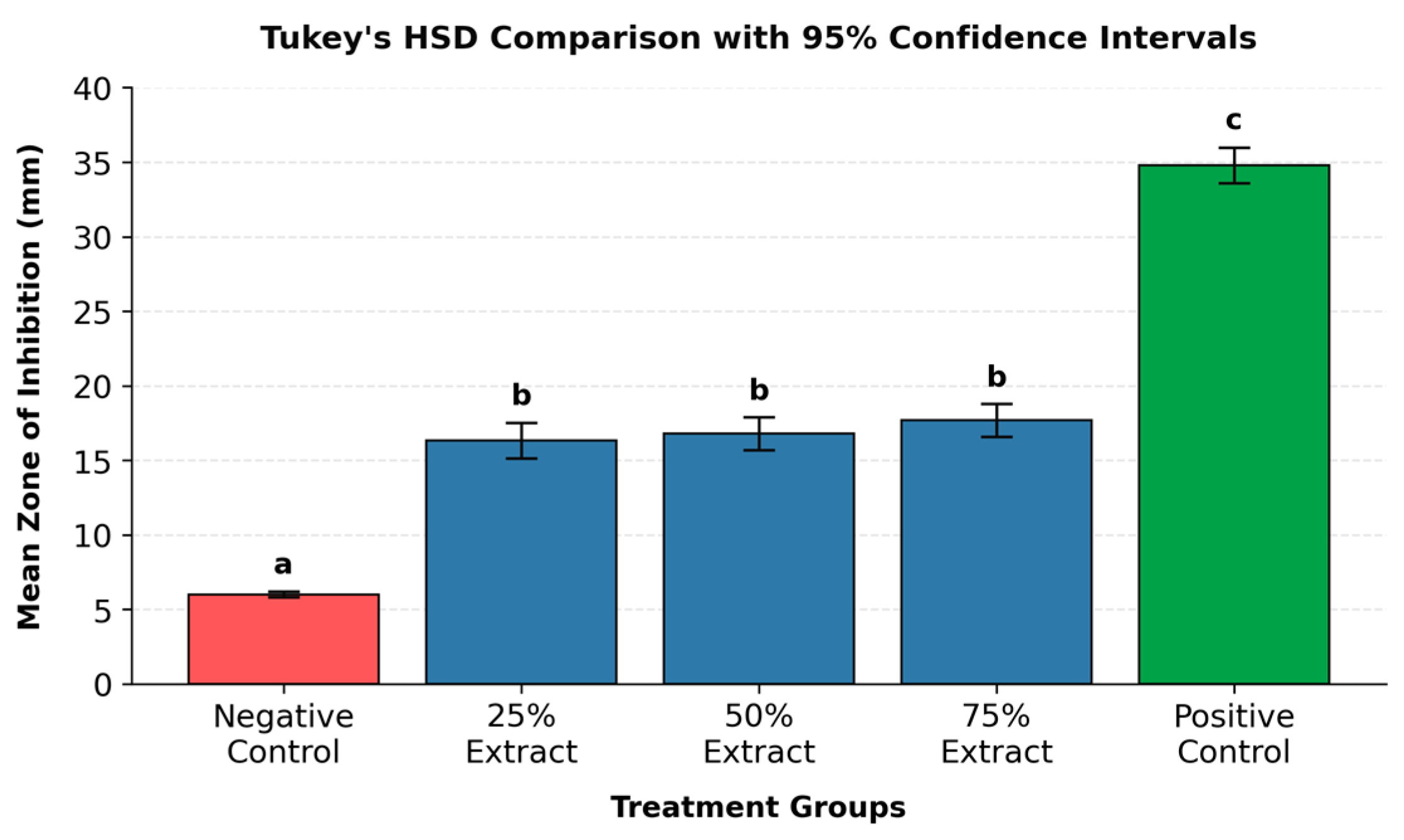
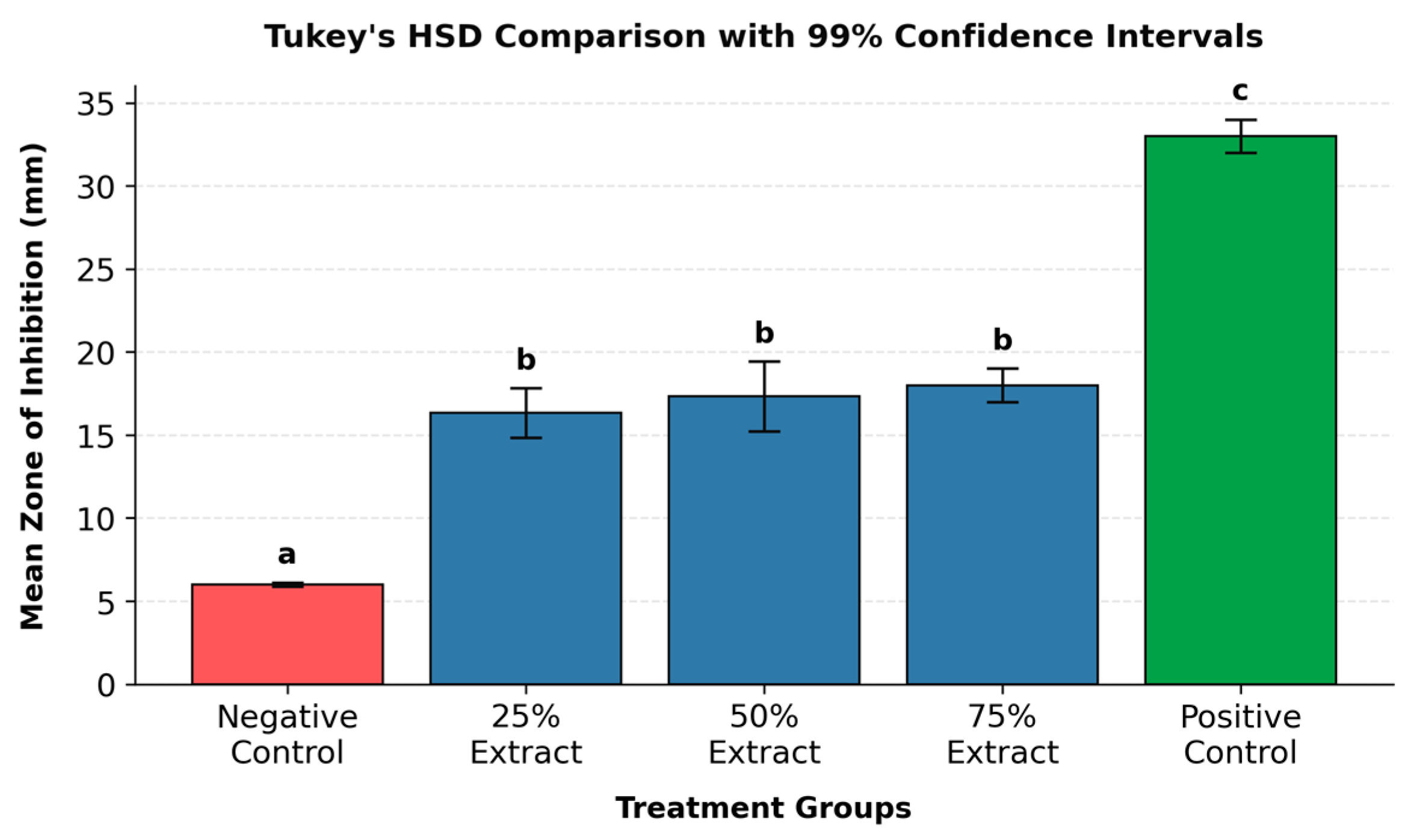
To visually assess and compare the antimicrobial efficacy of the various J. multifida extract concentrations, the zones of inhibition against Staphylococcus aureus and Escherichia coli are illustrated in Figure 1 and Figure 2, respectively.
Post hoc comparisons using Tukey’s Honestly Significant Difference (HSD) test revealed that all evaluated J. multifida extract concentrations of 25%, 50%, and 75% produced significantly larger zones of inhibition than the negative control (M = 6.00, SD = 0.00) against both S. aureus (Figure 1) and E. coli (Figure 2) (p < 0.001).
For S. aureus, the mean inhibition zones for the 25%, 50%, and 75% concentrations were 16.33 ± 1.15 mm, 16.67 ± 2.08 mm, and 17.67 ± 1.53 mm, respectively. A nearly identical baseline and upward trend were mirrored in the E. coli trials, yielding zones of 16.33 ± 1.53 mm, 17.33 ± 2.08 mm, and 18.00 ± 1.00 mm, respectively.
Despite this baseline efficacy, no statistically significant differences were observed among the extract groups themselves for either pathogen (p > 0.05). Pairwise comparisons confirmed statistical parity among all extract pairs for S. aureus (25% vs. 50%: p = 0.99; 50% vs. 75%: p = 0.92; 25% vs. 75%: p = 0.77) and E. coli (25% vs. 50%: p = 0.87; 50% vs. 75%: p = 0.96; 25% vs. 75%: p = 0.55), a lack of concentration-dependent differentiation that is visually reinforced by the heavily overlapping error bars (±1 SD) across all extract formulations. Conversely, the positive control demonstrated a significantly wider zone of inhibition than all extract concentrations and negative controls (p < 0.001), peaking at 34.67 ± 1.53 mm for S. aureus and 33.00 ± 1.00 mm for E. coli.
Ultimately, these comparative data demonstrate that while the extract-based soap formulations possess robust, functional antimicrobial activity against both Gram-positive and Gram-negative targets, increasing the extract concentration beyond 25% does not yield a statistically meaningful boost in performance.

4. Discussion

4.1. Functional Bioactivity of Secondary Metabolites of J. mutifida Extract

Qualitative phytochemical screening of the crude J. multifida leaf extract positively identified the presence of saponins, tannins, and flavonoids, also observed in previous studies [7,8,10,27]. These compound classes do not merely serve as passive markers but also function as active, synergistic ingredients that rationalize the therapeutic and antimicrobial efficacy of the extract when integrated into a cosmetic soap delivery vehicle [28].
The gravimetric crude extraction yield of 9.8% (recovering 4.9 g of crude mass from 50.0 g of initial Jatropha multifida leaf biomass) reflects a highly efficient and selective extraction process governed by the polarity dynamics of the solvent system and sequential processing mechanics. Utilizing 80% ethanol as a hydroalcoholic menstruum provides a dual-action mechanism that maximizes the recovery of secondary metabolites while minimizing the co-extraction of inactive structural plant matter. The use of 80% ethanol was also seen in the phytochemical extraction studies [29,30,31].
The organoleptic and physicochemical analysis of the cosmetic soap formulated with the 25% J. multifida extract proposes a potential alignment with quality control standards. True soaps are naturally alkaline due to the progressive hydrolysis of fatty acid salts, and the potentiometric determination of a safe, stable pH of 9.42 indicates that the high 25% botanical extract load did not induce erratic chemical drift, phase separation, or unintended ionization within the matrix [32]. This stable alkalinity profile is further corroborated by a negligible free caustic alkali content of 0.02% ± 0.005%. Because free caustic alkali tracks unreacted sodium hydroxide (NaOH) remaining post-saponification, remaining significantly below the standard industrial hazard threshold of 0.05% confirms an exceptionally high conversion efficiency. This ensures that the finished topical delivery system carries zero risk of causing dermal irritation, chemical dermatitis, or severe stratum corneum barrier disruption.
Furthermore, the structural integrity and economic–commercial viability of the bioactive formulation are validated by the total fatty matter (TFM) parameters. Petroleum ether extraction yielded a TFM content of 75.15% ± 1.15%, which acts as the primary global regulatory benchmark for evaluating cleansing performance and lipid retention capacity. Under international standardization frameworks, a TFM index spanning 70% to 75% categorizes the botanical matrix as a high-performing, Grade II cosmetic toilet soap [20]. This demonstrates that despite incorporating a dense 25% raw extract volume, the crystalline soap framework successfully maintained proper micellar networks, emollient characteristics, and optimal lathering kinetics. Consequently, these multi-parameter outcomes collectively imply that the soap formulation is a stable, non-hazardous, and highly reproducible delivery vehicle for plant-derived hygiene therapeutics.

4.2. Antibacterial Efficacy

Bioassay results have demonstrated that the soap formulations integrated with J. multifida L. (Coral Bush) leaf extract possess robust antibacterial properties. Notably, all experimental concentrations (25%, 50%, and 75%) consistently achieved an “active” qualitative interpretation against both S. aureus (Gram-positive) and E. coli (Gram-negative) bacteria. This broad-spectrum activity highlights the potential of J. multifida leaf extract as a functional, bioactive ingredient in antimicrobial hygiene products. The use of 25%, 50%, and 75% concentrations is a widely accepted approach in antimicrobial and phytopharmaceutical research to determine the minimum effective concentration and optimize product performance [33,34,35].
The distinct clearance zones observed in the treatment groups can be attributed to the rich profile of secondary metabolites typical of the Jatropha genus [36]. Phytochemical screening confirmed that J. multifida leaves contain potent components, including flavonoids, tannins, and saponins (Table 1). Flavonoids are molecules known to disrupt bacterial cell walls and precipitate cytoplasmic proteins [37]. Tannins form complex linkages with extracellular, soluble proteins and bacterial cell walls, thereby inactivating essential microbial enzymes [38]. Saponins function as surface-active agents that increase the permeability of cell membranes, causing the leakage of critical internal metabolites and subsequent cell lysis [39]. The successful expression of these mechanisms within a soap matrix indicates that the active phytochemical components remained chemically stable and bioavailable, surviving the saponification or blending process during soap preparation.

4.3. The Dynamics of Concentration and Diffusion Limits

A critical finding in this study is that while all extract-based soaps significantly outperformed the negative control (p < 0.001), increasing the extract concentration from 25% to 75% did not yield a statistically meaningful increase in antibacterial performance. For S. aureus, the mean zones ranged from 16.33 to 17.67 mm (Tukey’s HSD p = 0.77), and for E. coli, they ranged from 16.33 to 18.00 mm (Tukey’s HSD p = 0.55). This lack of statistical differentiation between concentrations suggests a plateau effect, which may be attributed to agar diffusion thresholds [40] and soap base entrapment [41].
Zone sizes in agar diffusion assays depend strictly on phytochemical molecular size and solubility. At a 25% concentration, the agar matrix may have reached saturation, which suggests that introducing higher concentrations of 50% and 75% did not expand the inhibition radius or accelerate diffusion. Moreover, the fatty acid salts that form dense micellar networks in saponified matrices can entrap excess lipophilic secondary metabolites, such as specific flavonoids or terpenoids [41]. While the exact physicochemical drivers behind the observed efficacy plateau remain to be fully elucidated, several mechanisms can be tentatively proposed. The increased extract fractions in the 50% and 75% formulations may have been entrapped within the soap matrices, preventing their free release into the aqueous agar testing medium. Alternatively, the lack of significant difference in antibacterial performance among the 25%, 50%, and 75% concentrations of extract-based soaps is that, at higher extract concentrations, the portion of extract that cannot be entrapped within the soap base matrix may leak out and degrade during the soap preparation process, leading to the observed lack of significant difference. From an industrial and economic perspective, this plateau is highly advantageous. The 25% extract concentration performance achieved in parity with the 75% formulation represents a cost-effective and resource-efficient choice for scale-up. This optimization could minimize raw material consumption without compromising product efficacy.

4.4. Comparative Efficacy Against Gram-Positive and Gram-Negative Pathogens

The J. multifida extract-infused soaps demonstrated remarkably similar inhibitory thresholds against both S. aureus (16.33 to 17.67 mm) and E. coli (16.33 to 18.00 mm), corroborating findings from similar studies [42,43]. This outcome is significant considering the structural differences between these two bacterial groups. Gram-positive bacteria (S. aureus) possess a thick, porous peptidoglycan cell wall that is typically highly receptive to plant-derived antimicrobials [44]. Conversely, Gram-negative bacteria (E. coli) feature a complex outer lipid membrane acting as an additional selective barrier that often restricts the influx of hydrophobic foreign compounds [44,45].
The ability of the extract to bypass this complex outer membrane in E. coli and match its performance against S. aureus may indicate the presence of highly permeable or amphiphilic agents within the J. multifida leaf matrix. Saponins, for instance, naturally lower surface tension and interact fluidly with lipid bilayers [36]. This structural disruption most likely destabilizes the Gram-negative outer membrane, clearing an entry path for companion metabolites like tannins and flavonoids to execute their intracellular mechanisms.

4.5. Validation of the Bioassay Parameters

Finally, the experimental infrastructure was strictly verified by the control groups. The positive control exhibited prominent mean zones of inhibition (34.67 mm for S. aureus and 33.00 mm for E. coli), confirming that both test strains were fully viable and highly susceptible to standard antibiotics. Remarkably, the negative control showed a completely flat, uniform baseline measurement of 6.00 mm across all replicates, which reflects the physical borders of the disc itself. This net-zero zone (0 mm of true clearance) may indicate that the base soap ingredients and the solvents used did not possess independent antimicrobial traits. Therefore, all antibacterial performance recorded across the 25%, 50%, and 75% groups can be attributed to the active phytochemical components of the J. multifida leaf extract.

4.6. Limitations of the Study

A limitation of this study is that the efficiency of the sequential drying steps in reducing residual solvent was not quantitatively verified. While these procedural steps were designed to mitigate solvent retention, the complete absence of ethanol was not confirmed via analytical testing. Future investigations should incorporate rigorous analytical residual solvent testing to definitively quantify trace levels and ensure safety compliance. Moreover, the mechanism of action involving agar diffusion restrictions and micelle entrapment was presented from a purely theoretical standpoint. Future studies are needed to validate these hypotheses, as the current work did not measure extract release, solubility, diffusion coefficients, or the compound’s distribution inside the soap matrix. Finally, a primary limitation of this research is that safety studies were outside the current scope. Future investigations must prioritize cytotoxicity assays, skin irritation tests, and evaluations of how the formulation affects the resident skin microbiota before human or commercial application can be considered.

5. Conclusions

In conclusion, this study demonstrates that the qualitative incorporation of plant extracts into a soap formulation provides a stable baseline for antibacterial activity, particularly against the tested strains. However, several limitations must be acknowledged: the precise diffusion coefficients, bioavailability, and exact mechanisms of action against Gram-negative bacteria were not empirically quantified, nor was the formulation commercially optimized. Our current findings suggest a potential for botanical additives in hygiene products, but definitive conclusions regarding their long-term stability and molecular interactions require further investigation. Future research will focus on quantifying the release kinetics of the active compounds, evaluating matrix stability over time, and performing minimum inhibitory concentration (MIC) assays to clarify the specific mechanisms of action.

Author Contributions

Conceptualization, software, validation, formal analysis, resources, data curation, writing, supervision, funding acquisition, project administration, A.M.A.; methodology, investigation, writing—original draft preparation, A.M.A. and T.J.B.G. All authors have read and agreed to the published version of the manuscript.

Funding

This research was funded by Apayao State College, Philippines under the annual Research fund for A.Y. 2025.

Institutional Review Board Statement

Not applicable as this study did not involve humans or animals.

Informed Consent Statement

Not applicable.

Data Availability Statement

The raw data supporting the conclusions of this article are already in the manuscript.

Acknowledgments

The authors would like to thank Apayao State College for the administrative and logistics support in the conduct of this study. During the preparation of this manuscript, the authors used Google Gemini for the purposes of verifying citation and reference formats, composition, and grammar revisions. The authors have reviewed and edited the output and take full responsibility for the content of this publication.

Conflicts of Interest

The authors declare no conflicts of interest.

Abbreviations

The following abbreviations are used in this manuscript:
EDTAEthylenediaminetetraacetic acid
hHour
gGram
minMinutes
mLMilliliter

References

  1. GBD 2019 Antimicrobial Resistance Collaborators. Global mortality associated with 33 bacterial pathogens in 2019: A systematic analysis for the Global Burden of Disease Study 2019. Lancet 2022, 400, 2221–2248. [Google Scholar] [CrossRef] [Scilit] [PubMed]
  2. Ventola, C.L. The antibiotic resistance crisis: Part 1: Causes and threats. Pharm. Ther. 2015, 40, 277–283. [Google Scholar]
  3. Hosseinzadeh, S.; Jafarikukhdan, A.; Hosseini, A.; Armand, R. The application of medicinal plants in traditional and modern medicine: A review of Thymus vulgaris. Int. J. Clin. Med. 2015, 6, 635–644. [Google Scholar] [CrossRef]
  4. Boy, H.I.A.; Rutilla, A.J.H.; Santos, K.A.; Ty, A.M.T.; Yu, A.I.; Mahboob, T.; Tang, L.Y.; Nissapatorn, V.; Galba, B.; de Lourdes Pereira, M.; et al. Recommended medicinal plants as source of natural products: A review. Digit. Chin. Med. 2018, 1, 131–142. [Google Scholar] [CrossRef] [Scilit]
  5. Cowan, M.M. Plant products as antimicrobial agents. Clin. Microbiol. Rev. 2009, 12, 564–582. [Google Scholar] [CrossRef] [Scilit]
  6. Lai, Z.F.; Kaur, S. Home hygiene and prevention of infections. Asia Pac. J. Health Manag. 2020, 15, 79–84. [Google Scholar] [CrossRef] [Scilit]
  7. Anani, K.; Adjrah, Y.; Améyapoh, Y.; Karou, S.D.; Agbonon, A.; de Souza, C.; Gbeassor, M. Antimicrobial, anti-inflammatory and antioxidant activities of Jatropha multifida L. (Euphorbiaceae). Pharmacogn. Res. 2016, 8, 142–146. [Google Scholar] [CrossRef] [Scilit]
  8. Aiyelaagbe, O.O.; Oguntuase, B.J.; Arimah, B.D.; Adeniyi, B.A. The antimicrobial activity of Jatropha multifida extracts and chromatographic fractions against sexually transmitted infections. J. Med. Sci. 2008, 8, 143–147. [Google Scholar] [CrossRef] [Scilit]
  9. Igbeneghu, O.A. The antimicrobial assessment of some Nigerian herbal soap. Afr. J. Tradit. Complement. Altern. Med. 2013, 10, 513–518. [Google Scholar] [CrossRef] [Scilit] [PubMed]
  10. Carvalho, C.; Mariano, L.V.; Negrão, V.S.; Gonçalves, P.; Marcucci, C. Phenols, flavonoids and antioxidant activity of Jatropha multifida L. collected in Pindamonhangaba, Sao Paulo State, Brazil. J. Anal. Pharm. Res. 2018, 7, 581–584. [Google Scholar] [CrossRef] [Scilit]
  11. Diwakar, A.; Sharma, H.; Yadav, T.; Chandel, R.; Sahu, G.K.; Sahu, P.; Nema, R.K. Development of Herbal Soap for Effective Cleansing and Skin Moisturizing. Int. J. Emerg. Technol. Innov. Res. 2024, 11, o518–o526. Available online: http://www.jetir.org/papers/JETIR2404F72.pdf (accessed on 28 May 2026). [CrossRef] [Scilit]
  12. Kareru, P.G.; Keriko, J.M.; Kenji, G.M.; Thiong’o, G.T.; Gachanja, A.N.; Mukiira, H.N. Antimicrobial activities of skincare preparations from plant extracts. Afr. J. Tradit. Complement. Altern. Med. 2010, 7, 214–218. [Google Scholar] [CrossRef] [Scilit] [PubMed]
  13. Mwambete, K.D.; Lyombe, F. Antimicrobial Activity of Medicated Soaps Commonly Used By Dar es Salaam Residents in Tanzania. Indian J. Pharm. Sci. 2011, 73, 92–98. [Google Scholar] [CrossRef] [PubMed]
  14. Sahle, H.G.; Sbahtu, D.B.; Gebreyohannes, G. Antibacterial properties of Aloe adigratana and Aloe elegans extracts and their potential applications in shampoo and soap development. Sci. Rep. 2025, 15, 37848. [Google Scholar] [CrossRef] [Scilit] [PubMed]
  15. Jamil, S.; Abdulrahman, T.T.; Hasan, R.U. Formulation and Evaluation of Anti-Microbial Herbal Soap Containing Neem (Azadirachta indica), Tulsi (Ocimum tenuiflorum), and Aloe Vera (Aloe barbadensis Miller). CME J. Geriatr. Med. 2025, 17, 1–7. [Google Scholar] [CrossRef] [Scilit]
  16. Kanyama, T.; Masunaga, A.; Kawahara, T.; Morita, H.; Akita, S. Natural soap is clinically effective and less toxic and more biodegradable in aquatic organisms and human skin cells than synthetic detergents. PLoS ONE 2025, 20, e0324842. [Google Scholar] [CrossRef] [Scilit] [PubMed]
  17. Gligor, O.; Clichici, S.; Moldovan, R.; Muntean, D.; Vlase, A.M.; Nadăș, G.C.; Filip, G.A.; Vlase, L.; Crișan, G. Influences of Different Extraction Techniques and Their Respective Parameters on the Phytochemical Profile and Biological Activities of Xanthium spinosum L. Extracts. Plants 2022, 12, 96. [Google Scholar] [CrossRef] [Scilit] [PubMed]
  18. Guevara, B.Q. (Ed.) A Guidebook to Plant Screening: Phytochemical and Biological; University of Santo Tomas Publishing House: Manila, Philippines, 2005. [Google Scholar]
  19. ISO 685:2020; Analysis of Soaps—Determination of Total Alkali Content and Total Fatty Matter Content. International Organization for Standardization (ISO): Geneva, Switzerland, 2020.
  20. ISO 456:1973; Surface Active Agents—Analysis of Soaps—Determination of Free Caustic Alkali. International Organization for Standardization (ISO): Geneva, Switzerland, 1973.
  21. Bauer, A.W.; Kirby, W.M.; Sherris, J.C.; Turck, M. Antibiotic susceptibility testing by a standardized single disk method. Am. J. Clin. Pathol. 1966, 45, 493–496. [Google Scholar] [CrossRef] [Scilit] [PubMed]
  22. Balouiri, M.; Sadiki, M.; Ibnsouda, S.K. Methods for in vitro evaluating antimicrobial activity: A review. J. Pharm. Anal. 2016, 6, 71–79. [Google Scholar] [CrossRef] [Scilit] [PubMed]
  23. Clinical and Laboratory Standards Institute (CLSI). Performance Standards for Antimicrobial Susceptibility Testing, 31st ed.; CLSI Supplement M100; CLSI: Wayne, PA, USA, 2021. [Google Scholar]
  24. Cohen, J. Statistical Power Analysis for the Behavioral Sciences, 2nd ed.; Lawrence Erlbaum Associates: Hillsdale, NJ, USA, 1988. [Google Scholar]
  25. Kim, H.Y. Statistical notes for clinical researchers: Post-hoc multiple comparisons. Restor. Dent. Endod. 2015, 40, 172–176. [Google Scholar] [CrossRef] [Scilit] [PubMed]
  26. Hendry, G.A.F.; Houghton, J.D. (Eds.) Natural Food Colorants, 2nd ed.; Blackie Academic & Professional: London, UK, 1996. [Google Scholar]
  27. Dah-Nouvlessounon, D.; Chokki, M.; Agossou, E.A.; Houédanou, J.B.; Nounagnon, M.; Sina, H.; Vulturar, R.; Heghes, S.C.; Cozma, A.; Mavoungou, J.F.; et al. Polyphenol Analysis via LC-MS-ESI and Potent Antioxidant, Anti-Inflammatory, and Antimicrobial Activities of Jatropha multifida L. Extracts Used in Benin Pharmacopoeia. Life 2023, 13, 1898. [Google Scholar] [CrossRef] [Scilit] [PubMed]
  28. Sitarek, P.; Merecz-Sadowska, A.; Kowalczyk, T.; Wieczfinska, J.; Zajdel, R.; Śliwiński, T. Potential Synergistic Action of Bioactive Compounds from Plant Extracts against Skin Infecting Microorganisms. Int. J. Mol. Sci. 2020, 21, 5105. [Google Scholar] [CrossRef] [Scilit] [PubMed]
  29. Gavrila, A.I.; Zalaru, C.M.; Tatia, R.; Seciu-Grama, A.-M.; Negrea, C.L.; Calinescu, I.; Chipurici, P.; Trifan, A.; Popa, I. Green Extraction Techniques of Phytochemicals from Hedera helix L. and In Vitro Characterization of the Extracts. Plants 2023, 12, 3908. [Google Scholar] [CrossRef] [Scilit] [PubMed]
  30. Kim, H.; Kim, J.; Cho, J.A.; Han, I. Effect of boiling and extraction methods using water or 80% ethanol on the physiological activities of the inner shell of chestnut (Castanea crenata). Food Chem. X 2025, 31, 103017. [Google Scholar] [CrossRef] [Scilit] [PubMed]
  31. El Mannoubi, I. Impact of different solvents on extraction yield, phenolic composition, in vitro antioxidant and antibacterial activities of deseeded Opuntia stricta fruit. J. Umm Al-Qura Univ. Appll. Sci. 2023, 9, 176–184. [Google Scholar] [CrossRef] [Scilit]
  32. Tarun, J.; Susan, J.; Suria, J.; Susan, V.J.; Criton, S. Evaluation of pH of Bathing Soaps and Shampoos for Skin and Hair Care. Indian J. Dermatol. 2014, 59, 442–444. [Google Scholar] [CrossRef] [Scilit] [PubMed]
  33. Capagas, R.J. Antimicrobial Activity of Kangkong and Paragis Leaf Extracts against Klebsiella oxytoca. J. Interdiscip. Perspect. 2024, 2, 206–215. [Google Scholar] [CrossRef] [Scilit]
  34. Allami, R.H.; Mouhamad, R.S.; Abdulateef, S.A.; abedalelah Al-Khafaji, K. Antimicrobial Activity of Herbal Mixture Extract Combination on Microorganisms Isolated from Urinary Tract infection. Bionatura 2020, 5, 1346–1351. [Google Scholar] [CrossRef] [Scilit]
  35. Zeleke, B.; Mekonnen, Z.; Bireda, M.; Yitbarek, M.; Dendir, A. Phytochemical screening and antimicrobial activity of Polygala sadebeckiana Gürke extracts on bacterial isolates from Wound samples of patients with “Shimetere”. BMC Complement. Med. Ther. 2024, 24, 72. [Google Scholar] [CrossRef] [Scilit] [PubMed]
  36. Cavalcante, N.B.; Santos, A.D.C.; Almeida, J.R.G.D. The genus Jatropha (Euphorbiaceae): A review on secondary chemical metabolites and biological aspects. Chem. Biol. Interact. 2020, 318, 108976. [Google Scholar] [CrossRef] [Scilit] [PubMed]
  37. Liu, Y.; Zhu, J.; Liu, Z.; Zhi, Y.; Mei, C.; Wang, H. Flavonoids as promising natural compounds for combating bacterial infections. Int. J. Mol. Sci. 2025, 26, 2455. [Google Scholar] [CrossRef] [Scilit] [PubMed]
  38. Govindarajan, R.K.; Revathi, S.; Rameshkumar, N.; Krishnan, M.; Kayalvizhi, N. Microbial tannase: Current perspectives and biotechnological advances. Biocatal. Agric. Biotechnol. 2016, 6, 168–175. [Google Scholar] [CrossRef] [Scilit]
  39. Shakeel, A.; Noor, J.J.; Jan, U.; Gul, A.; Handoo, Z.; Ashraf, N. Saponins, the unexplored secondary metabolites in plant defense: Opportunities in integrated pest management. Plants 2025, 14, 861. [Google Scholar] [CrossRef] [Scilit] [PubMed]
  40. Flanagan, J.N.; Steck, T.R. The relationship between agar thickness and antimicrobial susceptibility testing. Indian J. Microbiol. 2017, 57, 503–506. [Google Scholar] [CrossRef] [Scilit] [PubMed]
  41. Sharma, M.; Sharma, V.; Majumdar, D.K. Entrapment of α-amylase in agar beads for biocatalysis of macromolecular substrate. Int. Sch. Res. Not. 2014, 2014, 936129. [Google Scholar] [CrossRef] [Scilit]
  42. Gerald, E.; Alfonse, O. A comparative study of the effects of Jatropha multifida and Euphorbia hirta and their mixture on pathogenic growth rate. Eng. Proc. 2023, 37, 63. [Google Scholar] [CrossRef] [Scilit]
  43. Ongtengco, D.C. The in vitro antibacterial activity of Jatropha multifida Linn. latex against common bacterial wound isolates. Acta Manil. 1992, 40, 25–28. [Google Scholar]
  44. Sun, J.; Rutherford, S.T.; Silhavy, T.J.; Huang, K.C. Physical properties of the bacterial outer membrane. Nat. Rev. Microbiol. 2022, 20, 236–248. [Google Scholar] [PubMed]
  45. Gutsmann, T.; Seydel, U. Impact of the glycostructure of amphiphilic membrane components on the function of the outer membrane of Gram-negative bacteria as a matrix for incorporated channels and a target for antimicrobial peptides or proteins. Eur. J. Cell Biol. 2010, 89, 11–23. [Google Scholar] [CrossRef] [Scilit] [PubMed]
Figure 1. Mean zone of inhibition across treatment groups for antibacterial activity of J. multifida soap formulations against S. aureus. Bars represent mean values (n = 3). Error bars indicate ± 1 standard deviation. Columns sharing lowercase letter superscripts do not differ significantly at p > 0.05 based on Tukey’s Honestly Significant Difference (HSD) test.
Cosmetics 13 00193 g001
Figure 2. Mean zone of inhibition across treatment groups for antibacterial activity of J. multifida soap formulations against E. coli. Bars represent mean values (n = 3). Error bars indicate ± 1 standard deviation. Columns sharing lowercase letter superscripts do not differ significantly at p > 0.05 based on Tukey’s Honestly Significant Difference (HSD) test.
Cosmetics 13 00193 g002
Table 1. Qualitative screening parameters for the identification of saponins, tannins, and flavonoids in plant extracts.
Phytochemical GroupScreening TestReagents/Key StepsInference
Positive Criteria (+)Negative Criteria (−)
SaponinsFroth Test2 mL extract;
10 mL distilled H2O;
shaken vigorously for 60 s.
Honeycomb foam layer ≥ 1.2 cm in height persists for at least 10 min.Absence of foam, or complete collapse of a thin bubble layer in less than 10 min.
TanninsFerric Chloride Assay2 mL extract + 2 mL H2O;
3 drops of 5% aqueous FeCl3.
Deep brownish-green or blue-green coloration.Retention of baseline light amber/yellow hue or a transparent orange tint.
FlavonoidsShinoda/Bate-Smith & Metcalf2 mL extract;
0.5 cm Mg ribbon;
5 drops conc. HCl.
Gradual development of a distinct pink, scarlet, crimson, or cherry-red color.No change from baseline yellow-green extract tone or yellowing from the acid matrix.
Abbreviations: HCl, hydrochloric acid; FeCl3, ferric chloride; H2O, water; Mg, magnesium; s, seconds; min, minutes.
Table 2. Quantitative formulation and concentration of the soap batches.
Target Extract Concentration (% w/w)Mass of J. multifida Extract (g)Mass of Olive Oil Base (g)Total Active
Lipid Matrix Mass (g) *
Concentration
(% w/w)
25%8.3325.0033.3325%
50%8.338.3316.6650%
75%8.332.8011.1175%
* Note: This represents the core active organic phase before the introduction of the standardized aqueous alkali (NaOH/water) solution required to complete the saponification process.
Table 3. Phytochemical screening profile of J. multifida extract.
PhytochemicalsResultInferenceMethod Used
[20]
SaponinsHoneycomb foam layer ≥ 1.2 cm in height persists for at least 10 min.Positive (+)Froth Test
TanninsDeep brownish-green coloration.Positive (+)Ferric chloride Assay
FlavonoidsGradual development of a distinct crimson color.Positive (+)Bate-Smith & Metcalf/Shinoda Test
Note: (+): Positive result, indicating the presence of the phytochemical.
Table 4. Quality control metrics for the J. multifida extract and the formulated soap.
ParametersEvaluation MethodObservation/Descriptor/Result
  • Extract
ColorVisual inspection in natural daylightDark olive green
OdorOlfactory assessmentDistinctive herbal odor; mildly pungent
TextureTactile and physical inspectionViscous semi-solid paste
b.
Formulated Soap (25%)
pH (10% aqueous)Potentiometric protocol9.42 ± 0.12 *
Free Caustic AlkaliVolumetric titration [20]0.02% ± 0.005% *
Total Fatty MatterPetroleum Ether Extraction [19]75.15% ± 1.15% *
* Note: Values represent the mean of three independent experimental replicates (n = 3) ± standard deviation.
Table 5. Antibacterial activity of J. multifida soap formulations against S. aureus (mean ZOI in mm).
SampleZone of Inhibition (Mean ± SD, mm)Qualitative
Interpretation
25% Extract16.33 ± 1.15Active
50% Extract16.67 ± 2.08Active
75% Extract17.67 ± 1.53Active
Positive Control34.67 ± 1.53Very Active
Negative Control6.00 ± 0.00Inactive
Note: Values are expressed as mean ± standard deviation (SD) of three replicates (n = 3). Qualitative scale: inactive (<10 mm), active (10–19 mm), and very active (>19 mm).
Table 6. One-way ANOVA results for antibacterial activity of J. multifida soap formulations against S. aureus.
SourceSSDfMSFp-Value
Between Groups1278.274319.57154.63<0.001
Within Groups20.67102.07
Total1298.9414
Note: SS: sum of squares; Df: degrees of freedom; MS: mean square; F: Fisher’s F-ratio. F(4, 10) = 154.63, p < 0.001. The variance indicates a highly statistically significant difference in the zones of inhibition across the treatment groups.
Table 7. Antibacterial activity of J. multifida soap formulations against E. coli (mean ZOI in mm).
SampleZone of Inhibition
(Mean ± SD, mm)
Qualitative
Interpretation
25% Extract16.33 ± 1.53Active
50% Extract17.33 ± 2.08Active
75% Extract18.00 ± 1.00Active
Positive Control33.00 ± 1.00Very Active
Negative Control6.00 ± 0.00Inactive
Note: Values are expressed as mean ± standard deviation (SD) of three replicates (n = 3). Qualitative scale: inactive (<10 mm), active (10–19 mm), and very active (>19 mm).
Table 8. One-way ANOVA results for antibacterial activity of J. multifida soap formulations against E. coli.
SourceSSDfMSFp-Value
Between Groups1116.404279.10161.02<0.001
Within Groups17.33101.73
Total1133.7314
Note: SS: sum of squares; Df: degrees of freedom; MS: mean square; F: Fisher’s F-ratio. F(4, 10) = 161.02, p < 0.001. The variance indicates a highly statistically significant difference in the zones of inhibition across the treatment groups.
Disclaimer/Publisher’s Note: The statements, opinions and data contained in all publications are solely those of the individual author(s) and contributor(s) and not of MDPI and/or the editor(s). MDPI and/or the editor(s) disclaim responsibility for any injury to people or property resulting from any ideas, methods, instructions or products referred to in the content.