1. Introduction
Exposure to ultraviolet (UV) radiation is a primary cause of photoaging and a significant risk factor for various skin pathologies, including sunburn, hyperpigmentation, and skin cancer [
1]. The sun’s UV spectrum is broadly categorized into UVA (320–400 nm) and UVB (290–320 nm), both of which are known to cause damage to cellular DNA and collagen fibers [
2]. Consequently, the use of sun protection, particularly sunscreen, has become a cornerstone of public health recommendations for skin cancer prevention and skin health [
3]. However, Diffey et al. [
4] sampled 59 commercial sunscreen products in Cincinnati, Ohio, and analyzed their broad-spectrum protection (protection from both UVA and UVB), for which the critical wavelength must be at least 370 nm. The definition of critical wavelength refers to the wavelength at which the integral of the spectral absorbance curve reaches 90% of the integral from 290 to 400 nm. The results were surprising, as only five out of 59 products had a critical wavelength greater than or equal to 370 nm, equating to 8% of the samples [
4]. A sunscreen formulation must be broad-spectrum for the product to protect the skin, which is as important as the SPF and PA ratings often displayed on product labels.
Historically, sunscreens have relied on two main types of UV filters: organic (chemical) and inorganic (physical) [
5]. Organic filters, such as oxybenzone and octinoxate, absorb UV radiation and convert it into heat. In contrast, inorganic filters, such as zinc oxide and titanium dioxide, physically reflect and scatter UV rays [
6]. However, the effectiveness and safety of these conventional ingredients have come under increasing scrutiny [
7]. While effective, specific organic filters have been linked to potential endocrine-disrupting activity and allergic reactions. More critically, several organic UV filters have been identified as environmental pollutants, contributing to coral reef bleaching and marine ecosystem disruption, prompting global discussions and legislative action to ban their use in certain regions [
8]. This abundance of environmental pollutants has led to a growing demand for effective, broad-spectrum, and environmentally friendly sunscreen formulations. Claims of reef-safe or reef-friendly cosmetic products remain unclear, but regulations prohibiting the use of sunscreens containing banned ingredients have been implemented in certain regions, such as the U.S. Virgin Islands, Palau, and Hawaii. This has led to the development of reef-safe sunscreen formulations. In particular, organic UV filters such as avobenzone, homosalate, octisalate, and octocrylene are frequently utilized in reef-safe sunscreens instead of the prohibited filters oxybenzone and octinoxate. Evaluations of acceptable threshold levels for marine water obtained from current REACH registration documents could potentially protect corals. Miller et al. (2021) defined coral-safe or reef-friendly as efforts to reduce harmful sunscreen agents that flow into the reef environment [
9]. In addition, there are guidelines for assessing the environmental toxicity of cosmetic products by considering three factors, namely persistence, bioaccumulation, and toxicity (PBT), during the design of cosmetic formulations [
10]. Finally, Choi et al. (2022) introduced an algorithm that evaluates the design of cosmetic formulations with the aim of reducing their environmental impact based on the PBT database [
11].
The current scientific landscape is marked by divergent hypotheses regarding the optimal approach to sun protection. On one hand, some studies argue that the benefits of established organic filters in preventing skin cancer outweigh their potential risks, particularly in formulations that limit dermal absorption. On the other hand, a rapidly growing body of research emphasizes the need for natural, plant-based alternatives that are both safe for humans [
12] and non-toxic to the environment [
13]. These natural compounds often possess inherent antioxidant and anti-inflammatory properties, offering a multi-faceted approach to skin health beyond simple UV filtration [
14].
One such promising natural source is
Perilla frutescens, a plant from the mint family, whose seed extract is rich in phenolic compounds, triterpenoids, and polyunsaturated fatty acids [
15]. In a recent study, Hou et al. (2022) highlighted the potent antioxidant and anti-inflammatory activities of Perilla seed extract, suggesting its potential as a bioactive ingredient in dermatological and cosmetic applications [
16]. Furthermore, according to the study by Choi et al. [
17], cold-pressed perilla oil contains linolenic acid, oleic acid, and linoleic acid as key components. In their study, perilla oil was used on the dorsal skin of hairless mice to evaluate sun protection efficacy. It was found that the perilla seed oil has the potential to be an anti-wrinkle ingredient, reduce transepidermal water loss (TEWL), lower erythema values, and decrease melanin index [
17]. Beyond its direct dermatological benefits, cultivating
P. frutescens also supports sustainable agriculture and local economies. As a traditional crop in many parts of Asia, it provides a reliable cash crop for local farmers, promotes agricultural biodiversity, and strengthens regional economies. Its versatility across food, medicine, and cosmetics creates a stable market demand, making its integration into a high-value product a compelling case for a truly sustainable supply chain [
18].
To address the growing demand for sustainable yet high-performance photoprotection, this research focuses on the formulation of a broad-spectrum, reef-friendly sunscreen utilizing the bioactive potential of perilla seeds. Specifically, it is hypothesized that the incorporation of P. frutescens seed extract (1% w/w) into a reef-friendly sunscreen formulation will significantly enhance its biological photoprotective efficacy and skin-brightening properties while maintaining superior physical stability and dermatological safety under real-world usage conditions. Through this approach, this study seeks to establish a scientific basis for using P. frutescens as a functional ingredient in eco-conscious cosmetic applications.
2. Materials and Methods
2.1. Chemicals and Reagents
Perilla seeds (Perilla frutescens) were obtained from Phayao Province, Thailand. The perilla seeds used in this research had strain code CR10-4. The botanical information and reference data are maintained by the Highland Research and Development Institute (HRDI), Chiang Mai, Thailand. The plant material was compared and authenticated against the database. Food-grade ethanol (95%) was purchased from Liquor Distillery Organization (Chachoengsao, Thailand).
For the formulation of sunscreen products, the following ingredients were utilized: Deionized water (Aqua), Carbopol 940 (Carbomer), 1,3-Butylene Glycol, Emulgade 165 (Glyceryl Stearate and PEG-100 Stearate), Olivem 300 (Olive oil PEG-7 Esters), Dub Helioptima (Diisopropyl Adipate, Diisopropyl Sebacate, Propylene Glycol Dicaprylate/Dicaprate, and C12-15 Alkyl Benzoate), Tween 60 (Polysorbate 60), Homosalate, Ethylhexyl Salicylate, Avobenzone, Polyester-7, Triethanolamine, and SUNCAT JW03 (Water, C12-C15 Alkyl Benzoate, Diethylamino Hydroxybenzoyl Hexyl Benzoate, Ethylhexyl Triazone, Bis-Ethylhexyloxyphenol Methoxyphenyl Triazine, Lecithin, 1,3-Butylene Glycol, and Phenoxyethanol). Preservatives used included 1,2-Hexanediol and Ethylhexylglycerin.
Reagents for the cytotoxicity assay, including Dimethyl sulfoxide (DMSO), Dulbecco’s Modified Eagle Medium (DMEM), Fetal Bovine Serum (FBS), 3-(4,5-dimethylthiazol-2-yl)-2,5-diphenyltetrazolium bromide (MTT), and Sodium Dodecyl Sulfate (SDS), were of analytical grade.
2.2. Plant Extract Preparation
The extraction process was designed to optimize the sun protection factor (SPF) of the perilla extract by investigating two factors: extraction time and solvent concentration. Perilla seeds were washed and dried in a hot air oven at 60 °C for 24 h. The dried seeds were ground into a fine powder and sieved to control particle size.
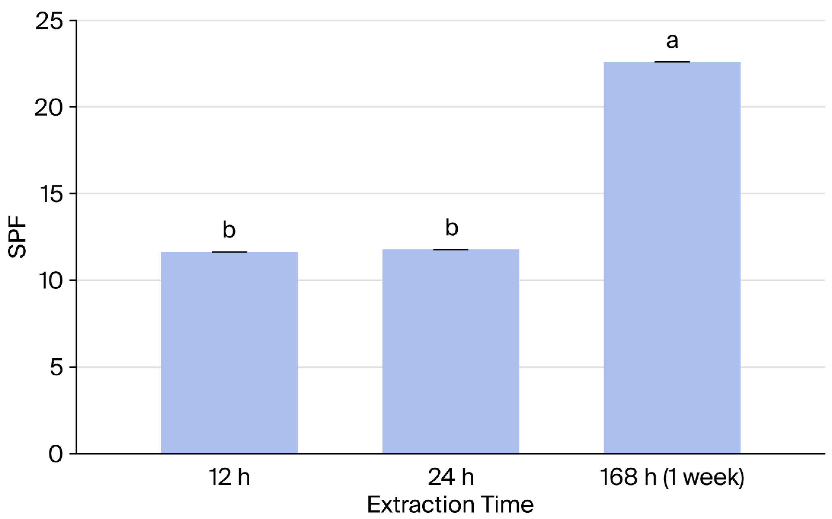
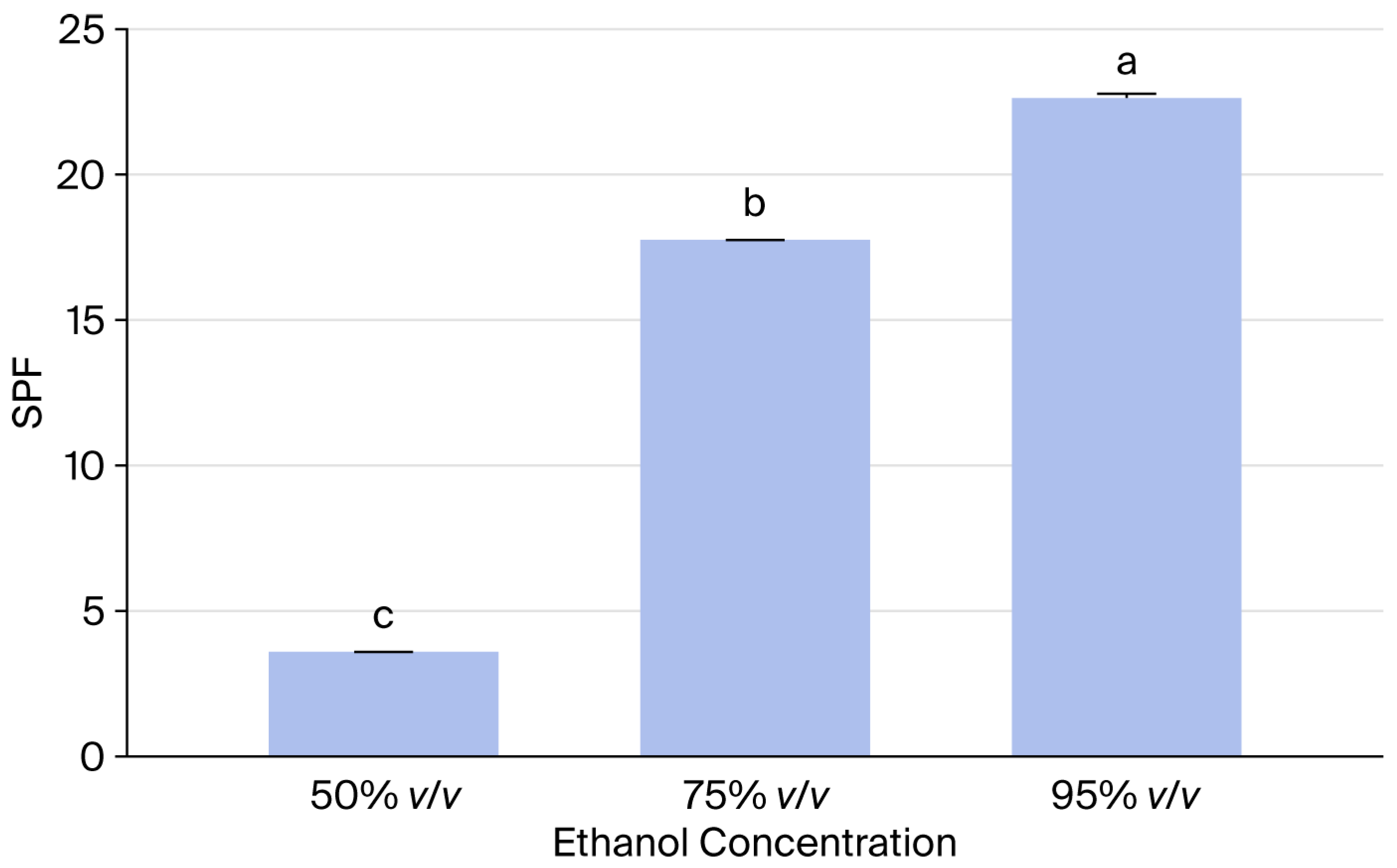
To study the effect of extraction time, the ground seeds were macerated in 95% v/v ethanol at a 1:10 (w/v) ratio for 12 h, 24 h, and 1 week (168 h). To study the effect of solvent concentration, the seeds were macerated in ethanol at varying concentrations (50% v/v, 75% v/v, and 95% v/v) for the optimal amount of time. To ensure data reliability and reproducibility, all extraction processes were performed in triplicate (n = 3) under identical experimental conditions on the same day. The mixtures were filtered through cheesecloth to remove coarse residue, then through Whatman No. 2 filter paper (125 mm). The filtrates were collected in Erlenmeyer flasks for further analysis.
The perilla seeds used in this research were sourced from a local market in Phayao from November to December 2024. The seeds were initially washed with tap water and subsequently dried in an oven at 60 °C until the moisture content was reduced to below 10% (
w/
v) [
19].
2.3. Evaluation of SPF of Perilla Extract
The
in vitro sun protection factor (SPF) of the perilla extracts (10 mg/mL) was evaluated spectrophotometrically in triplicate, utilizing the mathematical equation established by Mansur et al. (1986) [
20]. This spectrophotometric approach was strategically employed as a rapid and cost-effective screening tool to identify the optimal extraction conditions that yield the highest SPF value. Subsequently, the extract demonstrating the maximum photoprotective potential was selected for incorporation into the final sunscreen formulation. The SPF value was calculated using Equation (1) as follows:
where CF = 10 (correction factor) [
21]; EE(λ) = erythemogenic effect of radiation at wavelength λ; I(λ) = intensity of solar light at wavelength λ; and abs(λ) = absorbance of wavelength λ by a solution of the preparation.
The results from the experiments, which were evaluated three times per group, were analyzed using Microsoft Excel 365. The mean and standard deviation are presented as mean ± standard deviation (SD). Furthermore, the average of each experiment was tested for difference between groups using post hoc analysis (one-way ANOVA), with a confidence level of 99% (p-value < 0.01). The group with significantly different mean values is labeled with letters a, b, and c to indicate differences between levels. Identical letters in the output indicate no significant difference at p-value = 0.01.
2.4. Evaluation of Fatty Acid Composition
The fatty acid composition of the perilla extract was analyzed at the Halal Science Center, Chulalongkorn University. The analysis was performed using the method of Lepage & Roy (1984) [
22], based on triple quadrupole gas chromatography–mass spectrometry (GC-MS/MS).
The sample was prepared using a one-step direct transesterification method, all performed in the same test tube to skip the preliminary lipid extraction and purification steps. The process began by adding 500 μg each of internal standard consisting of nonanoic acid (C9:0), tridecanoic acid (C13:0), and tricosanoic acid (C23:0), dissolved in a 3:2 (
v/
v) methanol–benzene mixture, to the sample. Next, 1 mL of a freshly prepared 5:100 (
v/
v) acetylchloride–methanol mixture was added, along with a magnetic stirrer. The test tube was then sealed and heated to induce methanolysis at 100 °C for 1 h. This method does not require the addition of antioxidants to protect unsaturated fats [
22].
After the reaction was completed and the mixture was allowed to cool to room temperature, we added 500 μg of methyl pentadecanoate (methylated C15:0) external standard dissolved in 1 mL of hexane, followed by 1 mL of water. The test tube was shaken to mix and centrifuged to separate the liquid layers. Then, the hexane layer was stored at 4 °C until injected into the chromatograph [
22].
Fatty acid methyl ester derivatives (FAMEs) were analyzed using a GC-MS/MS. Separation was performed on a 10-foot glass column with an inner diameter of 2 mm. The stationary phase was equivalent to 5% SP-2340 on a Chromosorb W-AW support (100–120 mesh). Nitrogen was used as a carrier gas, with the flow rate maintained at 28 mL/min. The injection port temperature was set to 220 °C and the detector temperature to 300 °C. The column temperature program started at 80 °C for 2 min and gradually increased to a maximum of 215 °C. Fatty acids were quantified by determining the peak area using a computer processing system and the proportions were compared using the internal standard method [
22].
2.5. DPPH Radical Scavenging Activity
The DPPH assay methodology for evaluating the antioxidant activity of perilla seed extract was adapted from the research of Lee et al. [
23]. A 0.1 mM DPPH solution was prepared using methanol as the solvent. The prepared solution was stored in a dark glass bottle and wrapped in aluminum foil to prevent light exposure (photosensitivity), which affects the stability of the compound.
The positive control solution used for comparison in the experiment was L-ascorbic acid. The stock solution was prepared and subjected to serial dilution to obtain a series of concentrations: 0.15, 0.31, 0.61, 1.22, and 2.44 μg/mL. The data was then analyzed to determine the concentration that inhibits free radicals by 50% (IC
50) based on the linear relationship from the standard curve. The percentage of DPPH radical scavenging activity was calculated using Equation (2) as follows:
Stock solutions of perilla seed extract at a concentration of 100 g/mL were prepared via dissolution in methanol. Then, the solution was diluted to obtain a range of concentrations covering the range of 10 to 60 μg/mL.
2.6. Cytotoxicity Test
The cytotoxicity of the extract was evaluated using primary screening and the half-maximal inhibitory concentration (IC50) at the School of Science, King Mongkut’s Institute of Technology, Ladkrabang.
2.6.1. Preliminary Toxicity Testing
The perilla seed extract was subjected to preliminary toxicity testing. A 10 mg/mL extract was prepared and filtered using a 0.22 µm filter. The sample was then diluted to a concentration of 1000 µg/mL for testing. A cell line (HaCaT cells, iCell Bioscience Inc., Shanghai, China) was cultured at 1 × 10
5 cells/mL in a 96-well plate with a volume of 100 µL/well. The cells were incubated in an incubator at 37 °C with 5% CO
2 for 24 h, after which the medium was removed from each well. Next, 100 µL/well of the 1000 µg/mL sample was added, and the cells were again incubated at 37 °C with 5% CO
2 for 24 h. After this, 5 mg/mL of MTT was added at a concentration of 10 µL/well, and the cells were incubated at 37 °C with 5% CO
2 for 4 h. Then, the medium was carefully removed, and the resulting formazan crystals were solubilized by adding 100 µL/well of a mixture of DMSO and 10% (
w/
v) SDS solution (9:1,
v/
v). The optical density (OD) value was measured at a wavelength of 570 nm, with programmed shaking for 5 min before measurement. The % cytotoxicity of each sample was calculated using Equation (3).
where A = absorbance of the control (cells in culture medium) minus the blank and B = absorbance of the sample (cells treated with extract) minus the blank.
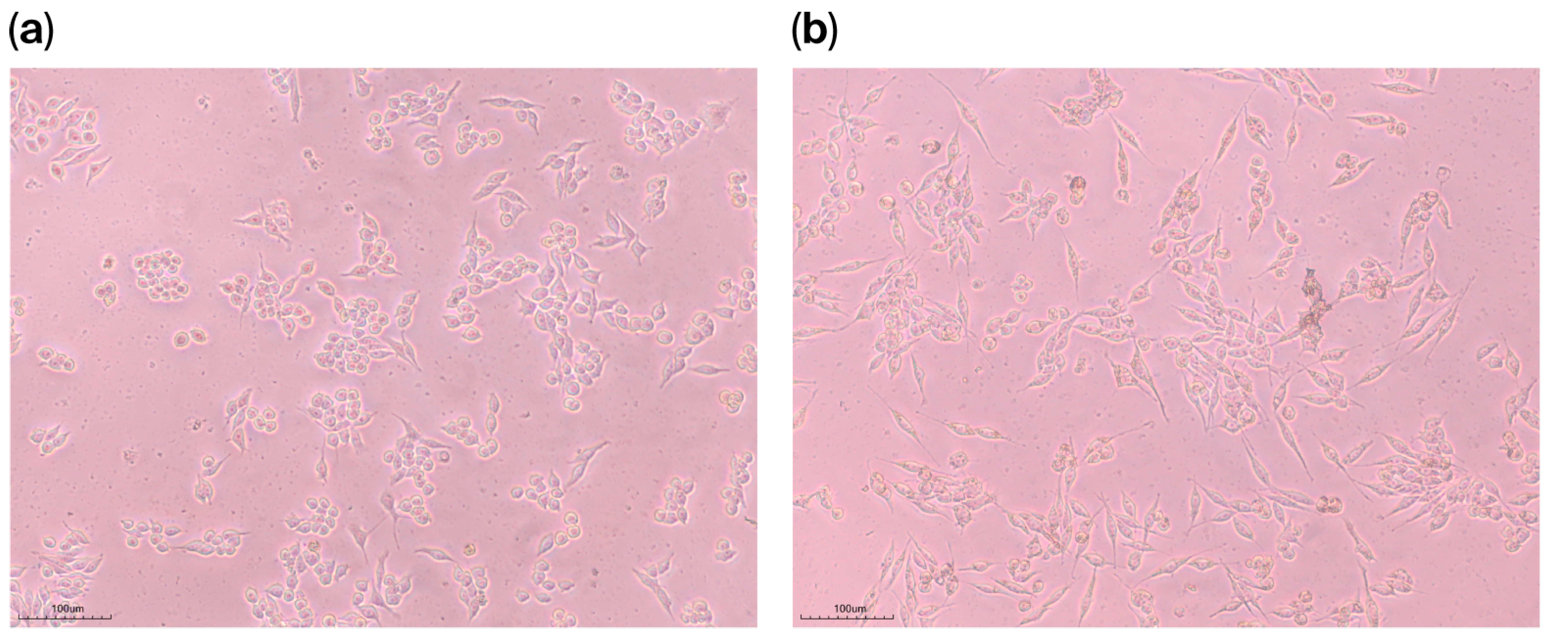
Cells were photographed using an imaging system (Lanoptik, Model: MC4KW-G1, Leeds, UK) after being exposed to the test substance for 24 h, under a Nikon ECLIPSE TS100 (Tokyo, Japan) inverted microscope at 100× magnification.
2.6.2. Determination of IC50
The cytotoxicity of the extract was evaluated using an MTT assay on HaCaT cells (iCell Bioscience Inc., Shanghai, China). Cells were seeded in 96-well plates at a density of 1 × 10⁵ cells/mL (100 µL/well) and incubated at 37 °C in a humidified atmosphere containing 5% CO2 for 24 h. After incubation, the culture medium was removed and replaced with 100 µL of the extract at the desired concentration. Cells were then incubated for an additional 24 h under the same conditions. Following treatment, 10 µL of MTT solution (5 mg/mL) was added to each well, and the plate was incubated for 4 h. The supernatant was carefully removed, and the resulting formazan crystals were dissolved in 100 µL of a solubilization solution consisting of DMSO and 10% (w/v) SDS (9:1, v/v). The absorbance was measured at 570 nm after shaking for 5 min. Cytotoxicity (%) was calculated according to Equation (3).
For IC
50 determination, the extracts were dissolved in DMSO and diluted in DMEM supplemented with 5% FBS to obtain final concentrations ranging from 12.5 to 200 mg/mL. The cytotoxicity assay was performed as described above [
24]. The IC
50 values were calculated from dose–response curves using GraphPad Prism 5.0 software.
The cytotoxicity of the extract toward the tested cell line was classified as follows:
IC50 < 5 µg/mL (high cytotoxicity);
5–10 µg/mL (moderate cytotoxicity);
10–25 µg/mL (low cytotoxicity);
>25 µg/mL (non-toxic) [
25].
2.7. Reef-Safe Sunscreen Formulation
To ensure the environmental safety of the sunscreen, an environmental risk assessment was conducted for two formulations: one without perilla extract (SN) and one containing perilla extract (SE). The evaluation focused on three necessary elements, which were persistence (P score), bioaccumulation (B score), and toxicity (T score), following the protocols described by Choi et al. (2023) [
11].
The algorithm by Choi et al. [
11] assesses hazards associated with the substances used in the cosmetic formulation in terms of persistence (P), bioaccumulation (B), and toxicity (T) by searching for information from the US Environmental Protection Agency (US EPA) and the European Chemical Agency (ECHA).
Persistence is assessed based on biodegradation, bioaccumulation is assessed based on the Bioconcentration Factor (BCF) value, and toxicity is assessed based on the value of predicted no effect concentration (PNEC) or the ecotoxicological Threshold of Toxicological Concern (Eco TTC).
The persistence score (P score) considers a substance’s degradability based on the chemical database, where a score of 0 means no degradation and a score of 100 means immediate degradation. Natural extracts that are extracted by water and ethanol are considered to be highly degradable; therefore, a score of 100 is considered for perilla seed extract.
The bioaccumulation score (B score) is based on the EU PBT Guideline and the NIER Notification 2020-8, with the scores classified as shown in
Table 1.
The toxicity score (T score) is based on information on the toxicity of the substances. Then, toxicity data is used to calculate the PNECwater value (mg/L) by dividing the LC50 or NOEC value by the Assessment Factor (AF). Once the PNECwater value is obtained from the calculation, it is substituted into the following equation: T Score = 4.3651ln(PNECwater value in mg/L) + 76.115.
Once the P, B, and T values for each substance are obtained, these values are used to calculate the substance score (Score S) using Equation (4).
The coefficients a, b, and c were assigned based on the ingredient category (organic, inorganic, or botanical). The weights of each group of substances are shown in
Table 2.
P score (persistence) is scored based on biodegradability (0–100).
B score (bioaccumulation) is determined using the BCF according to the EU PBT Guidelines (0–100).
T score (toxicity) is calculated using the PNEC in water, derived from LC50 or NOEC values and AF.
In this calculation, water is assigned an S score of 100.
After assessing the hazards of the formulation’s ingredients, the next step is to calculate the product’s potential environmental risk by determining the substance’s score in product, or Score (S in P), using Equation (5).
where Score (S) is the safety value of any ingredient in a formula, and content (%) is the percentage of any ingredient in a formula. The use factor depends on the formula’s concentration. If the general formula is 1.00, but the cosmetic formula has a high concentration, it is set to 0.5.
After calculating the Score (S in P) of each ingredient in the formulation, the Score P is calculated by summing the Score (S in P) of all ingredients in the formula, as shown in Equation (6). The closer the Score (S in P) is to 100, the safer the product.
Sunscreen formulations were designed in emulsion form, without sunscreen agents that pose a risk of harm to coral reefs, namely nano ZnO, Benzophenone-1 (BP-1) [
26], Benzophenone-2 (BP-2) [
27], Benzophenone-3 (BP-3) or Oxybenzone, Ethylhexyl Methoxycinnamate (EHMC) or Octinoxate, and Octocrylene (OC), based on the research of Rücker et al. (2025) [
28]. The formulations used in this study are shown in
Table 3, which compares formulations containing perilla seed extract (SE) and those without perilla seed extract (SE). The concentration of the extract was equivalent to the concentration used to test its effectiveness in sun protection.
The SE and SE were prepared as follows: First, phase A (water, carbomer, and 1,3-butylene glycol) was weighed into a mixing container and heated to 75–80 °C according to the formula (
Table 3). Phase B (Emulgade 165, Olivem 300, Dub Helioptima, Tween 60, homosalate, ethylhexyl salicylate, avobenzone, and polyester-7) was weighed in a separate mixing container according to the specified proportions, then heated to 75–80 °C. After both containers reached the desired temperatures, a homogenizer (IKA T25, Staufen, Germany) was used to mix them. Once the emulsion had cooled below 50 °C, the remaining ingredients were added and blended thoroughly.
2.8. Stability Evaluation
The stability of the sunscreen formulations (SN and SE) was evaluated under three storage conditions, 4 °C, 25 °C, and 45 °C, for a period of 2 months. Observations were recorded on the first day and 2 months after the products were formulated [
29]. Furthermore, the sunscreen formulations were evaluated based on their physicochemical properties, which included monitoring for separation and color changes (yellowness) using a colorimeter (3nh NR10QC, Guangzhou, China), and measuring viscosity using a viscometer (Brookfield DV2T, Middleboro, MA, USA) with Spindle LV-03.
When evaluating the color of sunscreen products, yellowing on shelves is a common issue. Therefore, this research focuses on assessing the yellowing of a formula compared to a freshly made product and a product stored for 2 months at different temperatures. The yellowing value (b-value; b*), measured using a colorimetric instrument [
30], is used to calculate the difference in yellowness (∆b) between the formula made on the first day and after two months of storage using Equation (7).
where ∆b is the total difference in yellowness between two samples; b*
initial is the yellow value (b*) of a fresh product measured using a colorimeter; and b*
final is the yellow value of the product after storage for a period of time.
2.9. SPF Test
The efficacy of the sunscreen formulations was tested using a UV2000S Ultraviolet Transmittance Analyzer (Labsphere, North Sutton, NH, USA). The samples (SN and SE) were applied onto a PMMA (Polymethylmethacrylate) substrate plate at a concentration of 1.3 mg/cm2. The samples were spread evenly and allowed to dry. The analyzer measured the UV transmission from 290 nm to 400 nm. The SPF, PA, and critical wavelength were calculated and the SPF values obtained from the tests were statistically analyzed for difference using a t-test, with a p-value of 0.05.
2.10. Clinical Test
This clinical study was conducted in accordance with ethical principles for human research. This research project has received accreditation for this study in accordance with international ethical standards for human research from the Academic and Ethics Committee of Phranakhon Rajabhat University. This accreditation follows the Declaration of Helsinki and the Belmont Report, the CIOM guideline, and the International Conference on Harmonization in Good Clinical Practice (ICH-GCP). The project number is 01.063/67. The study protocol included informed consent procedures, ensuring volunteers were fully informed of the objectives, methods, and potential risks.
This clinical study was conducted as a double-blind, non-randomized trial to assess skin irritation and photoprotective efficacy under real-world conditions. The double-blind methodology was strictly implemented to eliminate potential experimental bias from both investigators and participants. A non-randomized design was purposefully adopted as all volunteers were assigned to receive both formulations; this approach ensured ethical fairness by providing every participant with actual UV protection while facilitating a direct comparative analysis between the two sunscreen products. Participants were recruited from the Bang Khen district, Bangkok, Thailand, specifically targeting outdoor workers with daily sun exposure of at least four hours. A total of 30 volunteers were enrolled, a sample size strategically chosen to ensure that all skin evaluations could be completed within a single day, thereby allowing for data comparison under consistent environmental conditions. To maintain anonymity and uphold the integrity of the double-blind process, each volunteer was assigned a unique three-digit randomized identification code.
The inclusion criteria were healthy volunteers with Fitzpatrick skin types 1–3, aged between 20 and 60 years. Exclusion criteria included individuals with a history of skin cancer, excessive hair or nevi at the test sites, pre-existing pigmentation or photoaging diseases, or active skin conditions [
31]. Furthermore, participants who had undergone prolonged sun exposure within the three months prior to the study were excluded.
A primary irritation test was conducted on at least 10 volunteers. The sunscreen formulations were applied to the upper arm using Finn Chambers® (Hillerød, Denmark). The chambers remained in place for 24 h, during which volunteers were instructed not to wash the test area. Following the removal of the patches, skin reactions were monitored objectively using bioengineering methods to ensure data precision.
The photoprotective efficacy of the formulations (SN and SE) was evaluated on at least 30 volunteers under actual usage conditions. Volunteers applied the assigned products to both forearms daily during their outdoor work activities. To minimize subjective bias associated with traditional visual grading, objective skin bioengineering techniques were employed as the primary evaluative tool [
32]. Measurements were recorded at baseline (before application) and at the end of the study period.
To enhance data reliability and provide a more accurate assessment than visual observation, instrumental measurements were performed using bioengineering principles. This included the assessment of TEWL to monitor skin barrier integrity and the measurement of skin erythema (redness) using a Mexameter to detect subtle inflammatory changes. This quantitative approach allows for precise tracking of skin physiological parameters and facilitates robust statistical decision-making regarding product safety and efficacy.
All quantitative data were expressed as mean ± standard deviation (SD). Statistical significance was determined using a paired t-test to compare the differences between baseline and post-treatment values. The threshold for statistical significance was set at p-value < 0.05. Data analysis was performed using Microsoft Excel 365.
2.10.1. Safety Evaluation
A group of 17 healthy volunteers with skin types 1–3, without a history of skin cancer, excessive hair or nevi, pigmentation or photoaging diseases, or prolonged sun exposure within 3 months, was recruited [
31]. The volunteers’ skin color was classified using a Skin Colorimeter CL 400 (Courage-khazaka electronic GmbH, Cologne, Germany), measuring the Individual Typology Angle (ITA) according to Fitzpatrick skin phototype [
33]. Skin type 1 had an ITA greater than 55, skin type 2 had an ITA between 42 and 55, and skin type 3 had an ITA between 35 and 41. Volunteers with active skin diseases were excluded. The test was performed in a controlled room (20 °C, 35–45% RH).
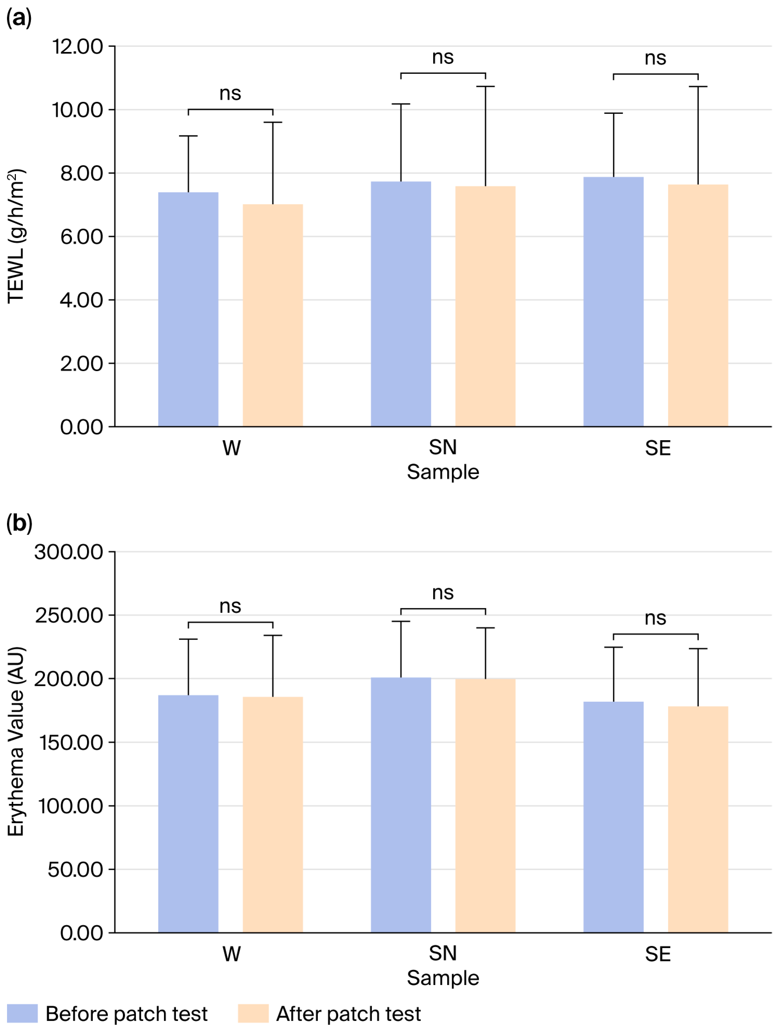
For skin irritation tests using the bioengineering method, TEWL and erythema were measured on the volar forearm before application, and the values were compared with those from the Finn Chamber
® patch. The sunscreen samples were applied to the upper arms and left for 24 h without washing. TEWL and erythema were measured again after 24 h [
31]. The results were then analyzed using a paired
t-test
via Microsoft Excel 365.
The percentage difference in TEWL before and after the patch test can be calculated using Equation (8).
2.10.2. Efficacy Evaluation
A total of 30 volunteers with skin types 1–3 participated in the efficacy test. To control the UV exposure (UV index was also recorded), the volunteers stayed in the same district, Bang Khen, Bangkok, Thailand, for 4 h (10 A.M. to 2 P.M.). To overcome human bias, bioengineering techniques were utilized in the efficacy tests. To compare the SE and SN formulas, a Sebumeter SM 815 (Courage-khazaka electronic GmbH, Cologne, Germany) was used to measure sebum levels on the left and right forearms after applying 5.2 mg of sunscreen in the controlled area (4 cm2) for 5 min. The difference in sebum secretion levels on the skin between two sunscreen formulations was analyzed using a t-test with a p-value set at 0.05. A Mexameter MX 18 (Courage-khazaka electronic GmbH, Cologne, Germany) was used to measure erythema and melanin levels before and after the application of the sunscreens. A Skin Colorimeter CL400 was used to measure the skin whiteness (L*) of the volunteers to compare the values before and after applying both sunscreens.
On the first day, erythema values, melanin values, and skin whiteness (L*) were measured. Then, the volunteers applied the sunscreens without covering their skin. After daily sunlight exposure, the skin parameters of the 30 volunteers were measured 24 h later. Finally, differences between pre- and post-exposure values were analyzed using a paired t-test via Microsoft Excel 365 with a p-value = 0.05.
4. Discussion
The present study demonstrates the potential of
P. frutescens seed extract as a bioactive ingredient in broad-spectrum, reef-friendly sunscreen formulations. The results indicate that the extraction method significantly influences photoprotective efficacy, with the 95% ethanolic extract obtained from a 1-week maceration process yielding the highest SPF value (22.61). This finding aligns with previous studies that suggest that ethanol is an effective solvent for extracting phenolic compounds and flavonoids, which are responsible for UV absorption and antioxidant activity [
38]. The prolonged maceration time likely facilitated the maximum diffusion of these bioactive compounds from the plant matrix into the solvent [
39].
The chemical analysis results using GC-MS/MS revealed that the perilla seed extract was rich in polyunsaturated fatty acids, predominantly linolenic acid (omega-3), followed by palmitic acid and linoleic acid. Linoleic acid’s potent anti-inflammatory and antioxidant properties are well documented [
40]. Antioxidant efficacy testing using a DPPH assay revealed that the perilla seed extract exhibited an IC
50 of 38.95 μg/mL. Based on antioxidant classification criteria, this extract is considered to have high antioxidant activity [
34]. This result provides significant empirical evidence confirming the efficacy of perilla seed extract as a bioactive ingredient in cosmetic formulations. In the context of sunscreen development, high antioxidant activity plays a crucial role, as ultraviolet (UV) radiation is a major trigger for the production of reactive oxygen species (ROS) within the skin, leading to oxidative stress, collagen damage, and photoaging [
41]. The relationship between the chemical composition and the skin-protective efficacy of
P. frutescens seed extract can be attributed to its high content of omega unsaturated fatty acids. Based on the GC-MS/MS analysis in this study, linolenic acid (C18:3) was identified as the predominant component (43.54%) of the extract. These findings align with the study by Choi et al. [
17], which investigated the effects of perilla in a hairless mouse model and demonstrated that these fatty acid components play a crucial role in mitigating UV-induced photoaging. Their research confirmed that the extract attenuates epidermal thickening and inhibits collagen degradation by downregulating the expression of matrix metalloproteinases (MMPs), which are the primary contributors to wrinkle formation and skin deterioration. Therefore, the abundance of linolenic acid in this formulation provides strong evidence for the extract’s role as an agent of biological photoprotection. Rather than merely acting as a surface-level UV filter, it actively prevents and repairs UV-induced cellular damage at the molecular level. Consequently, the developed sunscreen formulation offers dual benefits by delivering effective sun protection alongside nourishing and anti-aging properties.
An evaluation of the stability of the developed sunscreen product revealed that temperature plays a significant role in changes to physical characteristics, particularly changes in color and sun protection efficacy. This phenomenon aligns with research by Abud and Ibrahim (2025), which indicated that temperature fluctuations directly affect the degradation of UV filters and changes in color in cosmetic formulations, potentially leading to a long-term decrease in SPF value [
42]. Regarding the properties of perilla seed extract, while it exhibits notable benefits as an antioxidant and skin cell strengthening agent, as demonstrated by cytotoxicity tests, its high content of unsaturated fatty acids, especially linolenic acid (C18:3) and linoleic acid (C18:2), with its numerous double bonds, makes it highly susceptible to oxidation when exposed to heat and light. This results in the product turning a deep yellow over time. To address this color change and improve formulation stability in the future, further research is needed. Formulation strategies can be implemented in two main ways. The first method involves adding antioxidants, such as tocopherol (vitamin E), directly to the formulation to inhibit the oxidation of unsaturated fatty acids [
43]. The second method uses oleogel technology to encapsulate the extract. Developing a sunflower wax-based oleogel formulation allows for the encapsulation of the oil within a gel network structure. This strategy has proven highly effective with oils containing over 80% unsaturated fatty acids, such as olive oil and avocado oil. The oleogel structure reduces contact between the oil and external factors, significantly reducing the yellowing of the formulation [
44]. Therefore, applying oleogel techniques or balancing antioxidants can enhance the stability, effectiveness, and shelf life of sunscreen products containing perilla extract.
Viscosity stability test results revealed an interesting difference between SN and SE. In the SN formulation, viscosity tended to decrease under all tested temperatures, possibly due to the softening of the emulsion structure under continuous thermal activation. Conversely, the SE formulation exhibited specific flow behavior, with viscosity increasing at 4 °C and 25 °C but decreasing only slightly at 45 °C. This phenomenon can be explained by the chemical composition of the perilla seed extract, which, according to GC-MS/MS analysis, is rich in fatty acids and triglycerides. As reported by Valeri and Meirelles (1997), the viscosity of fatty acids and triglycerides is closely related to temperature, with viscosity decreasing significantly with increasing temperature due to increased thermal energy reducing intermolecular forces; this makes the formulation flow easier [
45]. However, at low temperatures (4 °C) or room temperature (25 °C), the fatty acids in the perilla seed extract act as bodying agents in cosmetic formulations, with fatty acid molecules arranged more densely in the emulsion network structure, resulting in a thicker formulation and better physical stability under normal conditions. However, when the temperature rises to 45 °C, the heat energy disrupts this arrangement, causing a slight decrease in viscosity, which is a natural characteristic of lipids. Therefore, the inclusion of perilla seed extract not only enhances the biological activity of sunscreen products but also plays an important role in improving rheological properties, helping to maintain texture and increase product stability at room temperatures and in chilled conditions. In addition to the chemical composition factors of the extract, the physical stability of the emulsion formulation can also be explained in depth through the principles of fluid dynamics, referencing Stokes’ law, which is crucial for evaluating the stability of cosmetics. According to Querol et al. (2017), who studied the relationship between viscosity and storage stability in emulsions, the law states that as viscosity increases, the movement rate of particles decreases significantly [
46]. The experimental results showed that the SE formulation had a significantly higher viscosity than the SN formulation (particularly at temperatures of 4 °C and 25 °C). This increased viscosity acts as a mechanical drag shield, slowing the movement of oil droplets within the emulsion structure. Therefore, the high viscosity of the SE formulation serves as a key indirect indicator of excellent formulation stability (high physical stability), as a high viscosity helps prevent particle collisions and coalescence, as well as phase separation.
Safety is a paramount concern for cosmetic products. The primary screening for cytotoxicity on HaCaT cells provided significant insights into the safety and biological activity of the
P. frutescens seed extract. The results indicated that the extract, which is rich in omega fatty acids, does not merely exhibit a lack of toxicity but potentially contributes to skin cell recovery and regeneration. The negative cytotoxicity values observed in the extract-treated group suggest a stimulatory effect on cell viability or metabolic activity. This finding is consistent with previous in vivo studies by Choi et al. [
17], which demonstrated that perilla seed oil, particularly due to its fatty acid composition, could enhance skin barrier functions and increase skin thickness in hairless mouse models. The alignment between our
in vitro results on human keratinocytes and the previously reported animal data underscores the potential of perilla seed extract as a bioactive ingredient capable of supporting skin health and structural integrity against environmental stressors. In addition, the cytotoxicity assay on HaCaT keratinocytes showed an IC
50 value of approximately 12.9 mg/mL, indicating that the extract is practically non-toxic to human skin cells according to standard toxicity classifications. Furthermore, the formulation addresses the growing global concern regarding the environmental impact of sunscreen. By avoiding oxybenzone and octinoxate [
47] and utilizing the reef-safe formulation criteria (persistence, bioaccumulation, and toxicity scoring) [
11], this product meets the eco-friendly standards required by emerging regulations in protected marine areas [
48].
A major strength of this study lies in the application of non-invasive bioengineering techniques coupled with robust statistical analyses for human safety evaluation. As an objective approach to assessing skin compatibility, TEWL was measured to monitor the integrity of the skin barrier, aligning with the evaluation principles described by Smith et al. (2004) [
37]. Our findings revealed that both the base formulation (SN) and the formulation containing perilla seed extract (SE) did not induce any severe adverse effects on the TEWL of the 17 human volunteers. The application of these formulations did not significantly disrupt the skin barrier function, as evidenced by the lack of statistically significant differences in TEWL values before and after application (
p-value > 0.05).
Furthermore, to strengthen the reliability of the safety profile, the study design was enhanced by incorporating an instrumental evaluation of skin erythema (redness) before and after the patch test. Traditionally, skin irritation and erythema are evaluated through visual grading, which is inherently subjective and susceptible to human error or environmental biases. By utilizing a Mexameter for this assessment, we were able to obtain precise, objective, and quantifiable measurements of skin redness. This instrumental approach not only eliminates the inaccuracies associated with visual scoring but also generates reliable continuous numerical data, thereby enabling a more rigorous and statistically sound decision-making process regarding the dermatological safety of the developed sunscreen.
The in-use clinical evaluation conducted on 30 volunteers provided a comprehensive comparison between the base formulation (SN) and the perilla seed extract-enhanced formulation (SE) under real-world conditions. Regarding consumer acceptability, sensory perception is a critical determinant of product compliance. Our findings indicated that while the SE formulation was perceived as being slightly more oily than the SN formulation—likely due to the lipid-rich nature of the 1% perilla seed extract—the difference was not statistically significant (
p > 0.05). This is a vital result for product development, as formulation esthetics, particularly greasiness and stickiness, are primary factors influencing consumer rejection of sunscreens, as reported by Diehl et al. (2021) [
49]. The fact that the addition of the extract did not significantly alter the perceived oiliness suggests that the SE formulation remains highly acceptable for daily use.
Interestingly, the SE formulation showed a slightly higher efficacy in preventing the skin dullness than the SN formulation. This superior performance can be attributed to the potent antioxidant properties of the perilla seed extract, which helps neutralize UV-induced oxidative stress and prevents the oxidative darkening of existing melanin. The significant reduction in melanin index and improvement in skin luminosity observed in the SE formulation aligned with previous reports on the biological activities of omega-3 fatty acids in skin health. For instance, while Choi et al. [
17] focused on the anti-photoaging effects of perilla oil in animal models, our findings extend this evidence to human subjects, demonstrating a tangible brightening effect under real-world conditions. Furthermore, the use of
P. frutescens as a natural SPF booster is consistent with the current trend in green cosmetology, where botanical extracts are utilized to minimize the concentration of chemical filters, thereby reducing both potential skin irritation and environmental toxicity.
Regarding skin inflammation, although both formulations led to a marginal reduction in erythema values, no statistically significant differences were observed (p > 0.05) between pre- and post-application measurements. This suggests that while the products are safe and non-irritating, the duration or conditions of this specific test may not have been sufficient to demonstrate a significant anti-inflammatory shift. The most notable evidence of the bioactive efficacy of the extract was observed in the changes to the melanin index. Volunteers using the base formulation (SN) exhibited a 0.64% increase in melanin levels, reflecting the natural tanning response to UV exposure. Conversely, those using the SE formulation, containing 1% perilla seed extract, showed a 1.32% reduction in melanin levels. This striking contrast highlights the extract’s dual functionality; it not only serves as a protective barrier but also acts as an active depigmenting agent that inhibits melanogenesis even under high-UV-index conditions. These results further validate the integration of perilla seed extract as a high-performance bioactive ingredient in photoprotective formulations.
Despite the promising results, certain limitations of this study must be acknowledged. First, the clinical evaluation period was relatively brief, focusing primarily on immediate and short-term photoprotective effects. Long-term studies are required to fully elucidate the cumulative benefits of perilla seed extract on skin barrier repair and chronic photoaging. Second, while the reef-friendly status of the formulation was determined based on the exclusion of known harmful filters and predictive environmental impact models, further direct ecotoxicological assessments on various coral species would provide more definitive evidence of its marine safety. Future research should aim to bridge these gaps through longitudinal clinical trials and longitudinal ecological monitoring in marine environments.
In conclusion, the developed sunscreen containing P. frutescens extract offers a dual benefit: effective photoprotection for human skin through UV absorption and cell proliferation, and environmental safety for marine ecosystems. This supports the utilization of botanical extracts as sustainable alternatives or boosters in modern cosmeceutical formulations.