Article

Preparation and Investigation of Nano-TiO2-Modified Silicone-Based Reflective Thermal Insulation Coatings


1
School of Material Science and Technology, Wuhan University of Technology, Wuhan 430070, China
2
Core Research Cluster for Materials Science (CRCMS), Advanced Institute for Materials Research, Tohoku University, Sendai 9808577, Japan
*
Authors to whom correspondence should be addressed.
Coatings 2026, 16(3), 319; https://doi.org/10.3390/coatings16030319
Submission received: 31 January 2026 / Revised: 24 February 2026 / Accepted: 3 March 2026 / Published: 5 March 2026
(This article belongs to the Special Issue Ceramic and Glass Material Coatings)

Abstract

A nano-TiO2-modified silicone-based reflective thermal insulation coating is successfully synthesized. The influence of the nano-TiO2 content on the microstructure, adhesion strength as well as near-infrared reflectivity (NIR) of the coatings before and after heat treatment is investigated. The results demonstrate that the coating is an organic/inorganic composite coating composed of muscovite, rutile-phase titanium dioxide and an organosilicon binder before heat treatment. The addition of an appropriate amount of nano-TiO2 helps fill the pores in the coating, resulting in a dense coating and improved adhesion. Meanwhile, due to the reduced average size of the pigment, the reflectance of the coating is maintained or enhanced. When the addition amount is 5.0 wt.%, the coating achieves the highest bonding strength of Grade 1 with a reflectivity of 0.830. After heat treatment at 1000 °C for an hour, the coating transforms into an inorganic coating composed of partially melted muscovite and rutile-phase TiO2. The nano-TiO2 promotes the formation of a molten phase, which further increases the coating density and makes the surface smoother. Consequently, the coating’s bonding strength and reflectance are further improved, reaching Grade 0 and 0.945 respectively.

1. Introduction

Modern industry has developed rapidly, leading to an increasing demand for equipment operating in high-temperature environments. Components such as aircraft engine casings, high-temperature furnaces, chimneys, and exhaust pipes are all subjected to high-temperature working conditions. However, prolonged exposure to elevated temperatures often results in metal aging and corrosion, thereby degrading equipment performance and even shortening service life [1]. As a result, effective thermal management and protection measures are becoming essential. In particular, high-temperature-resistant thermal insulation coatings have aroused extensive interest as an economical protection measure. Traditional high-temperature-resistant thermal insulation coatings include barrier-type, radiation-type and reflective-type [2]. Among them, reflective thermal insulation coatings have high reflectivity in the near-infrared band and are an important method to block radiation heat transfer in high-temperature environments, especially high-temperature vacuum environments. Reflective thermal insulation coatings can be applied to the inner walls of high-temperature furnaces to reduce energy loss, playing a significant role in energy conservation. Against the backdrop of “carbon peaking” and “carbon neutrality” initiatives, energy saving and consumption reduction, as well as cost reduction and efficiency improvement, represent fundamental requirements for enterprises under current market economic conditions. This study aims to develop a reflective thermal insulation coating for application on energy-saving furnace inner walls, thereby providing a technically feasible solution that combines economic viability with long-term effectiveness for the energy-saving retrofit of industrial high-temperature furnaces and kilns.
Generally, based on the binders, the reflective thermal insulation coatings could be classified into organic and inorganic coatings. The organic coatings account for the vast majority due to their high bonding strength and good construction performance [3]. Common organic binders include epoxy resin, phenolic resin and silicone resin [4,5,6]. Due to a higher chemical bond energy of Si-O than that of C-C and C-O, as well as a lower infrared absorption [7], silicone is more suitable as the binder for reflective thermal insulation coatings in a high-temperature environment. Meanwhile, during high-temperature pyrolysis, Si-O undergoes a high-temperature ceramization transformation through Si-O bonds to form a continuous SiO2 ceramic phase. In particular, the heat resistance of the organic/inorganic composite silicone resin is further enhanced. Yoshinaga et al. [8] introduced Si, Al, Ti and Nb into the main chain of silicone resin by the sol–gel method, and found the thermal stability of resin improved significantly. As for the functional fillers, Wu et al. [9] improved the thermal resistance of silicone resin by adding muscovite and low-melting-point glass powder. Research also indicates that incorporating nanoparticles can effectively enhance the thermal resistance. Mansouri et al. [10] incorporated muscovite into silicone rubber, achieving ceramization enhancement through the reaction between mica and SiO2 produced during silicone rubber pyrolysis. Suchikova et al. [11] reported that rutile-type TiO2 exhibits excellent thermal stability and a high refractive index, which can improve the overall reflectivity of coatings; however, pure rutile TiO2 nanoparticles are prone to agglomeration issues. Podelinska et al. [12] systematically compared TiO2 nanomaterials synthesized by various methods, revealing the key mechanisms by which multiple factors synergistically regulate crystal phase composition, nanoparticle packing density, and surface morphology. Zeng et al. [13] summarized the effects of various approaches on the synthesis of high-performance well-dispersed TiO2, providing a reference for the design and synthesis of high-performance TiO2. Huang et al. [14] incorporated nano-TiO2 into silicone resin. Due to the excellent compatibility between nano-TiO2 and the resin, the thermal decomposition rate of the resin was significantly reduced, leading to a substantial improvement in its thermal resistance. Yan Zhou et al. [15] incorporated TiO2 into the epoxy resin, enhancing the strength of the epoxy resin and endowing it with solar reflection ability. This approach yielded a high-reflectivity coating with a solar reflectance of 0.9. Peoples et al. [16] incorporated multi-scale TiO2 particles with different particle sizes into acrylic resin. Owing to the scattering synergistic effect among particles of varying sizes, the solar reflectance of the coating was significantly enhanced, thereby achieving efficient passive radiative cooling. Our former study developed an organic/inorganic hybrid binder by the sol–gel method using tetraethyl orthosilicate, methyl triethoxysilane, dimethyl diethoxysilane, and 3- (isobutylloxy) propyl trimethoxysilane as raw materials. A high-temperature silicone-based reflective thermal insulation coating was prepared by using micron-sized TiO2 and muscovite as the functional filler. The optimized coating had a reflectivity of 0.83 in the 0.75~2.5 μm band after heat treatment at 600 °C. Despite existing studies on silicone-based coatings, there is still a gap in research on the thermal protection performance and high-reflectivity coatings at high temperatures. Inspired by the above research, in order to further enhance the thermal resistance of the prepared silicone-based reflective thermal insulation coating, the micron-sized TiO2 was partially replaced with nano-TiO2 to construct a multi-scale particle system. Nano-TiO2 was expected to promote liquid-phase sintering and pore filling at a relatively lower temperature. A nano-TiO2-modified silicone-based reflective thermal insulation coating was expected to be synthesized, which would achieve the synergistic optimization of the microstructure, adhesion strength, and spectral reflection performance through room-temperature curing and high-temperature ceramization mechanisms, providing a novel strategy for the design of thermal protection materials under extreme temperature environments.

2. Materials and Methods

2.1. Coatings Preparation

Alumina ceramic plates with dimensions of 30 mm × 30 mm × 2 mm were used as the substrate. The organic precursors, namely tetraethyl orthosilicate (TEOS), methyltriethoxysilane (MTES), and dimethyldiethoxysilane (DMDES), were utilized in the system, where anhydrous ethanol (EtOH) served as the solvent. Muscovite powder and micron-sized TiO2 were selected as functional pigments, which are expected to endow the coating with the desired reflectivity. Nano-TiO2 was introduced as a modifier, aiming to enhance the thermal resistance of the coating. Meanwhile, 3-(methacryloyloxy) propyltrimethoxysilane (KH570) was utilized as a silane coupling agent, with hydrochloric acid as the catalyst, aiming to improve the interfacial adhesion between the coating and the substrate. Hydrochloric acid and anhydrous ethanol were obtained from Sinopharm Chemical Reagent Co., Ltd. (Shanghai, China), micron-sized TiO2 was sourced from CNNC Hua Yuan Titanium Dioxide Co., Ltd. (Changsha, Hunan, China), while the rest of the reagents were purchased from Shanghai Macklin Biochemical Co., Ltd. (Shanghai, China).
The organic binder was prepared via a sol–gel process. TEOS, MTES, and DMDES were mixed in a molar ratio of 3:2:1. Ethanol was used as the solvent, with a molar ratio of ethanol to silicon of 3:10. After stirring at room temperature for an hour, deionized water (equimolar to the solvent) and concentrated hydrochloric (HCl:H2O = 1:0.009) were added. The mixture was reacted in a 70 °C water bath for 3 h to obtain the organic precursor. After cooling, KH570, micron-sized TiO2, nano-TiO2, and muscovite were introduced and subjected to planetary ball milling for 6 h with a ball-to-powder ratio of 3:1 and a rotation speed of 260 r/min to prepare the coating slurry. The nano-TiO2 content was set at 2.5 wt.%, 5 wt.%, 7.5 wt.%, and 10 wt.%. The pigment-to-binder (P/B) ratio was 0.6. The coating slurry was applied to the alumina substrate surface by brush coating. After brushing the first coat, we waited for the surface to dry before brushing the second coat. This procedure was repeated until the coating thickness reached approximately 60~70 μm. The coating thickness was measured using a micrometer: first measuring the substrate thickness, then measuring the overall thickness after coating, with the difference representing the coating thickness. Five-point sampling was employed during measurement, and the average value was taken as the final thickness. After painting, the coating was dried at room temperature for 24 h to obtain room-temperature-cured coatings. The obtained coatings were then heat-treated in a muffle furnace in an air atmosphere at 600 °C, 800 °C, and 1000 °C for 1 h, followed by furnace cooling. The heating and cooling rates were both set at 5 °C/min.

2.2. Characterizations

Chemical phases of the coating were determined by an X-ray diffractometer (XRD, Empyrean, Thermo Fisher Scientific, Waltham, MA, USA) with Cu Kα radiation, a test angle range of 10–80°, a step size of 0.01°, and a scanning speed of 4° per minute. The chemical functional groups of the coating were analyzed using a Fourier transform infrared spectrometer (FT-IR, Nicolet 6700, Thermo Fisher Scientific, Waltham, MA, USA), with a scanning range of 4000 cm−1 to 400 cm−1. The microscopic morphology of the coating surface was observed using a cold-field emission scanning electron microscope (SEM, JSM-7500F, JEOL, Akishima, Tokyo, Japan) at an accelerating voltage of 5 kV. The thermal stability of the coating was tested using a comprehensive thermal analyzer (TGA, STA2500, NETZSCH, Selb, Germany), conducted from room temperature to 1000 °C at a heating rate of 10 °C/min in an air atmosphere. The adhesion strength of the coating was determined according to ISO 2409:2020, using the “cross-cut test method” [17]. A cross-cut tester (QFH-A, Zhejiang AIRUIPU Instrument Co., Ltd., Wenzhou, Zhejiang, China) with a blade spacing of 2 mm was employed for the test. The reflectance of the coating in the 0.75–2.5 μm band was tested using a UV-VISS-NIR spectrometer (UV3600, Shimadzu, Kyoto, Japan) with a signal acquisition step of 1 nm. A standard BaSO4 plate was used as the reference. The average reflectivity of the coating within the 0.75–2.5 μm wavelength range was calculated according to Equations (1)–(3).
ρ = 1 α τ ,
α = 0.75 2.5 α ( λ ) M b ( λ ) d λ 0.75 2.5 M b ( λ ) d λ ,
M b ( λ ) = 2 π h c 2 λ 5 e c h k b λ t 1
When light irradiates the material surface, three simultaneous processes occur: reflection, absorption, and transmission. Due to the opacity of the substrate, light energy cannot penetrate it, resulting in only reflection and absorption at the coating surface. Consequently, the transmittance (τ) is 0 [18]. In Equation (1), ρ, α, and τ represent the average reflectance, absorptance, and transmittance of the coating over the given wavelength range, respectively. The average absorptance across the specified band is calculated using Equation (2), where α(λ) denotes the absorptance of the coating at wavelength λ, and Mb(λ) is the spectral radiant exitance of a blackbody [19]. The latter is derived from Planck’s law of blackbody radiation, as expressed in Equation (3), where h is the Planck constant, c is the speed of light, kb is the Boltzmann constant and T = 1273.15 K [20].

3. Results

3.1. Microstructure and Chemicals of Coatings Before Heat Treatment

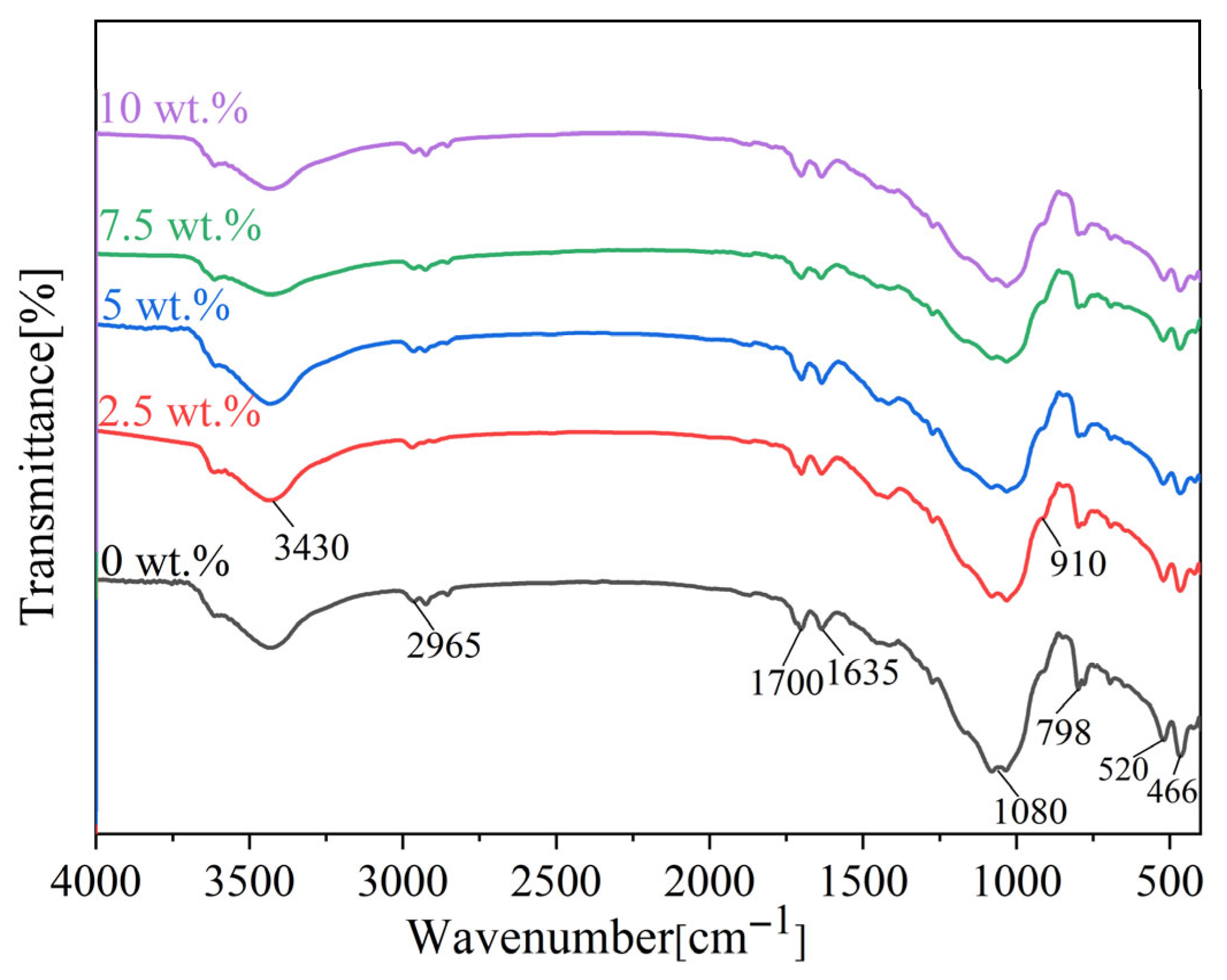
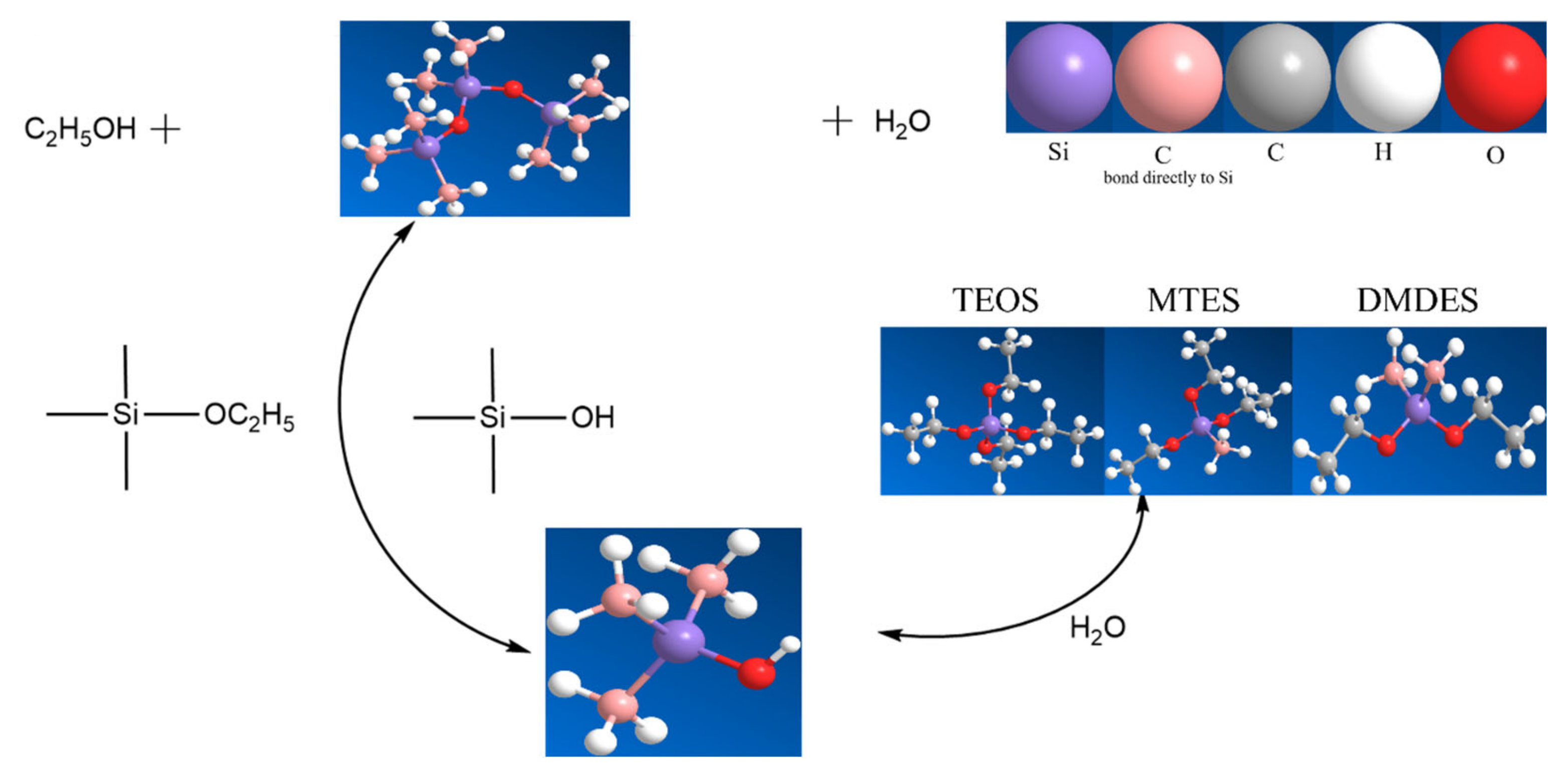
To identify the chemical composition of the prepared coating, infrared spectroscopy analysis was performed and the results are as shown in Figure 1. It can be seen that all the coatings exhibited similar infrared spectrums. There was a wide absorption peak near 3430 cm−1, which was caused by the -OH stretching vibration in chemical H2O [21]. Moreover, a very weak vibration absorption peak of Si-O-C2H5 appeared at 2965 cm−1 [22], indicating nearly complete hydrolysis with minimal ethoxy groups remaining, suggesting an adequate reaction time. The absorption peaks at 1700 cm−1 and 1635 cm−1 corresponded to the C=O stretching vibration and CH2=CH2 stretching vibration, respectively [23], which were attributed to the addition of KH570. Peaks at 798 cm−1 represented the absorption of Si-CH3 [24]. The absorption peaks among 1016 cm−1 to 1141 cm−1 were due to the Si-O-Si stretching vibration in the siloxane network [25]. Meanwhile, the peaks around 400–500 cm−1 are attributed to the Si-O-Si bending vibration, confirming the formation of Si-O-Si through the dehydration of Si-OH or reaction of Si-OH with Si-OR. Additionally, the peak at 910 cm−1 corresponded to the absorption of Al-OH in muscovite [26]. The alkoxysilane monomers would undergo hydrolysis and condensation reactions according to Figure 2. In the hydrolysis and polycondensation process of organo-siloxane, the oxyethyl group is first dehydrated to form a silicon hydroxyl group, and Si-OH is dehydrated with the Si-OH or dehydrated with the Si-OR to form a Si-O-Si bond.
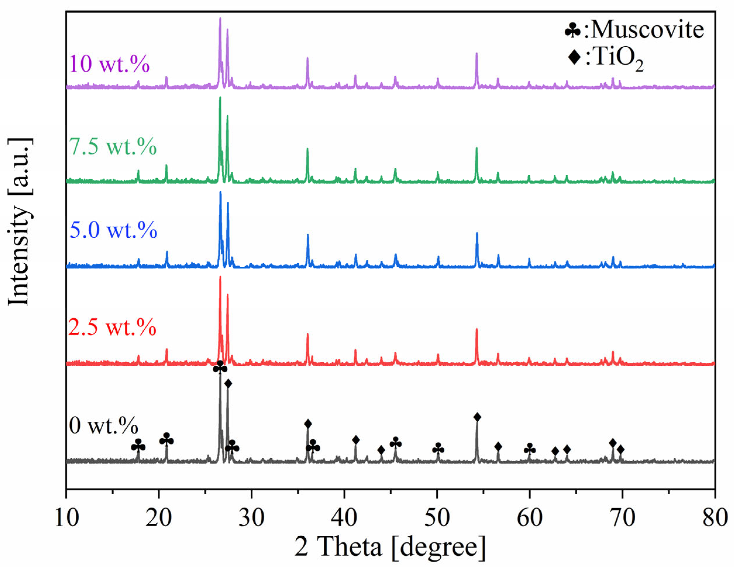
In order to identify the phase composition of the prepared coating, X-ray diffraction (XRD) analysis was performed on the coatings after curing at room temperature. Figure 3 shows the obtained XRD patterns, which displayed that all the coatings possessed a similar phase composition, with the presence of muscovite and rutile-phase titanium dioxide. No other crystalline phases were observed. The Si-O in the organic binder lacks a long-range ordered structure, thus exhibiting no sharp peaks; only possible broadened humps may appear, which would also be masked by the intense rutile titanium dioxide peaks.
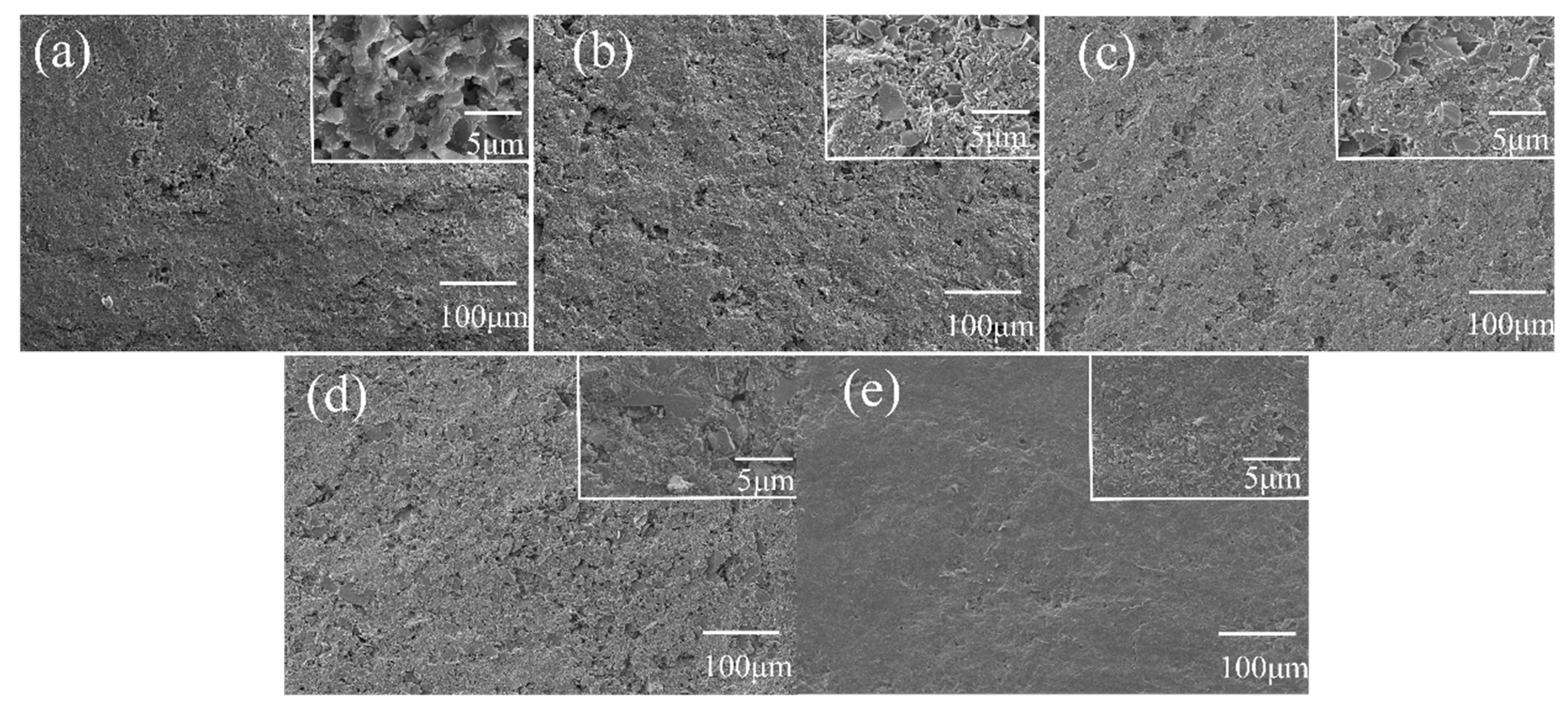
Figure 4 shows the surface morphology of the coatings modified by different proportions of nano TiO2. It can be observed that the coating without nano-TiO2, as shown in Figure 4a, was free of cracks. Meanwhile, it appeared rough with apparent micro-sized pores, which might be the ones left after water/alcohol evaporation during the curing process. Quantitative characterization of the micropore area fraction through image analysis revealed that the micropore area fractions for coatings doped with 0 wt.%, 2.5 wt.%, 5.0 wt.%, 7.5 wt.%, and 10 wt.% TiO2 were 6.4%, 6.1%, 3.7%,3.8%, and 0.7%, respectively. The 10 wt.%-doped coating exhibited the lowest porosity. With increasing addition amounts of nano-TiO2 particles, the coating surface became progressively denser and smoother, indicating that the nano-TiO2 particles might effectively fill the pores formed during the curing process.

3.2. Reflectance and Adhesion Strength of Coatings Before Heat Treatment

Figure 5 shows the near-infrared (NIR) reflection spectra of the coatings modified with different weight ratios of nano-TiO2 particles. The calculated reflectivity of the coatings is summarized in Table 1. To ensure data reliability, all reflectance curves were obtained by averaging the results of three measurements. The reflectivity initially decreases slightly then gradually increases to a maximum of 0.841 with 7.5 wt.% nano-TiO2 particles and subsequently drops with 10 wt.% nano-TiO2 particles, indicating an optimal addition amount for maximum reflectance. The variation in reflectivity with the content of nano-TiO2 particles was considered to be related to two factors. On one side, the nano particles might have more crystal defects such as vacancies, which will introduce impurity energy levels, promote the absorption in the NIR band, and thus reduce the reflectivity. On the other side, the scattering of light is closely related to the particle size of the pigment: The near-infrared reflectivity of coatings is mainly influenced by the Mie scattering theory, which states that the scattering efficiency is highest when the particle diameter is about half the wavelength of the incident light [27]. The closer the particle size is to it, the higher the reflectivity will be. For the NIR range (0.75~2.5 μm), the optimal pigment particle size corresponds to 0.375~1.25 μm. When the content of nano TiO2 is low, it promotes absorption in the near-infrared band, and the decrease in reflectivity is greater than the positive effect of the particle size on it. Therefore, the reflectivity is low. However, it should be noted that the study was unable to fully distinguish between absorption and scattering contributions, making it difficult to accurately attribute the observed reflectivity changes solely to either mechanism. The average size of the pigment decreased with increasing the content of nano-TiO2 particles. It was deduced that the coating with 7.5 wt.% nano-TiO2 particles might have the closest pigment particle size and therefore had the highest reflectivity.
Table 2 shows the adhesion strength of the coatings with different weight ratios of nano-TiO2 particles. Samples of each component were tested in three independent replicates, and the test results showed good stability. The results indicated that with the incorporation of nano-TiO2, the adhesion strength of the coatings initially increased and then decreased. The coatings with 2.5 wt.%, 5.0 wt.%, and 7.5 wt.% nano-TiO2 particles achieved an adhesion grade of level 1, which demonstrated that an appropriate addition of nano-TiO2 particles could enhance the adhesion between the coating and the substrate. On the one hand, the addition of nanoparticles promoted the densification of the coating, as shown in Figure 4, which improved the cohesive force of the coating and made it hard to be destroyed and peeled off. On the other hand, excessive addition of nanoparticles will introduce more interfaces. To some extent, these interfaces can be regarded as defects within the coatings, thereby reducing the bonding strength of the coating.

3.3. Microstructure and Chemicals of Coatings After Heat Treatment

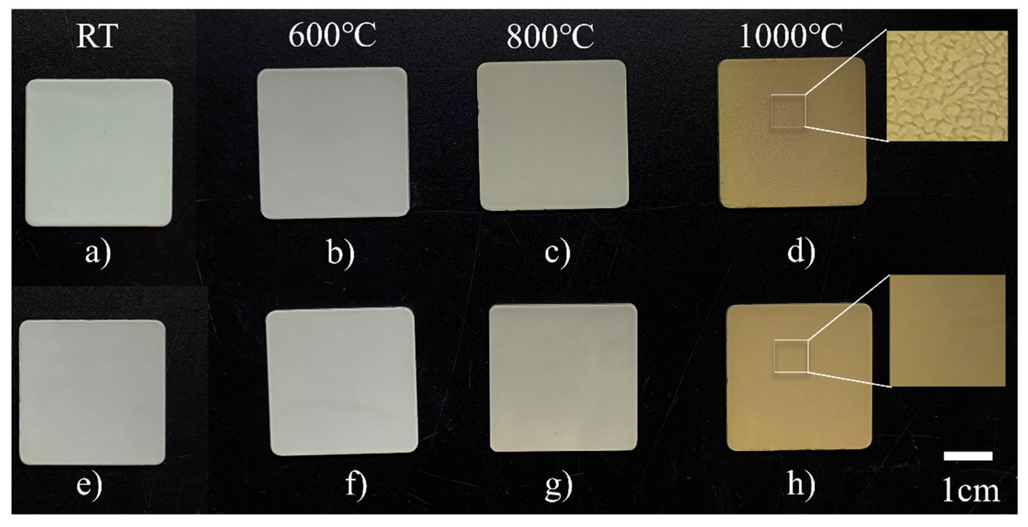
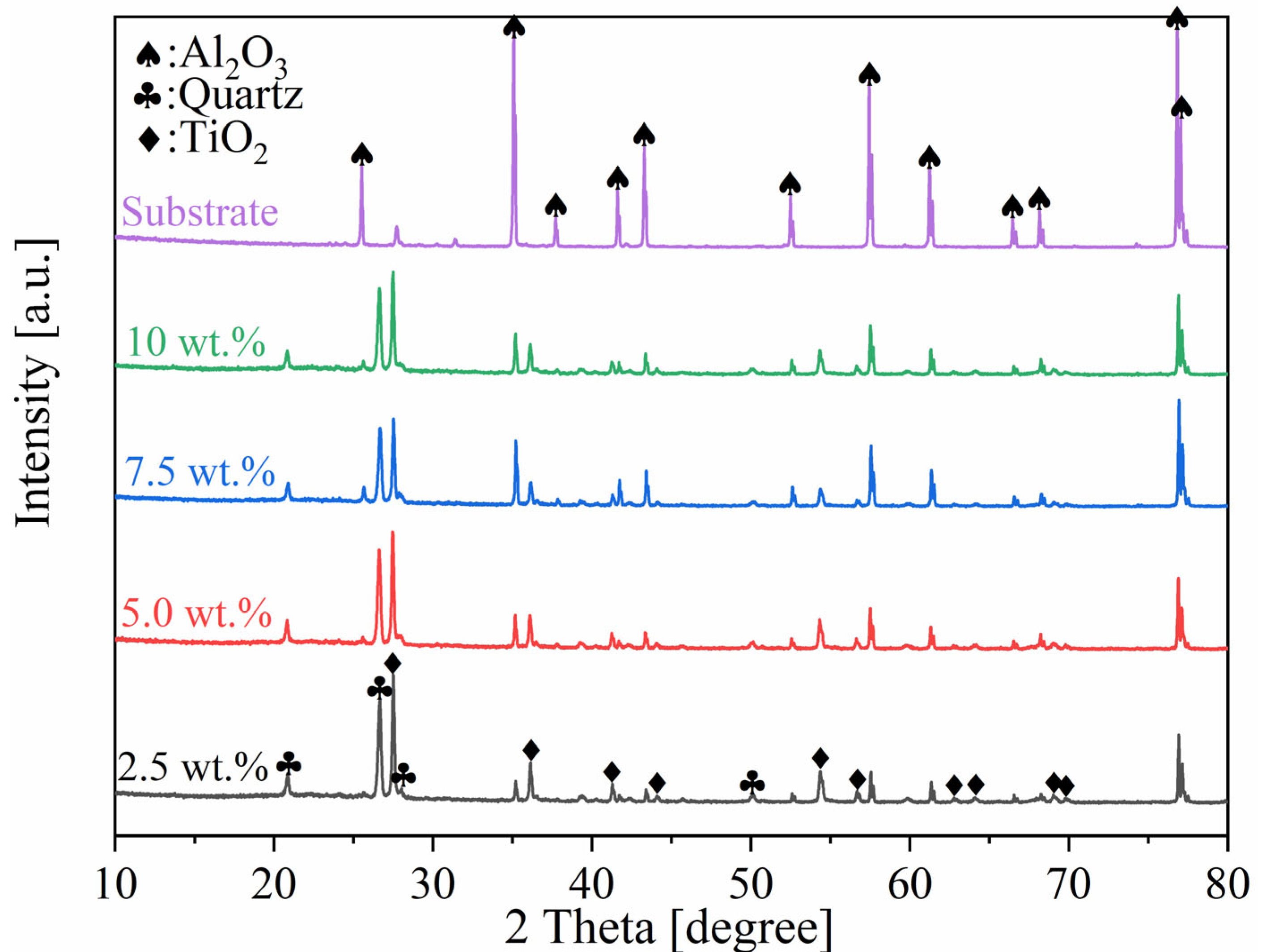
The five groups of coatings, cured at room temperature (RT) for 24 h, were subjected to heat treatment with a holding time of 1 h at 600 °C, 800 °C and 1000 °C respectively. The test results demonstrated all coatings have good thermal resistance at 600 °C and 800 °C, with no cracks or bubbles occurring on the coating surface. However, coatings without nano-TiO2 particles failed after being heat-treated at 1000 °C. Since the TiO2-incorporated coatings shared similar macroscopic features, the 5 wt.% formulation is presented as a representative example. Figure 6 shows that after heat treatment at 1000 °C, the coating without nano-TiO2 developed numerous microcracks on its surface. In contrast, the coating with 5.0 wt.% nano-TiO2 particles remained macroscopically dense and smooth, free from defects such as bubbles or cracks. The X-ray diffraction (XRD) patterns of the coating after treatment at 1000 °C with 1 h holding are shown in Figure 7. The primary components of the coating remain muscovite and TiO2, with no significant changes observed. The disappearance of muscovite peaks at 2θ = 17.8°, 36.5°and 45.5° indicates that the interlayer water in the muscovite crystal lattice was removed at high temperatures, leading to the destruction of the layered structure; muscovite may have partially decomposed and melted, resulting in severe distortion of the crystal structure. The failure to observe amorphous SiO2 or glassy eutectic phases is attributed to the fact that the diffuse peaks of the amorphous phase and the low crystallinity of glassy eutectic phases produce weak peak intensities, which were masked by the overly sharp peaks of rutile titanium dioxide [28].
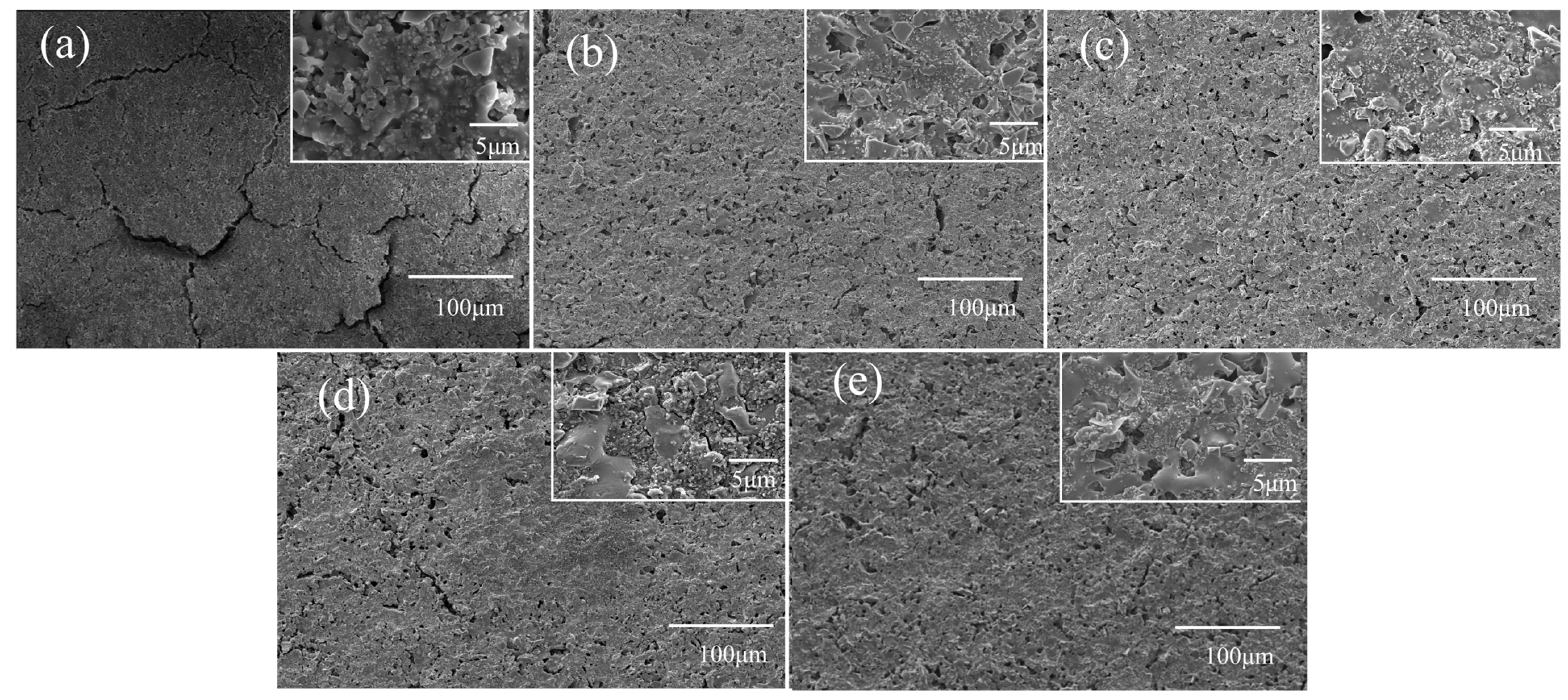
Figure 8 shows the surface morphology of the coatings after heat treatment at 1000 °C. As shown in Figure 8a, the unmodified coatings developed cracks and suffered complete surface destruction after heat treatment. In contrast, the coatings with nano-TiO2 shown in Figure 8b–e developed numerous micropores on the surface after heat treatment. These micropores are attributed to the decomposition of the binder, which released small gas molecules during heating. However, the integrity of the coatings was preserved, and no delamination or failure occurred, indicating that the addition of nano-TiO2 significantly enhanced the thermal resistance of the coatings. Quantitative characterization of the micropore area fraction through image analysis revealed that the micropore area fractions for coatings doped with 2.5 wt.%, 5.0 wt.%, 7.5 wt.%, and 10 wt.% TiO2 were 9.1%, 5.1%, 7.5%, and 8.6%, respectively. The 5.0 wt.%-doped coating exhibited the lowest porosity. Microscopic observation indicated that coatings with lower doping ratios displayed finer and more uniform micropore distributions; as the nano-TiO2 content increased, the micropore size enlarged and tended to form elongated cavities. This phenomenon can be attributed to the increased liquid-phase viscosity at high temperatures resulting from a higher nanoparticle addition, which prevented the timely filling of pores formed by gas evolution. High-magnification microscopy further revealed that the nano-TiO2 particles were most uniformly distributed in the 5.0 wt.%-doped coating, with homogeneous dispersion throughout the molten liquid phase, whereas coatings with higher doping ratios showed obvious local agglomeration of nanoparticles. Additionally, partial melting occurred at the mica edges, where K+ enrichment at the sheet edges formed low-melting-point eutectics with surrounding amorphous SiO2 [10]. This caused originally sharp pores to be filled by the molten liquid phase and become smoother, thereby significantly reducing the crack tendency induced by stress concentration. Simultaneously, the molten liquid phase acted as a binding phase, not only interconnecting TiO2 particles but also firmly bonding them with unmelted muscovite particles, effectively enhancing the structural integrity of the coating and ultimately improving its high-temperature resistance.
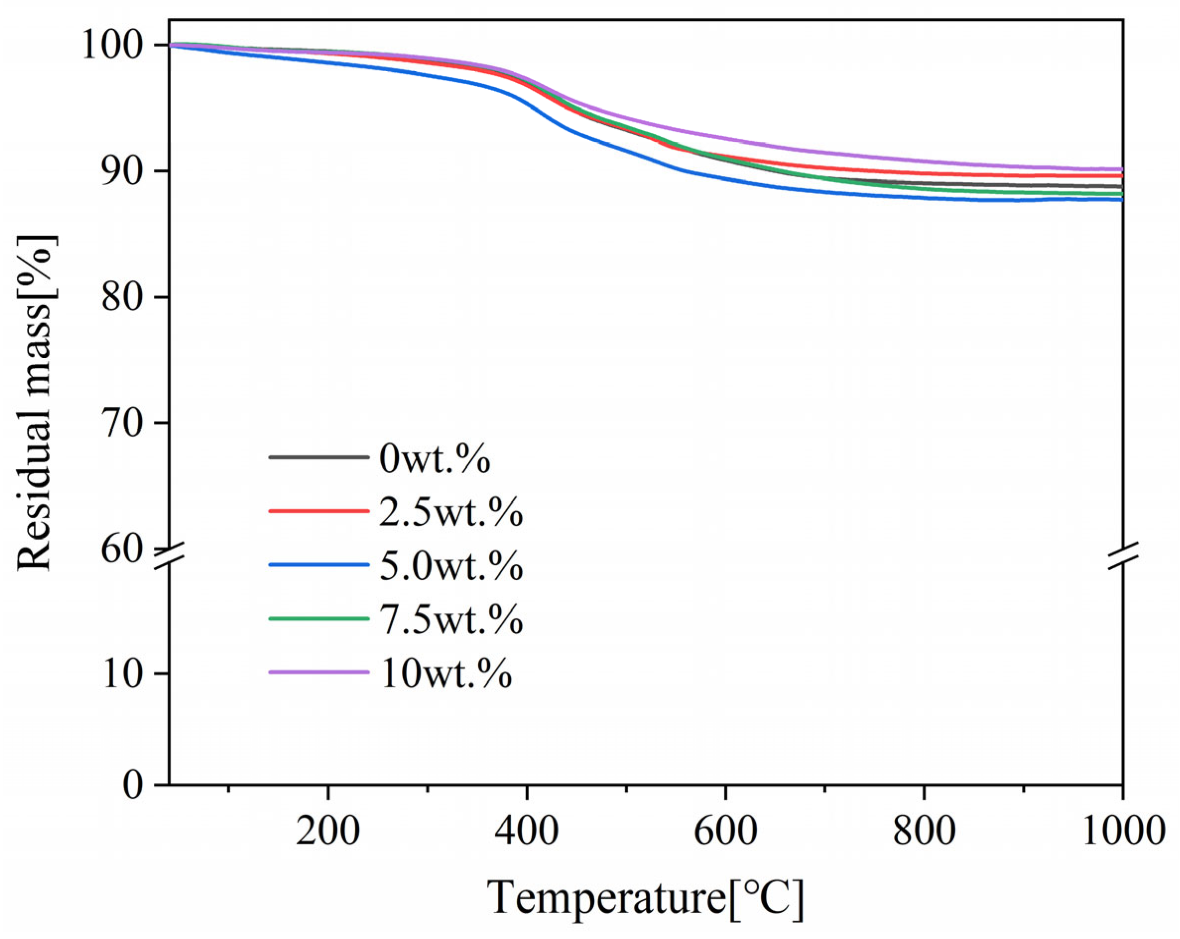
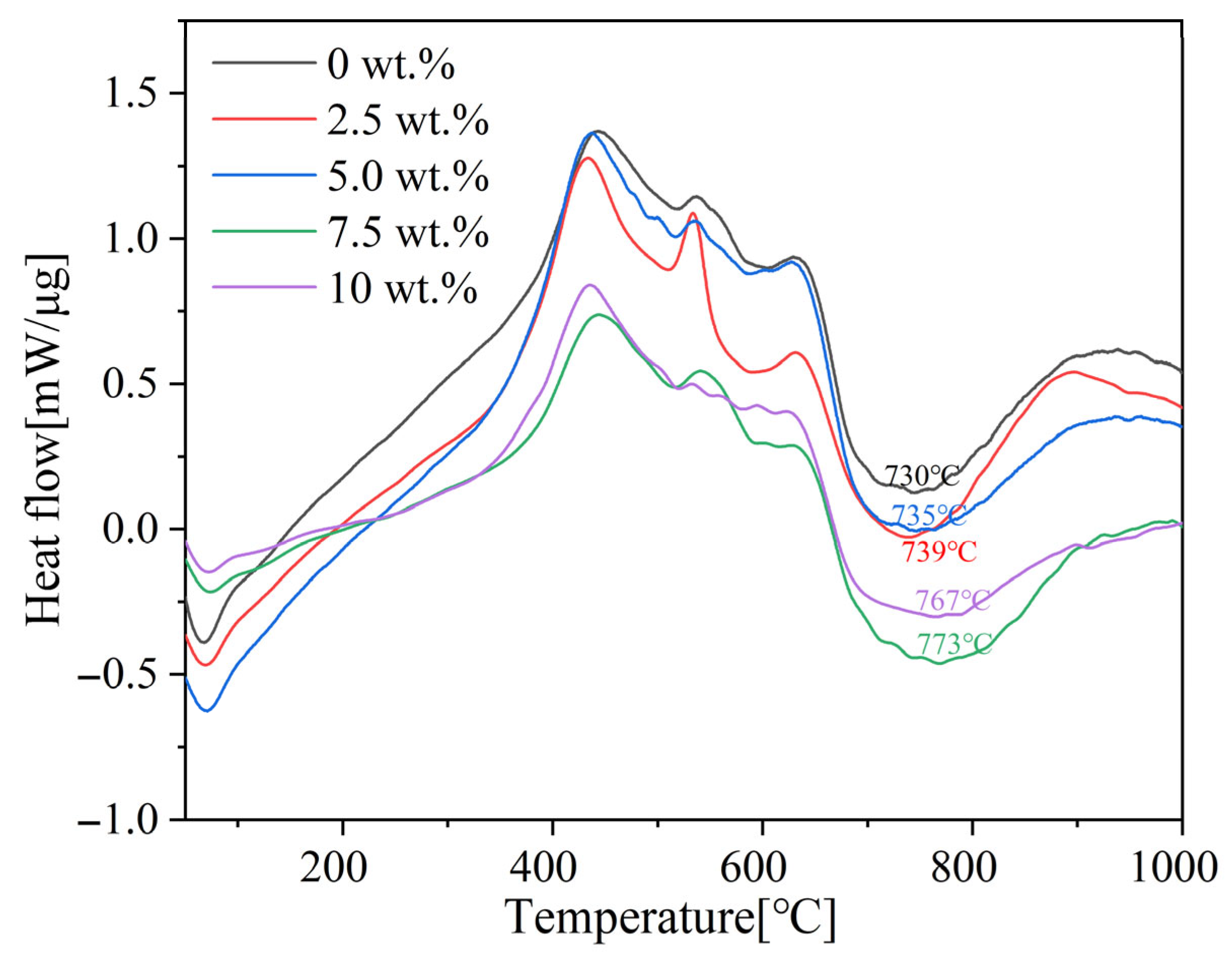
To investigate the thermal degradation behavior of nano-TiO2 particle-modified coatings with different weight ratios under high-temperature conditions, thermogravimetric (TG) and differential scanning calorimetry (DSC) were conducted in an air atmosphere; the results are shown in Figure 9 and Figure 10. The TG curves showed the residual mass of the coatings. The mass loss below 100 °C was mainly attributed to the evaporation of the solvent (ethanol) and adsorbed water. A slight mass loss was observed for all coatings before 300 °C, which was due to dehydration between Si-OH groups and dealcoholization between Si-OH and Si-OR groups, leading to the formation of free terminal hydroxyl groups, accompanied by gradual exothermic behavior. Around 400 °C, a significant mass loss occurred along with a strong exothermic peak, resulting from the thermal oxidative degradation of organic groups attached to the silicone resin backbone. This is primarily caused by the vigorous oxidation of Si-CH3 groups [29,30]. The bond energy of Si-C is lower than that of Si-O, leading to the preferential oxidative combustion of Si-C bonds that releases significant heat [31]. In the temperature range of 500~600 °C, an endothermic reaction occurred accompanied by continuous mass loss shown in the TG curve. This is attributed to the “unzipping” degradation of Si-O-Si chains induced by unreacted terminal hydroxyl groups, generating low-molecular-weight cyclic siloxanes [32]. Around 600 °C, mass loss continued together with an endothermic peak, mainly due to the “thermal rearrangement degradation” of siloxane bonds, leading to the cleavage of long Si-O-Si chains and the release of small Si-O-Si species. A strong endothermic peak was observed at approximately 750 °C without significant mass loss. This is attributed to the endothermic dehydroxylation of muscovite and its partial edge melting at this temperature [33]. With increasing addition amounts of nano-TiO2 particles, the endothermic peak gradually shifted toward higher temperatures. This is because well-dispersed nano-TiO2 particles effectively block the contact sites between muscovite and amorphous SiO2 generated from silicone pyrolysis, increasing the diffusion distance of reactants and thereby delaying the onset temperature of the reaction. Meanwhile, as the reaction proceeded at a higher temperature, the melting eutectic reaction became more complete. Consequently, an increasing amount of molten liquid phase was observed, as shown in Figure 8. Above 800 °C, the residual mass remained virtually constant, which was attributed to the formation of a stable molten liquid phase from siloxanes and muscovite, resulting in excellent thermal stability. The final residual mass for all coating compositions was very similar, approximately 90%.
Based on the comprehensive thermal analysis, the incorporation of nano-TiO2 does not significantly alter the overall reaction pathway during the heat treatment process. However, the addition of nano-TiO2 increased the starting temperature of the melting eutectic reaction, resulting in a more thorough reaction with more liquid phases. Consequently, the coating surface becomes smoother and denser, as shown in Figure 8, thereby blocking the entry of oxygen and improving the overall heat resistance performance.

3.4. Reflectance and Adhesion Strength of Coatings After Heat Treatment

Table 3 shows the adhesion strength of the coatings with different weight ratios of nano-TiO2 particles after heat treatment. It suggests that the adhesion strength of each group of coatings increased significantly after heat treatment; this is because the coating formed a dense ceramic layer, establishing a more stable chemical bond with the substrate.
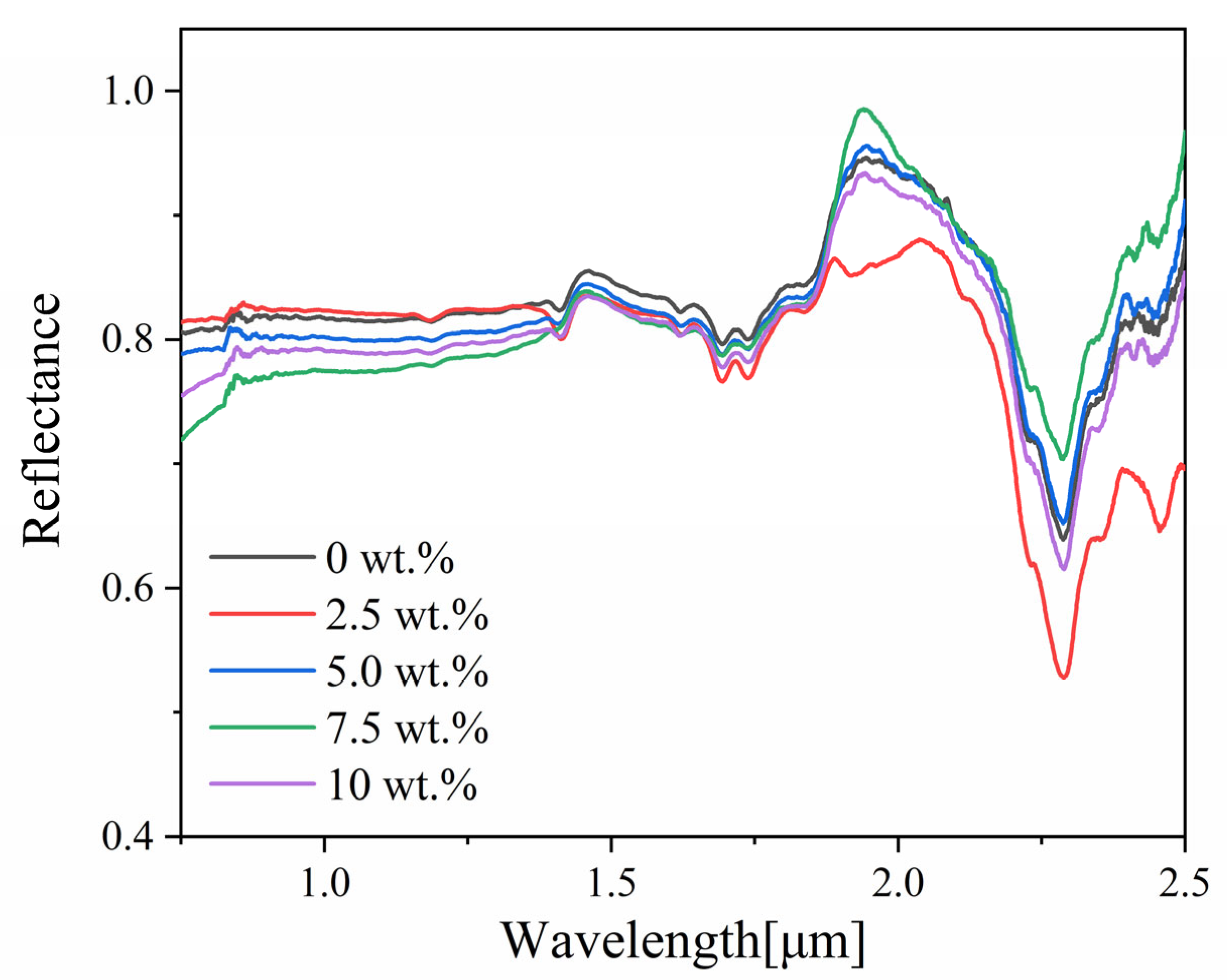
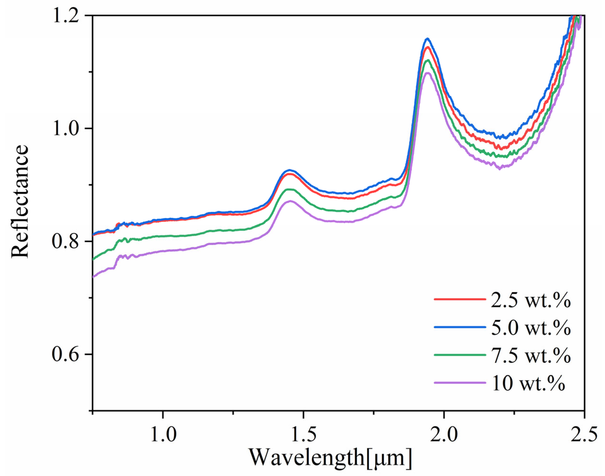
The NIR reflectance of coatings with varying amounts of nano-TiO2 after 1000 °C thermal treatment was measured and is shown in Figure 11. The calculated reflectivity is summarized in Table 4. To ensure data reliability, all reflectance curves were obtained by averaging the results of three measurements. It is noteworthy that the anomalous phenomenon of coating reflectance exceeding 1 at approximately 2 μm is not attributable to experimental error. This phenomenon originates from the spectral characteristics of the reference standard BaSO4: the BaSO4 reference plate exhibits hygroscopicity and readily adsorbs trace environmental moisture, forming characteristic absorption bands in the near-infrared region [34]. In contrast, the ceramic coating surface becomes smooth and hydrophobic after high-temperature heat treatment, with negligible moisture adsorption. Furthermore, under the integrating sphere test mode including specular component included (SCI), the coating surface is smoother than the standard white plate, and its directional reflection efficiency may be higher than that of the standard diffuse reflectance plate, resulting in elevated relative reflectance in this wavelength band. When calculating reflectance, this anomalous value was corrected to 0.99. The increasement in reflectivity after heat treatment was thought to be attributed to the formation of numerous smooth planes upon cooling of the molten phase, resulting in more specular reflection. In contrast, coatings before heat treatment primarily exhibited diffuse reflection, resulting in a lower reflectance. As the addition amount increased, the overall reflectance of the coating after heat treatment exhibited a decreasing trend. This phenomenon can be attributed to two main factors. On one side, despite the enhanced specular reflection from the formed molten liquid phase, the agglomeration of nano-TiO2 particles reduced the light-scattering interfaces, decreasing reflectance [35]. On the other side, at high temperatures, SiO2 migrates toward the surface to form a dense layer. The molten liquid phase on the surface may exhibit a certain degree of absorption in the NIR band, increasing the probability of incident light being absorbed and ultimately reducing the reflectance.

4. Conclusions

In this study, a nano-TiO2-modified silicone-based reflective thermal insulation coating was successfully synthesized to enhance its thermal resistance and reflective performance. Systematic investigation of the nano-TiO2 content revealed that the coating with 5.0 wt.% nano-TiO2 particles exhibited the optimal overall performance. Quantitative analysis showed that at room temperature, the coating achieved an adhesion strength of Grade 1 and a solar reflectance of 0.830. After heat treatment at 1000 °C for 1 h, the coating underwent a ceramization transformation with further improved performance: the adhesion strength increased to Grade 0 and the reflectance rose to 0.945.
The performance enhancement is attributed to the synergistic effects of physical and chemical processes. Before heat treatment, well-dispersed nano-TiO2 acted as a filler, reducing micropores in the silicone binder and densifying the composite structure, thereby improving the mechanical properties. Meanwhile, the incorporation of nano-TiO2 reduced the average particle size of the pigment system to the optimal scattering size, ensuring excellent reflective performance. Under high-temperature conditions at 1000 °C, an appropriate amount of nano-TiO2 delayed the formation temperature of stable molten phases, enhancing the thermal stability of the coating. Simultaneously, the uniformly dispersed liquid phase resulted in a smooth and flat surface, further improving the coating’s reflective performance. The coating system developed in this study combines the dual advantages of convenient room-temperature curing for easy application and high-temperature ceramization for thermal protection, demonstrating broad application potential in industrial furnace energy saving, aerospace thermal protection, and concentrated solar power generation.

Author Contributions

Conceptualization, X.Z. and X.C.; methodology, S.K. and X.Z.; validation, X.Z. and S.K.; formal analysis, S.K.; investigation, S.K.; resources, S.K. and X.X.; data curation, S.K.; writing—original draft preparation, S.K. and X.Z.; writing—review and editing, X.Z.; visualization, X.X.; supervision, X.Z. and R.-Z.W.; project administration, X.Z. and R.-Z.W.; funding acquisition, X.Z. and R.-Z.W. All authors have read and agreed to the published version of the manuscript.

Funding

This work was financially supported by the National Natural Science Foundation of China (Grant No. 52473280), Guangxi Science and Technology Department (Grant Numbers: AA23062025-3), MEXT Strategic Professional Development Program for Young Researchers of Japan (TI-FRIS Fellow, No. J250000163).

Institutional Review Board Statement

Not applicable.

Informed Consent Statement

Not applicable.

Data Availability Statement

The data presented in this study are available from the corresponding author upon request. The data are not publicly available due to privacy.

Conflicts of Interest

The authors declare no conflicts of interest.

References

  1. Wang, Y.X.; Yin, J.W.; Ren, J.T. Research Progress of Silicone High Temperature Resistant Coatings. Mod. Paint Finish. 2023, 26, 11–14. [Google Scholar]
  2. Lu, H.; Chen, J. Thermal Insulation Mechanisms of Heat-Resistant Coatings and Their Research Advancements. Mater. Rep. 2005, 19, 71–73. [Google Scholar]
  3. Liu, Z.; Di, Z.; Gou, Y.; He, Y.; Shi, L.; Tan, W. Research Progress in Silicone High Temperature Resistant Coatings. Paint Coat. Ind. 2019, 49, 83–87. [Google Scholar]
  4. Deng, B. Duplex Corrosion Protection to Steel Substrates by Zinc (Aluminum) and Organic Coatings. Coat. Prot. 2021, 42, 48–56. [Google Scholar]
  5. Wang, J.; Jiang, Y.; Xiong, Y.; Wang, F.; Ma, H.; Li, W.; Lei, L.; Chen, Z. Toughening and Modification of Organic Silicone Heat-Resistant Micro-Wave Absorbing Coatings with Heat-Resistant Linear Molecules. Package. Eng. 2023, 44, 81–90. [Google Scholar]
  6. Niu, Z.; Li, G.; Ma, X.; Shen, S.; Xin, Y.; Cai, L.; Chen, F.; Zhang, C.; Hou, X. Synergetic Effect of O-POSS and T-POSS to Enhance Ablative Resistance of Phenolic-Based Silica Fiber Composites via Strong Interphase Strength and Ceramic Formation. Compos. Part A Appl. Sci. Manuf. 2022, 155, 106855. [Google Scholar] [CrossRef] [Scilit]
  7. Zhang, L.; Chen, R. TiO2-Siloxane Thermal Control Coatings for Protection of Spacecraft Polymers. Chin. J. Aeronaut. 2004, 17, 53–59. [Google Scholar] [CrossRef] [Scilit]
  8. Yoshinaga, I.; Yamada, N.; Katayama, S. Effect of Inorganic Components on Thermal Stability of Methylsiloxane-Based Inorganic/Organic Hybrids. J. Sol-Gel Sci. Technol. 2005, 35, 21–26. [Google Scholar] [CrossRef] [Scilit]
  9. Wu, L.; Wang, F.; Wang, X. Development of Long-Term High-Temperature Resistant Silicone Coatings. China Coat. 2021, 36, 14–18. [Google Scholar]
  10. Mansouri, J.; Burford, R.P.; Cheng, Y.B. Pyrolysis Behaviour of Silicone-Based Ceramifying Composites. Mater. Sci. Eng. A 2006, 425, 7–14. [Google Scholar] [CrossRef] [Scilit]
  11. Suchikova, Y.; Nazarovets, S.; Konuhova, M.; Popov, A.I. Binary Oxide Ceramics (TiO2, ZnO, Al2O3, SiO2, CeO2, Fe2O3, and WO3) for Solar Cell Applications: A Comparative and Bibliometric Analysis. Ceramics 2025, 8, 119. [Google Scholar] [CrossRef] [Scilit]
  12. Podolińska, A.; Neilande, E.; Pankratova, V.; Serga, V.; Bandarenka, H.; Burko, A.; Piskunov, S.; Pankratov, V.A.; Sarakovskis, A.; Popov, A.I.; et al. Structural and Spectroscopic Characterization of TiO2 Nanocrystalline Materials Synthesized by Different Methods. Nanomaterials 2025, 15, 498. [Google Scholar] [CrossRef] [Scilit] [PubMed]
  13. Zeng, K.; Cheng, L.; Hu, W.; Li, J. Synthesis, Stability, and Tribological Performance of TiO2 Nanomaterials for Advanced Applications. Lubricants 2023, 11, 56. [Google Scholar] [CrossRef] [Scilit]
  14. Huang, P.; Shi, H.; Xiao, H.-M.; Li, Y.-Q.; Hu, N.; Fu, S.-Y. High Performance Surface-Modified TiO2/Silicone Nanocomposite. Sci. Rep. 2017, 7, 5951. [Google Scholar] [CrossRef] [Scilit] [PubMed]
  15. Zhou, Y.; Yang, J.; Jia, X.; Ni, J.; Han, N.; Song, H. Durable Daytime Radiative Cooling Achieved by Super-Slippery Coatings Containing Polydimethylsiloxane Brushes. Prog. Org. Coat. 2024, 194, 108631. [Google Scholar] [CrossRef] [Scilit]
  16. Peoples, J.; Li, X.; Lv, Y.; Qiu, J.; Huang, Z.; Ruan, X. A Strategy of Hierarchical Particle Sizes in Nanoparticle Composite for Enhancing Solar Reflection. Sol. Energy Mater. Sol. Cells 2018, 182, 226–234. [Google Scholar] [CrossRef] [Scilit]
  17. ISO 2409:2020; Paints and Varnishes—Cross-Cut Test. International Organization for Standardization: Geneva, Switzerland, 2020.
  18. ASTM E903-20; Standard Test Method for Solar Absorptance, Reflectance, and Transmittance of Materials Using Integrating Spheres. ASTM International: West Conshohocken, PA, USA, 2020.
  19. Siegel, R.; Howell, J.R. Thermal Radiation Heat Transfer, 6th ed.; CRC Press: Boca Raton, FL, USA, 2015; pp. 112–115. [Google Scholar]
  20. Planck, M. The Theory of Heat Radiation; Dover Publications: New York, NY, USA, 1959. [Google Scholar]
  21. Andre, S.; Guida-Pietrasanta, F.; Ratsimihety, A.; Rousseau, A.; Boutevin, B. Synthesis, Characterization and Thermal Properties of SiH and SiOH Terminated Hybrid Polysiloxanes. Macromol. Chem. Phys. 2000, 201, 67–81. [Google Scholar] [CrossRef]
  22. Hu, C.; Xu, G.; Shen, X.; Shao, C.; Yan, X. The Epoxy-Siloxane/Al Composite Coatings with Low Infrared Emissivity for High Temperature Applications. Appl. Surf. Sci. 2010, 256, 3459–3463. [Google Scholar] [CrossRef] [Scilit]
  23. Lee, J.H.; Kang, D.W.; Kang, H.J. Characteristics of Heat Curable Polyorganosiloxane Coating Materials. Polym. Korea 2015, 39, 499–505. [Google Scholar] [CrossRef] [Scilit]
  24. Sakai, R.T.; Da Cruz, F.M.D.L.; De Melo, H.G.; Benedetti, A.; Santilli, C.; Suegama, P. Electrochemical Study of TEOS, TEOS/MPTS, MPTS/MMA and TEOS/MPTS/MMA Films on Tin Coated Steel in 3.5% NaCl Solution. Prog. Org. Coat. 2012, 74, 288–301. [Google Scholar] [CrossRef] [Scilit]
  25. Jovanovic, J.D.; Govedarica, M.N.; Dvornic, P.R.; Popovic, I.G. The Thermogravimetric Analysis of Some Polysiloxanes. Polym. Degrad. Stab. 1998, 61, 57–66. [Google Scholar] [CrossRef] [Scilit]
  26. Akinay, Y.; Akku, I.N. Synthesis and Characterization of the Pearlescent Pigments Based on Mica Deposited with SiO2, AlN and TiO2: First Report of Its Dielectric Properties. Ceram. Int. 2020, 46, 17735–17740. [Google Scholar] [CrossRef] [Scilit]
  27. Zhao, X.; Mofid, S.A.; Jelle, B.P.; Tan, G.; Yin, X.; Yang, R. Optically-Switchable Thermally-Insulating VO2-Aerogel Hybrid Film for Window Retrofits. Appl. Energy 2020, 278, 115663. [Google Scholar] [CrossRef] [Scilit]
  28. Li, Y.; Deng, C.; Wang, Y.-Z. A Novel High-Temperature-Resistant Polymeric Material for Cables and Insulated Wires via the Ceramization of Mica-Based Ceramifiable EVA Composites. Compos. Sci. Technol. 2016, 132, 116–122. [Google Scholar] [CrossRef] [Scilit]
  29. Sun, J.; Huang, Y.; Cao, H.; Gong, G. Synthesis of High Temperature Resistant Silicone Resin and Its Heat Resistance and Curing Properties. J. Aeronaut. Mater. 2005, 25, 25–29+35. [Google Scholar]
  30. You, Y.; Chen, J.; Zheng, A.; Wei, D.; Xu, X.; Guan, Y. Effect of Silanol on the Thermal Stability of Poly[methyl(trifluoropropyl)siloxane]. J. Appl. Polym. Sci. 2020, 137, 49347. [Google Scholar] [CrossRef] [Scilit]
  31. Grassie, N.; Francey, K.; Macfarlane, I.M. The Thermal Degradation of Polysiloxanes—Part 4: Poly(dimethyl/diphenylsiloxane). Polym. Degrad. Stab. 1980, 2, 67–83. [Google Scholar] [CrossRef] [Scilit]
  32. Dai, G.; Pei, Y.; Chu, J.; Luo, Y.; Xie, Y.; Xie, D.; Mei, Y. Research Progress on High Temperature Resistance Modification Method and Mechanism of Silicone Resin. Polym. Mater. Sci. Eng. 2022, 38, 174–182. [Google Scholar]
  33. Meng, P. Preparation and Study on Properties of Ablation Resistant and Ceramizable Silicone Rubber Composites. Ph.D. Thesis, Wuhan University of Technology, Wuhan, China, 2016. [Google Scholar]
  34. Poh, A.H.; Jamaludin, M.F.; Fadzallah, I.A.; Nik Ibrahim, N.M.J.; Yusof, F.; Adikan, F.R.M.; Moghavvemi, M. Diffuse Reflectance Spectroscopic Analysis of Barium Sulfate as a Reflection Standard within 173–2500 nm: From Pure to Sintered Form. J. Near Infrared Spectrosc. 2019, 27, 393–401. [Google Scholar] [CrossRef] [Scilit]
  35. Yang, X.; Xu, G.; Tan, S.; Wu, S.; Liang, J.; Shen, S. Study on Influential Factors on Heat Reflectance of TiO2 Coating. Paint Coat. Ind. 2018, 48, 64–68. [Google Scholar]
Figure 1. Infrared spectra of nano-TiO2 particle-modified coatings with different weight ratios.
Coatings 16 00319 g001
Figure 2. Hydrolytic polycondensation of siloxane.
Coatings 16 00319 g002
Figure 3. X-ray diffraction of nano-TiO2 particle-modified coatings with different weight ratios.
Coatings 16 00319 g003
Figure 4. Surface morphology of nano-TiO2 particle-modified coatings with different weight ratios: (a) 0 wt.%; (b) 2.5 wt.%; (c) 5.0 wt.%; (d) 7.5 wt.%; (e) 10 wt.%.
Coatings 16 00319 g004
Figure 5. Near-infrared reflectance spectra of coatings with different content of nano-TiO2 particles.
Coatings 16 00319 g005
Figure 6. Macroscopic surface morphology of the heat-treated coatings. (a) RT, 0 wt.%; (b) 600 °C, 0 wt.%; (c) 800 °C, 0 wt.%; (d) 1000 °C, 0 wt.%; (e) RT, 5 wt.%; (f) 600 °C, 5 wt.%; (g) 800 °C, 5 wt.%; (h) 1000 °C, 5 wt.%.
Coatings 16 00319 g006
Figure 7. X-ray diffraction of nano TiO2 particle-modified coatings with different weight ratios after heat treatment at 1000 °C.
Coatings 16 00319 g007
Figure 8. Surface morphology of coatings with different content of nano-TiO2 particles after heat treatment at 1000 °C: (a) 0 wt.%; (b) 2.5 wt.%; (c) 5.0 wt.%; (d) 7.5 wt.%; (e) 10 wt.%.
Coatings 16 00319 g008
Figure 9. TG curves of nano-TiO2 particle-modified coatings with different weight ratios.
Coatings 16 00319 g009
Figure 10. DTG curves of nano-TiO2 particle-modified coatings with different weight ratios.
Coatings 16 00319 g010
Figure 11. The 1000 °C near-infrared band reflectance spectra of nano-TiO2 particle-modified coatings with different weight ratios after heat treatment at 1000 °C.
Coatings 16 00319 g011
Table 1. Near-infrared reflectivity of coatings with different content of nano-TiO2 particles.
Addition Amount of Nano TiO2 [wt.%]Reflectivity
00.831
2.50.777
5.00.830
7.50.841
100.812
Table 2. Adhesion strength of coatings with different weight ratios of nano-TiO2 particles.
Addition Amount of Nano-TiO2 [wt.%]Adhesion Levels
02
2.51
51
7.51
102
Table 3. Adhesion strength of coatings with different weight ratios of nano-TiO2 particles after heat treatment at 1000 °C.
Addition Amount of Nano-TiO2 [wt.%]Adhesion
2.50
5.00
7.51
101
Table 4. Near-infrared reflectivity of nano-TiO2 particle-modified coatings with different weight ratios after heat treatment at 1000 °C.
Addition Amount of Nano-TiO2 [wt.%]Reflectivity
2.50.939
5.00.945
7.50.925
100.911
Disclaimer/Publisher’s Note: The statements, opinions and data contained in all publications are solely those of the individual author(s) and contributor(s) and not of MDPI and/or the editor(s). MDPI and/or the editor(s) disclaim responsibility for any injury to people or property resulting from any ideas, methods, instructions or products referred to in the content.