Article

Preliminary Study on the Role of Humic Substances in the Early Corrosion Behavior of High-Tin Bronze Alloys Under Simulated Soil Conditions

School of Cultural Heritage, Northwest University, Xi’an 710127, China
*
Author to whom correspondence should be addressed.
Coatings 2026, 16(3), 320; https://doi.org/10.3390/coatings16030320
Submission received: 24 January 2026 / Revised: 25 February 2026 / Accepted: 4 March 2026 / Published: 6 March 2026
(This article belongs to the Section Corrosion, Wear and Erosion)

Highlights

What are the main findings?
Electrochemical tests reproduced early-stage corrosion features of high-tin bronze.
A bilayer structure is formed: surface corrosion layer and semi-corroded transition zone.
Weakly acidic conditions promote Cu leaching and in situ SnO2 formation.
What are the implications of the main findings?
Soil pH is the dominant factor controlling early corrosion of high-tin bronze.
Humus shows a limited, pH-dependent influence on corrosion behavior.
Results clarify the role of humus in archaeological bronze burial environments.

Abstract

To investigate the influence of humus on the corrosion behavior of high-tin bronze in soil environments, potentiostatic polarization was applied to simulate early-stage corrosion under controlled conditions. Open-circuit potential and potentiodynamic polarization tests were performed, and corrosion products were characterized by stereo microscopy, SEM-EDS, and confocal Raman spectroscopy. A Cu–Sn–Pb ternary alloy was examined in simulated archaeological soil solutions with selective humus addition at different pH values. A bilayer structure, consisting of a secondary corrosion layer and a semi-corroded transition zone, developed in all media, with more extensive corrosion under weakly acidic conditions. In acidic environments, humus enhanced preferential α-phase corrosion, associated with copper depletion and tin enrichment as SnO2. Under weakly alkaline conditions, humus mainly affected surface color and micro-morphology without altering the overall corrosion pattern. Electrochemical testing reproduced corrosion layer structures similar to those formed during early burials, but differences in morphology were observed. The results suggest that, as an accelerated corrosion technique, electrochemical methods can reproduce key features of early-stage corrosion in high-tin bronze and serve as an effective tool for monitoring corrosion behavior.

1. Introduction

Bronze is an alloy primarily composed of copper, with the addition of tin and lead. It has been widely used to make ancient artifacts such as ritual vessels, weapons, and currency. After long-term burial, the surface corrosion of bronze artifacts exhibits various morphologies [1]. The corrosion layer is typically composed of rough, reddish-brown and blue-green substances [2]. Some bronze artifacts also exhibit a dense and smooth corrosion product, often appearing as lustrous dark brown or black layers. Due to its lacquer-like appearance, this type of patina was historically termed “black lacquer” by earlier researchers; today, it is commonly referred to in academic circles as the “high-tin patina layer.” Its main characteristics include a surface enriched with dense cassiterite, which preserves the original metal surface of the bronze artifact and exhibits excellent wear resistance and corrosion resistance [3]. Since the high-tin patina possesses various distinctive properties, scholars have been studying this corrosion product since 1926 [4]. It is generally believed that the high-tin patina consists of a tin-rich layer that preserves the original metal surface and a cuprous oxide layer beneath it [5,6]. On some bronze artifacts, secondary corrosion products such as cuprous oxide, malachite, and azurite are deposited on top of the tin-rich layer. In a few cases, intergranular corrosion may develop along cracks in the high-tin patina and extend into the metal substrate. In studies of bronze artifacts, the term patina has traditionally been used to describe surface corrosion layers, often characterized by tin enrichment, especially on high-tin bronze objects [7,8]. Since secondary corrosion or transgranular corrosion does not occur universally in bronze artifacts, high-tin corrosion is often characterized by a black, relatively smooth patina that largely preserves the original metallic surface [5]. Studies on bronze corrosion are increasingly conducted within the framework of an integrated metal–patina–environment system, emphasizing that the formation and evolution of corrosion layers are governed not only by alloy composition but also by surrounding environmental conditions [9]. During the burial process, selective corrosion of bronze causes copper to be oxidized into ions and leached into the surrounding environment [10], whereas tin—due to the extremely low solubility of its oxides—undergoes in situ deposition and forms a dense layer [11]. However, the factors governing selective corrosion and in situ deposition remain subject to debate. Some scholars point out that this corrosion process is influenced by the burial environment, and that the slow leaching of copper ions is critical for the formation of high-tin corrosion products [12]. Meanwhile, based on archaeological excavation contexts, soils that are acidic and have high water content are more conducive to the formation of high-tin patina [13]. Previous studies have demonstrated that environmental parameters such as pH, soil ionic composition, dissolved oxygen availability, and organic matter content play critical roles in governing the corrosion behavior of copper alloys in soil environments [14,15]. To quantitatively assess the influence of environmental conditions on metal degradation, corrosion indices have been employed to integrate water chemistry parameters, thereby establishing correlations between environmental factors and metal corrosion processes [16]. Notably, the burial environment of bronze artifacts is often rich in humus, either inherently present in the soil or derived from the decomposition of organic matter within archaeological remains. Accordingly, some scholars have conducted a series of experiments to investigate the promoting effect of humus on the formation of the high-tin patina on bronze mirrors. These experiments have yielded corrosion products that not only resemble high-tin patina in appearance but also exhibit mineralogical similarity to those found in archaeologically excavated samples [17,18,19,20,20]. Under the combined influence of oxygenated groundwater and humus generated from the decomposition of organic matter, copper at the surface of high-tin bronze undergoes oxidation. Concurrently, the strong complexing ability of humus for copper ions promotes the selective leaching of copper from the metal substrate [21].
Existing studies on the formation mechanisms of tin-enriched corrosion layers in high-tin bronzes have primarily focused on inorganic environmental factors such as soil ionic composition and pH. In contrast, humic substances—ubiquitous and chemically active components of soil environments—have received comparatively limited attention. In many corrosion studies, natural organic matter is treated as a bulk or undifferentiated background constituent of soils, without systematic consideration of its specific chemical characteristics and metal-binding behavior [14,22]. Furthermore, the aforementioned research on simulating the interaction between humus and bronze specimens has employed accelerated protocols, such as elevated concentrations and heated immersion, which differ significantly from the corrosion processes occurring in natural soil burial environments. Based on archaeological contexts, this study aims to establish corrosion conditions that closely approximate those of natural soil burial environments, thereby investigating the corrosion behavior of ancient-style high-tin bronze in a simulated soil medium containing humus, and exploring the key factors governing the formation of high-tin patina on bronze artifacts. Significantly, a substantial number of bronze artifacts unearthed from the Guanzhong Plain in Shaanxi Province exhibit typical high-tin patina features. This region is characterized by a warm temperate semi-humid climate, with a mean annual temperature ranging from 12 to 13.6 °C and an annual precipitation of approximately 604 mm. Its surface is covered by a thick layer of clayey loess. Building upon these observations, the present study selects the Guanzhong Loess in Shaanxi as a representative burial environment for northern Chinese bronze artifacts. By referencing its chemical composition, a simulated soil solution containing soil-derived ions and humus was formulated to replicate the natural burial environment. Open-circuit potential (OCP) and potentiodynamic polarization measurements were subsequently performed on cast high-tin bronze alloys in this corrosive medium, followed by characterization of the corrosion products in terms of morphology, elemental distribution, and phase composition.

2. Materials and Methods

2.1. Corrosion Conditions and Bronze Specimens

The dissolved inorganic salts in the shallow groundwater of the Loess Plateau are predominantly composed of bicarbonate ions, with minor concentrations of chloride, sulfate, and nitrate ions. Due to leaching processes, the groundwater is generally weakly alkaline [23]. The extractable fraction of soil humus comprises two main components: humic acid (HA) and fulvic acid (FA). In the dominant soil types of the Loess Plateau, the ratio of HA and FA to organic carbon exhibits minimal variation throughout the soil profile and tends to stabilize at depths below 120 cm [24].
Based on the physicochemical properties of the aforementioned soil, simulated soil ion solutions were prepared using deionized water and analytical-grade chemical reagents, with pH values adjusted to 5.5 and 8.5 using H2SO4 and NaOH. To avoid introducing additional ionic species that might interfere with the corrosion process, no buffer system was employed, and the solution pH was not actively controlled during the experiments. Commercial analytical-grade HA and FA were used to ensure that the organic components in the corrosion solutions originated solely from purified soil-derived soluble humus. The concentrations were selected with reference to reported humic acid levels in loess soils at a depth of approximately 120 cm in the Guanzhong region, in order to enhance the environmental relevance of the simulated system [24]. It should be noted that adjustment of pH using dilute H2SO4 inevitably introduces a small amount of sulfate ions into the solution. Therefore, the ion concentrations listed in Table 1 represent the nominal target values prior to pH adjustment, while slight deviations in the actual solution composition may occur after pH regulation.
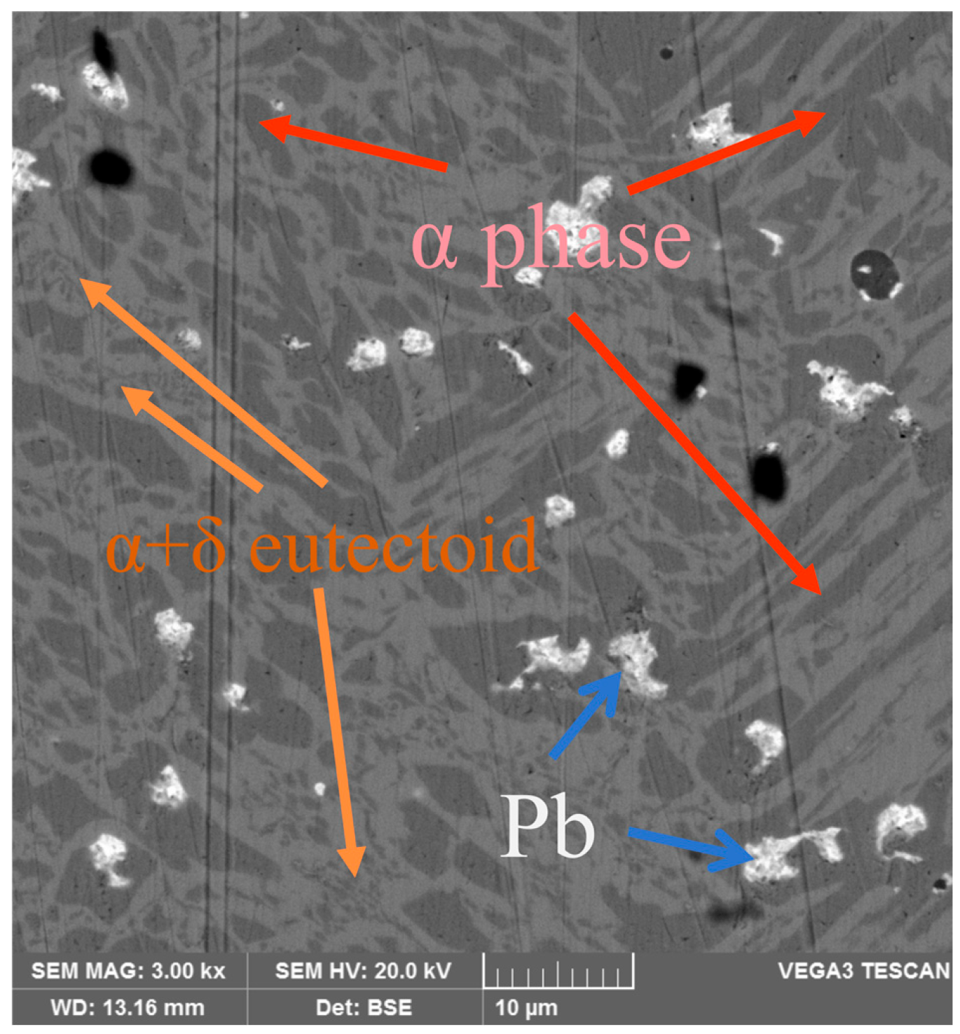
Given that high-tin patina is more commonly observed on the surfaces of high-tin bronze artifacts—such as bronze mirrors and weapons—the bronze specimens used in this study were sand-cast simulated high-tin bronze mirrors. The elemental composition of the polished and ground bronze matrix was determined by EDS area scanning, yielding mass fractions of Cu 72.5%, Sn 24.5%, and Pb 3.0%. The metallographic image of the sample shown in Figure 1 reveals that the α phase appears as acicular grains with pointed ends, distributed within a continuous eutectoid matrix [25]. The Cu-rich α phase exhibits a darker contrast and a needle-like morphology, whereas the Sn-rich eutectoid matrix appears as a lighter, continuous region. The brightest features correspond to discrete particles of metallic lead [26].
The central region of the bronze mirror was cut into specimens measuring 10 mm × 10 mm × 5 mm. A copper wire was welded to the back surface of each specimen, and the non-working surfaces were then sealed with epoxy resin, leaving only a 10 mm × 10 mm working surface exposed. The exposed surfaces were sequentially ground using silicon carbide papers of 300-, 600-, 1000-, 1500-, and 2000-grit grades, followed by polishing with a 1 μm diamond paste. Subsequently, the specimens were rinsed repeatedly with deionized water, ultrasonically cleaned in anhydrous ethanol for 10 min, dried under ambient conditions, and hermetically sealed for subsequent use.

2.2. Electrochemical Tests

Electrochemical tests were conducted using a Scott CS310M (Scott, Wuhan, China) electrochemical workstation in a three-electrode electrolytic cell with an effective volume of 300 mL, maintained at a controlled laboratory temperature of 25 °C. The bronze specimen, with an exposed working area of 1 cm2, served as the working electrode (WE); a 15 mm × 15 mm platinum sheet was used as the counter electrode (CE); and a saturated calomel electrode (SCE) was employed as the reference electrode (RE), which has a potential of +0.241 V versus the standard hydrogen electrode (SHE) at 25 °C. After immersion in the simulated ion solution, the specimen was allowed to stabilize for 5000 s to establish a stable open-circuit potential (OCP) and achieve steady-state surface conditions. Potentiodynamic polarization measurements were then performed by scanning from −0.6 V to +0.6 V vs. SCE at a sweep rate of 0.25 mV/s. All electrochemical measurements were conducted under naturally aerated conditions at room temperature. The electrolyte solutions were neither deaerated nor subjected to forced aeration during the experiments. The solutions were maintained in a static state and remained in contact with laboratory air, resulting in dissolved oxygen levels close to air-saturated conditions.

2.3. Analysis of Corrosion Products

Following the completion of the electrochemical measurements, the epoxy resin encapsulation and copper wires attached to the backside of the bronze specimens were carefully removed. The specimens were then thoroughly rinsed with deionized water and anhydrous ethanol, followed by drying under a stream of nitrogen. The morphological characteristics of the surface corrosion products were examined using a Zeiss optical microscope (Carl Zeiss AG, Oberkochen, Germany). Phase composition analysis of the corrosion products was performed using a Renishaw InVia Qontor micro-confocal Raman spectrometer (Renishaw plc, Wotton-under-Edge, UK) equipped with laser excitation at wavelengths of 532 nm and 785 nm, with adjustable laser power ranging from 0.1 to 5 mW. Spectral data were collected over a wavenumber range of 100–3200 cm−1, using a 50× long-working-distance objective lens (Leica Microsystems, Wetzlar, Germany).
A TESCAN VEGA 3 scanning electron microscope (SEM, TESCAN, Brno, Czech Republic) equipped with an energy-dispersive X-ray spectrometer (EDS, Oxford Instruments, High Wycombe, UK) was used to characterize the surface and cross-sectional microstructures, corrosion layer morphology, and elemental composition of the corroded bronze specimens. Each specimen was symmetrically sectioned along its central axis into two halves: one half was reserved for analysis of the surface corrosion products, while the other was designated for cross-sectional examination. For cross-sectional analysis, the latter half was mounted in epoxy resin with the cross-section facing downward, sequentially ground using silicon carbide papers from 300 to 2000 grit, and then polished with a 1 μm diamond paste on a flannel pad to expose the cross-section of the corrosion layer. Backscattered electron (BSE) imaging was employed to observe microstructural features. The operating parameters were as follows: accelerating voltage of 20 kV, working distance of 15 mm, and acquisition time of 50 s. EDS analysis was performed to obtain semi-quantitative elemental composition and elemental distribution information from selected surface regions of the corrosion layers. Due to the inherent limitations of the EDS technique, particularly for light elements such as O and C, the obtained results were used only for qualitative and semi-quantitative analysis.

3. Results and Discussion

3.1. Microscopic Morphology of Corrosion Products Surface

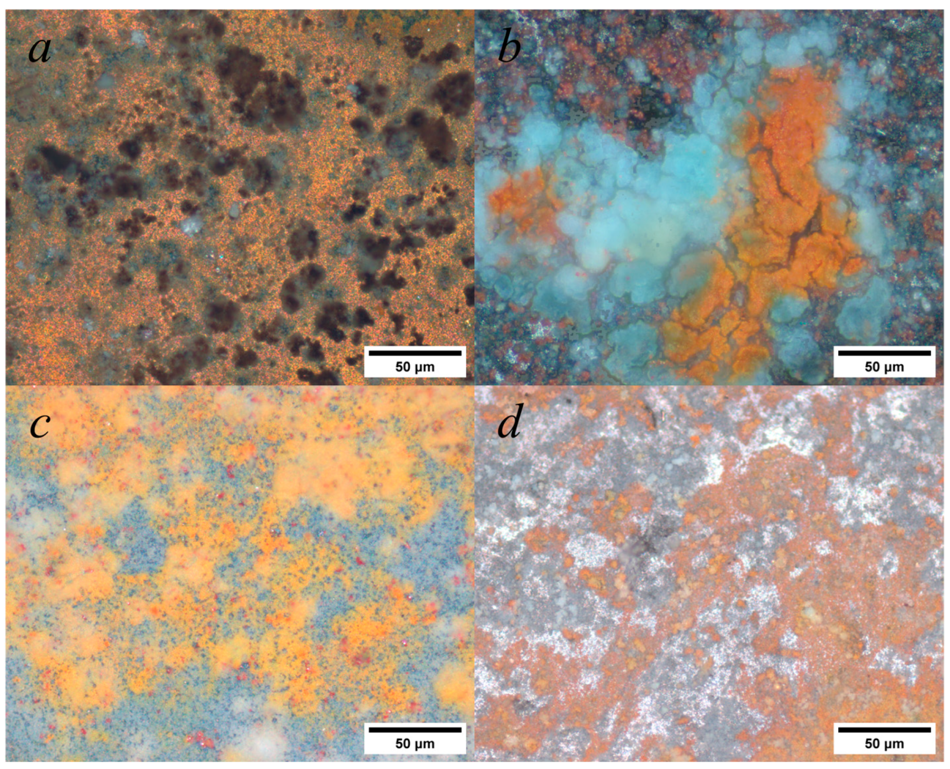
To investigate the micro-morphology of corrosion products on bronze surfaces, a super depth-of-field microscope was used to capture micrographs of the corrosion layers formed under four distinct corrosive environments; the results are presented in Figure 2.
In Figure 2a, for Electrode 1—corroded in a weakly alkaline medium containing HA and FA—the surface is dominated by orange-red corrosion products, while the outer layer is covered with patchy black corrosion products overlying the orange-red layer, along with minor blue corrosion products. In Figure 2b, Electrode 2, which was corroded in a weakly alkaline medium without HA and FA, is largely covered by intermingled red and blue corrosion products, with no clear stratification between the two phases. Some regions still expose the original metallic surface. In Figure 2c, for Electrode 3 corroded in a weakly acidic medium with HA and FA, the outer layer consists predominantly of orange-red corrosion products, interspersed with scattered red crystalline particles. The surface reveals small areas of exposed deep-blue corrosion products, and no portion of the metallic substrate is visible. In Figure 2d, for Electrode 4 corroded in a weakly acidic medium without HA and FA, the corrosion consists of continuously distributed orange-red products, with a brown corrosion layer intercalated in the intermediate region, while a small area of the metallic substrate remains exposed.
All corrosion conditions resulted in the formation of extensive orange-red corrosion products deposited on the original bronze surface. In the absence of humic substances, corrosion products were sparsely distributed across the electrode surface, with limited coverage and partial exposure of the underlying metal substrate. Upon the introduction of humic substances, the surface morphology changed markedly. The coverage of corrosion products increased significantly, forming a more continuous deposit layer that partially masked and protected the underlying metal matrix. In addition, black to brown precipitates were observed on the surface. Under weakly acidic conditions, a greater abundance of orange-red corrosion products was detected, suggesting enhanced copper oxidation and dissolution, likely associated with increased anodic activity in the acidic environment.

3.2. Electron Microscopic Morphology and Elemental Distribution of Corrosion Products

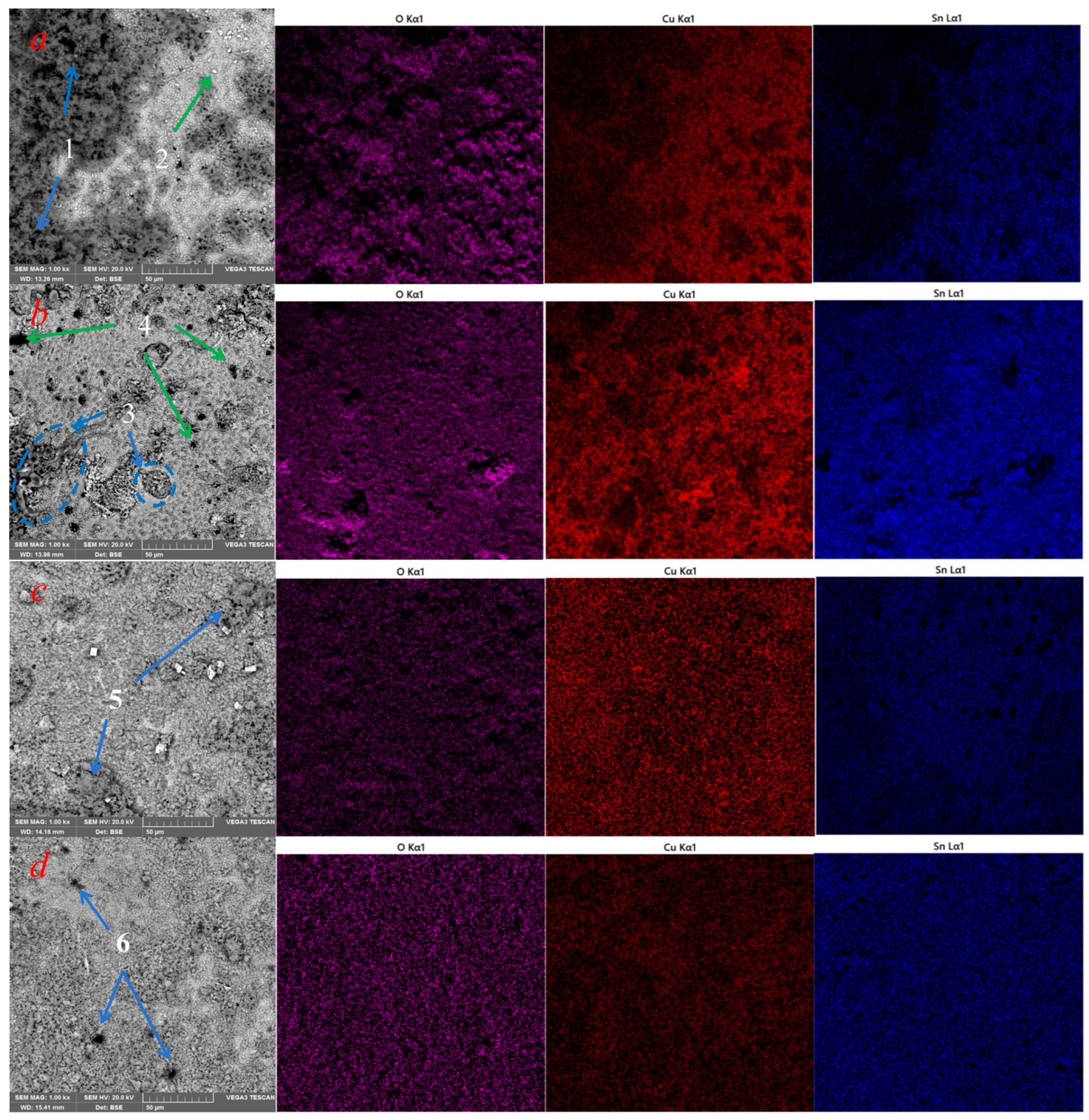
In Figure 3, the corrosion morphology of the bronze surfaces was observed, and elemental maps of Cu, Sn, and O were acquired at 1000× magnification. The EDS data presented here correspond to area-mapping analyses acquired from the same regions observed by SEM and are primarily intended to illustrate the distribution of elements within the surface corrosion layers.
As shown in Figure 3a,b, Electrodes 1 and 2—both exposed to weakly alkaline conditions—exhibited clumpy surface deposits. For Electrode 1 (corroded in a weakly alkaline medium containing HA and FA), approximately half of the surface was covered by continuous (point 1), loose, and rough deposits, while the remaining area consisted of flat corrosion products intermixed with fine particles (point 2), consistent with optical microscopy observations. In contrast, Electrode 2, which was tested in the absence of HA and FA, exhibits a lower degree of surface coverage by corrosion products. Several island-like agglomerates are distributed across different regions of the surface (point 3), without forming a continuous layer, and granular crystalline features are scattered over the surface (point 4). Dark pits associated with the detachment of metallic lead particles can also be observed [27].
Figure 3c and Figure 2d reveal that Electrodes 3 and 4—exposed to weakly acidic environments—were predominantly covered by thick, film-like deposits. For Electrode 3, which was tested in the presence of HA and FA, the surface corrosion layer appears uniform and continuous, with an overall dense morphology. Patchy deposits are locally developed (point 5), and no clearly exposed metallic microstructures are observed. In comparison, Electrode 4 (without HA and FA) exhibited thinner and less extensive deposits. In addition, a small number of annular pits associated with the detachment of lead particles can be observed (point 6). A comparative analysis indicates that bronze surfaces exposed to humus-containing electrolytes are more prone to developing thick, continuous corrosion layers. Consequently, the original metallic surfaces of Electrodes 1 and 3 were more extensively obscured than those of Electrodes 2 and 4.
The surface EDS data of corrosion products formed under the four corrosion conditions are summarized in Table 2. The EDS analyses were performed using area-mapping scans on representative regions of the corrosion layers corresponding to the SEM observation areas. The reported values represent normalized weight percentages (wt%) of the detected elements. Compared to the bulk bronze composition, Electrode 1 exhibited elevated Cu and significantly increased C contents. In regions corresponding to clumpy deposits, Cu and Sn distributions were sparse, with pronounced Sn depletion. In contrast, flat corrosion product areas showed higher Sn signals. Combined with BSE images, the surface deposits observed on Electrode 1 may consist of agglomerated precipitates of HA and FA, together with widely distributed copper-containing corrosion products. Under alkaline conditions, deprotonation of functional groups in HA and FA enhances their ability to complex and adsorb copper ions, which may promote localized accumulation and deposition of copper-rich phases on the metal surface. As a result, regions covered by these deposits exhibit relatively higher copper content, while the uncovered areas display apparent tin enrichment. However, at this stage, a continuous and homogeneous tin-enriched layer has not yet developed across the surface.
Electrodes 2, 3, and 4 all showed significant Sn enrichment, with similar C and O levels across specimens. Their surface corrosion layers lacked the high-Cu and high-C features seen on Electrode 1, suggesting that deposits consist mainly of alloy-derived oxides and salts. Notably, Electrode 3—exposed to weakly acidic simulated soil electrolyte containing humus—displayed higher Sn than Cu content and greater O content than Electrodes 2 and 4. This trend indicates enhanced formation of Sn on Electrode 3. These observations suggest that the weakly acidic, humus-containing electrolyte may promote copper oxidation and tin enrichment during corrosion.
The observed trends in surface morphology and elemental distribution indicate that both pH conditions and humic substances jointly influence the corrosion behavior of high-tin bronze. During corrosion, the metal substrate exhibits evident copper depletion accompanied by relative tin enrichment within the corrosion layer. In the presence of humic substances, this selective dissolution trend may be further enhanced, as humic compounds can promote copper dissolution and/or stabilize dissolved copper species through complexation.

3.3. Microstructural Characteristics and Elemental Composition of the Corrosion Cross-Section

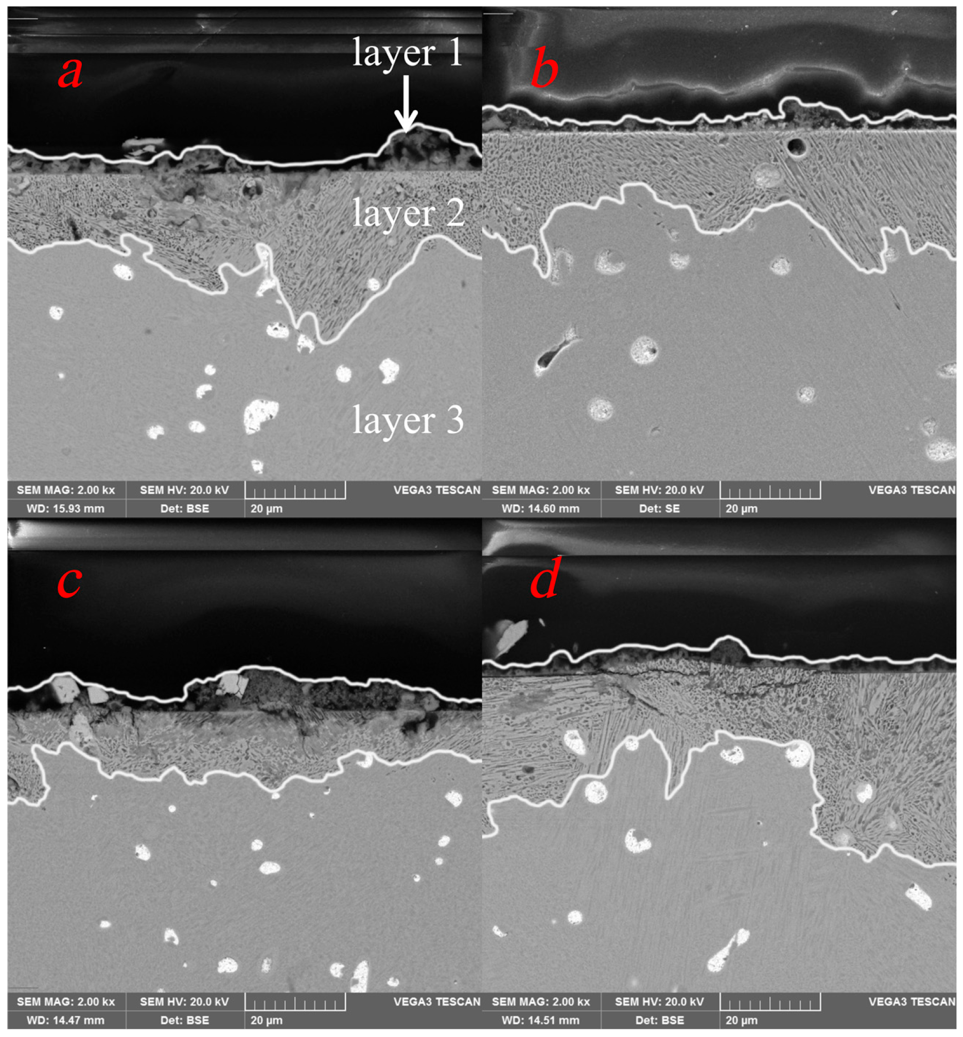
To characterize the patina layer structure and the progression of internal corrosion within the metal matrix under the four distinct corrosion conditions, BSE micrographs of the corroded electrodes were obtained using SEM, accompanied by EDS elemental mapping. The BSE micrographs are presented in Figure 4. The cross-sectional analysis mainly focuses on the regions highlighted by the white line boxes in the figure, which correspond to the corroded zones.
Cross-sectional BSE micrographs analyses reveal a consistent three-layer structure in all four specimens, extending from the surface inward (as shown in Figure 4a): an adherent secondary corrosion product layer (layer 1), a semi-corroded transition zone beneath the original metal surface (layer 2), and an uncorroded bulk matrix (layer 3). The corroded zones observed in the cross-sections of the four electrodes were non-uniform and exhibited localized penetration features. Based on measurements from the BSE, the maximum corrosion depths were 33.2 μm, 25.6 μm, 12.9 μm, and 35.8 μm, respectively. The regions enclosed by white lines correspond to the combined surface deposits and underlying transition layer. As shown in Figure 4a,c, Electrodes 1 and 3 exhibit relatively thick and continuous secondary corrosion layers—consistent with the extensive surface coverage observed earlier. Sectioned cubic crystalline particles are observed, whose morphology is similar to that of the crystalline features present on the sample surface. In contrast, the corrosion layers on Electrodes 2 and 4 (Figure 4b,d) are thinner and more discontinuous, aligning with their planar, patchy distribution patterns. A semi-corroded transition zone is present beneath all 4 samples, albeit with variable thickness. Due to the short corrosion duration, no fully mineralized layers—indicative of complete metal degradation—were observed [28].
The BSE images reveal that the α + δ eutectoid structures within the semi-corroded transition zone retain a light gray contrast and morphological features consistent with those of the uncorroded bulk matrix, indicating negligible elemental loss or corrosion [29]. In contrast, the acicular α-phase regions exhibit a darker contrast, which is attributed to the oxidation and leaching of copper ions. During the corrosion of the α phase within the semi-corroded zone, the high-tin eutectoid matrix remains largely intact. For Electrodes 1 and 2, pores formed by the depletion of metallic lead particles are observed, while the underlying metal surface remains structurally intact. Both Electrodes 3 and 4 display cracks originating at the surface and propagating inward, showing a tendency to compromise the integrity of the metallic surface. Notably, the semi-corroded transition zone of Electrode 3 is the most extensively developed: the α-phase at the surface is almost completely corroded, the eutectoid structures have transformed into a dark gray appearance, and their morphology progressively evolves toward mineralized corrosion products [29].
The EDS results for bronze specimens under the four corrosion conditions are summarized in Table 3. These data confirm selective corrosion of the α-phase within the semi-corroded transition zones of all four electrodes [30]. Within the regions analyzed by EDS, all four electrodes exhibit an increased Sn content. Compared with the uncorroded metal matrix, a pronounced decrease in Cu content is observed in these regions, resulting in a significantly reduced Cu/Sn ratio, whereas the variations in Sn and Pb contents remain relatively limited. The elemental changes observed in the cross-sections of all four electrodes indicate that corrosion initiates preferentially in the Cu-rich α phase and subsequently extends into the eutectoid matrix, with the corrosion process being dominated by the loss of copper. This elemental migration pattern aligns with that reported for high-tin bronzes during early-stage soil burial corrosion [31].
It is noteworthy that carbon signals are markedly enhanced on Electrodes 1 and 3, tentatively attributed to the co-deposition of humus (HA and FA) with surface corrosion products. Concurrently, detectable Cu signals in the outer layers reflect ongoing metallic ion diffusion during corrosion. Among the four electrodes, Electrodes 1 and 3 exhibit lower cross-sectional Cu contents, indicating that HA and FA promote Cu leaching from the alloy surface. In particular, Electrode 3 shows the most severe Cu depletion in its semi-corroded zone, accompanied by a significant increase in oxygen—consistent with its surface composition. This further supports the role of humus substances in accelerating Cu dissolution and facilitating in situ SnO2 formation under weakly acidic conditions.

3.4. Corrosion Product Phases

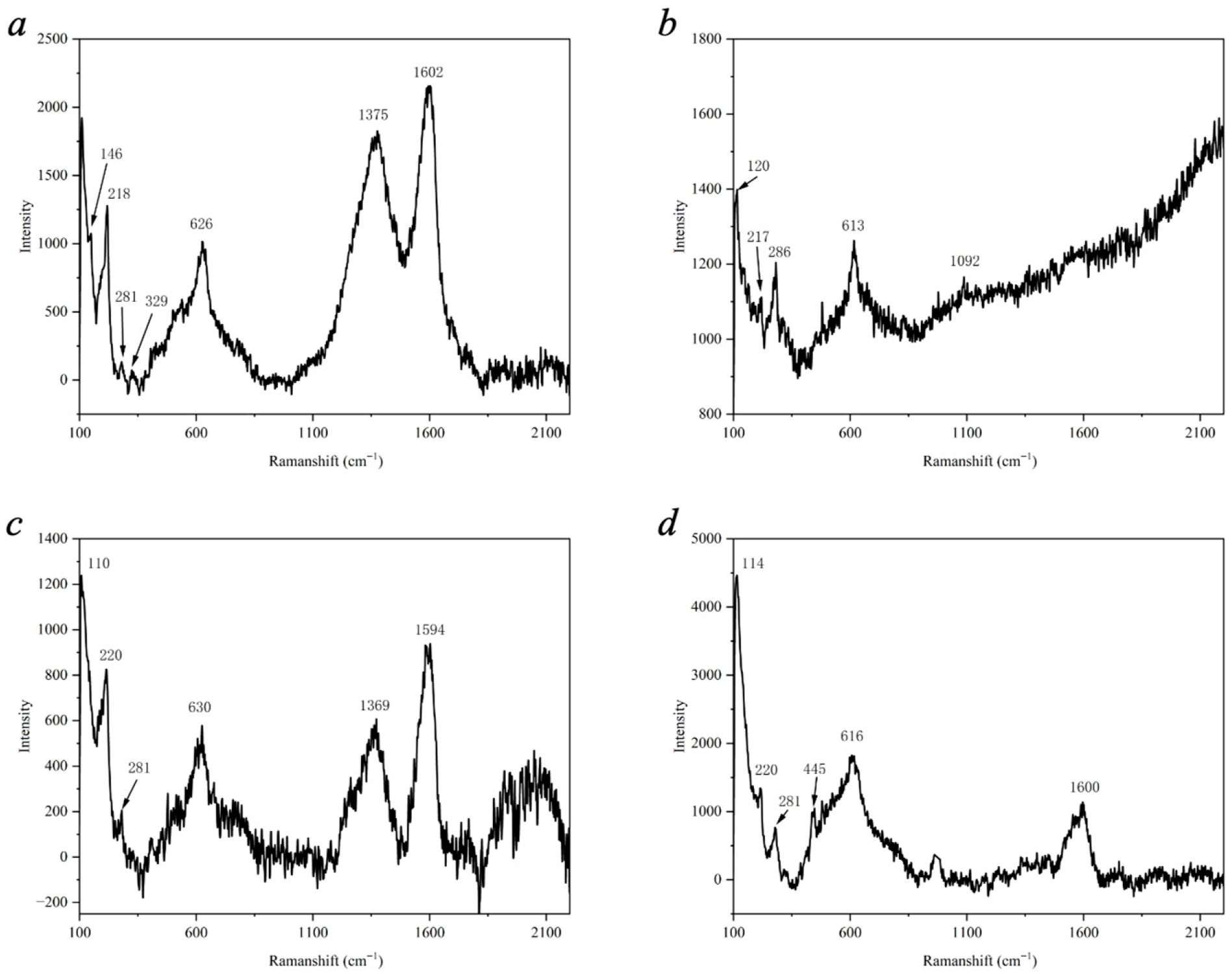
Figure 5 presents the Raman spectra of the corrosion products formed on the electrode surfaces. In studies focusing on high-tin corrosion layers on bronze surfaces, Raman spectroscopy is commonly employed as a phase identification technique, particularly for the analysis of cassiterite (SnO2) and cuprite (Cu2O). Due to the localized nature of Raman measurements, the identified compounds cannot be directly assigned to specific structural components or microstructural regions. Therefore, the Raman results are interpreted as phase information representative of the probed surface areas, rather than as evidence for a direct spatial correspondence between specific compounds and distinct morphological features. The characteristic Raman signals in spectra 4a and 4c support the hypothesis of humus co-deposition within the surface corrosion products. Specifically, two broad peaks centered at 1375 cm−1 and 1602 cm−1 are assigned to the D/G bands of the carbon signal [30]. When combined with the observation of black agglomerates in the corresponding micrographs and the elevated carbon content detected by EDS, these results confirm the presence of humus deposits on the surface of Electrode 1.
The shoulder peak at 146 cm−1 and the sharp peaks at 218 cm−1 (Figure 5a), 217 cm−1 (Figure 5b), and 220 cm−1 (Figure 5c,d) are attributed to the low-frequency vibrational modes of cuprite [31,32,33]. Corroborated by the microscopic imaging results, these signals identify the orange-colored corrosion products as Cu2O. Additionally, the sharp peaks at 110 cm−1 and 114 cm−1 are associated with lead dioxide [34], which correlates with the mild corrosion of the lead constituent in the bronze matrix.
All four bronze corrosion product samples exhibit a distinct, broad Raman peak centered around 620 cm−1. This characteristic signal corresponds to the unique broad spectral band spanning 300–698 cm−1, which is attributed to the coexistence of amorphous Cu2O and SnO2 [35,36]. Notably, this specific Raman signature has been detected in high-tin corrosion layers on numerous bronze artifacts and is frequently used as a diagnostic indicator for identifying the formation of high-tin patina [37].
To summarize, Raman spectroscopy revealed the presence of cassiterite characteristic peaks on all four bronze electrodes, with spectral features comparable to those observed in high-tin patina of archaeological bronzes. For samples corroded in humic media, adsorption and deposition of humic substances on the metal surface promoted the formation of brown to black agglomerates within the corrosion products, accompanied by pronounced surface carbon enrichment. Raman signals corresponding to copper and lead corrosion products were also detected.

3.5. Electrochemical Corrosion Behavior

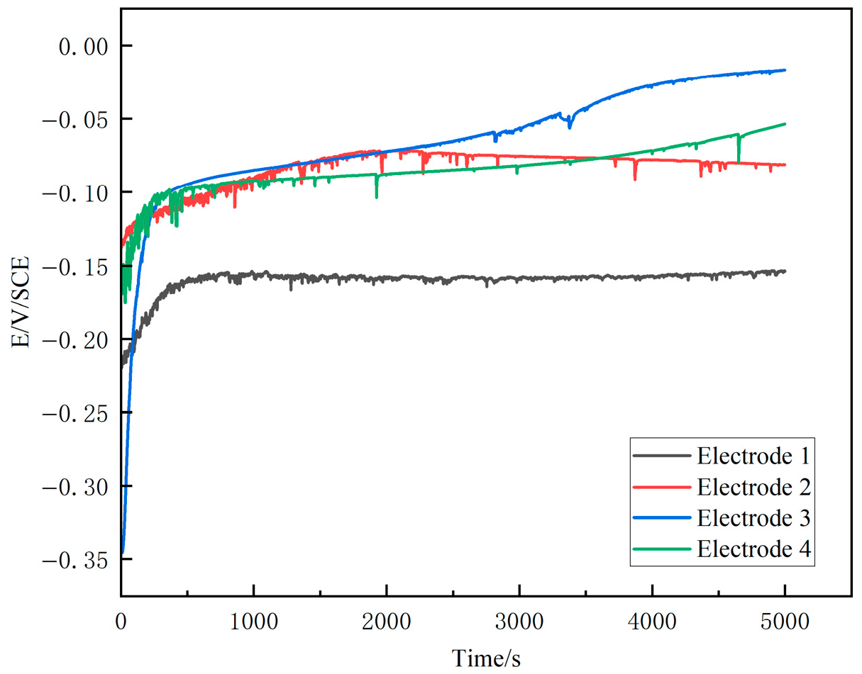
The OCP of the four electrodes initially shifted in the positive direction during the early immersion stage and eventually reached a relatively stable state after approximately 5000 s, facilitating the establishment of an accurate and stable system for subsequent electrochemical measurements. The alloy electrodes attained steady-state conditions after 595 s, 1800 s, 358 s, and 405 s, respectively, with corresponding stable potentials of −0.2 V vs. SCE, −0.1 V vs. SCE, −0.1 V vs. SCE, and −0.02 V vs. SCE. Bronze undergoes corrosion immediately upon immersion in the solution, and as surface corrosion products gradually accumulate, a deposited layer with a certain protective effect is formed, which retards further corrosion and leaching of the metal [38]. Under weakly acidic conditions, the magnitude of the positive shift in the bronze’s OCP is more pronounced. This is attributed to the enhanced feasibility of oxygen depolarization reactions in acidic media, which accelerate copper corrosion and dissolution. The thicker corrosion layers observed on the surfaces of Electrodes 3 and 4 under weakly acidic conditions further support that bronze tends to dissolve more rapidly in this environment, thereby promoting the formation of a corrosion film, which is consistent with the trend observed in the OCP curves [39]. The naturally aerated condition was selected to approximate oxygen availability in moist soil environments; however, variations in dissolved oxygen may still influence cathodic reactions and represent an inherent limitation of the present experimental design [40]. The OCP results reflect changes in the electrochemical state of the bronze surface under different conditions (see Figure 6).
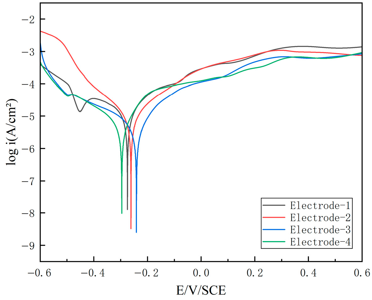
Figure 7 presents the polarization curves of the bronze electrodes in four simulated soil solutions. All samples exhibit weak passivation behavior in their respective media. As the polarization potential is swept in the anodic direction, the curves sequentially traverse the active region, the active-to-passive transition region, and the passive region. Beyond a potential of −0.1 V vs. sce, the current density increases with increasing potential, indicating enhanced dissolution of the bronze matrix, and no distinct passive plateau is observed [41]. Electrode 3, exposed to the weakly acidic medium containing humus, exhibited a positive shift in corrosion potential, likely due to the protective effect of its more continuous and compact surface corrosion layer [42]. During polarization, localized depletion of dissolved oxygen may occur near the electrode surface. This phenomenon can also lead to fluctuations in current density, particularly under unstirred solution conditions. All samples displayed similar polarization behavior, indicating that factors such as solution pH and humic acid content have negligible influence on the overall electrochemical response [43].
The electrochemical parameters, including the corrosion potential (Ecorr) and the estimated corrosion current density (iset), are summarized in Table 4. In this study, the corrosion current density was estimated from the polarization curves in the vicinity of Ecorr, rather than being strictly determined by Tafel extrapolation. Moderate variations in Ecorr were observed among the four electrodes, consistent with the trends shown in the polarization curves. In particular, the electrode corroded under weakly acidic conditions in the presence of humic substances exhibited a slight positive shift in Ecorr, which is in agreement with the relatively continuous corrosion layer observed in the cross-sectional analysis. As an important parameter reflecting the corrosion rate under near-equilibrium conditions, it showed only limited variation across the four environments. Although minor differences were detected, the values remained within the same order of magnitude, indicating that under the present experimental conditions, solution pH and humic substance content did not significantly alter the overall corrosion kinetics of high-tin bronze.

3.6. Initial Corrosion Behavior of High-Tin Bronze

Four bronze electrodes were subjected to potentiostatic polarization in soil ionic solutions with varying pH values and selective addition of humic acid. Secondary corrosion layers formed on the surface. In the semi-corroded transition zone, the α-phase within the metal acted as the anodic phase and underwent preferential corrosion, leading to the initial formation of the semi-corroded layer [31]. Although the crystallinity of the initial corrosion products was limited and the addition of humic acid did not reproduce the typical “black lacquer-like” appearance, corrosion progressed slowly and the original metal surface was largely preserved. The resulting surface morphology and elemental distribution show similarities to the tin-enriched corrosion layers observed in high-tin archaeological bronzes.
Attributed to the predominance of sulfate and carbonate ions in the solution with relatively low chloride concentrations, the corrosiveness toward the metal is limited; consequently, corrosion progresses more slowly and the original metallic surface can be preserved [44]. Copper on the surface of the bronze electrode undergoes the following reactions: Cu→Cu++e-, 2Cu++H2O→Cu2O+2H+, Cu+→Cu2+. Tin undergoes the following reactions: Sn→Sn2+, Sn2+→Sn4+, Sn4++2H2O→SnO2+4H+. During corrosion, metallic lead is oxidized first, and pits formed by lead corrosion can be observed on the electrode surface. Subsequently, the α phase, which has a lower electrochemical potential, undergoes preferential corrosion, followed by the eutectoid matrix [5]. Regarding the copper element, metallic Cu tends to be oxidized to Cu2O at lower potentials, which subsequently transforms into Cu2+ species as the potential increases. In alkaline environments, the formation of Cu(OH)2 and its further conversion into CuO are thermodynamically favored [45]. However, these species did not yield distinct Raman signals, potentially due to the relatively low concentration of Cu2+ near the electrode surface caused by mass diffusion and other transport processes. When the concentration of anions in the environment reaches the threshold for solid-phase precipitation, secondary corrosion products form on the surface of the corrosion layer. In contrast, under conditions of relatively low anion concentrations, cuprite is preferentially formed [46]. Therefore, when the Cu-rich α phase undergoes oxidation, a Cu2O corrosion layer initially forms on the metal surface due to the relatively low anion concentrations established in the simulated solution. Subsequently, as anions accumulate in the vicinity of the corroding electrode, a portion of the released Cu2+ ions is converted into secondary corrosion products. Within the pH and potential ranges applied in the present experiments, according to the Pourbaix diagrams of the Sn–H2O systems, the thermodynamically stable tin phases are SnO2 and Sn(OH)4 [47]. However, the latter is unstable and tends to undergo dehydration, further transforming into SnO2 [48]. Therefore, insoluble SnO2 forms on the metal surface, precipitates in situ, and gradually develops into a dense corrosion layer, thereby inhibiting further degradation of the metal.
Dissolved oxygen in the system can enhance the overall oxidizing power and corrosion driving force by promoting the cathodic oxygen reduction reaction. At the same time, it influences the oxidation state of corrosion products and thereby affects the structure and evolution of the corrosion layer. Compared to alkaline environments, oxygen depolarization reactions are enhanced in acidic media [49], accelerating the dissolution and leaching rates of copper. Consequently, Corrosion progression on both the surface and interior of the metal proceeds more rapidly. Meanwhile, cuprite is relatively more stable under alkaline conditions, which corresponds well with the greater abundance of orange corrosion products observed in the microstructural morphology under weakly alkaline environments [50].
The surface corrosion morphology and composition of Electrode 1 differ from those obtained in the simulated soil solution without humic substances. This difference is mainly attributed to the relatively abundant deposition of humic acid on its surface. Under weakly acidic conditions, Electrode 3 exhibited the most pronounced tin enrichment at the surface, indicating the influence of humic acid addition. However, based on cross-sectional analysis, phase characterization, and electrochemical results, the overall corrosion pattern of the four bronze samples remained unchanged. The presence of humic acid primarily acted to promote the corrosion reactions rather than fundamentally alter the corrosion mechanism. Humic substances can accelerate copper dissolution as well as tin oxidation and deposition, thereby promoting the formation of a tin-enriched layer beneath the original metal surface during the early stages of corrosion. Under weakly alkaline conditions, humic substances mainly act through complexation with Cu2+ ions and subsequent deposition on the metal surface [51].
HA and FA contain oxygen-bearing functional groups, primarily including carboxyl (–COOH), hydroxyl (–OH), and carbonyl (–C=O) groups. These functional groups confer strong metal-binding capacity, particularly a high affinity for transition metal ions such as Cu2+ through complexation. They are pH-sensitive organic substances whose ionization degree, molecular conformation, and solubility strongly depend on solution pH. Under weakly acidic conditions, HA and FA are partially protonated, resulting in reduced solubility and an increased tendency to adsorb onto metal surfaces or form surface-associated aggregates. Such behavior may enhance complexation with Cu species and facilitate the dissolution and migration of copper during the early stages of corrosion. In contrast, under weakly alkaline conditions, humic substances predominantly exist in a deprotonated form, exhibiting higher solubility and weaker interactions with the metal surface. As a result, their influence on corrosion processes is relatively limited compared with acidic environments. The pH-dependent chemical state of humic substances therefore provides a plausible explanation for the different corrosion responses observed under acidic and alkaline conditions in the present study.

4. Conclusions

Electrochemical accelerated corrosion tests conducted in four simulated soil media reproduced key features of early-stage corrosion in high-tin bronze. All conditions produced a two-layer structure consisting of a secondary corrosion layer and a semi-corroded transition zone, with the original metal surface largely preserved. Preferential corrosion of the Cu-rich α-phase resulted in copper depletion and relative tin enrichment, with tin stabilized as SnO2 and copper reprecipitated mainly as Cu2O.
Corrosion behavior was primarily controlled by soil ionic composition and pH. Humic substances did not alter the fundamental corrosion mechanism or stratigraphic structure but influenced surface morphology and elemental distribution to a limited extent. Under weakly acidic conditions, HA and FA at natural concentrations slightly enhanced tin enrichment but did not reproduce the classical morphology of high-tin archaeological patina. Finally, it should be noted that this work represents a preliminary study conducted on a limited number of samples. Each of the four simulated environments was evaluated using a single electrode. While the results offer initial insights into early-stage corrosion behavior, further investigations with expanded sample sizes are necessary to confirm the long-term stability and statistical reproducibility of these observations.

Author Contributions

Conceptualization, L.Y. and Y.M.; methodology, Y.M.; validation, Y.M.; formal analysis, Y.M.; investigation, Y.M.; resources, Y.M.; data curation, Y.M.; writing—original draft preparation, Y.M.; writing—review and editing, L.Y.; visualization, Y.M.; supervision, L.Y.; project administration, L.Y.; funding acquisition, L.Y. All authors have read and agreed to the published version of the manuscript.

Funding

This work was supported by two funding. Key R&D Project in Shaanxi Province (2023-GHZD-34), supported by Education Department of Shaanxi Provincial Government. Scientific Research Project on Cultural Relics Inventory and Quantification (2023ZCK029), supported by National Cultural Heritage Administration. The funding source had no role in the design of the study; in the collection, analysis, or interpretation of data; in the writing of the manuscript; or in the decision to submit the manuscript for publication.

Institutional Review Board Statement

Not applicable.

Informed Consent Statement

Not applicable.

Data Availability Statement

The original contributions presented in this study are included in the article. Further inquiries can be directed to the corresponding author.

Conflicts of Interest

The authors declare no conflict of interest.

References

  1. Guadagnini, L.; Chiavari, C.; Martini, C.; Bernardi, E.; Morselli, L.; Tonelli, D. The Use of Scanning Electrochemical Microscopy for the Characterisation of Patinas on Copper Alloys. Electrochim. Acta 2011, 56, 6598–6606. [Google Scholar] [CrossRef] [Scilit]
  2. Metikos-Hukovic, M.; Babic, R.; Paic, I. Copper Corrosion at Various pH Values with and without the Inhibitor. J. Appl. Electrochem. 2000, 30, 617–624. [Google Scholar] [CrossRef] [Scilit]
  3. Liu, W.; Chen, J. A Review of High-Tin Patina on Ancient Bronze Artifacts. J. Natl. Mus. China 2019, 146, 146–160. (In Chinese) [Google Scholar]
  4. Karlbeck, O. Notes on Some Early Chinese Bronze Mirrors. China J. Sci. Arts 1926, 4, 4. [Google Scholar]
  5. Robbiola, L.; Blengino, J.M.; Fiaud, C. Morphology and Mechanisms of Formation of Natural Patinas on Archaeological Cu–Sn Alloys. Corros. Sci. 1998, 40, 2083–2111. [Google Scholar] [CrossRef] [Scilit]
  6. Robbiola, L.; Hurtel, L.-P. Standard nature of the passive layers of buried archaeological bronze—The example of two Roman half-length portraits. In Proceedings of the International Conference on Metal Conservation (METAL 95); MacLeod, I., Penney, C., Eds.; James &James Science Pub: London, UK, 1995; pp. 109–117. [Google Scholar]
  7. Oudbashi, O.; Hasanpour, A.; Davami, P. Investigation on Corrosion Stratigraphy and Morphology in Some Iron Age Bronze Alloys Vessels by OM, XRD and SEM-EDS Methods. Appl. Phys. A 2016, 122, 262. [Google Scholar] [CrossRef] [Scilit]
  8. Oudbashi, O.; Naseri, R.; Asadi Hasanvand, P. Long-Term Corrosion of Copper Alloys in the Soil: New Aspects of Corrosion Morphology in Archaeological Vessels from South-Western Iran. Herit. Sci. 2024, 12, 73. [Google Scholar] [CrossRef] [Scilit]
  9. Robbiola, L.; Portier, R. A global approach to the authentication of ancient bronzes based on the characterization of the alloy–patina–environment system. J. Cult. Herit. 2006, 7, 1–12. [Google Scholar] [CrossRef] [Scilit]
  10. Piccardo, P.; Mille, B.; Robbiola, L. Tin and Copper Oxides in Corroded Archaeological Bronzes. In Corrosion of Metallic Heritage Artefacts; Elsevier: Amsterdam, The Netherlands, 2007; pp. 239–262. ISBN 978-1-84569-239-1. [Google Scholar]
  11. Oddy, A.; Scott, D.A. Copper and Bronze in Art: Corrosion, Colorants, Conservation. Stud. Conserv. 2002, 47, 277. [Google Scholar] [CrossRef] [Scilit]
  12. He, L.; Liang, J.; Zhao, X.; Jiang, B. Corrosion Behavior and Morphological Features of Archeological Bronze Coins from Ancient China. Microchem. J. 2011, 99, 203–212. [Google Scholar] [CrossRef] [Scilit]
  13. Yang, X.; Wu, W.; Chen, K. A Preliminary Study on the Structural Characteristics and Classification of Corrosion Layers on Ancient Bronze Artifacts. Corros. Prot. 2023, 44, 42–50. (In Chinese) [Google Scholar]
  14. Abdelbar, M.; El-Shamy, A.M. Understanding Soil Factors in Corrosion and Conservation of Buried Bronze Statuettes: Insights for Preservation Strategies. Sci. Rep. 2024, 14, 19230. [Google Scholar] [CrossRef] [Scilit]
  15. Oudbashi, O. A Methodological Approach to Estimate Soil Corrosivity for Archaeological Copper Alloy Artefacts. Herit. Sci. 2018, 6, 2. [Google Scholar] [CrossRef] [Scilit]
  16. Al-Rawajfeh, A.E.; Al-Shamaileh, E.M. Assessment of Tap Water Resources Quality and Its Potential of Scale Formation and Corrosivity in Tafila Province, South Jordan. Desalination 2007, 206, 322–332. [Google Scholar] [CrossRef] [Scilit]
  17. Ma, Z.; Du, L.; Fang, C.; Yin, X.; Jin, L. Structure of the “Heiqigu” Surface Layer on Bronze Mirrors. J. Univ. Sci. Technol. Beijing (Engl. Ed.) 1996, 126, 121–126. (In Chinese) [Google Scholar]
  18. Ma, Z.; Yin, X.; Jin, L. Polyphenolic Substances Induce the Formation of “Heiqigu” on Bronze Mirrors and the Structure of Heiqigu—A Study on Bronze Mirror Patina (II). Kao Gu 1995, 1037–1046, 1051. (In Chinese) [Google Scholar]
  19. Ma, Z. Advances in the Study of “Heiqigu” Bronze Mirrors. Univ. Chem. 1995, 58–62. (In Chinese) [Google Scholar]
  20. Ma, Z.; Jin, L.; Yin, X. Humic Acid Induces the Formation of “Heiqigu” on the Surface of Tin-Bronze Mirrors. Kao Gu 1994, 261–273. (In Chinese) [Google Scholar]
  21. Wang, A. “Heiqigu” Bronze Mirrors and the Humic Acid Enrichment Hypothesis. Collect. Invest. 2021, 12, 119–122. (In Chinese) [Google Scholar] [CrossRef]
  22. Nord, A.G.; Mattsson, E.; Tronner, K. Factors Influencing the Long-Term Corrosion of Bronze Artefacts in Soil. Prot. Met. 2005, 41, 309–316. [Google Scholar] [CrossRef] [Scilit]
  23. Song, C.; Wang, P.; Han, G.; Shi , Y. Chemical Characteristics of Shallow Groundwater in the Loess Tableland and Its Significance for the Carbon Cycle. South-to-North Water Transf. Water Sci. Technol. 2017, 15, 121–126. (In Chinese) [Google Scholar] [CrossRef]
  24. Dang, Y.; Li, S.; Wang, G. Distribution Characteristics of Humus Fraction in Soil Profile for the Rypical Regions in the Loess Plateau. Acta Ecol. Sin. 2012, 32, 1820–1829. (In Chinese) [Google Scholar] [CrossRef] [Scilit]
  25. Institute for the History of Natural Sciences; Chinese Academy of Sciences. Atlas of Fiber Microstructures of Ancient Chinese Metallic Materials: Non-Ferrous Metals; Science Press: Beijing, China, 2011; pp. 33–35. (In Chinese) [Google Scholar]
  26. Jia, M.; Xu, H.; Hu, P.; Hu, G. Microstructures and Electrochemical Corrosion Behaviors of Bronze Mirror. npj Herit. Sci. 2025, 13, 405. [Google Scholar] [CrossRef] [Scilit]
  27. Cho, N.C.; Jang, M.K.; Huh, I.K. A Study on the Microstructure and Corrosion Characteristics of Early Iron Age Bronze Mirrors Excavated from the Korean Peninsula. Appl. Sci. 2021, 11, 2441. [Google Scholar] [CrossRef] [Scilit]
  28. Hu, Y.; Zhang, B.; Xia, P.; Shao, A.; Chen, J. Study on the Corrosion Causes of Bronze Artifacts Unearthed from the Liujiwa Site in Chengcheng County. Sci. Conserv. Archaeol. 2022, 34, 78–87. (In Chinese) [Google Scholar] [CrossRef]
  29. Mu, Y.; Luo, W.; Li, L.; Huang, F.; Wang, C. Comprehensive Analysis of the Corrosion Layer Structure of Bronze Artifacts Unearthed from the Western Zhou Cemetery at Yejiashan, Suizhou, Hubei. Sci. Conserv. Archaeol. 2020, 32, 8–16. (In Chinese) [Google Scholar] [CrossRef]
  30. He, Y.; Qu, X.; Liu, Y.; Li, Y.; Liu, C. Technologies and Sources of the Earliest Chinese Bronze Horse Sculptures Unearthed on the Loess Plateau. npj Herit. Sci. 2025, 13, 165. [Google Scholar] [CrossRef] [Scilit]
  31. Tang, Q.; Wang, J.; Ma, J. Morphology Change and Elements Migration of Bronze with High Tin Content after Soil Corrosion. J. Chin. Soc. Nonferrous Met. 2011, 21, 3175–3181. (In Chinese) [Google Scholar] [CrossRef]
  32. Bouchard, M.; Smith, D.C. Catalogue of 45 Reference Raman Spectra of Minerals Concerning Research in Art History or Archaeology, Especially on Corroded Metals and Coloured Glass. Spectrochim. Acta Part A Mol. Biomol. Spectrosc. 2003, 59, 2247–2266. [Google Scholar] [CrossRef] [Scilit]
  33. Kareem, K.; Sultan, S.; He, L. Fabrication, Microstructure and Corrosive Behavior of Different Metallographic Tin-Leaded Bronze Alloys Part II: Chemical Corrosive Behavior and Patina of Tin-Leaded Bronze Alloys. Mater. Chem. Phys. 2016, 169, 158–172. [Google Scholar] [CrossRef] [Scilit]
  34. Li, Y.; Bao, Z.; Wu, T.; Jiang, J.; Chen, G.; Pan, C. Specific Corrosion Product on Interior Surface of a Bronze Wine Vessel with Loop-Handle and Its Growth Mechanism, Shang Dynasty, China. Mater. Charact. 2012, 68, 88–93. [Google Scholar] [CrossRef] [Scilit]
  35. Ospitali, F.; Chiavari, C.; Martini, C.; Bernardi, E.; Passarini, F.; Robbiola, L. The Characterization of Sn-Based Corrosion Products in Ancient Bronzes: A Raman Approach. J. Raman Spectrosc. 2012, 43, 1596–1603. [Google Scholar] [CrossRef] [Scilit]
  36. Tang, Q.; Wang, J.; Ma, J. Review of the Research on Tin Corrosion Products of Ancient Bronze: Properties, Characterization Methods and Corrosion Models. J. Chin. Soc. Nonferrous Met. 2011, 21, 3175–3181. (In Chinese) [Google Scholar] [CrossRef]
  37. Marušić, K.; Otmačić-Ćurković, H.; Horvat-Kurbegović, Š.; Takenouti, H.; Stupnišek-Lisac, E. Comparative Studies of Chemical and Electrochemical Preparation of Artificial Bronze Patinas and Their Protection by Corrosion Inhibitor. Electrochim. Acta 2009, 54, 7106–7113. [Google Scholar] [CrossRef] [Scilit]
  38. Li, B.; Xia, Q.; Dong, W. Comparative Study of the Corrosive Behaviors of Rust Layers on Bronze Ware in Different Corrosive Environments. Materials 2025, 18, 1359. [Google Scholar] [CrossRef] [Scilit] [PubMed]
  39. Shaik, M.A.; Syed, K.H.; Golla, B.R. Electrochemical Behavior of Mechanically Alloyed Hard Cu-Al Alloys in Marine Environment. Corros. Sci. 2019, 153, 249–257. [Google Scholar] [CrossRef] [Scilit]
  40. Yun, T.H.; Kim, T.; Kim, S.; Kim, J. Investigation of Corrosion Behavior of Oxygen-Free Copper in Oxygen-Containing Systems. Materials 2023, 17, 74. [Google Scholar] [CrossRef] [Scilit] [PubMed]
  41. Chang, T.; Herting, G.; Goidanich, S.; Sánchez Amaya, J.M.; Arenas, M.A.; Le Bozec, N.; Jin, Y.; Leygraf, C.; Odnevall Wallinder, I. The Role of Sn on the Long-Term Atmospheric Corrosion of Binary Cu-Sn Bronze Alloys in Architecture. Corros. Sci. 2019, 149, 54–67. [Google Scholar] [CrossRef] [Scilit]
  42. Kapitanović, A.; Ćurković, H.O. The Effect of Corrosion Conditions on Aging of Artificial Patina on Three Bronzes. Coatings 2022, 12, 936. [Google Scholar] [CrossRef] [Scilit]
  43. Liang, Z.; Jiang, K.; Feng, B.; Lin, S.; Chao, X.; Sui, Q.; Zhang, T. Corrosion Evolution of Cu-Pb Alloys from the Western Zhou Dynasty in Simulated Archaeological Soil Environment. J. Electroanal. Chem. 2021, 899, 115688. [Google Scholar] [CrossRef] [Scilit]
  44. Kwon, H. Corrosion Behaviors of Artificial Chloride Patina for Studying Bronze Sculpture Corrosion in Marine Environments. Coatings 2023, 13, 1630. [Google Scholar] [CrossRef] [Scilit]
  45. Beverskog, B.; Puigdomenech, I. Revised Pourbaix Diagrams for Copper at 25 to 300 °C. J. Electrochem. Soc. 1997, 144, 3476–3483. [Google Scholar] [CrossRef] [Scilit]
  46. Scott, D.A. Periodic corrosion phenomena in bronze antiquities. Stud. Conserv. 1985, 30, 49–57. [Google Scholar] [CrossRef] [Scilit]
  47. House, C.I.; Kelsall, G.H. Potential—pH Diagrams for the Sn/H2O-Cl System. Electrochim. Acta 1984, 29, 1459–1464. [Google Scholar] [CrossRef] [Scilit]
  48. May, P.M.; Filella, M. Thermodynamic Data for Sn(IV) Species in Aqueous Solution: A Matter of Controversy and Error. J. Solut. Chem. 2023, 52, 754–761. [Google Scholar] [CrossRef] [Scilit]
  49. Fang, Y.; Han, R.; Fan, S.; Wu, W.; Ma, T.; Zhu, J. Corrosion State Simulation of Excavated Bronze Artifacts in Different Soil Media. Corros. Prot. 2025, 46, 1–8. (In Chinese) [Google Scholar]
  50. Zindrou, A.; Deligiannakis, Y. Quantitative In Situ Monitoring of Cu-Atom Release by Cu2O Nanocatalysts under Photocatalytic CO2 Reduction Conditions: New Insights into the Photocorrosion Mechanism. Nanomaterials 2023, 13, 1773. [Google Scholar] [CrossRef] [Scilit]
  51. Zhuang, X.; Yu, T.; Hu, G.; Ma, C.; Zhao, B.; Sun, X. Effect of Iron Ions, Copper Ions and Humic Acid on Nonylphenol’s Photodegradation. J. Liaoning Univ. 2017, 44, 75–80. (In Chinese) [Google Scholar] [CrossRef]
Figure 1. Backscattered electron image of the metal matrix.
Coatings 16 00320 g001
Figure 2. Optical micrographs of corrosion products on electrode surfaces: (a) weakly alkaline solution with humus; (b) weakly alkaline solution without humus; (c) weakly acidic solution with humus; (d) weakly acidic solution without humus.
Coatings 16 00320 g002
Figure 3. BSE micrograph and EDS elemental distribution maps of O, Cu, and Sn for the corroded bronze electrodes: (a) weakly alkaline solution with humus; (b) weakly alkaline solution without humus; (c) weakly acidic solution with humus; (d) weakly acidic solution without humus.
Coatings 16 00320 g003
Figure 4. BSE images in the cross-section of the corroded bronze: (a) weakly alkaline solution with humus; (b) weakly alkaline solution without humus; (c) weakly acidic solution with humus; (d) weakly acidic solution without humus.
Coatings 16 00320 g004
Figure 5. Raman spectra of the corrosion products on the four electrodes: (a) weakly alkaline solution with humus; (b) weakly alkaline solution without humus; (c) weakly acidic solution with humus; (d) weakly acidic solution without humus.
Coatings 16 00320 g005
Figure 6. Open circuit potential curves of the bronze electrodes in four simulated soil solutions.
Coatings 16 00320 g006
Figure 7. Polarization curves of the bronze electrodes in four simulated soil solutions.
Coatings 16 00320 g007
Table 1. Designation of bronze electrodes and their corrosion parameters.
ElectrodesHCO3 (mg/L)Cl (mg/L)SO42− (mg/L)NO3 (mg/L)HA (mg/L)FA (mg/L)pH
1320.05.512.210.00.380.338.5
2320.05.512.210.0008.5
3320.05.512.210.00.380.335.5
4320.05.512.210.0005.5
Table 2. Four electrodes EDS data for electrode corrosion products.
ElementalElement Content/wt%
Cu KSn LPb MO KC K
1471232315
230374245
322354309
430298249
Table 3. EDS data of the corrosion cross-sections from the four electrodes.
ElectrodeElement Content/wt%
Cu KSn LPb MO KC K
1352731025
242262822
3242651629
442303916
Table 4. Corrosion potential (Ecorr/V vs. SCE) and corrosion current density (icorr/A·cm−2) of 4 electrodes under four simulated soil corrosion conditions.
Electrode1234
Ecorr/V vs. SCE −0.3−0.275−0.25−0.3
iest/A·cm−21.5 × 10−50.1 × 10−55 × 10−70.1 × 10−7
Disclaimer/Publisher’s Note: The statements, opinions and data contained in all publications are solely those of the individual author(s) and contributor(s) and not of MDPI and/or the editor(s). MDPI and/or the editor(s) disclaim responsibility for any injury to people or property resulting from any ideas, methods, instructions or products referred to in the content.