

Cell Response on Laser-Patterned Ti/Zr/Ti and Ti/Cu/Ti Multilayer Systems

 , , , and
Abstract
1. Introduction
2. Materials and Methods
3. Results and Discussion
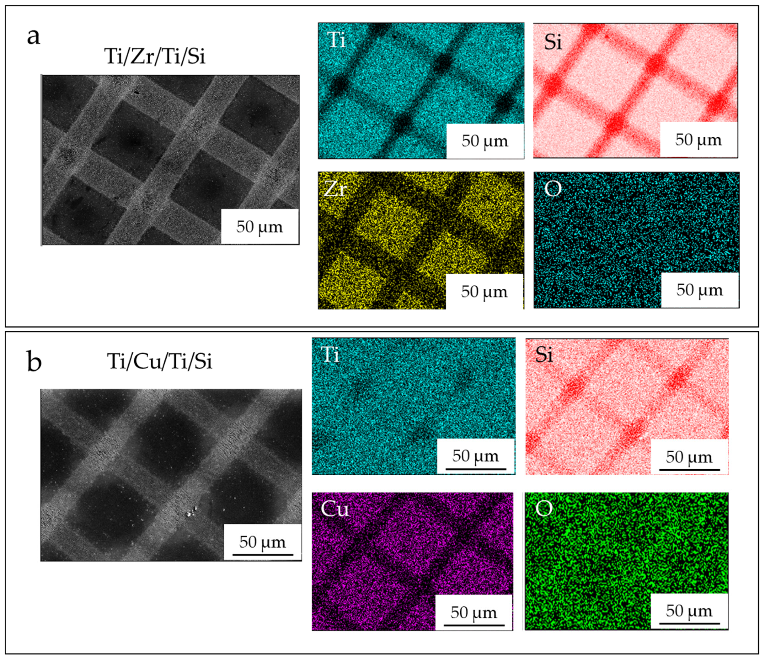
Morphological Characterization of Ti/Zr/Ti/Si and Ti/Cu/Ti/Si Thin-Film Systems
4. Conclusions
Author Contributions
Funding
Institutional Review Board Statement
Informed Consent Statement
Data Availability Statement
Acknowledgments
Conflicts of Interest
References
- Asri, R.I.M.; Harun, W.S.W.; Samykano, M.; Lah, N.A.C.; Ghani, S.A.C.; Tarlochan, F.; Raza, M.R. Corrosion and surface modification on biocompatible metals: A review. Mater. Sci. Eng. C 2017, 77, 1261–1274. [Google Scholar] [CrossRef] [PubMed]
- Rack, H.J.; Qazi, J.I. Titanium alloys for biomedical applications. Mater. Sci. Eng. C 2006, 26, 1269–1277. [Google Scholar] [CrossRef]
- Shalabi, M.; Gortemaker, A.; Hof, M.V.; Jansen, J.; Creugers, N. Implant Surface Roughness and Bone Healing: A Systematic Review. J. Dent. Res. 2006, 85, 496–500. [Google Scholar] [CrossRef]
- Szczęsny, G.; Kopec, M.; Politis, D.J.; Kowalewski, Z.L.; Łazarski, A.; Szolc, T. A Review on Biomaterials for Orthopaedic Surgery and Traumatology: From Past to Present. Materials 2022, 15, 3622. [Google Scholar] [CrossRef]
- Benea, L.; Simionescu-Bogatu, N. Reactivity and Corrosion Behaviors of Ti6Al4V Alloy Implant Biomaterial under Metabolic Perturbation Conditions in Physiological Solutions. Materials 2021, 14, 7404. [Google Scholar] [CrossRef] [PubMed]
- Liu, C.; Matsunami, C.; Shirosaki, Y.; Miyazaki, T. Bioactive Co-Cr alloy for biomedical applications prepared by surface modification using self-assembled monolayers and poly-γ-glutamic acid. Dent. Mater. J. 2015, 34, 707–712. [Google Scholar] [CrossRef] [PubMed]
- Wang, L.; Xie, L.; Shen, P.; Fan, Q.; Wang, W.; Wang, K.; Lu, W.; Hua, L.; Zhang, L.-C. Surface microstructure and mechanical properties of Ti-6Al-4V/Ag nanocomposite prepared by FSP. Mater. Charact. 2019, 153, 175–183. [Google Scholar] [CrossRef]
- Feng, J.; Wei, D.; Zhang, P.; Yu, Z.; Liu, C.; Lu, W.; Wang, K.; Yan, H.; Zhang, L.; Wang, L. Preparation of TiNbTaZrMo high-entropy alloy with tunable Young’s modulus by selective laser melting. J. Manuf. Process. 2023, 85, 160–165. [Google Scholar] [CrossRef]
- Dong, J.; Pacella, M.; Liu, Y.; Zhao, L. Surface engineering and the application of laser-based processes to stents—A review of the latest development. Bioact. Mater. 2022, 10, 159–184. [Google Scholar] [CrossRef]
- Wang, W.; Zhang, X.; Sun, J. Phase stability and tensile behavior of metastable β Ti-V-Fe and Ti-V-Fe-Al alloys. Mater. Charact. 2018, 142, 398–405. [Google Scholar] [CrossRef]
- Jung, I.; Jang, H.; Oh, M.; Lee, J.; Wee, D. Microstructure control of TiAl alloys containing β stabilizers by directional solidification. Mater. Sci. Eng. A 2002, A329–A331, 13–18. [Google Scholar] [CrossRef]
- Donato, T.A.G.; de Almeida, L.H.; Nogueira, R.A.; Niemeyer, T.C.; Grandini, C.R.; Caram, R.; Schneider, S.G.; Santos, A.R., Jr. Cytotoxicity study of some Ti alloys used as biomaterial. Mater. Sci. Eng. C 2009, 29, 1365–1369. [Google Scholar] [CrossRef]
- Carvalho, A.; Cangueiro, L.; Oliveira, V.; Vilar, R.; Fernandes, M.H.; Monteiro, F.J. Femtosecond laser microstructured Alumina toughened Zirconia: A new strategy to improve osteogenic differentiation of hMSCs. Appl. Surf. Sci. 2018, 435, 1237–1245. [Google Scholar] [CrossRef]
- Putra, N.E.; Mirzaali, M.J.; Apachitei, I.; Zhou, J.; Zadpoor, A.A. Multi-material additive manufacturing technologies for Ti-, Mg-, and Fe-based biomaterials for bone substitution. Acta Biomater. 2020, 109, 1–20. [Google Scholar] [CrossRef] [PubMed]
- Correa, D.; Kuroda, P.; Lourenço, M.; Fernandes, C.; Buzalaf, M.; Zambuzzi, W.; Grandini, C. Development of Ti-15Zr-Mo alloys for applying as implantable biomedical devices. J. Alloys Compd. 2018, 749, 163–171. [Google Scholar] [CrossRef]
- Zhao, D.; Chen, C.; Yao, K.; Shi, X.; Wang, Z.; Hahn, H.; Gleiter, H.; Chen, N. Designing biocompatible Ti-based amorphous thin films with no toxic element. J. Alloys Compd. 2017, 707, 142–147. [Google Scholar] [CrossRef]
- Gordin, D.M.; Gloriant, T.; Texier, G.; Thibon, I.; Ansel, D.; Duval, J.L.; Nagel, M.D. Development of a β-type Ti–12Mo–5Ta alloy for biomedical applications: Cytocompatibility and metallurgical aspects. J. Mater. Sci. Mater. Med. 2004, 15, 885–891. [Google Scholar] [CrossRef]
- Wang, L.; Lu, W.; Qin, J.; Zhang, F.; Zhang, D. The characterization of shape memory effect for low elastic modulus biomedical β-type titanium alloy. Mater. Charact. 2010, 61, 535–541. [Google Scholar] [CrossRef]
- Ikarashi, Y.; Toyoda, K.; Kobayashi, E.; Doi, H.; Yoneyama, T.; Hamanaka, H.; Tsuchiya, T. Improved Biocompatibility of Titanium–Zirconium (Ti–Zr) Alloy: Tissue Reaction and Sensitization to Ti–Zr Alloy Compared with Pure Ti and Zr in Rat Implantation Study. Mater. Trans. 2005, 46, 2260–2267. [Google Scholar] [CrossRef]
- Barter, S.; Stone, P.; Brägger, U. A pilot study to evaluate the success and survival rate of titanium-zirconium implants in partially edentulous patients: Results after 24 months of follow-up. Clin. Oral Implant. Res. 2011, 23, 873–881. [Google Scholar] [CrossRef]
- Grandin, H.M.; Berner, S.; Dard, M. A Review of Titanium Zirconium (TiZr) Alloys for Use in Endosseous Dental Implants. Materials 2012, 5, 1348–1360. [Google Scholar] [CrossRef]
- Liu, Z.; Liu, X.; Ramakrishna, S. Surface engineering of biomaterials in orthopedic and dental implants: Strategies to improve osteointegration, bacteriostatic and bactericidal activities. Biotechnol. J. 2021, 16, 2000116. [Google Scholar] [CrossRef] [PubMed]
- Wu, W.; Cheng, R.; das Neves, J.; Tang, J.; Xiao, J.; Ni, Q.; Liu, X.; Pan, G.; Li, D.; Cui, W.; et al. Advances in biomaterials for preventing tissue adhesion. J. Control. Release 2017, 261, 318–336. [Google Scholar] [CrossRef] [PubMed]
- Frostevarg, J.; Olsson, R.; Powell, J.; Palmquist, A.; Brånemark, R. Formation mechanisms of surfaces for osseointegration on titanium using pulsed laser spattering. Appl. Surf. Sci. 2019, 485, 158–169. [Google Scholar] [CrossRef]
- Berg, Y.; Kotler, Z.; Shacham-Diamand, Y. Holes generation in glass using large spot femtosecond laser pulses. J. Micromech. Microeng. 2018, 28, 035009. [Google Scholar] [CrossRef]
- Jenko, M.; Gorenšek, M.; Godec, M.; Hodnik, M.; Batič, B.; Donik, Č.; Grant, J.T.; Dolinar, D. Surface chemistry and microstructure of metallic biomaterials for hip and knee endoprostheses. Appl. Surf. Sci. 2018, 427, 584–593. [Google Scholar] [CrossRef]
- Simitzi, C.; Ranella, A.; Stratakis, E. Controlling the morphology and outgrowth of nerve and neuroglial cells: The effect of surface topography. Acta Biomater. 2017, 51, 21–52. [Google Scholar] [CrossRef]
- Bonse, J.; Koter, R.; Hartelt, M.; Spaltmann, D.; Pentzien, S.; Höhm, S.; Rosenfeld, A.; Krüger, J. Tribological performance of femtosecond laser-induced periodic surface structures on titanium and a high toughness bearing steel. Appl. Surf. Sci. 2015, 336, 21–27. [Google Scholar] [CrossRef]
- Gnilitskyi, I.; Derrien, T.J.-Y.; Levy, Y.; Bulgakova, N.M.; Mocek, T.; Orazi, L. High-speed manufacturing of highly regular femtosecond laser-induced periodic surface structures: Physical origin of regularity. Sci. Rep. 2017, 7, 8485. [Google Scholar] [CrossRef]
- Kirner, S.V.; Wirth, T.; Sturm, H.; Krüger, J.; Bonse, J. Nanometer-resolved chemical analyses of femtosecond laser-induced periodic surface structures on titanium. J. Appl. Phys. 2017, 122, 104901. [Google Scholar] [CrossRef]
- Li, C.L.; Fisher, C.J.; Burke, R.; Andersson-Engels, S. Orthopedics-Related Applications of Ultrafast Laser and Its Recent Advances. Appl. Sci. 2022, 12, 3957. [Google Scholar] [CrossRef]
- Stasić, J.; Gaković, B.; Perrie, W.; Watkins, K.; Petrović, S.; Trtica, M. Surface texturing of the carbon steel AISI 1045 using femtosecond laser in single pulse and scanning regime. Appl. Surf. Sci. 2011, 258, 290–296. [Google Scholar] [CrossRef]
- Petrović, S.; Peruško, D.; Kovač, J.; Panjan, P.; Mitrić, M.; Pjević, D.; Kovačević, A.; Jelenković, B. Design of co-existence parallel periodic surface structure induced by picosecond laser pulses on the Al/Ti multilayers. J. Appl. Phys. 2017, 122, 115302. [Google Scholar] [CrossRef]
- Babaliari, E.; Kavatzikidou, P.; Angelaki, D.; Chaniotaki, L.; Manousaki, A.; Siakouli-Galanopoulou, A.; Ranella, A.; Stratakis, E. Engineering Cell Adhesion and Orientation via Ultrafast Laser Fabricated Microstructured Substrates. Int. J. Mol. Sci. 2018, 19, 2053. [Google Scholar] [CrossRef]
- Kovačević, A.G.; Petrović, S.; Petrović, S.; Mimidis, A.; Stratakis, E.; Pantelić, D.; Kolaric, B. Molding wetting by laser-induced nanostructures. Appl. Sci. 2020, 10, 6008. [Google Scholar] [CrossRef]
- Petrović, S.; Peruško, D.; Mimidis, A.; Kavatzikidou, P.; Kovač, J.; Ranella, A.; Novaković, M.; Popović, M.; Stratakis, E. Response of NIH 3T3 fibroblast cells on laser-induced periodic surface structures on a 15×(Ti/Zr)/Si multilayer system. Nanomaterials 2020, 10, 2531. [Google Scholar] [CrossRef]
- Amigó-Mata, A.; Haro-Rodriguez, M.; Vicente-Escuder, Á.; Amigó-Borrás, V. Development of Ti–Zr alloys by powder metallurgy for biomedical applications. Powder Met. 2021, 65, 31–38. [Google Scholar] [CrossRef]
- Liu, J.; Zhang, X.; Wang, H.; Li, F.; Li, M.; Yang, K.; Zhang, E. The antibacterial properties and biocompatibility of a Ti–Cu sintered alloy for biomedical application. Biomed. Mater. 2014, 9, 025013. [Google Scholar] [CrossRef]
- Zhang, E.; Wang, X.; Chen, M.; Hou, B. Effect of the existing form of Cu element on the mechanical properties, bio-corrosion and antibacterial properties of Ti-Cu alloys for biomedical application. Mater. Sci. Eng. C 2016, 69, 1210–1221. [Google Scholar] [CrossRef]
- National Institute of Health, USA. Available online: https://rsb.info.nih.gov/ij/ (accessed on 28 March 2023).
- Van Oss, C.J.; Chaudhury, M.K.; Good, R.J. Monopolar surfaces. Adv. Colloid Interface Sci. 1987, 28, 35–64. [Google Scholar] [CrossRef]
- Aleksić, M.; Stanisavljević, D.; Smiljković, M.; Vasiljević, P.; Stevanović, M.; Soković, M.; Stojković, D. Pyrimethanil: Between efficient fungicide against Aspergillus rot on cherry tomato and cytotoxic agent on human cell lines. Ann. Appl. Biol. 2019, 175, 228–235. [Google Scholar] [CrossRef]
- Gastaldi, D.; Baleani, M.; Fognani, R.; Airaghi, F.; Bonanni, L.; Vena, P. An experimental procedure to perform mechanical characterization of small-sized bone specimens from thin femoral cortical wall. J. Mech. Behav. Biomed. Mater. 2020, 112, 104046. [Google Scholar] [CrossRef] [PubMed]
- Lee, S.H.; Kwon, S.Y.; Ham, H.J. Thermal conductivity of tungsten–copper composites. Thermochim. Acta 2012, 542, 2–5. [Google Scholar] [CrossRef]
- Klemens, P.G.; Williams, R.K. Thermal conductivity of metals and alloys. Int. Met. Rev. 1986, 31, 197–215. [Google Scholar] [CrossRef]
- Petrović, S.; Peruško, D.; Skoulas, E.; Kovač, J.; Mitrić, M.; Potočnik, J.; Rakočević, Z.; Stratakis, E. Laser-Assisted Surface Texturing of Ti/Zr Multilayers for Mesenchymal Stem Cell Response. Coatings 2019, 9, 854. [Google Scholar] [CrossRef]
- Petrović, S.; Tsibidis, G.D.; Kovačević, A.; Božinović, N.; Peruško, D.; Mimidis, A.; Manousaki, A.; Stratakis, E. Effects of static and dynamic femtosecond laser modifications of Ti/Zr multilayer thin films. Eur. Phys. J. D 2021, 75, 304. [Google Scholar] [CrossRef]
- Kisić, D.; Nenadović, M.; Barudžija, T.; Noga, P.; Vaňa, D.; Muška, M.; Rakočević, Z. Modification of polyethylene’s surface properties by high fluence Fe implantation. Nucl. Instrum. Methods Phys. Res. Sect. B Beam Interact. Mater. Atoms 2020, 462, 143–153. [Google Scholar] [CrossRef]
- Vogler, E.A. Structure and reactivity of water at biomaterial surfaces. Adv. Colloid Interface Sci. 1998, 74, 69–117. [Google Scholar] [CrossRef]
- Xu, L.-C.; Siedlecki, C.A. Effects of surface wettability and contact time on protein adhesion to biomaterial surfaces. Biomaterials 2007, 28, 3273–3283. [Google Scholar] [CrossRef]
- Monfared, A.; Faghihi, S.; Karami, H. Biocorrosion and surface wettability of Ni-free Zr-based bulk metallic glasses. Int. J. Electrochem. Sci. 2013, 8, 7744–7752. [Google Scholar]
- Xiao, K.; Wen, L.; Jiang, L. Bioinspired Super Wettability Materials; Wiley Online Library: Hoboken, NJ, USA, 2016. [Google Scholar]
- Zhigarkov, V.; Volchkov, I.; Yusupov, V.; Chichkov, B. Metal Nanoparticles in Laser Bioprinting. Nanomaterials 2021, 11, 2584. [Google Scholar] [CrossRef]
- Abdal Dayem, A.; Lee, S.B.; Cho, S.-G. The Impact of Metallic Nanoparticles on Stem Cell Proliferation and Differentiation. Nanomaterials 2018, 8, 761. [Google Scholar] [CrossRef]
- Medici, S.; Peana, M.; Pelucelli, A.; Zoroddu, M.A. An updated overview on metal nanoparticles toxicity. Semin. Cancer Biol. 2021, 76, 17–26. [Google Scholar] [CrossRef] [PubMed]
- Cai, K.; Hou, Y.; Li, J.; Chen, X.; Hu, Y.; Luo, Z.; Ding, X.; Xu, D.; Lai, M. Effects of titanium nanoparticles on adhesion, migration, proliferation, and differentiation of mesenchymal stem cells. Int. J. Nanomed. 2013, 8, 3619–3630. [Google Scholar] [CrossRef] [PubMed]
- Jiao, Y.; Brousseau, E.; Ayre, W.N.; Gait-Carr, E.; Shen, X.; Wang, X.; Bigot, S.; Zhu, H.; He, W. In vitro cytocompatibility of a Zr-based metallic glass modified by laser surface texturing for potential implant applications. Appl. Surf. Sci. 2021, 547, 149194. [Google Scholar] [CrossRef]
- Busuioc, C.; Voicu, G.; Zuzu, I.D.; Miu, D.; Sima, C.; Iordache, F.; Jinga, S.I. Vitroceramic coatings deposited by laser ablation onTi-Zr substrates for implantable medical applications with improved biocompatibility. Ceram. Int. 2017, 43, 5498–5504. [Google Scholar] [CrossRef]


| Ti/Zr/Ti/Si | Ti | Si | Zr | O |
|---|---|---|---|---|
| As-deposited | 77.03 | 16.21 | 5.81 | 0.95 |
| Center | 76.60 | 16.75 | 5.67 | 0.98 |
| Line | 49.37 | 47.88 | 0.74 | 2.01 |
| Intersection | 43.91 | 53.63 | / | 2.46 |
| Ti/Cu/Ti/Si | Ti | Si | Cu | O |
|---|---|---|---|---|
| As-deposited | 72.92 | 14.85 | 10.91 | 1.32 |
| Center | 73.09 | 15.04 | 10.51 | 1.36 |
| Line | 74.37 | 17.92 | 5.08 | 2.63 |
| Intersection | 74.62 | 22.35 | / | 3.03 |
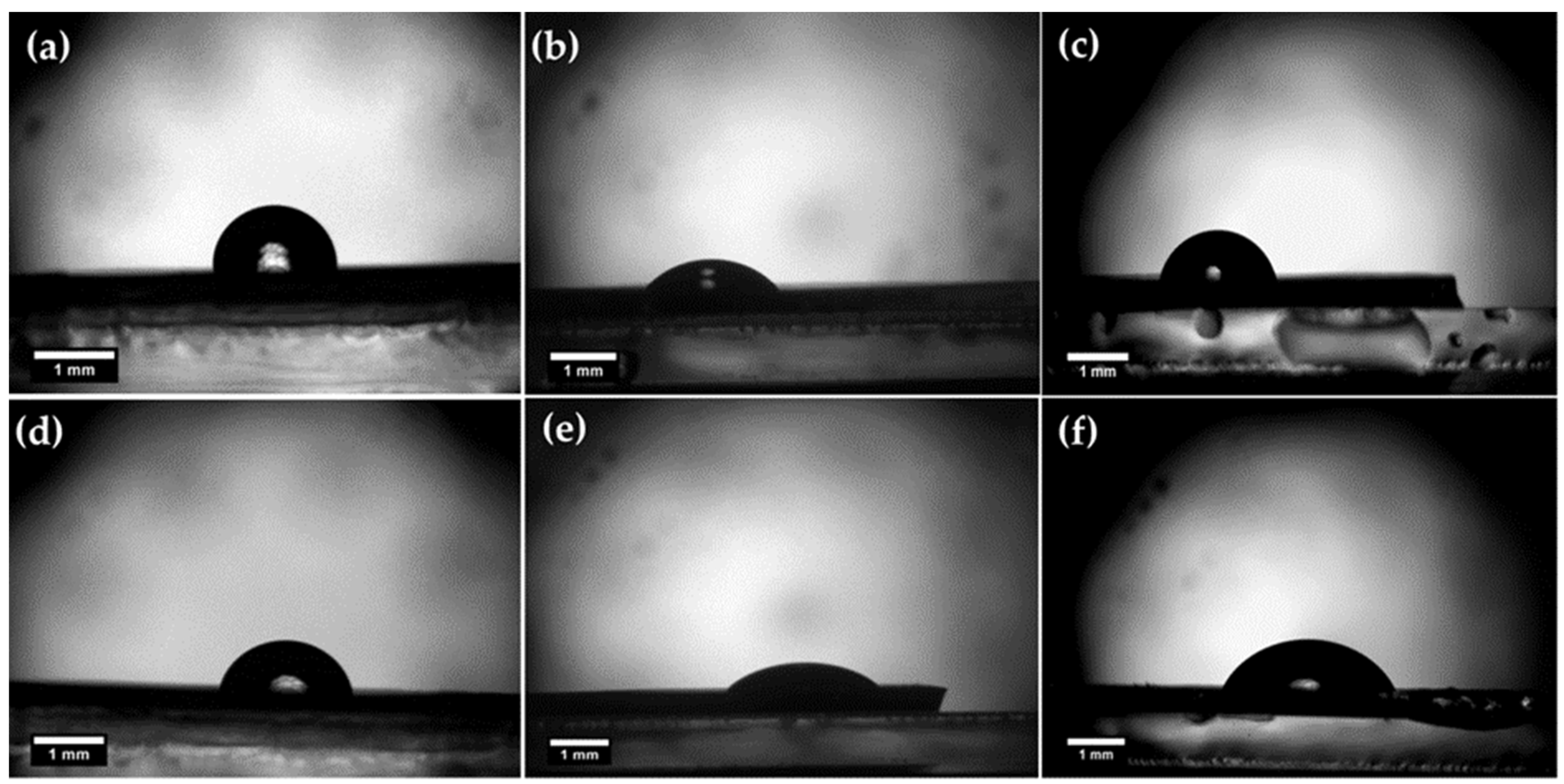
| Specimen | Contact Angle [°] | Surface Free Energy [mJ/m2] | ||||
|---|---|---|---|---|---|---|
| Deionized Water | Diiodmethane | Ethylene Glycol | Polar Part | Nonpolar Part | Total (γs) | |
| Ti/Cu/Ti as-prepared | 84 ± 3 | 50 ± 3 | 54 ± 2 | 1.7 | 33.7 | 35.4 |
| Ti/Cu/Ti laser modified | 86 ± 4 | 51 ± 3 | 63 ± 3 | 0.6 | 34.3 | 34.9 |
| Ti/Zr/Ti as-prepared | 82 ± 2 | 37 ± 2 | 58 ± 3 | 3.3 | 40.1 | 43.4 |
| Ti/Zr/Tu laser modified | 86 ± 3 | 40 ± 2 | 70 ± 3 | 6.3 | 40.1 | 46.4 |
| Sample | ||
|---|---|---|
| Ti/Cu/Ti as-Prepared | 0.77 ± 0.03 | 0.92 ± 0.03 |
| Ti/Cu/Ti laser modified | 0.02 ± 0.04 | 4.82 ± 0.03 |
| Ti/Zr/Ti as-prepared | 0.29 ± 0.02 | 9.14 ± 0.02 |
| Ti/Zr/Tu laser modified | 1.30 ± 0.03 | 7.53 ± 0.02 |
Disclaimer/Publisher’s Note: The statements, opinions and data contained in all publications are solely those of the individual author(s) and contributor(s) and not of MDPI and/or the editor(s). MDPI and/or the editor(s) disclaim responsibility for any injury to people or property resulting from any ideas, methods, instructions or products referred to in the content. |
© 2023 by the authors. Licensee MDPI, Basel, Switzerland. This article is an open access article distributed under the terms and conditions of the Creative Commons Attribution (CC BY) license (https://creativecommons.org/licenses/by/4.0/).
Share and Cite
Petrović, S.; Božinović, N.; Rajić, V.; Stanisavljević Ninković, D.; Kisić, D.; Stevanović, M.J.; Stratakis, E. Cell Response on Laser-Patterned Ti/Zr/Ti and Ti/Cu/Ti Multilayer Systems. Coatings 2023, 13, 1107. https://doi.org/10.3390/coatings13061107
Petrović S, Božinović N, Rajić V, Stanisavljević Ninković D, Kisić D, Stevanović MJ, Stratakis E. Cell Response on Laser-Patterned Ti/Zr/Ti and Ti/Cu/Ti Multilayer Systems. Coatings. 2023; 13(6):1107. https://doi.org/10.3390/coatings13061107
Chicago/Turabian StylePetrović, Suzana, Nevena Božinović, Vladimir Rajić, Danijela Stanisavljević Ninković, Danilo Kisić, Milena J. Stevanović, and Emmanuel Stratakis. 2023. "Cell Response on Laser-Patterned Ti/Zr/Ti and Ti/Cu/Ti Multilayer Systems" Coatings 13, no. 6: 1107. https://doi.org/10.3390/coatings13061107
APA StylePetrović, S., Božinović, N., Rajić, V., Stanisavljević Ninković, D., Kisić, D., Stevanović, M. J., & Stratakis, E. (2023). Cell Response on Laser-Patterned Ti/Zr/Ti and Ti/Cu/Ti Multilayer Systems. Coatings, 13(6), 1107. https://doi.org/10.3390/coatings13061107