Abstract
MoS2 is a two-dimensional layered transition metal dichalcogenide with unique electronic and optical properties. The fabrication of ultrathin MoS2 is vitally important, since interlayer interactions in its ultrathin varieties will become thickness-dependent, providing thickness-governed tunability and diverse applications of those properties. Unlike with a number of studies that have reported detailed information on direct bandgap emission from MoS2 monolayers, reliable experimental evidence for thickness-induced evolution or transformation of the indirect bandgap remains scarce. Here, the sulfurization of MoO3 thin films with nominal thicknesses of 30 nm, 5 nm and 3 nm was performed. All sulfurized samples were examined at room temperature with spectroscopic ellipsometry and photoluminescence spectroscopy to obtain information about their dielectric function and edge emission spectra. This investigation unveiled an indirect-to-indirect crossover between the transitions, associated with two different Λ and K valleys of the MoS2 conduction band, by thinning its thickness down to a few layers.
1. Introduction
Group-VI layered two-dimensional (2D) transition metal dichalcogenides (TMDs) (e.g., MoS2, WS2, MoSe2 and WSe2) exhibit very interesting semiconducting properties and are attracting a lot of attention that has been increasingly growing after the discovery of metallic two-dimensional graphene. As graphene, TMD monolayers have hexagonal symmetry and show a direct bandgap in the range of 1–2 eV [1]. Bi- or multilayers of TMDs are indirect bandgap semiconductors [1]. The remarkable versatility of monolayer and multilayer TMDs as a viable alternative to graphene [2] arises from their unique crystal structure. Their bandgaps and chemical properties [3] can be effectively tuned using different approaches (varying the number of layers, intercalation, alloying, mechanical stress, doping, etc.) [4,5].
As far as MoS2 is concerned, numerous studies have revealed its intricate electronic spectrum and optical properties, surpassing the limitations of the conventional one-electron band structure [6]. The substantial influence of multiexcitonic effects has highlighted the importance of thorough experimental studies on electronic excitations [7,8,9,10,11], placing them at the forefront of the current scientific research concerning MoS2. According to a series of theoretical works [1,2,3,4,5,12], the reported tunability of the direct bandgap due to indirect-to-direct crossover [13,14] in atomically thin MoS2 is directly related to the variable orbital composition of the involved electronic states. For the same reason, the indirect bandgap of few-layer MoS2 will also vary with the thinning of its thickness, and even an indirect-to-indirect crossover between the transitions associated with two different valleys of the conduction band may occur. While few-layer MoS2 exhibits two valleys along the Γ–K line with similar energy, as highlighted by Zhao et al. [15], the particular valley responsible for forming the conduction band minimum remains poorly understood. Fundamental questions persist regarding the circumstances under which the indirect Λ−Γ transition will shift to indirect K−Γ band alignment and whether this shift will occur at all, because the valley responsible for forming the conduction band minimum is yet to be specified.
Until now, MoS2 monolayer and few-layer films were obtained using the exfoliation technique [10,13,16,17], chemical vapor deposition (CVD) [11,18,19,20,21,22,23], the sulfurization of CVD-deposited MoCl5 [24] and MoO3 [25] and the sulfurization of Mo [26] and MoO3 [27] thin films obtained using vapor phase growth.
In this work, we report the preparation of MoS2 ultrathin films by sulfurization of few-layer MoO3 films that were preliminary obtained on a SiO2/Si substrate by plasma-enhanced atomic layer deposition (PE-ALD). All sulfurized samples were examined at room temperature with spectroscopic ellipsometry and photoluminescence spectroscopy to obtain information about their dielectric functions (DF) and emission spectra. The latter unveiled the competition of two indirect transitions from the Λ and K valleys of the conduction band by thinning the MoS2 thickness down to a few layers.
2. Experimental Section
2.1. MoO3 Thin Films
The MoO3 films were deposited in a PE-ALD system (SI ALD LL, SENTECH Instruments, Berlin, Germany), as described in detail in [28,29]. The films were grown on thermal oxidized (SiO2 with a nominal thickness of 105 nm) p-type Si (100) wafers (thickness ~ 650–700 µm) with a resistance of <0.005 Ohm × cm (high-doped substrates).
X-ray diffraction (XRD) patterns were recorded on a Malvern PANalytical X’Pert Pro MRD diffractometer(Malvern Panalytical Ltd., Bristol, UK) equipped with a fast PIXcel detector, using CuKα radiation generated at 40 kV and 40 mA. Grazing incidence XRD (GIXRD) patterns were recorded at an incident angle of 0.5°, with a step size of 0.01 °2θ and a counting time of 0.5 s, over a 5–55 °2θ interval. Typical GIXRD patterns and dielectric functions (DFs) retrieved using spectroscopic ellipsometry (SE), performed with the aid of a rotating-compensator M 2000DI (J.A. Woollam, Lincoln, NE, USA) ellipsometer at different incident angles over the photon energy range of 0.7–6.5 eV, are given in Figure S1a and Figure S1b, respectively. All the obtained MoO3 films, subjected to further sulfurization, were crystalline, and their DFs were indicative of the absence of the oxygen deficiency, as discussed in the supplementary information S1 Section. Their thicknesses, found in an X-ray reflectometry (XRR) examination were 3, 5 and 30 nm.
2.2. MoS2 Thin Films
The sulfurization of the MoO3 films was carried out in a 2-inch single-zone tube furnace (OTF-1200X-S, MTI Corporation, Richmond, CA, USA). For this purpose, the MoO3 films were placed at the center of the furnace. The furnace and samples were purged multiple times under Ar flow (250 sccm 99.999% Ar). The temperature of the furnace was ramped up from room temperature to 700 °C over 15–20 min and maintained at that temperature for 60 min under atmospheric pressure. During heating, H2S (10 sccm 99.5% H2S) was introduced at 300 °C; it was removed at the end of the 60 min thermal treatment. Afterward, the furnace was purged with Ar (250 sccm) during the cool-down process. After cooling to ~400 °C, the furnace was opened for rapid cooling at ~100 °C over ~10 min.
The topographies of the prepared 30, 5 and 3 nm-thick MoS2 films (plan view) were examined using a Zeiss UltraPlus scanning electron microscope (Carl Zeiss, Oberkochen, Germany). The corresponding scanning electron microscopy (SEM) images are given in Figure S2b–d.
Examinations using X-ray reflectivity and SE revealed that sulfurization induces changes in film thickness. Specifically, the thickness of 30 nm-thick film was reduced by nearly 30% after sulfurization. On the other hand, the films with nominal thicknesses of 3 and 5 nm each experienced less than a 15% change in thickness after sulfurization. To simplify the references in this text, each of the MoS2 films studied will be identified based on its nominal thickness, which corresponds to the thickness of the source MoO3 film before sulfurization. The variation in thickness among the resulting MoS2 films is distinctly evident in their Raman spectra (Figure S2a). These were recorded using back-scattering geometry on a Nanofinder-30 confocal Raman system (Tokyo Instrument Inc., Tokyo, Japan) equipped with a Juno 3050 GS-11 (Kyocera Soc Corporation, Yokohama, Japan) Nd:yttrium–aluminum–garnet laser (second harmonic, 532 nm). The maximum output power of the excitation source was 10 mW. The cross-sectional beam diameter was 4 μm. Diffraction grating with 1800 grooves per mm provided a spectral resolution of 0.5 cm−1. The spectral signal was detected using a photon-counting charge-coupled device (CCD) camera “Andor” (Andor Technology, Belfast, Ireland) cooled down to −100 °C.
As shown in Supplementary S2, the observed decreases in intensity and disappearance of the 521 cm−1 Raman line of the Si substrate with increasing thickness of the MoS2 film (Figure S2a) corroborates the value of the absorption coefficient (Figure S3) extracted from the DFs of the 3, 5 and 30 nm MoS2 films.
Despite the large number of theoretical works on ultrathin MoS2, only a few address its DFs. So far, as the thicknesses of the obtained 3 and 5 nm MoS2 samples exceeded one layer (L) and approximately corresponded to 4 L and 8 L MoS2 (Supplementary S2), respectively, they were of primary importance in the context of the present work. Nevertheless, along with the latter two, bulk and 2 L MoS2 were also included in the band structure calculations to obtain a more complete picture and unravel the main trends that band structure and DF show upon thinning of the MoS2 thickness.
We used the full-potential linearized augmented plane wave (FP LAPW) method implemented in the scheme reported in [30]. The exchange-correlation interactions were described as within the generalized gradient approximation (GGA), using the strategy reported in [31]. The convergence parameter RmtKmax, where Rmt is the smallest atomic sphere radius and Kmax is the largest K-vector of the plane wave expansion of the wave function, was set to 7.0. Within the atomic spheres, the partial waves were expanded up to lmax = 10, where lmax is the highest value of the orbital angular-momentum quantum number used for partial waves inside atomic spheres. Integrations over the first Brillouin zone (BZ) were performed using the tetrahedron method, with 60 points in the irreducible part of the BZ. The Rmt values for Mo and S were set to 2.34 and 2.08 a. u. (atomic unit), respectively. The value of −6.0 Ry of cut-off energy was used for the separation of the core and valence states. The imaginary part of the DF was calculated using the joint density of the states for optical transitions between the valence and conduction bands, using the Monkhorst–Pack technique for integration over the Brillouin zone [32]. The real part of the dielectric function was calculated from the imaginary part using the Kramers–Kronig relation.
While SE is a powerful tool for studying direct optical transitions, photoluminescence (PL) is a well-endorsed technique for studying indirect-gap emissions in MoS2.
In the present work, PL studies were performed on an Infrared PL/PLE/Raman spectrometer (Tokyo Instrument Inc., Tokyo, Japan) using lasers with two excitation wavelengths, 785 nm (NovaPro 785–250) and 532 nm (NovaPro PB 532–200 DPss), to embrace a possibly wider range of intrinsic electronic excitations that may decay radiatively and contribute to the PL of MoS2. The obtained PL spectra are given and discussed together with the other main results in the next section.
3. Results and Discussion
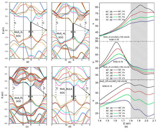
Among the significant parameters in the resulting electronic energy spectrum of MoS2 (Figure 1a–d) is the splitting between adjacent bands at the K-point of the BZ. As highlighted in Figure 1, for each MoS2 layer count, it arises from interlayer interactions and spin–orbit coupling (SOC). The splitting magnitude was reduced in the 8, 4 and 2 L MoS2 compared to the bulk material, primarily due to the finite number of layers in ultrathin MoS2. In a 1 L MoS2 limit, the splitting solely originates from SOC [3].

Figure 1.
Electronic band structures of 2 L (a), 4 L (b), 8 L (c) and bulk (d) MoS2. Valence band splitting is encircled; possible indirect radiative transitions are indicated by arrows; and bold and dashed vertical lines show direct transitions A, B and C, respectively. (e) Ellipsometric parameter Ψ as a function of photon energy for the obtained MoS2/SiO2/Si structures with 3 (top plot), 5 (middle plot) and 30 nm (bottom plot) MoS2. On the top, colored open circles and solid lines represent experimental data and the fits to these data, respectively; each color corresponds to one of the four accessed angles of incidence.
As shown in Figure 1e (shaded area), this splitting manifests itself through the irregular behavior of the ellipsometric parameter Ψ within the narrow photon energy gap spanning from 1.8 to 2 eV.
Such peculiar behavior was observed across all MoS2/SiO2/Si stacks studied in this work and for all incident angles accessed during the SE measurements. Note that the mean square error (MSE) given in Figure 1e for each studied MoS2/SiO2/Si structure is related to the model that was fitted to the ellipsometric parameters not only within the 1.4 to 2.1 eV range shown in Figure 1 but also for the entire accessible photon energy range spanning from 0.7 to 6.5 eV. The obtained MSE values fell below eight, indicating that the fit was acceptable and the retrieved DF was accurate enough.
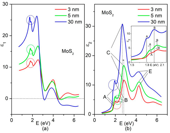
The DF across the entire range of photon energies is displayed in Figure 2, with separate representation for the real (a) and imaginary (b) parts. The data for the MoS2 films with thicknesses of 30, 5, and 3 nm are indicated by the blue, green and red lines, respectively.

Figure 2.
Real (a) and imaginary (b) parts of the experimental DFs retrieved for 30 (blue lines), 5 (green lines) and 5 nm (red lines)-thick MoS2 in a wide photon energy range. The splitting-related features of the spectra are encircled. Notations A, B, C and E, commonly used for MoS2, are related to the particular peaking structures in the spectrum of each of the considered MoS2 thin films. The vertical dashed lines and small horizontal arrows are given for convenience to show how A, B, and C exciton peak positions shift with changing the thickness of MoS2. As illustrated in the inset in (b), A- and B-excitons also exhibited shifts toward higher photon energies as the thickness of the obtained MoS2 was reduced to 5 or 3 nm.
Based on its crystal structure and symmetry [33], MoS2 is a uniaxial crystal, and its optic axis is normal to the layer plane (LP). As reported in Figures S4 and S5, the calculated DF indeed shows different dispersions for light polarized parallel and perpendicular to the layer plane. Comparison between the SE-based (Figure 2) and calculated (Figure S4) data drove us to the conclusion that the obtained SE-based DF is overwhelmingly determined by dielectric response to the light polarized in the LP for all MoS2 films studied in this work. The descending trend in the intensity of the main features of the SE-based DFs of the 30, 5 and 3 nm MoS2 was reproduced using the calculated DFs obtained before and after rescaling (Figure S6). The latter mitigated the vacuum dilution effect, since the band structure calculations for MoS2 with a finite number of layers were conducted using supercells that included vacuum space.
The A, B and C transitions depicted in Figure 1 and reproduced by SE-based DFs in Figure 2 are commonly observed in the majority of the related studies. The exciton peaks (Figure 2) related to these transitions clearly exhibit blue shifts with the thinning of the MoS2 samples. The E-exciton, which is also frequently observed in optical studies [34] does not show noticeable shift. The most significant shift was observed for the C-excitons, which are not originating from transitions between parabolic bands with opposite concavities, like A- and B-excitons, but resulting from transitions between the valence and conduction band regions with similar concavities, known as the nested band regions. The comparison of the C-exciton position in 30 nm MoS2 with those in 5 and 3 nm MoS2 indicated a considerable blue shift upon thinning, exceeding 100 meV (Figure 2b). As illustrated in the inset of Figure 2b, the A- and B-excitons also exhibited shifts toward higher photon energies as the thickness of the obtained MoS2 was reduced to 5 or 3 nm. In comparison to the C-excitons, the B- and A-excitons experienced smaller shifts (approximately 50 and 20 meV, respectively).
Overall, as stated before, the above analysis confirms that the obtained 30 nm MoS2 is a good counterpart to bulk MoS2. Along with the already mentioned Raman spectra (Supplementary S2 Section), this assertion was corroborated by the PL spectra taken for the studied MoS2 thin films and shown in Figure S7.
The exciton landscape in MoS2 is highly intricate, embracing neutral, charged and dark excitons, all of which directly or indirectly contribute to the DFs. This landscape is dynamic, with various components influenced by numerous factors, including the fabrication process [20]. Therefore, when the retrieved DFs are evaluated, they should be analyzed in conjunction with PL data, comparing the absorption coefficient derived from the DFs to the PL spectra within the same spectral range. Since our primary focus was on few-layer MoS2, we initially concentrated on MoS2/SiO2/Si structures featuring 3 and 5 nm-thick MoS2 layers.
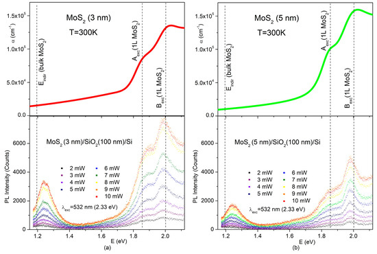
Figure 3 displays the photon energy dependencies of the absorption coefficients (α) and PLs of the few-layer MoS2 films. The PLs under the excitation wavelength of 532 nm (2.33 eV) are given for various levels of excitation power. The normalized PL spectrum (shape function) for each excitation level underwent little change, with increasing excitation power in the range of 2–10 mW (Figure S8), and the positions of the emission lines remained unchanged.

Figure 3.
Top part: SE-based absorption coefficients of the 3 (a) and 5 nm (b) MoS2, respectively; bottom part: PL spectra at various excitation densities under the excitation wavelength of 532 nm (2.33 eV) for MoS2/SiO2/Si thin film structures with 3 (a) and 5 nm (b) MoS2 on the top. Vertical dashed lines, given for eye guidance, show the reported positions of the A- and B-excitons in a single layer of MoS2 [13,34,35,36,37] and the reported value of the indirect gap for bulk MoS2 [38].
A- and B-excitons, which are positioned above 1.8 eV in MoS2 [13,33,34,35,36], clearly manifested themselves in the spectral features of the α and PL values of the studied films. Comparison with the reported energy positions of Aexc. (1L MoS2) and Bexc. (1L MoS2) for a single layer showed that the energy gap between the A- and B-excitons in our case did not exceed approximately 150 meV. This value is noticeably smaller than 200 meV that is the value of the splitting between A- and B-excitons in bulk MoS2 [3]. This observation is directly related to a few layers’ thickness of the prepared films. In the case of the 5 nm MoS2, the energy position of the emission line ascribed to the B-excitons was red-shifted by nearly 20 meV from its energy position in the MoS2 monolayer and from its absorption peak as reported in Figure 2b. For the 3 nm MoS2, the shift was definitely less pronounced (see Figure 2a). The energy difference between absorption and emission, known as the Stokes shift, is inherent to all materials and varies widely depending on the material type (semiconductor, molecular crystal) and internal electrical fields. For typical semiconductors like GaAs, this shift is only 4 meV [39]. There are no reports on the intrinsic Stokes shift value in MoS2. However, as the shift is decreased in 3 nm MoS2 compared to 5 nm MoS2, the Stokes shift in the latter is non-intrinsic and is likely caused by some strain or inhomogeneity in 5 nm MoS2.
Strong enhancement in PL intensity related to the direct exciton radiative transitions in MoS2 when MoS2 thickness goes down to a few nanometers is a well-known and solid fact. According to the PL spectra in Figure 3, some enhancement also takes place at the nanoscale when MoS2 thickness is changed from 5 to 3 nm. It can be clearly seen that under the same excitation levels, the PL intensity in 3 nm MoS2 was noticeably stronger than in the 5 nm MoS2.
While α is structureless and, for both the 3 and 5 nm MoS2, showed only absorption tails descending toward lower photon energies, the PL spectrum showed an indirect-gap emission band centered around 1.25 eV (Figure 3).
It has been established in many experimental and theoretical works [2,5,14,15,18,38,40,41] that MoS2 remains an indirect semiconductor even when reduced to bilayers. Observation of the indirect-gap emission around 1.25 eV on 3 nm-thick MoS2 (Figure 3) has revealed that MoS2 retains its indirect characteristics even when its thickness is reduced to just four layers. This agrees well with the already-quoted works.
However, as noted by Zhao et al. [15], few-layer MoS2 exhibits two valleys along the Γ–K line with similar energy, and there is limited understanding of which valley forms the conduction band minimum. It is important to highlight that the indirect-gap value of 1.20 eV [38] characterizes bulk MoS2 and corresponds to the I1 transition in the band structure shown in Figure 1. This implies that, like in previous calculations, including the cited work [15], the band structure in Figure 1 captures the main trends in the evolution of band structure with the thinning of MoS2. It indicates a higher energy of K–Γ transitions (I2) compared to Λ–Γ transitions (I1) and even predicts a crossover between the Λ and K conduction band valleys. However, the precise temperature at which this crossover occurs in few-layer MoS2 is beyond the predictive capabilities of the performed calculations. Nevertheless, discovering experimental conditions that would enable the simultaneous generation of both I1 and I2 radiative transitions at room temperature would greatly assist in addressing this issue.
The PL spectra displayed in Figure 3 were excited with a 532 nm (2.33 eV) laser and, along with direct exciton lines, showed only indirect I1 (Λ–Γ) emission. The photon energy of 2.33 eV considerably exceeds the direct energy gap between the conduction and valence bands at the K point, and this may have prevented excited carriers from emitting I2 (K-Γ).
Note that some relaxation paths for excited electrons can be blocked or very slow before emission, as has been observed in a case of high energy C-excitons in MoS2 [42]. However, considering the blocking of carriers excited at 2.33 eV from relaxation down to the K–point to prevent K–Γ indirect emission seems implausible. Such a blocking mechanism would contradict the observation of the intense direct gap exciton emission (Figure 3) associated with the same K point in the band structure (Figure 1). It is more reasonable to assume that the direct emission channel for the K-point is considerably more effective than the indirect emission channel and the K–Γ indirect emission does not manifest itself under 2.33 eV excitation. This assumption has received strong experimental evidence in the PL spectra taken under excitation with a wavelength of 785 nm or photon energy of 1.57 eV, which was below the direct bandgap energy to exclude the excitation of direct emission.
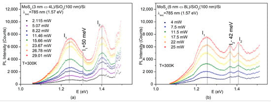
Indeed, as shown in Figure 4a, b for the 3 and 5 nm MoS2, respectively, both the I1 (Λ–Γ) and I2 (K–Γ) indirect-gap emissions, with experimental energies corresponding to 1.25 and 1.4 eV, respectively, were observed in the PL spectra under 1.57 eV excitation.

Figure 4.
PL spectra at various excitation densities under the excitation wavelength of 785 nm (1.57 eV) for MoS2/SiO2/Si thin-film structures with 3 (a) and 5 nm (b) MoS2 on the top, respectively.
It is important to stress that although both the Λ and K valleys were involved in the indirect emission of the obtained few-layer MoS2, the PL spectra of the 3 (Figure 4a) and 5 nm (Figure 4b) MoS2 differed in some details.
Along with the emission line related to the K–Γ transition, I2 and besides the emission line associated with the Λ-Γ transition, I1, the room-temperature PL spectra of the 3 nm MoS2 also showed a small satellite (I1+50 meV) positioned 50 meV higher than I1 (Figure 4a). On the other hand, the K–Γ emission I2 in the 5 nm MoS2 was split into two components, denoted as I2 and I2–42 meV, while the I1+50 meV satellite was no longer seen in the spectra (Figure 4b).
The disparity observed in the PL between the 3 and 5 nm-thick MoS2 highlights the complicated and thickness-dependent character of the radiative processes in ultrathin MoS2. Further investigation is required to understand their origin. However, when the intensity ratio between the I1 and I2 emissions was compared under similar excitation powers (11.46 mW for the 3 nm MoS2 and 11.5 mW for the 5 nm MoS2), the results strongly suggested that at room temperature, 3 nm (4 L) MoS2 is more likely to be closer to the K-Λ crossover than 5 nm (8 L) MoS2. Preliminary studies on temperature-dependent PL further support this assumption.
Until now, the concurrent generation of PL related to both the I1 and I2 indirect transitions had not been detected in MoS2 thin films. In a work by Luo et al. [43], the simultaneous generation of similar indirect-gap emissions was reported in multilayer MoS2 bubbles prepared from exfoliated MoS2 thin films. However, the exfoliated thin films themselves did not exhibit any PL [43]. This effect was achieved through the introduction of surface strain in multilayer bubbles and has little in common with our case. Lastly, it is essential to note that the simultaneous observation of indirect-gap emissions I1 (Λ-Γ transitions) at 1.25 eV and I2 (K-Γ transitions) at 1.4 eV occurred exclusively under excitation with a photon energy of 1.57 eV, which is below the energy gap for direct transitions in MoS2. For excitation with a photon energy of 2.33 eV or higher, beyond the energy gap for direct transitions in MoS2, the K-Γ indirect emission channel would become ineffective.
4. Conclusions
The sulfurization process applied to ultrathin MoO3 initially deposited on SiO2/Si substrates using plasma-enhanced atomic layer deposition has transformed MoO3 into ultrathin MoS2. The achieved material, in the form of a few layers, was deeply investigated using spectroscopic ellipsometry and photoluminescence with different excitation energies, supported by first-principles DFT-based calculations. This study has given rise to the first observation of the simultaneous generation of the indirect-gap PL caused by the indirect radiative transitions involving two distinct valleys (Λ and K) within the conduction band. This discovery provides a fresh insight into the electronic band structure of ultrathin MoS2 and a new platform for experimental studies into its bandgap.
Supplementary Materials
The following supporting information can be downloaded at: https://www.mdpi.com/article/10.3390/nano14010096/s1, Figure S1: (a): Typical XRD patterns of thin films with crystalline c-MoO3 (black line) and amorphous a-MoO3, obtained at different deposition temperatures; (b): Absorption coefficient of c-MoO3 (black line) and a-MoO3 (red line); Figure S2: Left panel (a): Raman spectra of the obtained MoS2/SiO2 (100 nm)/SiO2 structures with different thickness of MoS2 layer (blue, green, and red lines). Inset shows Raman spectra in a wavenumber range above 510 cm−1; Right panel: SEM images (top view) of the produced MoS2 layers with thicknesses of 30 (b), 5 (c), and 3 nm (d); Figure S3: Absorption coefficient as a function of the photon energy for 30 (blue), 5 (green) and 3 nm (red) MoS2 thin films. Inset shows the details of the photon energy dependence of the absorption coefficient, together with the parameters (excitation wavelength, photon energy) of the lasers used in the present work. Further explanation is given in the text; Figure S4: Calculated dielectric function of 4L (black lines), 8L (red lines) and bulk (green lines) MoS2 for light polarized in the layer plane; Figure S5: Calculated dielectric function of 4L(black lines), 8L(red lines) and bulk (green lines) MoS2 for light polarized perpendicular to the layer plane; Figure S6: Real and imaginary parts of the experimental (black lines) and calculated (red lines) DF obtained for 30 (a and b) 5 (c and d), and 3 nm (e and f) MoS2 for light with electrical vector E polarized in the layer plane (LP). The blue curves represent a correction to the calculated DF in order to take into account effects related to the finite number of layers in 5 and 3 nm MoS2; Figure S7: PL spectra of 30 (top part), 5 (middle part) and 30 nm (bottom part) MoS2. Vertical dashed lines are given for convenience and indicate positions of the indirect transitions I1, A and B excitons in one monolayer of MoS2 and shift of the A and B exciton positions in 30 nm MoS2 as compared to 5 and 3 nm MoS2; Figure S8: Normalized spectra (line-shape function) of the PL spectra in Figure 3 (main text); (a) MoS2(3 nm)/SiO2(100 nm)/Si thin film, (b) MoS2(5 nm)/SiO2(100 nm)/Si thin film. Two vertical dashed lines on the right and one on the left in each figure are given for comvenience and indicate the positions of excitons (A and B) in monolayer and indirect gap energy in bulk of MoS2. References [44,45,46,47] are cited in the supplementary materials.
Author Contributions
Conceptualization, N.T.M., M.C. and A.H.B.; methodology: N.T.M., M.C. and A.H.B.; data curation, software: Z.A.J. and E.A.; validation, A.H.B. and S.S.; formal analysis, N.T.M., Z.A.J., E.H.A. and D.A.-R.; investigation, E.H.A., J.N.J., S.G.A., Y.N.A., Z.A.J., D.A.-R., D.L., M.E., D.S., D.M.T., G.B. and E.A.B.; resources, A.H.B. and S.S.; data curation, A.H.B., S.S. and D.M.T.; writing—original draft preparation, N.T.M.; writing—review and editing, N.T.M., M.C., D.M.T., A.H.B., D.A.-R. and S.S.; visualization, E.H.A., J.N.J., D.A.-R., M.E., D.S. and D.M.T.; supervision, N.T.M., M.C., A.H.B. and S.S. All authors have read and agreed to the published version of the manuscript.
Funding
This research received no external funding.
Data Availability Statement
These data are available upon request.
Acknowledgments
We acknowledge that this work is an independent co-product of the research activities undertaken within the National project “NFFA-DI—Nano Foundries and Fine Analysis–Digital Infrastructure” Code: IR0000015, CUP: B53C22004310006, and the National project “Fit4MedRob–Fit for Medical Robotics” Grant: PNC0000007, both under the National Recovery and Resilience Plan (NRPP), co-financed by the Next Generation EU; the “Tecnopolo per la Medicina di Precisione” (TecnoMed Puglia)—Regione Puglia: DGR no. 2117 del 21/11/2018, CUP: B84I18000540002; the International cooperation project “HYSPID—Hybrid 3D Chiral Metamaterial/2D MoS2 Phototransistors for Circularly Polarized Light Detection”, CUP: B86C18000430006—funded by the Italian National Research Council and the Ministry of Science and Education of Azerbaijan (Institute of Physics); and the Regional (Puglia) project Innonetwork “IN-AIR”, CUP: B37H17004840007. N.M. is thankful to Mikhail Otrokov (Centro de Fisica de Materiales, San Sebastian, Spain) for reading the manuscript and useful comments. We are very much obliged to Iolena Tarantini (University of Salento) and Gianmichele Epifani (CNR NANOTEC) for their technical support.
Conflicts of Interest
The authors declare no conflicts of interest.
References
- Kuc, A.; Zibouche, N.; Heine, T.T. Influence of quantum confinement on the electronic structure of the transition metal sulfide TS2. Phys. Rev. B 2011, 83, 245213. [Google Scholar] [CrossRef]
- Ellis, J.K.; Lucero, M.J.; Scuseria, G.E. The indirect to direct band gap transition in multilayered MoS2 as predicted by screened hybrid density functional theory. Appl. Phys. Lett. 2011, 99, 261908. [Google Scholar] [CrossRef]
- Molina-Sanchez, A.; Sangalli, D.; Hummer, K.; Marini, A.; Wirtz, L. Effect of spin-orbit interaction on the optical spectra of single-layer, double-layer, and bulk MoS2. Phys. Rev. B 2013, 88, 045412. [Google Scholar] [CrossRef]
- Defo, R.K.; Fang, S.; Shirodkar, S.N.; Tritsaris, G.A.; Athanasios Dimoulas, A.; Kaxiras, E. Strain dependence of band gaps and exciton energies in pure and mixed transition-metal dichalcogenides. Phys. Rev. B 2016, 94, 155310. [Google Scholar] [CrossRef]
- Sun, Y.; Wang, D.; Shuai, Z. Indirect-to-direct band gap crossover in few-layer transition metal dichalcogenides: A theoretical prediction. J. Phys. Chem. C 2016, 120, 21866–21870. [Google Scholar] [CrossRef]
- Lebègue, S.; Eriksson, O. Electronic structure of two-dimensional crystals from ab initio theory. Phys. Rev. B 2009, 79, 115409. [Google Scholar] [CrossRef]
- Drüppel, M.; Deilmann, T.; Krüger, P.; Rohlfing, R. Diversity of trion states and substrate effects in the optical properties of an MoS2 monolayer. Nat. Commun. 2017, 8, 2117. [Google Scholar] [CrossRef] [PubMed]
- Malic, E.; Selig, M.; Feierabend, M.; Brem, S.; Christiansen, D.; Wendler, F.; Knorr, A.; Berghäuser, G. Dark excitons in transition metal dichalcogenides. Phys. Rev. B 2018, 2, 014002. [Google Scholar]
- Park, S.; Mutz, N.; Schultz, T.; Blumstengel, S.; Han, A.; Aljarb, A.; Li, L.-J.; List-Kratochvil, E.J.W.; Amsalem, P.; Koch, N. Direct determination of monolayer MoS2 and WSe2 exciton binding energies on insulating and metallic substrates. 2D Mater. 2018, 5, 025003. [Google Scholar] [CrossRef]
- Vaquero, D.; Clericò, V.; Salvador-Sánchez, J.; Martín-Ramos, A.; Díaz, E.; Domínguez-Adame, F.; Meziani, Y.M.; Diez, E.; Quereda, J. Excitons, trions and Rydberg states in monolayer MoS2 revealed by low-temperature photocurrent spectroscopy. Commun. Phys. 2020, 3, 194. [Google Scholar] [CrossRef]
- Paradisanos, I.; Shree, S.; George, A.; Leisgang, N.; Robert, C.; Watanabe, K.; Taniguchi, T.; Warburton, R.J.; Turchanin, A.; Marie, X.; et al. Controlling interlayer excitons in MoS2 layers grown by chemical vapor deposition. Nat. Commun. 2020, 11, 2391. [Google Scholar] [CrossRef] [PubMed]
- Zhu, B.; Lang, J.; Hu, Y.H. S-Vacancy induced indirect-to-direct band gap transition in multilayer MoS2. Phys. Chem. Chem. Phys. 2020, 22, 26005–26014. [Google Scholar] [CrossRef] [PubMed]
- Mak, K.F.; Lee, C.; Hone, J.; Shan, J.; Heinz, T.F. Atomically thin MoS2: A new direct-gap semiconductor. Phys. Rev. Lett. 2010, 105, 136805. [Google Scholar] [CrossRef] [PubMed]
- Kopaczek, J.; Zelewski, S.J.; Polak, M.P.; Gawlik, A.; Chiappe, D.; Schulze, A.; Caymax, M.; Kudrawiec, R. Direct and indirect optical transitions in bulk and atomically thin MoS2 studied by photoreflectance and photoacoustic spectroscopy. J. Appl. Phys. 2019, 125, 135701. [Google Scholar] [CrossRef]
- Zhao, W.; Ribeiro, R.M.; Toh, M.; Carvalho, A.; Kloc, C.; Castro Neto, A.H.; Eda, G. Origin of indirect optical transitions in few-layer MoS2, WS2, and WSe2. Nano Lett. 2013, 13, 5627–5634. [Google Scholar] [CrossRef] [PubMed]
- Wierzbowski, J.; Klein, J.; Sigger, F.; Straubinger, C.; Kremser, M.; Taniguchi, T.; Watanabe, K.; Wurstbauer, U.; Holleitner, A.W.; Kaniber, M.; et al. Direct exciton emission from atomically thin transition metal dichalcogenide heterostructures near the lifetime limit. Sci. Rep. 2017, 7, 12383. [Google Scholar] [CrossRef] [PubMed]
- Saigal, N.; Ghosh, S. Phonon induced luminescence decay in monolayer MoS2 on SiO2/Si substrates. Appl. Phys. Lett. 2015, 107, 242103. [Google Scholar] [CrossRef]
- Fu, L.; Wan, Y.; Tang, N.; Ding, Y.; Gao, J.; Yu, J.; Guan, H.; Zhang, K.; Wang, W.; Zhang, C.; et al. K-Λ crossover transition in the conduction band of monolayer MoS2 under hydrostatic pressure. Sci. Adv. 2017, 3, e1700162. [Google Scholar] [CrossRef]
- Golovynskyi, S.; Irfan, I.; Bosi, M.; Seravalli, L.; Datsenko, O.I.; Golovynska, I.; Li, B.; Lin, D.; Qu, J. Exciton and trion in few-layer MoS2: Thickness- and temperature-dependent photoluminescence. Appl. Surf. Sci. 2020, 515, 146033. [Google Scholar] [CrossRef]
- McCreary, K.M.; Hanbicki, A.T.; Sivaram, S.V.; Jonker, B.T. A- and B-exciton photoluminescence intensity ratio as a measure of sample quality for transition metal dichalcogenide monolayers. APL Mater. 2018, 6, 111106. [Google Scholar] [CrossRef]
- Lin, Y.; Ling, X.; Yu, L.; Huang, S.; Hsu, A.L.; Lee, Y.-H.; Kong, J.; Dresselhaus, M.S.; Palacios, T. Dielectric screening of excitons and trions in single-layer MoS2. Nano Lett. 2014, 14, 5569–5576. [Google Scholar] [CrossRef] [PubMed]
- Kumar, V.K.; Dhar, S.; Choudhury, T.H.; Shivashankar, S.A.; Raghavan, S. A predictive approach to CVD of crystalline layers of TMDs: The case of MoS2. Nanoscale 2015, 7, 7802–7810. [Google Scholar] [CrossRef] [PubMed]
- Kaupmees, R.; Komsa, H.-P.; Krustok, J. Photoluminescence study of B-trions in MoS2 monolayers with high density of defects. Phys. Stat. Sol. B 2019, 256, 1800384. [Google Scholar]
- Yu, Y.; Li, C.; Liu, Y.; Su, L.; Zhang, Y.; Cao, L. Controlled scalable synthesis of uniform, high-quality monolayer and few-layer MoS2 films. Sci. Rep. 2013, 3, 1866. [Google Scholar] [CrossRef] [PubMed]
- Lee, Y.-H.; Zhang, X.-Q.; Zhang, W.; Chang, M.-T.; Lin, C.-T.; Chang, K.-D.; Yu, Y.-C.; Tse-Wei Wang, J.; Chang, C.-S.; Li, L.-J.; et al. Synthesis of large-area MoS2 atomic layers with chemical vapor deposition. Adv. Mater. 2012, 24, 2320–2325. [Google Scholar] [CrossRef]
- Zhan, Y.; Liu, Z.; Najmaei, S.; Ajayan, P.M.; Lou, J. Large-area vapor-phase growth and characterization of MoS2 atomic layers on a SiO2 substrate. Small 2012, 8, 966–971. [Google Scholar] [CrossRef]
- Lin, Y.-C.; Zhang, W.; Huang, J.-K.; Liu, K.-K.; Lee, Y.-H.; Liang, C.-T.; Chu, C.W.; Li, L.-J. Wafer-scale MoS2 thin layers prepared by MoO3 sulfurization. Nanoscale 2013, 4, 66376641. [Google Scholar] [CrossRef]
- Lorenzo, D.; Tobaldi, D.M.; Tasco, V.; Esposito, M.; Passaseo, A.; Cuscunà, M. Molybdenum precursor delivery approaches in atomic layer deposition of α-MoO3. Dalton Trans. 2023, 52, 902–908. [Google Scholar] [CrossRef]
- Lorenzo, D.; Riminucci, F.; Manoccio, M.; Balestra, G.; Simeone, D.; Tobaldi, D.M.; Esposito, M.; Passaseo, A.; Tasco, V.; Cuscunà, M. Molybdenum oxide functional passivation of aluminum dimers for enhancing optical-field and environmental stability. Photonics 2022, 9, 523–535. [Google Scholar] [CrossRef]
- Blaha, P.; Schwarz, K.; Madsen, G.K.H.; Kvasnicka, D.; Luitz, J. WIEN2k, An Augmented Plane Waves + Local Orbitals Program for Calculating Crystal Properties, Rev. ed.; Vienna University of Technology: Vienna, Austria, 2008. [Google Scholar]
- Perdew, J.P.; Zunger, A. Self-interaction correction to density-functional approximations for many-electron systems. Phys. Rev. B 1981, 23, 5048–5079. [Google Scholar] [CrossRef]
- Monkhorst, H.; Pack, J. Special points for Brillouin-zone integrations. Phys. Rev.B 1976, 13, 5188–5192. [Google Scholar] [CrossRef]
- Li, X.; Zhu, H. Two-dimensional MoS2: Properties, preparation, and applications. J. Mater. 2015, 1, 33–44. [Google Scholar] [CrossRef]
- Shen, C.-C.; Hsu, Y.-T.; Li, L.-J.; Liu, H.-L. Charge dynamics and electronic structures of monolayer MoS2 films grown by chemical vapor deposition. Appl. Phys. Express 2013, 6, 125801. [Google Scholar] [CrossRef]
- Ermolaev, G.A.; Stebunov, Y.V.; Vyshnevyy, A.A.; Tatarkin, D.E.; Yakubovsky, D.I.; Novikov, S.M.; Baranov, D.G.; Shegai, T.; Nikitin, A.Y.; Arsenin, A.V.; et al. Broadband optical properties of monolayer and bulk MoS2. NPJ 2D Mater. Appl. 2020, 4, 21. [Google Scholar] [CrossRef]
- Ramasubramaniam, A. Large excitonic effects in monolayers of molybdenum and tungsten dichalcogenides. Phys. Rev. B 2012, 86, 115409. [Google Scholar] [CrossRef]
- Qiu, D.Y.; Da Jornada, F.H.; Louie, S.G. Optical spectrum of MoS2: Many-body effects and diversity of exciton states. Phys. Rev. Lett. 2013, 111, 216805. [Google Scholar] [CrossRef]
- Kopaczek, J.; Zelewski, S.; Yumigeta, K.; Sailus, R.; Tongay, S.; Kudrawiec, R.R. Temperature dependence of the indirect gap and the direct optical transitions at the high-symmetry point of the Brillouin zone and band nesting in MoS2, MoSe2, MoTe2, WS2, and WSe2 crystals. J. Phys. Chem. C 2022, 126, 5665–5674. [Google Scholar] [CrossRef]
- Ullrich, B.; Singh, A.K.; Barik, P.; Haowen, X.; Bhowmick, M. Inherent photoluminescence Stokes shift in GaAs. Optıcs Lett. 2015, 40, 2580–2583. [Google Scholar] [CrossRef]
- Park, Y.; Chan, C.C.S.; Taylor, R.A.; Kim, Y.; Jo, N.Y.; Lee, S.W.; Yang, W.; Im, H.; Lee, G. Temperature induced crossing in the optical bandgap of mono and bilayer MoS2 on SiO2. Sci. Rep. 2018, 8, 5380. [Google Scholar] [CrossRef]
- Tongay, S.; Zhou, J.; Ataca, C.; Lo, K.; Matthews, T.S.; Li, J.B.; Grossman, J.C.; Wu, J.Q. Thermally driven crossover from indirect toward direct bandgap in 2D semiconductors: MoSe2 versus MoS2. Nano Lett. 2012, 12, 5576–5580. [Google Scholar] [CrossRef]
- Wang, L.; Wang, Z.; Wang, H.Y.; Grinblat, G.; Huang, Y.-L.; Wang, D.; Ye, X.-H.; Li, X.-B.; Bao, Q.; Thye-Shen Wee, A.; et al. Slow cooling and efficient extraction of C-exciton hot carriers in MoS2 monolayer. Nat. Commun. 2017, 8, 13906. [Google Scholar] [CrossRef] [PubMed]
- Luo, H.; Li, X.; Zhao, Y.; Yang, R.; Bao, L.; Hao, Y.; Gao, Y.; Shi, N.N.; Guo, Y.; Liu, G.; et al. Simultaneous generation of direct- and indirect-gap photoluminescence in multilayer MoS2 bubbles. Phys. Rev. Mater. 2020, 4, 074006. [Google Scholar] [CrossRef]
- Ferlauto, A.S.; Ferreira, G.M.; Pearce, J.M.; Wronski, C.R.; Collins, R.W. Analytical model for the optical functions of amorphous semiconductors from the near-infrared to ultraviolet: Applications in thin film Photovoltaics. J. Appl. Phys. 2002, 92, 2424–2436. [Google Scholar] [CrossRef]
- Mohamed, S.H.; Kappertz, O.; Ngaruiya, J.M.; Leervad-Pedersen, T.P.; Drese, R.; Wuttig, M. Correlation between structure, stress and optical properties in direct current sputtered molybdenum oxide films. TSF 2003, 429, 135–143. [Google Scholar] [CrossRef]
- Oiwake, K.; Nishigaki, Y.; Shohei Fujimoto, S.; Maeda, S.; Fujiwara, H. Fully automated spectroscopic ellipsometry analyses: Application to MoOx thin films. J. Appl. Phys. 2021, 129, 243102. [Google Scholar] [CrossRef]
- Lee, C.; Yan, H.; Brus, L.E.; Heinz, T.F.; Hone, J.; Ryu, S. Anomalous lattice vibrations of single and few-layer MoS2. ACS Nano 2010, 4, 2695–2700. [Google Scholar] [CrossRef]
Disclaimer/Publisher’s Note: The statements, opinions and data contained in all publications are solely those of the individual author(s) and contributor(s) and not of MDPI and/or the editor(s). MDPI and/or the editor(s) disclaim responsibility for any injury to people or property resulting from any ideas, methods, instructions or products referred to in the content. |
© 2023 by the authors. Licensee MDPI, Basel, Switzerland. This article is an open access article distributed under the terms and conditions of the Creative Commons Attribution (CC BY) license (https://creativecommons.org/licenses/by/4.0/).