
A Five-Year Retrospective Study from a Single Center on the Location, Presentation, Diagnosis, and Management of 110 Patients with Aneurysms of the Femoral and Popliteal Arteries of the Lower Limb
 ,
,
Abstract
1. Introduction
2. Materials and Methods
2.1. Study Design and Population
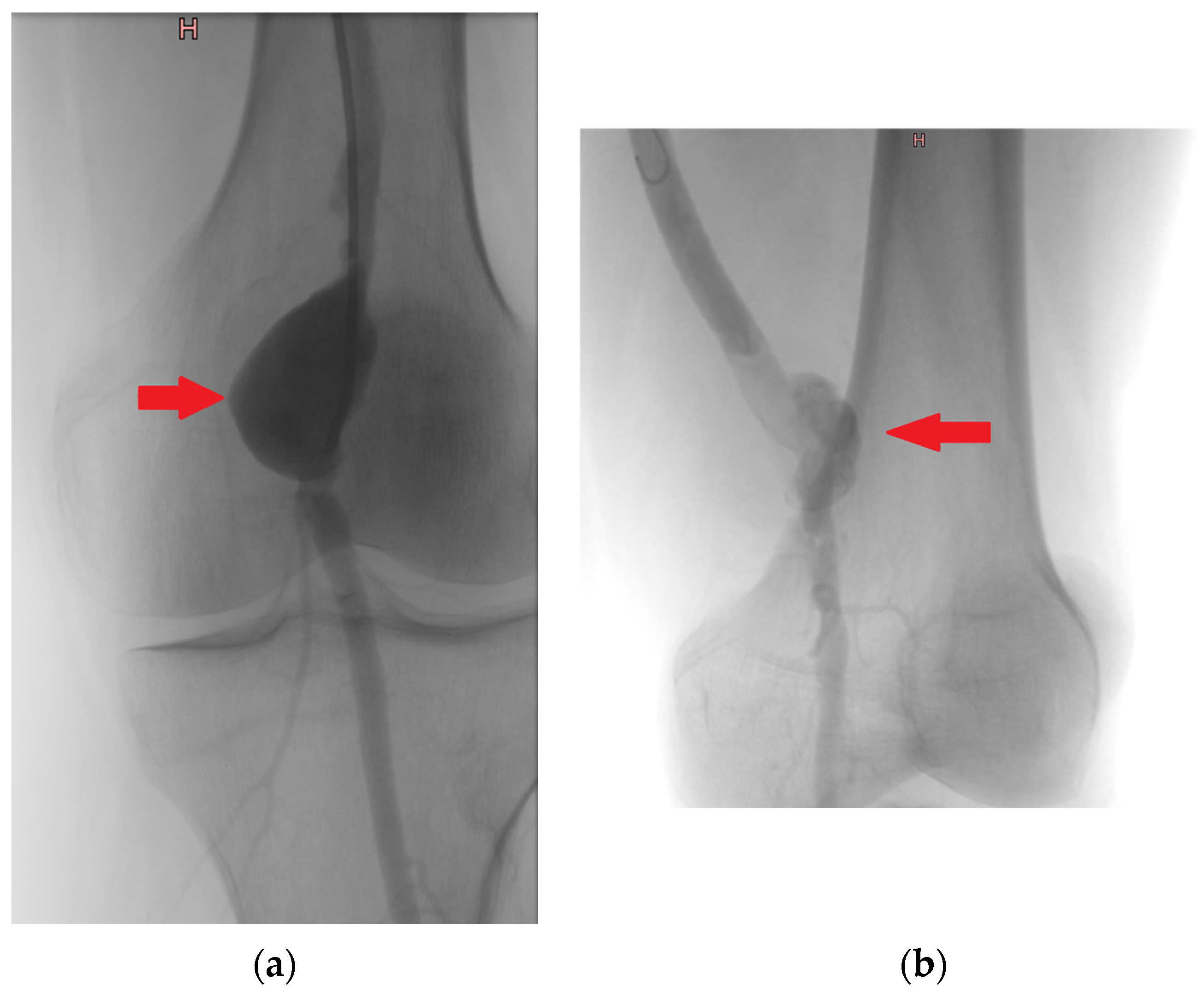
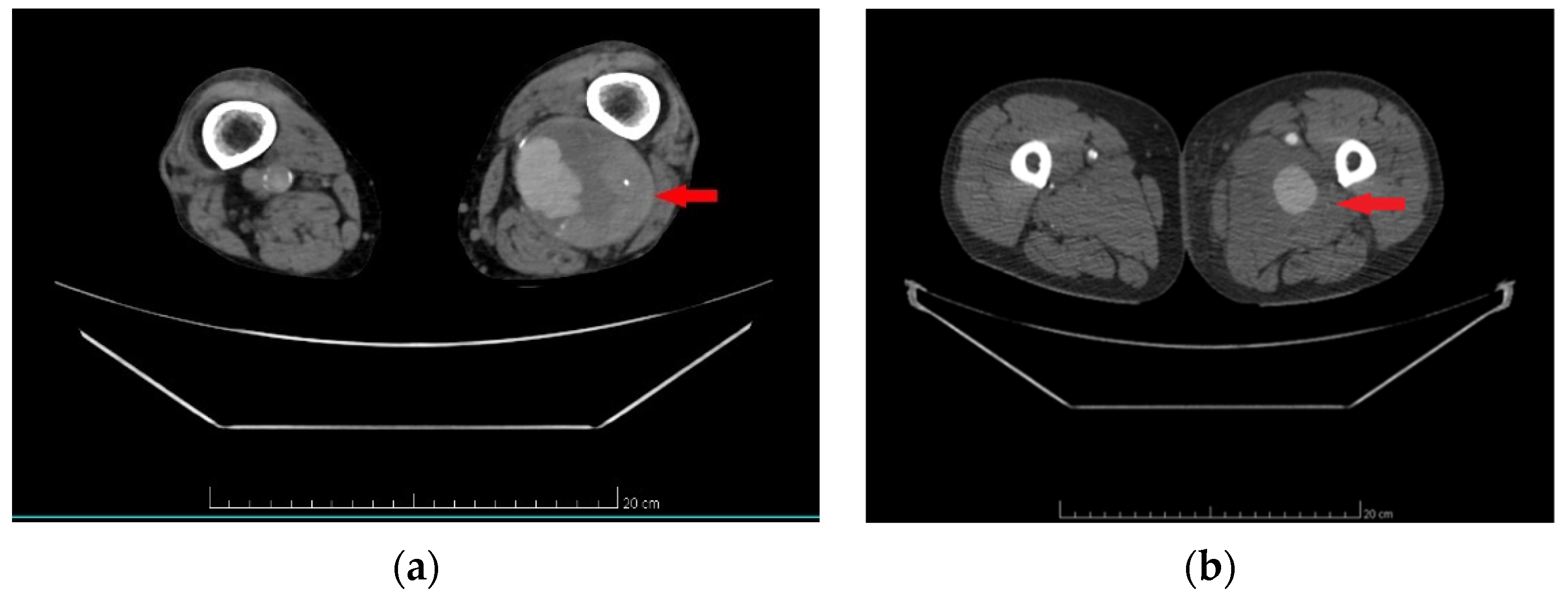
2.2. Diagnostic Criteria
2.3. Methods of Treatment
2.4. Definitions
2.5. Analyzed Data
2.6. Statistical Analysis
3. Results
3.1. Patient Demographics and Descriptive Data
3.2. Characteristics of Aneurysms in the Study Population
3.3. Procedural Characteristics and Outcomes in Patients Undergoing Aneurysm Repair
3.4. Long-Term Follow-Up and Outcomes in Patients Undergoing Aneurysm Repair
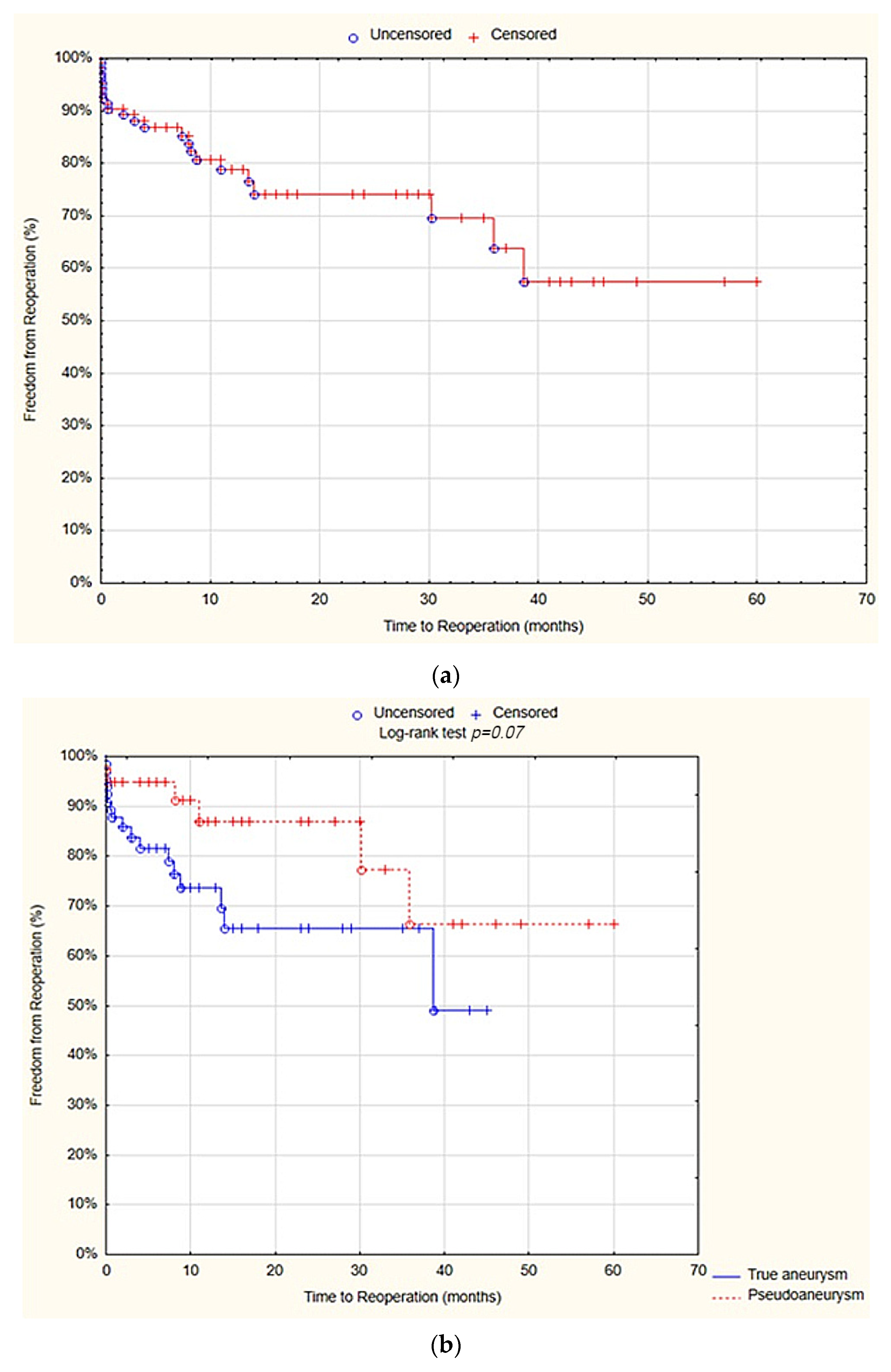
3.5. Reoperations
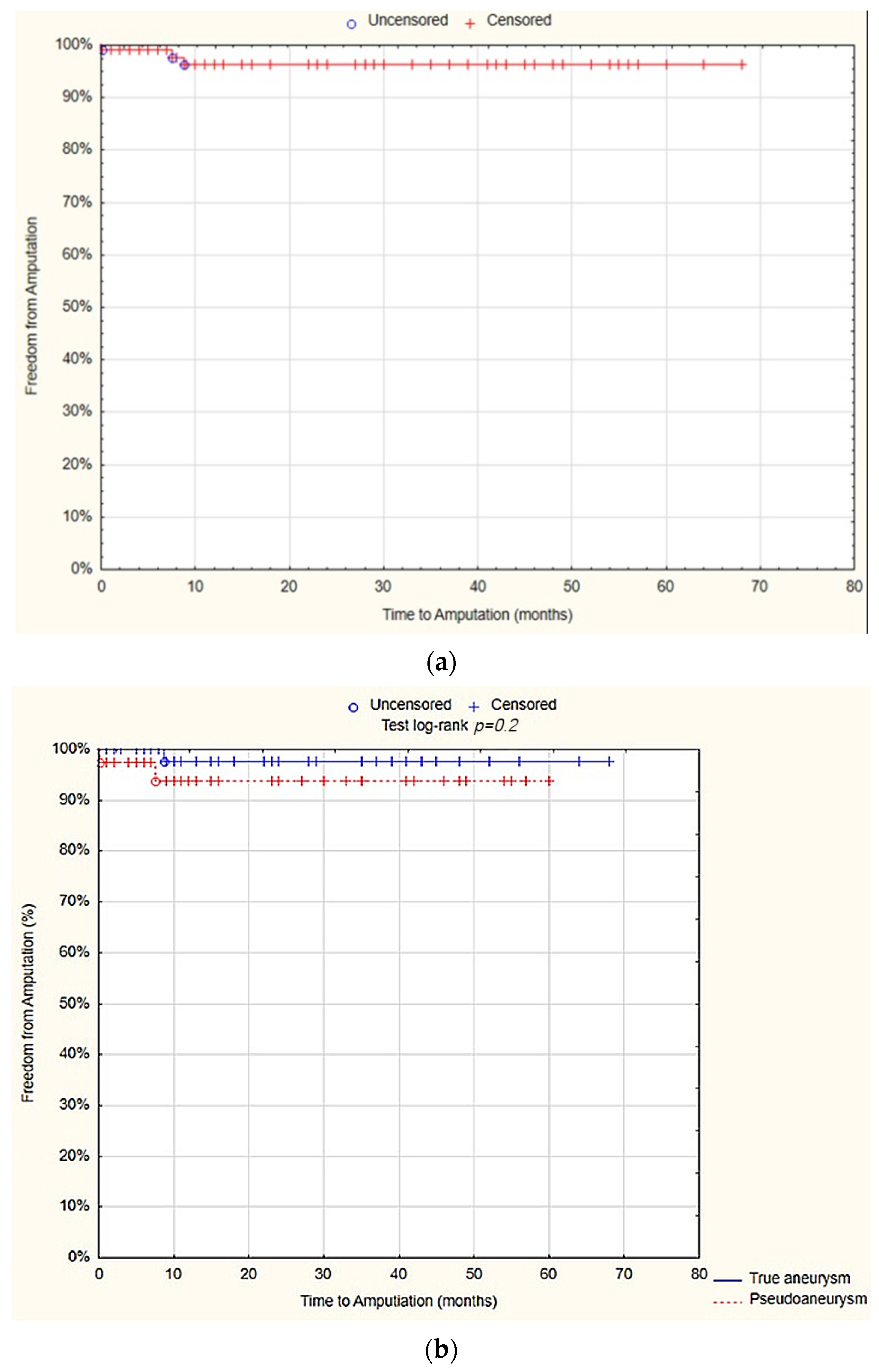
3.6. Amputations
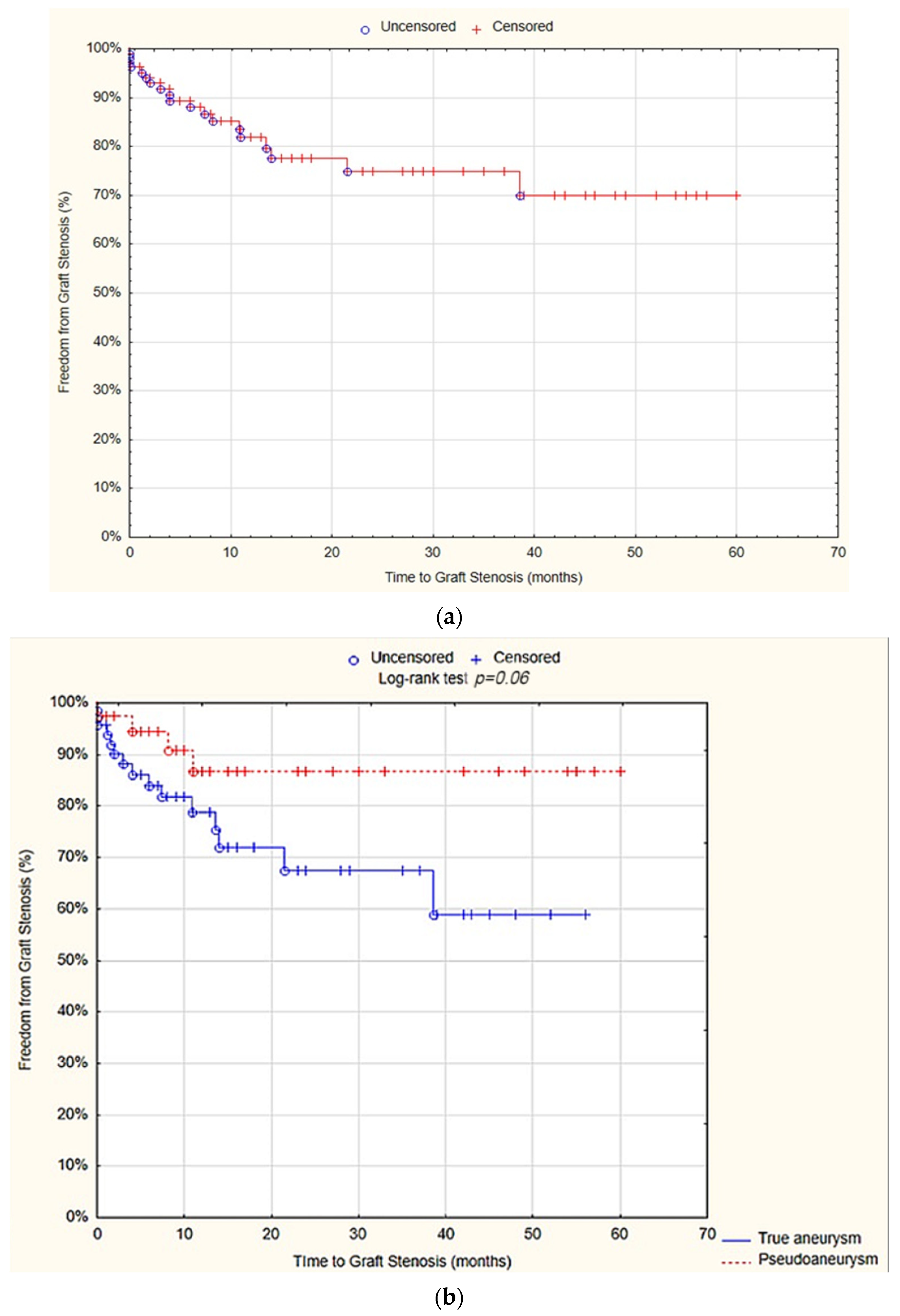
3.7. Graft Stenosis
3.8. RAS Events
3.9. Overall Survival (OS)
4. Discussion
Limitations of the Study
5. Conclusions
Author Contributions
Funding
Institutional Review Board Statement
Informed Consent Statement
Data Availability Statement
Acknowledgments
Conflicts of Interest
References
- Galland, R.B. History of the management of popliteal artery aneurysms. Eur. J. Vasc. Endovasc. Surg. 2008, 35, 466–472. [Google Scholar] [CrossRef] [PubMed]
- Diwan, A.; Sarkar, R.; Stanley, J.C.; Zelenock, G.B.; Wakefield, T.W. Incidence of femoral and popliteal artery aneurysms in patients with abdominal aortic aneurysms. J. Vasc. Surg. 2000, 31, 863–869. [Google Scholar] [CrossRef]
- Sokhal, B.S.; Ma, Y.; Rajagopalan, S. Femoral artery aneurysms. Br. J. Hosp. Med. 2022, 83, 1–10. [Google Scholar] [CrossRef] [PubMed]
- Rowlands, C.; Youssef, S.; Rajagopalan, S. Popliteal arterial aneurysms. Br. J. Hosp. Med. 2022, 83, 1–7. [Google Scholar] [CrossRef] [PubMed]
- Kassem, M.M.; Oropallo, A.; Gonzalez, L. Popliteal Artery Aneurysm. In StatPearls [Internet]; StatPearls Publishing: Treasure Island, FL, USA, 2024. Available online: https://www.ncbi.nlm.nih.gov/books/NBK430863/ (accessed on 16 February 2024).
- Fioranelli, A.; Carpentieri, E.A.; Wolosker, N.; Castelli, V., Jr.; Caffaro, R.A. Rupture of Thrombosed Popliteal Aneurysm: A Case Report. Ann. Vasc. Surg. 2018, 51, 324.e7–324.e10. [Google Scholar] [CrossRef] [PubMed]
- Stone, P.A.; Campbell, J.E.; AbuRahma, A.F. Femoral pseudoaneurysms after percutaneous access. J. Vasc. Surg. 2014, 60, 1359–1366. [Google Scholar] [CrossRef] [PubMed]
- Saleem, T.; D’Cruz, J.R.; Baril, D.T. Femoral Aneurysm Repair. In StatPearls [Internet]; StatPearls Publishing: Treasure Island, FL, USA, 2024. Available online: https://www.ncbi.nlm.nih.gov/books/NBK470329/ (accessed on 14 July 2023).
- Wright, L.B.; Matchett, W.J.; Cruz, C.P.; James, C.A.; Culp, W.C.; Eidt, J.F.; McCowan, T.C. Popliteal artery disease: Diagnosis and treatment. Radiographics 2004, 24, 467–479. [Google Scholar] [CrossRef] [PubMed]
- Golchehr, B.; Zeebregts, C.J.; Reijnen, M.M.P.J.; Tielliu, I.F.J. Long-term outcome of endovascular popliteal artery aneurysm repair. J. Vasc. Surg. 2018, 67, 1797–1804. [Google Scholar] [CrossRef] [PubMed]
- Farber, A.; Angle, N.; Avgerinos, E.; Dubois, L.; Eslami, M.; Geraghty, P.; Haurani, M.; Jim, J.; Ketteler, E.; Pulli, R.; et al. The Society for Vascular Surgery clinical practice guidelines on popliteal artery aneurysms. J. Vasc. Surg. 2022, 75, 109S–120S. [Google Scholar] [CrossRef]
- Lawrence, P.F.; Harlander-Locke, M.P.; Oderich, G.S.; Humphries, M.D.; Landry, G.J.; Ballard, J.L.; Abularrage, C.J. The current management of isolated degenerative femoral artery aneurysms is too aggressive for their natural history. J. Vasc. Surg. 2014, 59, 343–349. [Google Scholar] [CrossRef]
- Mohan, I.V.; Stephen, M.S. Peripheral arterial aneurysms: Open or endovascular surgery? Prog. Cardiovasc. Dis. 2013, 56, 36–56. [Google Scholar] [CrossRef] [PubMed]
- Tamashiro, G.A.; Tamashiro, A.; Villegas, M.O.; Dini, A.E.; Mollón, A.P.; Zelaya, D.A.; Soledispa-Suarez, C.I.; Diaz, J.A. Flexions of the popliteal artery: Technical considerations of femoropopliteal stenting. J. Invasive Cardiol. 2011, 23, 431–433. [Google Scholar]
- Shah, N.G.; Rokosh, R.S.; Garg, K.; Safran, B.; Rockman, C.B.; Maldonado, T.S.; Sadek, M.; Lamparello, P.; Jacobowitz, G.R.; Barfield, M.E.; et al. Endovascular treatment of popliteal artery aneurysms has comparable long-term outcomes to open repair with shorter lengths of stay. J. Vasc. Surg. 2021, 74, 1565–1572.e1. [Google Scholar] [CrossRef] [PubMed]
- Hirsch, A.T.; Haskal, Z.J.; Hertzer, N.R.; Bakal, C.W.; Creager, M.A.; Halperin, J.L.; Hiratzka, L.F.; Murphy, W.R.; Olin, J.W.; Puschett, J.B.; et al. ACC/AHA 2005 Practice Guidelines for the management of patients with peripheral arterial disease (lower extremity, renal, mesenteric, and abdominal aortic): A collaborative report from the American Association for Vascular Surgery/Society for Vascular Surgery, Society for Cardiovascular Angiography and Interventions, Society for Vascular Medicine and Biology, Society of Interventional Radiology, and the ACC/AHA Task Force on Practice Guidelines (Writing Committee to Develop Guidelines for the Management of Patients With Peripheral Arterial Disease): Endorsed by the American Association of Cardiovascular and Pulmonary Rehabilitation; National Heart, Lung, and Blood Institute; Society for Vascular Nursing; TransAtlantic Inter-Society Consensus; and Vascular Disease Foundation. Circulation 2006, 113, e463–e654. [Google Scholar] [PubMed]
- Moll, F.L.; Powell, J.T.; Fraedrich, G.; Verzini, F.; Haulon, S.; Waltham, M.; van Herwaarden, J.A.; Holt, P.J.E.; van Keulen, J.W.; Rantner, B.; et al. Management of abdominal aortic aneurysms clinical practice guidelines of the European society for vascular surgery. Eur. J. Vasc. Endovasc. Surg. 2011, 41 (Suppl. S1), S1–S58. [Google Scholar] [CrossRef] [PubMed]
- Wanhainen, A.; Verzini, F.; Van Herzeele, I.; Allaire, E.; Bown, M.; Cohnert, T.; Dick, F.; van Herwaarden, J.; Karkos, C.; Koelemay, M.; et al. Editor’s Choice—European Society for Vascular Surgery (ESVS) 2019 Clinical Practice Guidelines on the Management of Abdominal Aorto-iliac Artery Aneurysm. Eur. J. Vasc. Endovasc. Surg. 2019, 57, 8–93. [Google Scholar] [CrossRef] [PubMed]
- Peeran, S.; DeMartino, R.R.; Huang, Y.; Fleming, M.; Kalra, M.; Oderich, G.S.; Duncan, A.A.; Bower, T.C.; Gloviczki, P. Outcomes of women treated for popliteal artery aneurysms. Ann. Vasc. Surg. 2016, 34, 187–192. [Google Scholar] [CrossRef] [PubMed]
- Kent, K.C. Abdominal Aortic Aneurysms. N. Engl. J. Med. 2014, 371, 2101–2108. [Google Scholar] [CrossRef] [PubMed]
- Lederle, F.A.; Johnson, G.R.; Wilson, S.E.; Chute, E.P.; Littooy, F.N.; Bandyk, D.; Krupski, W.C.; Barone, G.W.; Acher, C.W.; Ballard, D.J. Prevalence and associations of abdominal aortic aneurysm detected through screening. Aneurysm Detection and Management (ADAM) Veterans Affairs Cooperative Study Group. Ann. Intern. Med. 1997, 126, 441–449. [Google Scholar] [CrossRef]
- Kuivaniemi, H.; Platsoucas, C.D. Aortic Aneurysms: Pathogenesis and Mechanisms. Eur. J. Vasc. Endovasc. Surg. 2012, 43, 261–267. [Google Scholar]
- Hall, H.A.; Minc, S.; Babrowski, T. Peripheral artery aneurysm. Surg. Clin. N. Am. 2013, 93, 911–923. [Google Scholar] [CrossRef] [PubMed]
- Ravn, H.; Bergqvist, D.; Björck, M.; Swedish Vascular Registry. Nationwide study of the outcome of popliteal artery aneurysms treated surgically. Br. J. Surg. 2007, 94, 970–977. [Google Scholar] [CrossRef]
- Costantino, T.G.; Bruno, E.C.; Handly, N.; Dean, A.J. Accuracy of emergency medicine ultrasound in the evaluation of abdominal aortic aneurysm. J. Emerg. Med. 2005, 29, 455–460. [Google Scholar] [CrossRef] [PubMed]
- Gabriel, M.; Pawlaczyk, K.; Waliszewski, K.; Krasiński, Z.; Majewski, W. Location of femoral artery puncture site and the risk of postcatheterization pseudoaneurysm formation. Int. J. Cardiol. 2007, 120, 167–171. [Google Scholar] [CrossRef] [PubMed]
- Demarche, M.; Waltregny, D.; van Damme, H.; Limet, R. Femoral anastomotic aneurysms: Pathogenic factors, clinical presentations and treatment. A study of 142 cases. Cardiovasc. Surg. 1999, 7, 315–322. [Google Scholar] [CrossRef] [PubMed]
- Marković, D.M.; Davidović, L.B.; Kostić, D.M.; Maksimović, Ž.V.; Činara, I.S.; Cvetković, S.D.; Marković, M.D.; Dragaš, M.V. Anastomotic pseudoaneurysms. Srpski Arhiv za Celokupno Lekarstvo 2006, 134, 114–121. [Google Scholar] [CrossRef] [PubMed]
- Pogorzelski, R.; Fiszer, P.; Toutounchi, S.; Krajewska, E.; Szostek, M.M.; Tworus, R.; Jakuczun, W.; Skórski, M. Anastomotic aneurysms—20-years of experience from one center. Pol. Przegl Chir. 2013, 85, 181–191. [Google Scholar] [CrossRef] [PubMed]
- Galiñanes, E.L.; Dombrovskiy, V.Y.; Graham, A.M.; Vogel, T.R. Endovascular versus open repair of popliteal artery aneurysms: Outcomes in the US Medicare population. Vasc. Endovasc. Surg. 2013, 47, 267–273. [Google Scholar] [CrossRef] [PubMed]
- Naazie, I.N.; Khan, M.A.; Gupta, J.D.; Patel, R.; AbuRahma, A.; Malas, M.B. Open Repair Versus Endovascular Repair in the Treatment of Symptomatic Popliteal Artery Aneurysms. Ann. Vasc. Surg. 2022, 86, 77–84. [Google Scholar] [CrossRef]
- Koksoy, C.; Gyedu, A.; Alacayir, I.; Bengisun, U.; Uncu, H.; Anadol, E. Surgical treatment of peripheral aneurysms in patients with Behcet’s disease. Eur. J. Vasc. Endovasc. Surg. 2011, 42, 525–530. [Google Scholar] [CrossRef]
- Aronow, W.S. Peripheral arterial disease of the lower extremities. Arch. Med. Sci. 2012, 8, 375–388. [Google Scholar] [CrossRef] [PubMed]


| Type of the Aneurysm | True Aneurysm (n = 71; 64.5%) | Pseudoaneurysm (n = 39; 35.5%) | Total (n = 110) | p |
|---|---|---|---|---|
| Age | 67 (51–98) IQR 11 | 68 (15–83) IQR 9 | 67.5 (15–98) IQR 10 | 0.9 |
| Gender | 0.03 | |||
| Male | 65 (91.6%) | 30 (76.9%) | 95 (86.4%) | |
| Female | 6 (8.5%) | 9 (23.1%) | 15 (13.6%) | |
| Presence of comorbidities | 56 (78.8%) | 34 (87.2%) | 90 (81.8%) | 0.3 |
| Arterial hypertension | 50 (70.4%) | 27 (69.2%) | 77 (70%) | 0.9 |
| Generalized atherosclerosis | 20 (28.2%) | 23 (59%) | 43 (39.1%) | 0.002 |
| Coronary artery disease | 16 (22.5%) | 13 (33.3%) | 29 (26.4%) | 0.2 |
| History of myocardial infarction | 14 (19.7%) | 13 (33.3%) | 27 (24.5%) | 0.1 |
| Dyslipidemia | 11 (15.5%) | 8 (20.5%) | 19 (17.3%) | 0.5 |
| Diabetes mellitus | 9 (12.7%) | 10 (25.6%) | 19 (17.3%) | 0.09 |
| Heart failure | 5 (7%) | 7 (18%) | 12 (10.9%) | 0.08 |
| COPD | 5 (7%) | 1 (2.6%) | 6 (5.5%) | 0.4 |
| Dialysis dependence | 4 (5.6%) | 0 (0%) | 4 (3.6%) | 0.3 |
| History of cigarette smoking (yes) | 33 (46.48%) | 19 (48.72%) | 52 (47.3%) | 0.8 |
| Current cigarette smoking (yes) | 23 (32.39%) | 15 (38.46%) | 38 (34.5%) | 0.5 |
| Clinical symptoms (yes) | 47 (66.2%) | 24 (61.5%) | 71 (64.5%) | 0.6 |
| Lower limb pain | 27 (38%) | 16 (41%) | 43 (39.1%) | 0.8 |
| Acute limb ischemia | 18 (25.4%) | 3 (7.7%) | 21 (19.1%) | 0.02 |
| Intermittent claudication | 12 (16.9%) | 5 (12.8%) | 17 (15.5%) | 0.6 |
| Limb swelling | 5 (7%) | 2 (5.1%) | 7 (6.5%) | 1 |
| Ulcer | 1 (1.4%) | 3 (7.7%) | 4 (3.6%) | 0.1 |
| Aneurysm rupture | 2 (2.8%) | 1 (2.6%) | 3 (2.7%) | 1 |
| Gangrene | 0 (0%) | 2 (5.1%) | 2 (1.8%) | 0.1 |
| Presence of other aneurysms (yes) | 42 (59.2%) | 15 (38.5%) | 57 (51.8%) | 0.04 |
| Abdominal aorta aneurysm | 15 (21.1%) | 6 (15.4%) | 21 (19.1%) | 0.5 |
| Popliteal artery aneurysm | 15 (21.1%) | 3 (7.7%) | 18 (16.4%) | 0.1 |
| Common femoral artery aneurysm | 7 (9.9%) | 4 (10.3%) | 11 (10%) | 1 |
| Superficial femoral artery aneurysm | 4 (5.6%) | 2 (5.1%) | 6 (5.5%) | 1 |
| Common iliac artery aneurysm | 1 (1.4%) | 0 (0.0%) | 1 (0.9%) | 1 |
| Type of the Aneurysm | True Aneurysm (n = 71; 64.5%) | Pseudoaneurysm (n = 39; 35.5%) | Total (n = 110) | p |
|---|---|---|---|---|
| Aneurysm localization (artery) | ||||
| Popliteal artery | 52 (73.2%) | 2 (5.1%) | 54 (49.1%) | <0.001 |
| Common femoral artery | 17 (23.9%) | 36 (92.3%) | 53 (48.2%) | <0.001 |
| Superficial femoral artery | 2 (2.8%) | 0 (0%) | 2 (1.8%) | 1 |
| Deep femoral artery | 0 (0%) | 1 (2.6%) | 1 (0.9%) | 0.4 |
| Aneurysm size (mm) | 36 IQR 21.5 | 25 IQR 34.5 | 36 IQR 20 mm | 0.5 |
| True aneurysm etiology | ||||
| Idiopathic | 51 (81.8%) | - | 51 (46.4%) | - |
| Atherosclerosis | 20 (28.2%) | - | 20 (18.2%) | - |
| Pseudoaneurysm etiology | ||||
| Arterial anastomosis | 37 (94.9%) | 37 (38.2%) | - | - |
| • Aortobifemoral bypass | 24 (61.5%) | 24 (21.8%) | - | - |
| • Femoropopliteal bypass | 10 (25.6%) | 10 (9.1%) | - | - |
| • Aortofemoral bypass | 6 (15.4%) | 6 (5.5%) | - | - |
| • Iliofemoral bypass | 2 (5.1%) | 2 (1.81%) | - | - |
| Injury | 2 (5.1%) | 2 (1.81%) | - | - |
| Type of the Aneurysm | True Aneurysm (n = 71; 64.5%) | Pseudoaneurysm (n = 39; 35.5%) | Total (n = 110) | p |
|---|---|---|---|---|
| Duration of procedure (minutes) | 170 (45–315) IQR 97.5 | 150 (60–365) IQR 79 | 165.5 (45–365) IQR 77.5 | 0.2 |
| Type of the procedure | ||||
| Aneurysmectomy | 39 (54.9%) | 34 (87.2%) | 73 (66.4%) | <0.001 |
| Endovascular treatment | 16 (22.5%) | 1 (2.6%) | 17 (15.5%) | 0.01 |
| Surgical bypass | 15 (21.1%) | 0 (0%) | 15 (13.6%) | <0.001 |
| Aneurysmectomy with bypass | 1 (1.4%) | 4 (10.3%) | 5 (4.5%) | 0.05 |
| Intraoperative blood loss | 0.4 | |||
| <400 mL | 65 (91.6%) | 38 (97.4%) | 103 (93.7%) | |
| >400 mL | 6 (4.2%) | 1 (2.6%) | 7 (5.4%) | |
| Transfusion of red blood cells (RBC) | 5 (7%) | 1 (2.6%) | 6 (5.5%) | 0.4 |
| Transfusion of fresh frozen plasma (FFP) | 1 (1.4%) | 1 (2.6%) | 2 (1.8%) | 1 |
| Graft material | 0.05 | |||
| Synthetic | 67 (94.37%) | 32 (82.1%) | 99 (90%) | |
| Patient’s vein | 4 (5.63%) | 7 (18) | 11 (10%) | |
| Duration of hospitalization (days) | 8 (5–26) IQR 5 | 9 (4–100) IQR 4 | 8 (4–100) IQR 4 | 0.3 |
| Duration of postoperative hospitalization (days) | 4 (1–25) IQR 3 | 5 (2–90) IQR 5 | 4 (1–90) IQR 4 | 0.2 |
| Early postoperative complications | 16 (22.5%) | 9 (23.1%) | 25 (22.7%) | 1 |
| Surgical site infection | 4 (5.6%) | 6 (15.4%) | 10 (9.1%) | 0.2 |
| Hematoma | 7 (9.8%) | 1 (2.6%) | 8 (7.3%) | 0.3 |
| Acute limb ischemia | 3 (3.2%) | 1 (2.6%) | 4 (3.6%) | 0.7 |
| Myocardial infarction | 0 (0%) | 1 (2.6%) | 1 (0.9%) | 0.4 |
| Femoral abscess | 1 (1.4%) | 0 (0%) | 1 (0.9%) | 1 |
| C.Difficile infection | 1 (1.4%) | 0 (0%) | 1 (0.9%) | 1 |
| Early reoperations | 8 (13.8%) | 2 (5.7%) | 10 (9.1%) | 0.3 |
| Univariable Analysis | ||||
|---|---|---|---|---|
| Variable | n (%) | OR | 95% CI | p |
| Pre-operative | ||||
| Age | 1 | 1–1.1 | 0.2 | |
| Gender | 0.7 | |||
| Male | 95 (86.4%) | 1 | ||
| Female | 15 (13.6%) | 1.2 | 0.3–4.7 | |
| Presence of comorbidities | 90 (81.8%) | 1.8 | 0.5–7 | 0.4 |
| Arterial hypertension | 77 (70%) | 1.1 | 0.4–3 | 0.8 |
| Generalized atherosclerosis | 43 (39.1%) | 3 | 1.2–7.7 | 0.02 |
| Coronary artery disease | 29 (26.4%) | 1.4 | 0.5–3.8 | 0.5 |
| History of myocardial infarction | 27 (24.5%) | 1.6 | 0.6–4.4 | 0.3 |
| Dyslipidemia | 19 (17.3%) | 1.3 | 0.4–4 | 0.7 |
| Diabetes mellitus | 19 (17.3%) | 1.3 | 0.4–4 | 0.7 |
| Heart failure | 12 (10.9%) | 1.8 | 0.5–6.8 | 0.4 |
| COPD | 6 (5.5%) | 3.7 | 0.7–20 | 0.1 |
| History of cigarette smoking (yes) | 52 (47.3%) | 1.6 | 0.6–3.9 | 0.3 |
| Current cigarette smoking(yes) | 38 (34.5%) | 1.08 | 0.4–2.8 | 0.8 |
| Aneurysm localization | ||||
| Popliteal artery | 54 (49.1%) | 0.6 | 0.2–1.5 | 0.2 |
| Common femoral artery | 53 (48.2%) | 1.33 | 0.54–3.3 | 0.5 |
| Intra-operative | ||||
| Type of procedure | ||||
| Aneurysmectomy | 73 (66.4%) | 1.1 | 0.4–2.9 | 0.8 |
| Endovascular treatment | 17 (15.5%) | 1.2 | 0.4–3.6 | 0.8 |
| Surgical bypass | 15 (13.6%) | 0.7 | 0.2–2.7 | 0.6 |
| Aneurysmectomy with bypass | 5 (4.5%) | 2.4 | 0.4–15 | 0.4 |
| Blood loss | 0.7 | |||
| >400 mL | 7 (6.3%) | 1.7 | 0.1–20.5 | |
| <400 mL | ||||
| Duration of procedure | 1 | 0.9–1 | 1 | |
| Type of the Aneurysm | True Aneurysm (n = 71; 64.5%) | Pseudoaneurysm (n = 39; 35.5%) | Total (n = 110) | p |
|---|---|---|---|---|
| Follow-up time (months) | 16 (1–68) IQR 31 | 17 (1–60) IQR 33 | 16 (1–68) IQR 31 | 0.6 |
| Late postoperative complications | 18 (25.4%) | 9 (23.1%) | 27 (24.5%) | 0.7 |
| Acute limb ischemia | 14 (19.7%) | 3 (7.7%) | 17 (15.5%) | 0.1 |
| Chronic limb ischemia | 3 (4.2%) | 1 (2.6%) | 4 (3.6%) | 1 |
| Popliteal abscess | 1 (1.4%) | 0 (0.0%) | 1 (0.9%) | 0.5 |
| Prosthesis infection | 0 (0.0%) | 2 (5.1%) | 2 (1.8%) | 0.1 |
| Myocardial infarction | 0 (0.0%) | 1 (2.6%) | 1 (0.9%) | 0.4 |
| Recurrence of pseudoaneurysm | 0 (0.0%) | 1 (2.6%) | 1 (0.9%) | 0.4 |
| Intracerebral hemorrhage | 0 (0.0%) | 1 (2.6%) | 1 (0.9%) | 0.4 |
| Late reoperations | 10 (14.1%) | 3 (7.7%) | 13 (11.8%) | 0.1 |
| Mortality | 1 (1.4%) | 2 (5.1%) | 3 (2.7%) | 0.3 |
| Univariable Analysis | ||||
|---|---|---|---|---|
| Variable | Freedom from Reoperations (Months) | HR | 95% CI | p |
| Age | 1 | 1–1.1 | 0.3 | |
| Gender | 0.1 | |||
| Male | 8 IQR 14.5 | 0.5 | 0.2–1.3 | |
| Female | 7.4 IQR 15 | 1 | ||
| History of cigarette smoking | 0.6 | |||
| Yes | 7.5 IQR 12.5 | 0.8 | 0.4–1.9 | |
| No | 8.9 IQR 21.8 | 1 | ||
| Current cigarette smoking | 0.2 | |||
| Yes | 8 IQR 14 | 0.5 | 0.2–1.4 | |
| No | 8.1 IQR 15 | 1 | ||
| Presence of comorbidities | ||||
| Arterial hypertension | 0.3 | |||
| Yes | 8 IQR 13 | 1.7 | 0.6–4.6 | |
| No | 8 IQR 22 | 1 | ||
| Generalized atherosclerosis | 0.2 | |||
| Yes | 7 IQR 14.5 | 1.7 | 0.7–3.8 | |
| No | 8.8 IQR 14.5 | 1 | ||
| Coronary artery disease | 0.5 | |||
| Yes | 11 IQR 17 | 0.7 | 0.3–1.9 | |
| No | 8 IQR 14 | 1 | ||
| History of myocardial infarction | 0.5 | |||
| Yes | 6 IQR 10.5 | 0.7 | 0.2–2.1 | |
| No | 8.2 IQR 15.5 | 1 | ||
| Localization | ||||
| Popliteal artery | 3 IQR 11.3 | 1.7 | 0.7–3.8 | 0.2 |
| Common femoral artery | 10 IQR 20.5 | 0.5 | 0.2–1.3 | 0.2 |
| Superficial femoral artery | 9.9 IQR 1.1 | 2.3 | 0.3–17.4 | 0.4 |
| Deep femoral artery | 12 | 1 | ||
| Aneurysm size | 1.1 | 1–1.1 | 0.06 | |
| Type of the procedure | ||||
| Aneurysmectomy | 8 IQR 17 | 0.6 | 0.3–1.4 | 0.3 |
| Endovascular treatment | 4 IQR 28 | 1.5 | 0.6–4.2 | 0.4 |
| Bypass | 8 IQR 12.8 | 2.06 | 0.8–5.6 | 0.2 |
| Aneurysmectomy with bypass | 12 | 1 | ||
| Graft material | ||||
| Synthetic | 11 IQR 19.5 | 1 | ||
| Patient’s vein | 3 IQR 22.95 | 4.64 | 0.51–42.56 | 0.17 |
| Univariable Analysis | ||||
|---|---|---|---|---|
| Variable | Freedom from Amputations (Months) | HR | 95% CI | p |
| Age | 1 | 0.9–1.1 | 0.9 | |
| Gender | 0.3 | |||
| Male | 11 IQR 29.5 | 0.3 | 0.02–3.3 | |
| Female | 10 IQR 23.5 | 1 | ||
| History of cigarette smoking | 0.7 | |||
| Yes | 10 IQR 17.5 | 0.6 | 0.1–6.5 | |
| No | 15 IQR 36.5 | 1 | ||
| Current cigarette smoking | 1 | |||
| Yes | 10 IQR 17.5 | 1 | 0.1–11 | |
| No | 11 IQR 32.8 | 1 | ||
| Presence of comorbidities | ||||
| Arterial hypertension | 0.8 | |||
| Yes | 11 IQR 25 | 0.7 | 0.1–8.3 | |
| No | 9 IQR 32 | 1 | ||
| Generalized atherosclerosis | 0.3 | |||
| Yes | 11 IQR 31.5 | 3.2 | 0.3–35.7 | |
| No | 11 IQR 27 | 1 | ||
| Coronary artery disease | 1 | |||
| Yes | 13 IQR 27 | 1 | ||
| No | 10 IQR 28 | 1 | ||
| History of myocardial infarction | 1 | |||
| Yes | 11 IQR 21 | 1 | ||
| No | 11 IQR 30.5 | 1 | ||
| Localization | ||||
| Popliteal artery | 9 IQR 24 | 1 | ||
| Common femoral artery | 13 IQR 32 | 2.2 | 0.2–24 | 0.5 |
| Superficial femoral artery | 9.9 IQR 1.1 | 1 | ||
| Deep femoral artery | 12 | 1 | ||
| Aneurysm size | 1 | 1–1.1 | 0.2 | |
| Type of the procedure | ||||
| Aneurysmectomy | 10 IQR 23 | 1.1 | 0.1–12.1 | 0.9 |
| Endovascular treatment | 22 IQR 37 | 2.9 | 0.3–32.1 | 0.4 |
| Bypass | 15 IQR 20 | 1 | ||
| Aneurysmectomy with bypass | 12 | 1 | ||
| Graft material | ||||
| Synthetic | 11 IQR 29.8 | 1 | ||
| Patient’s vein | 3 IQR 22.3 | 6.4 | 0.6–73.2 | 0.6 |
| Univariable Analysis | Multivariable Analysis | ||||||
|---|---|---|---|---|---|---|---|
| Variable | Freedom from Graft Stenosis (Months) | HR | 95% CI | p | HR | 95% CI | p |
| Age | 1 | 1–1.1 | 0.7 | ||||
| Gender | 0.2 | ||||||
| Male | 10 IQR 20.3 | 0.5 | 0.2–1.5 | ||||
| Female | 7.4 IQR 15 | 1 | |||||
| History of cigarette smoking | 0.4 | ||||||
| Yes | 9.5 IQR 12.5 | 0.7 | 0.3–1.7 | ||||
| No | 9.5 IQR 21.8 | 1 | |||||
| Current cigarette smoking | 0.2 | ||||||
| Yes | 10 IQR 13.5 | 0.5 | 0.2–1.5 | ||||
| No | 9 IQR 21 | 1 | |||||
| Presence of comorbidities | |||||||
| Arterial hypertension | 0.8 | ||||||
| Yes | 10 IQR 16 | 0.9 | 0.3–2.3 | ||||
| No | 8 IQR 22 | 1 | |||||
| Generalized atherosclerosis | 0.4 | ||||||
| Yes | 10 IQR 16 | 0.6 | 0.2–1.7 | ||||
| No | 9 IQR 20.5 | 1 | |||||
| Coronary artery disease | 0.2 | ||||||
| Yes | 11 IQR 22 | 0.4 | 0.1–1.5 | ||||
| No | 9 IQR 14.4 | 1 | |||||
| History of myocardial infarction | 0.2 | ||||||
| Yes | 10 IQR 15.8 | 0.4 | |||||
| No | 9 IQR 21 | 1 | |||||
| Localization | |||||||
| Popliteal artery | 5 IQR 11.3 | 5.1 | 1.8–14.5 | 0.002 | 1.2 | 0.2–1.4 | 0.8 |
| Common femoral artery | 13 IQR 27 | 0.1 | 0.04–0.5 | 0.001 | 0.2 | 0.03–1.4 | 0.1 |
| Superficial femoral artery | 26.5 IQR 15.5 | 1 | |||||
| Deep femoral artery | 12 | 1 | |||||
| Aneurysm size | 1 | 1–1.1 | 0.3 | ||||
| Type of the aneurysm | |||||||
| True aneurysm | 8 IQR 14.5 | 1 | |||||
| Pseudoaneurysm | 11 IQR 19 | 0.4 | 0.1–1.1 | 0.08 | |||
| Type of the procedure | |||||||
| Aneurysmectomy | 10 IQR 19 | 0.3 | 0.1–0.8 | 0.01 | 0.8 | 0.2–2.5 | 0.7 |
| Endovascular treatment | 4 IQR 36 | 2.2 | 0.8–6 | 0.1 | |||
| Bypass | 6 IQR 12.8 | 3.8 | 1.4–10.2 | 0.001 | 1.7 | 0.2–6 | 0.4 |
| Aneurysmectomy with bypass | 11 IQR 1 | 1 | |||||
| Graft material | |||||||
| Synthetic | 10 IQR 18.4 | 1 | |||||
| Patient’s vein | 3 IQR 22.3 | 0.6 | 0.1–4.5 | 0.6 | |||
| Univariable Analysis | Multivariable Analysis | ||||||
|---|---|---|---|---|---|---|---|
| Variable | Freedom from RAS Events (Months) | HR | 95% CI | p | HR | 95% CI | p |
| Age | 1 | 1–1.1 | 0.5 | ||||
| Gender | 0.4 | ||||||
| Male | 8.8 IQR 15 | 0.7 | 0.25–1.8 | ||||
| Female | 7.4 IQR 22 | 1 | |||||
| History of cigarette smoking | 0.9 | ||||||
| Yes | 8.1 IQR 14 | 1.1 | 0.5–2.1 | ||||
| No | 8.4 IQR 21.4 | 1 | |||||
| Current cigarette smoking | 0.5 | ||||||
| Yes | 8.5 IQR 16 | 0.8 | 0.4–1.7 | ||||
| No | 8.1 IQR 14.2 | 1 | |||||
| Presence of comorbidities | |||||||
| Arterial hypertension | 1 | ||||||
| Yes | 9 IQR 13 | 1.1 | 0.5–2.2 | ||||
| No | 7 IQR 22 | 1 | |||||
| Generalized atherosclerosis | 0.4 | ||||||
| Yes | 7 IQR 15 | 1.4 | 0.7–2.8 | ||||
| No | 9 IQR 14.4 | 1 | |||||
| Coronary artery disease | 0.6 | ||||||
| Yes | 11 IQR 14 | 0.8 | 0.3–1.9 | ||||
| No | 8 IQR 14 | 1 | |||||
| History of myocardial infarction | 0.8 | ||||||
| Yes | 6 IQR 11 | 0.9 | 0.4–2.2 | ||||
| No | 8.8 IQR 15.8 | 1 | |||||
| Localization | |||||||
| Popliteal artery | 4 IQR 12.5 | 2 | 1–4.3 | 0.04 | 0.24 | 0.1–1.1 | 0.06 |
| Common femoral artery | 10 IQR 22 | 0.4 | 0.2–0.9 | 0.02 | 0.1 | 0.02–0.5 | 0.005 |
| Superficial femoral artery | 9.9 IQR 1.1 | 1.8 | 0.2–13.4 | 0.6 | |||
| Deep femoral artery | 12 | 1 | |||||
| Aneurysm size | 1.1 | 1–1.1 | 0.01 | 1.1 | 1–1.1 | 0.01 | |
| Type of the aneurysm | |||||||
| True aneurysm | 6 IQR 13 | 1 | |||||
| Pseudoaneurysm | 11 IQR 22 | 0.5 | 0.2–1.1 | 0.07 | |||
| Type of the procedure | |||||||
| Aneurysmectomy | 8.2 IQR 16 | 0.6 | 0.3–1.3 | 0.2 | |||
| Endovascular treatment | 4 IQR 28 | 1.6 | 0.7–3.8 | 0.3 | |||
| Bypass | 6 IQR 13 | 2 | 0.8–4.9 | 0.1 | |||
| Aneurysmectomy with bypass | 11 IQR 1 | 1 | 1 | ||||
| Graft material | |||||||
| Synthetic | 8.9 IQR 13.3 | 1 | |||||
| Patient’s vein | 3 IQR 22.3 | 0.3 | 0.04–2.5 | 0.3 | |||
| Univariable Analysis | ||||
|---|---|---|---|---|
| Variable | Survival Time (Months) | HR | 95% CI | p |
| Age | 1.2 | 1–1.3 | 0.01 | |
| Gender | 0.4 | |||
| Male | 11 IQR 31 | 0.3 | 0.03–3.7 | |
| Female | 10 IQR 25 | 1 | ||
| History of cigarette smoking | 1 | |||
| Yes | 10 IQR 18 | 1 | ||
| No | 15.5 IQR 37 | 1 | ||
| Current cigarette smoking | 1 | |||
| Yes | 10 IQR 19 | 1 | ||
| No | 13.5 IQR 34 | 1 | ||
| Presence of comorbidities | ||||
| Arterial hypertension | 0.2 | |||
| Yes | 11 IQR 29 | 0.2 | 0.02–2.5 | |
| No | 15 IQR 32 | 1 | ||
| Generalized atherosclerosis | 0.8 | |||
| Yes | 11 IQR 33 | 0.76 | 0.07–8.4 | |
| No | 11 IQR 31 | 1 | ||
| Coronary artery disease | 0.9 | |||
| Yes | 13 IQR 27 | 1.2 | 0.1–13.1 | |
| No | 10 IQR 33 | 1 | ||
| History of myocardial infarction | 0.6 | |||
| Yes | 11 IQR 24 | 1.8 | 0.2–20.4 | |
| No | 13 IQR 30 | 1 | ||
| Localization | ||||
| Popliteal artery | 9 IQR 27 | 0.8 | 0.07–9.1 | 0.9 |
| Common femoral artery | 15 IQR 32 | 1.8 | 0.2–19.8 | 0.6 |
| Superficial femoral artery | 26.5 IQR 15.5 | 1 | ||
| Deep femoral artery | 12 | 1 | ||
| Aneurysm size | 0.8 | 0.2–3.4 | 0.7 | |
| Type of the aneurysm | ||||
| True aneurysm | 10 IQR 33 | 1 | ||
| Pseudoaneurysm | 13 IQR 28 | 3 | 0.3–33.4 | 0.4 |
| Type of the procedure | ||||
| Aneurysmectomy | 10 IQR 23 | 1 | 0.1–11.4 | 1 |
| Endovascular treatment | 29 IQR 37 | 1.9 | 0.2–21.7 | 0.6 |
| Bypass | 15 IQR 32 | 1 | 1 | |
| Aneurysmectomy with bypass | 11 IQR 1 | 1 | 1 | |
| Graft material | ||||
| Synthetic | 11.5 IQR 31.3 | 1 | ||
| Patient’s vein | 5.5 IQR 22.3 | 1 | 1 | |
Disclaimer/Publisher’s Note: The statements, opinions and data contained in all publications are solely those of the individual author(s) and contributor(s) and not of MDPI and/or the editor(s). MDPI and/or the editor(s) disclaim responsibility for any injury to people or property resulting from any ideas, methods, instructions or products referred to in the content. |
© 2024 by the authors. Licensee MDPI, Basel, Switzerland. This article is an open access article distributed under the terms and conditions of the Creative Commons Attribution (CC BY) license (https://creativecommons.org/licenses/by/4.0/).
Share and Cite
Serafin, M.; Łyko-Morawska, D.; Szostek, J.; Stańczyk, D.; Mąka, M.; Kania, I.; Kuczmik, W. A Five-Year Retrospective Study from a Single Center on the Location, Presentation, Diagnosis, and Management of 110 Patients with Aneurysms of the Femoral and Popliteal Arteries of the Lower Limb. J. Clin. Med. 2024, 13, 4323. https://doi.org/10.3390/jcm13154323
Serafin M, Łyko-Morawska D, Szostek J, Stańczyk D, Mąka M, Kania I, Kuczmik W. A Five-Year Retrospective Study from a Single Center on the Location, Presentation, Diagnosis, and Management of 110 Patients with Aneurysms of the Femoral and Popliteal Arteries of the Lower Limb. Journal of Clinical Medicine. 2024; 13(15):4323. https://doi.org/10.3390/jcm13154323
Chicago/Turabian StyleSerafin, Michał, Dorota Łyko-Morawska, Julia Szostek, Dariusz Stańczyk, Magdalena Mąka, Iga Kania, and Wacław Kuczmik. 2024. "A Five-Year Retrospective Study from a Single Center on the Location, Presentation, Diagnosis, and Management of 110 Patients with Aneurysms of the Femoral and Popliteal Arteries of the Lower Limb" Journal of Clinical Medicine 13, no. 15: 4323. https://doi.org/10.3390/jcm13154323
APA StyleSerafin, M., Łyko-Morawska, D., Szostek, J., Stańczyk, D., Mąka, M., Kania, I., & Kuczmik, W. (2024). A Five-Year Retrospective Study from a Single Center on the Location, Presentation, Diagnosis, and Management of 110 Patients with Aneurysms of the Femoral and Popliteal Arteries of the Lower Limb. Journal of Clinical Medicine, 13(15), 4323. https://doi.org/10.3390/jcm13154323