Featured Application
The prepared carbon nitride dots have potential applications in fluorescent sensing, bioimaging and photocatalysis.
Abstract
Carbon nitride dots (CNDs) were obtained via carbonization of polymer formed via the reaction of 1, 2-ethylenediamine (EDA) and CCl4. The average diameter of CNDs is calculated to be ca. 44 nm. They possess solubility in water and polar organic solvents.
1. Introduction
Carbon nitride dots (CNDs) mostly consist of carbon and nitrogen and possess similar properties to carbon dots. Due to theirlow cost, easy preparation, good biocompatibility, and excellent photoluminescence (PL), they have been explored for applications in fluorescent sensing [1,2,3], bioimaging [4,5,6], photocatalysis [7,8], etc. The preparation of CNDs usually relies on the carbonization of C, N-containing precursors, including various organic amines/amides in the presence of acids or other additives [1,9,10,11,12], chitosan [13], folic acid [5], ionic liquid [14], etc. Besides, CNDs can also be synthesized via etching a larger carbon nitride structure such as graphitic carbon nitride (g-C3N4) into small nanoparticles [15]. However, work on the preparation of CNDs is still limited. Sizes for CNDs obtained by previous methods are generally smaller than 10 nm, and their PL reveals single blue color at ca. 360 nm excitation. A scalable method for preparing CNDs with sizes of several tens of nanometers is needed to exploit their applications. Here, we improve thesynthesis of CNDs from 1, 2-ethylenediamine (EDA) and CCl4. After polymerization of EDA and CCl4, polymer precursor was further heated in a tube furnace under Ar protection. After the heating of thepolymer, the size of theCNDs increased again. As a result, we obtain CNDs with sizes from 23 to 70 nm.
2. Experimental
2.1. Synthesis of CNDs
CNDs were prepared according to the literature with a few modifications [9,16]. Firstly, a mixture of 8 mL EDA and 10.5 mL CCl4 (bothfromBeijing Chemical Reagent Co., Beijing, China) was refluxed and stirred at 90 °C for 6 h. The obtained thick paste was thendried at 120 °C for 12 h, forming a dark-brown solid. Subsequently, this was placed in a tube furnace and heated in Ar flow at 300 °C for 2 h. The yellow solid thatformed at the top of theinner wall of thequartz tube was collected as the resulting CNDs. These can be stably dispersed in water.
2.2. Characterization
The optical properties of the CNDs were measured by UV-visible spectrometer (U-3010, Hitachi, Tokyo, Japan) and fluorescence spectrometer (LS 55, PerkinElmer, Waltham, MA, USA), respectively. The morphology and structure of CNDs were observed by atomic force microscope (AFM, SPM-9600, Shimadzu, Tokyo, Japan), scanning electron microscope (SEM, Sirion 200s, JEOL, Tokyo, Japan), and transmission electron microscope (TEM, H-7560B, Hitachi, Tokyo, Japan) at 80 kV, respectively.
3. Results and Discussion
As shown in Figure 1, the synthesis of CNDs was carried out by thermal treatment of carbon nitride polymer formed via the reaction of EDA and CCl4. After heat treatment, the polymer underwent cross-linking and ring-formation [16]. The yellow solid at the top of the inner wall of the quartz tube was collected as our CNDs. These have solubility in water and polar organic solvents. The residual solid with a black color at the bottom of the inner wall of the quartz tube was discarded. This does not dissolve in water, however can disperse in polar organic solvents such as N, N-dimethylformamide and ethanol.
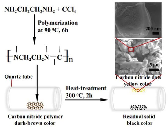
Figure 1.
Schematic illustration of the procedure for preparing carbon nitride dots (CNDs).
SEM observation reveals that the yellow product is composed of micro-grade particles (Figure 1). On higher magnification, these particles are seen to be formed via high packing of smaller particles with sizes of several tens of nanometers. However, the residual black solid has structure of a similar scale (Figure 2a), which shows a glabrous surface after magnification (Figure 2b). This structure may result from excessive cross-linking and ring-formation of carbon nitride polymer.
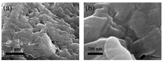
Figure 2.
Scanning electron microscope (SEM) images with different magnifications of the residual black solid at the bottom of the inner wall of the quartz tube. Low magnification (a), and high magnification (b).
TEM image of CNDs and their size distribution are shown in Figure 3a,b, respectively. The size of CNDs is mostly distributed in the range 23–70 nm and the average calculated diameter is ca. 44 nm, which is consistent with SEM observation. The height of CNDs was measured by AFM, as shown in Figure 3c–f. The synthesized CNDs have heights between 0.6 and 2.5 nm, indicating that they are composed of 1–4 layers of CN.
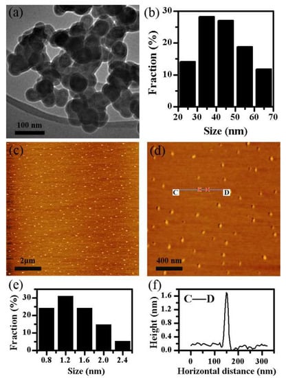
Figure 3.
TEM image (a), and the corresponding size distribution (b), of CNDs; AFM images of CNDs with different magnifications (c,d), and their height distribution (e) and height profile along the line CD (f).
Figure 4 shows the optical properties of CNDs aqueous dispersion. It is seen that there are two typical absorption peaks at 275 and 325 nm, attributed to π–π* transition of s-triazine and n–π* transition of defect state on the surface of the CNDs, respectively (Figure 4a) [17]. CNDs emit blue fluorescence at ca. 420 nm under the excitation of 360 nm irradiation (Figure 4b). Similar to most of carbon dots, the PL of the obtained CNDs is also excitation wavelength-dependent. The emission peak of CNDs is red-shifted from 375 to 450 nm with the excitation wavelength changing from 300 to 400 nm.

Figure 4.
Optical properties of CNDs aqueous dispersion. UV-vis absorption spectrum (a), and PL emission spectra (b).
4. Conclusions
CNDs with an average diameter of ca. 44 nm were synthesized via carbonization of a polymer precursor formed via the polymerization of EDA and CCl4. The prepared CNDs have sizes ranging from 23 to 70 nm and emit strong blue light under 360 nm excitation. In addition to their good solubility in water and polar organic solvents, these properties benefit the CNDs for further applications in bioimaging, fluorescence sensing, and optoelectronics devices. The method reported in this work for preparing CNDs is scalable and expands the size range of synthesized CNDs.
Author Contributions
P.L. designed the experiments; P.L. performed the experiments; P.L., Y.Y., and X.G. analyzed the data; P.L. wrote the paper.
Funding
This research was funded by the National Natural Science Foundation of China (51503036), Program for New Century Excellent Talents in Fujian Province University (Minjiaoke [2017] No. 52), Training Program for Distinguished Young Scholars in Fujian Province University (Minjiaoke [2016] No. 23), and Scientific and Technological Project of Fuzhou City (2017-G-97).
Conflicts of Interest
The authors declare no conflict of interest.
References
- Liu, S.; Tian, J.; Wang, L.; Luo, Y.; Sun, X. A general strategy for the production of photoluminescent carbon nitride dots from organic amines and their application as novel peroxidase-like catalysts for colormetric detection of H2O2 and glucose. RSC Adv. 2012, 2, 411–413. [Google Scholar] [CrossRef]
- Tang, Y.; Su, Y.; Yang, N.; Zhang, L.; Lv, Y. Carbon nitride quantum dots: A novel chemiluminescence system for selective detection of free chlorine in water. Anal. Chem. 2014, 86, 4528–4535. [Google Scholar] [CrossRef] [PubMed]
- Barman, S.; Sadhukhan, M. Facile bulk production of highly blue fluorescent graphitic carbon nitride quantum dots and their application as highly selective and sensitive sensors for the detection of mercuric and iodide ions in aqueous media. J. Mater. Chem. 2012, 22, 21832–21837. [Google Scholar] [CrossRef]
- Li, H.; Shao, F.Q.; Huang, H.; Feng, J.J.; Wang, A.J. Eco-friendly and rapid microwave synthesis of green fluorescent graphitic carbon nitride quantum dots for vitro bioimaging. Sensors Actuat. B Chem. 2016, 226, 506–511. [Google Scholar] [CrossRef]
- Guan, W.; Gu, W.; Ye, L.; Guo, C.; Su, S.; Xu, P.; Xue, M. Microwave-assisted polyol synthesis of carbon nitride dots from folic acid for cell imaging. Int. J. Nanomedicine 2014, 9, 5071–5078. [Google Scholar] [PubMed]
- Lu, Y.C.; Chen, J.; Wang, A.J.; Bao, N.; Feng, J.J.; Wang, W.; Shao, L. Facile synthesis of oxygen and sulfur co-doped graphitic carbon nitride fluorescent quantum dots and their application for mercury(II) detection and bioimaging. J. Mater. Chem. C 2015, 3, 73–78. [Google Scholar] [CrossRef]
- Lin, X.; Xu, D.; Zheng, J.; Song, M.; Che, G.; Wang, Y.; Yang, Y.; Liu, C.; Zhao, L.; Chang, L. Graphitic carbon nitride quantum dots loaded on leaf-like InVO4/BiVO4 nanoheterostructures with enhanced visible-light photocatalytic activity. J. Alloy. Compd. 2016, 688, 891–898. [Google Scholar] [CrossRef]
- Qin, X.; Liu, S.; Lu, W.; Li, H.; Chang, G.; Zhang, Y.; Tian, J.; Luo, Y.; Asiri, A.M.; Al-Youbi, A.O.; Sun, X. Submicrometre-scale polyaniline colloidal spheres: Photopolymerization preparation using fluorescent carbon nitride dots as a photocatalyst. Catal. Sci. Technol. 2012, 2, 711–714. [Google Scholar] [CrossRef]
- Liu, S.; Tian, J.; Wang, L.; Luo, Y.; Zhai, J.; Sun, X. Preparation of photoluminescent carbon nitride dots from CCl4 and 1, 2-ethylenediamine: A heat-treatment-based strategy. J. Mater. Chem. 2011, 21, 11726–11729. [Google Scholar] [CrossRef]
- Yu, Y.; Niu, X.; Zhang, H.; Xu, L.; Zhao, S.; Chen, X.; Chen, H. Switch-on fluorescence sensing of glutathione in food samples based on a g-CNQDs-Hg2+ chemosensor. J. Agric. Food Chem. 2015, 63, 1747–1755. [Google Scholar]
- Zhou, J.; Yang, Y.; Zhang, C. A low-temperature solid-phase method to synthesize highly fluorescent carbon nitride dots with tunable emission. Chem. Commun. 2013, 49, 8605–8607. [Google Scholar] [CrossRef] [PubMed]
- Liu, S.; Wang, L.; Tian, J.; Zhai, J.; Luo, Y.; Lu, W.; Sun, X. Acid-driven, microwave-assisted production of photoluminescent carbon nitride dots from N, N-dimethylformamide. RSC Adv. 2011, 1, 951–953. [Google Scholar] [CrossRef]
- Xiao, D.; Yuan, D.; He, H.; Lu, J. Microwave-assisted one-step green synthesis of amino-functionalized fluorescent carbon nitride dots from chitosan. Luminescence 2013, 28, 612–615. [Google Scholar] [CrossRef] [PubMed]
- Xiao, D.; Li, S.; Liu, S.; He, H.; Lu, J. One-step hydrothermal synthesis of photoluminescent carbon nitride dots derived from ionic liquid. New J. Chem. 2016, 40, 320–324. [Google Scholar] [CrossRef]
- Wang, X.; Wang, L.; Zhao, F.; Hu, C.; Zhao, Y.; Zhang, Z.; Chen, S.; Shi, G.; Qu, L. Monoatomic-thick graphitic carbon nitride dots on graphene sheets as an efficient catalyst in the oxygen reduction reaction. Nanoscale 2015, 7, 3035–3042. [Google Scholar] [CrossRef] [PubMed]
- Qiu, Y.; Gao, L. Chemical synthesis of turbostratic carbon nitride, containing C-N crystallites, at atmospheric pressure. Chem. Commun. 2003, 2378–2379. [Google Scholar] [CrossRef]
- Miller, D.R.; Wang, J.; Gillan, E.G. Rapid, facile synthesis of nitrogen-rich carbon nitride powders. J. Mater. Chem. 2002, 12, 2463–2469. [Google Scholar] [CrossRef]


© 2019 by the authors. Licensee MDPI, Basel, Switzerland. This article is an open access article distributed under the terms and conditions of the Creative Commons Attribution (CC BY) license (http://creativecommons.org/licenses/by/4.0/).