Article

Immediate Myofascial Responses to PFRT in Adolescent Endurance Runners: A Dorsal Chain Perspective


by
Kübra Sarıoğlu
* and
Volga Bayrakcı Tunay
Faculty of Physical Therapy and Rehabilitation, Hacettepe University, 06100 Ankara, Turkey
*
Author to whom correspondence should be addressed.
Appl. Sci. 2026, 16(2), 1017; https://doi.org/10.3390/app16021017
Submission received: 29 October 2025 / Revised: 14 January 2026 / Accepted: 15 January 2026 / Published: 19 January 2026
(This article belongs to the Special Issue Advanced Physical Therapy for Rehabilitation)

Featured Application

Regular self-massage under the foot may enhance hamstring and lumbar mobility due to the continuity of the dorsal kinetic chain. PFRT is more likely to improve flexibility and movement quality rather than explosive or strength-dependent per-formance. PFRT appeared to support posterior chain stabilization during recovery, but its effects were limited and not superior to natural recovery processes.

Abstract

Background: This study examined the acute effects of post-run plantar fascia recovery training (PFRT) on dorsal kinetic chain performance (DKCP) in adolescent long-distance runners. Methods: Thirty-four adolescent runners were randomly assigned to a PFRT group (n = 17) or a control group (n = 17). Following a standardized running session, the PFRT group received bilateral PFRT. Assessments were performed on the dominant side at three time points: pre-training, post-training, and post-PFRT. DKCP was evaluated using the Bunkie Test for the posterior stabilization line (PSL) and posterior power line (PPL), Myoton measurements of the latissimus dorsi, erector spinae, hamstrings, and gastrocnemius, the Sit-and-Reach Test for hamstring/lumbar flexibility, and the Modified Schober Test for lumbar mobility. Results: No significant group × time interactions were observed for any outcome except lumbar mobility. PSL performance increased significantly following PFRT compared with post-training (p = 0.016), whereas PPL performance did not change. Lumbar mobility improved significantly over time (p < 0.05). Although latissimus dorsi stiffness and hamstring and gastrocnemius stiffness were lower in the PFRT group at baseline, no significant within-group changes were observed following PFRT. Conclusions: PFRT may acutely improve lumbar mobility as a recovery intervention in adolescent runners. Further research is needed to clarify its short- and long-term effects within structured recovery programs during adolescence.

1. Introduction

The mechanical properties of the myofascia—tone, elasticity, and stiffness—play a crucial role in joint mobility, tissue nutrition, proprioception, and performance [1]. Self-myofascial release, commonly performed with foam rollers or massage balls, aims to reduce increased myofascial viscosity caused by elevated hyaluronic acid concentration and to enhance fascial fluid circulation. As a result, tone, elasticity, and stiffness may improve [2]. Excess hyaluronic acid can impair tissue gliding, restrict joint motion, and weaken motor control, which is why clinicians often recommend foam rolling to improve mobility, enhance performance, increase local blood flow, reduce delayed-onset muscle soreness (DOMS), and accelerate recovery both before and after exercise [3,4].
Fascia functions as a connective and communicative bridge between tissues, facilitating coordinated movement and force transmission [5,6,7]. Tissue excursion, defined as the gliding between adjacent fascial layers, is influenced by the viscoelastic properties of the extracellular matrix, in which hyaluronic acid plays a key role. Alterations in hyaluronic acid viscosity have been proposed to affect fascial sliding and tissue mobility [8,9]. Although direct evidence for acute changes in hyaluronic acid viscosity following low-intensity mechanical loading in humans is limited, experimental and theoretical models suggest that gentle, sustained loading may transiently modify fascial hydration and viscoelastic behavior, potentially improving tissue gliding [10,11]. Therefore, myofascial release techniques such as foam rolling are hypothesized to acutely influence fascial mechanics rather than induce structural changes.
Importantly, during adolescence the muscle–tendon–fascia system is exposed to rapid growth-related changes that may temporarily disrupt the balance between force production, tissue stiffness, and neuromuscular control. In this developmental phase, increases in muscle strength may not be matched by proportional adaptations in tendon and fascial stiffness, potentially altering load distribution and increasing reliance on compensatory muscle tone to maintain movement stability. As neuromuscular coordination and connective tissue properties are still maturing, even subtle impairments in fascial gliding or elastic energy transfer may have disproportionate effects on movement efficiency and mechanical loading patterns in young endurance athletes. Consequently, alterations in myofascial tone, elasticity, and stiffness may not only influence performance-related outcomes such as running economy but also contribute to an elevated susceptibility to overuse injuries during periods of high training volume [12,13,14,15,16].
The tensile load generated in the plantar fascia is transmitted as passive tension through serially connected collagenous tissues extending from the crural fascia to the hamstring fascia and ultimately to the thoracolumbar fascia. Low-friction gliding interfaces between these fascial layers facilitate efficient force propagation along the posterior chain. In parallel, mechanoreceptor activation within the plantar region elicits reflex-mediated modulation of muscle tone, reinforcing mechanical force transmission with a neurophysiological contribution [17]. Within this integrated system, the plantar fascia underpins two key functional mechanisms: the arch-spring and the windlass. During early stance, the arch-spring mechanism permits controlled elastic deformation of the medial longitudinal arch, allowing effective shock absorption and elastic energy storage [18]. In contrast, during terminal stance and push-off, dorsiflexion of the metatarsophalangeal joints activates the windlass mechanism, increasing arch stiffness and thereby enhancing force transmission and propulsive efficiency [18]. Through myofascial continuity with the Achilles tendon, the gastrocnemius–soleus complex, and the hamstrings, elastic energy stored at the plantar level can be transmitted proximally, supporting coordinated posterior chain activation and overall locomotor efficiency [19]. Functional impairments at the level of the plantar fascia may disrupt this force transmission, leading to compensatory movement strategies and reduced mechanical efficiency. Both contribute to efficient locomotion and are functionally integrated with the dorsal kinetic chain (DKC), extending through the superficial back line [18,19].
Evidence shows that local interventions on the plantar surface can temporarily improve flexibility in distant regions such as the hamstrings and lumbar spine, highlighting the fascial chain effect [20,21,22,23]. The plantar fascia also interacts with structures like the Achilles tendon, facilitating force transmission [21]. Yet, findings on self-massage effects remain mixed: while some report small reductions in strength or jump performance, others show no impact on fatigue or sprint ability [24,25].
It is still unclear whether plantar fascia self-release meaningfully alters force transmission along the superficial back line or improves DKC mobility [24,25]. Our study therefore aims to assess the acute effects of PFRT on DKC biomechanics by comparing pre-training, post-training, and post-PFRT measurements. Such insights may guide novel therapeutic strategies, optimize training, and support injury prevention.
Focusing on adolescent long-distance runners provides a unique contribution, as this population experiences substantial loading along the posterior kinetic chain and exhibits a heightened risk of overuse injuries. Adolescent fascia has been reported to exhibit higher water content and lower collagen cross-linking density, which may compromise optimal fascial gliding and elastic recoil under repetitive loading. Impairments in fascial sliding or elastic energy transfer can reduce the efficiency of elastic energy storage and return during the stretch–shortening cycle, thereby increasing the reliance on active muscle contraction to sustain movement. Over time, this compensatory strategy may impair movement economy, elevate local tissue stress, and increase the risk of overuse injuries, particularly during periods of high training volume. Notably, there is a striking lack of research investigating plantar-fascia–specific SMR interventions in this age group. By examining the acute effects of PFRT on posterior chain flexibility, lumbar and hamstring mobility, and functional performance parameters, the present study offers valuable insights into the functional implications of myofascial force transmission along the dorsal chain. Taken together, the study is expected to shed light on previously unexplored aspects of plantar-based myofascial interventions and contribute to a deeper understanding of chain-based recovery strategies in young endurance athletes.

2. Materials and Methods

The central research question explored whether PFRT, when applied bilaterally to the plantar surfaces of the lower extremities, produces acute effects on the functional performance of the DKC, specifically evaluated in the dominant leg. This assessment included examining the mechanical properties, such as muscle tone, stiffness, and elasticity, in muscles along the superficial back line and the functional back fascial line, namely the hamstrings, gastrocnemius, and latissimus dorsi. Therefore, the study tested the subsequent hypotheses:
H1. 
PFRT will acutely improve flexibility and posterior chain mobility in adolescent runners.
H2. 
PFRT will reduce muscle stiffness and tone more effectively than the control condition.
H3. 
PFRT will enhance posterior chain performance during recovery.
This study was designed as a randomized controlled trial with repeated measures over time, approved by the Hacettepe University Ethics Committee (FTREK 25_03), investigated the acute effects of PFRT on DKC performance in adolescent long-distance runners. Participants were randomly assigned to either a PFRT or control group. Inter-limb variability was controlled by assessing only the dominant limb. Assessments were limited to the dominant limb to reduce variability associated with habitual motor patterns and load distribution between limbs. While this approach may overlook potential asymmetrical responses, it improves interpretability of intervention effects in this preliminary study. Although measurements were repeated across three time points, participants were allocated to independent intervention and control groups; therefore, the design does not represent a within-subject comparison.
In total of 17 adolescent runners who received PFRT (mean ± SD, age, 14.65 ± 0.21 years; VKI, 18.83 ± 0.51 N/m2) and 17 adolescent runners in the control group (mean ± SD, age, 14.71 ± 0.34 years; VKI, 19.39 ± 0.42 N/m2) between 15 and 18 years participated in this study. Runners were recruited from various sports clubs through a social media announcement for this randomized controlled trial. The majority of participants in the study were adolescent long-distance runners from different clubs. A physiotherapist conducted evaluations at the athletes’ training grounds before training, after training, and following the recovery training session. Inclusion criteria included: being between 14 and 18 years old, participating as a long-distance runner, running a weekly distance between 30–60 km, and having at least 2 years of regular running training experience. Exclusion criteria included: having any chronic illness or a history of lower extremity and/or spinal injury in the past 6 months, no female athlete who was in her menstrual cycle and participants who had to interrupt or prematurely stop their training session were excluded from the study.
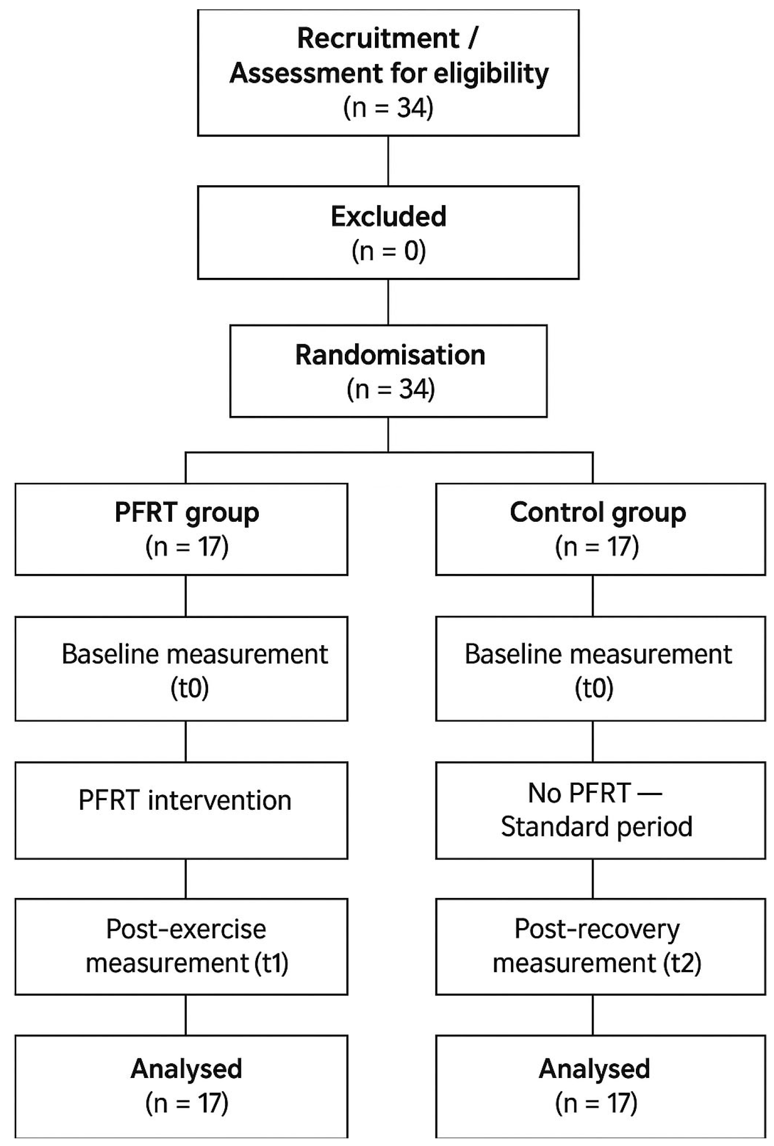
Participants provided demographic and training data and were instructed to avoid caffeine and meals 2 h before testing. Assessments (Bunkie Test, MyotonPRO, sit-and-reach, modified Schober) were performed pre-training, post-training, and post-intervention. Interventions were bilateral, but data were recorded on the dominant limb. A total of 34 athletes were randomized into either the intervention group (PFRT) or the control group (no intervention). Each assessment lasted ~10 min. The PFRT group completed post-training evaluation, a 10 min recovery intervention, and post-intervention assessment within ~30 min. The control group was evaluated at the same time points (immediately post-training and at the 20th minute). Interventions were applied bilaterally, but assessments were performed unilaterally on the dominant leg. All procedures were administered or supervised by a physical therapist following established protocols [24]. The schematic representation of the study design is shown in Figure 1.
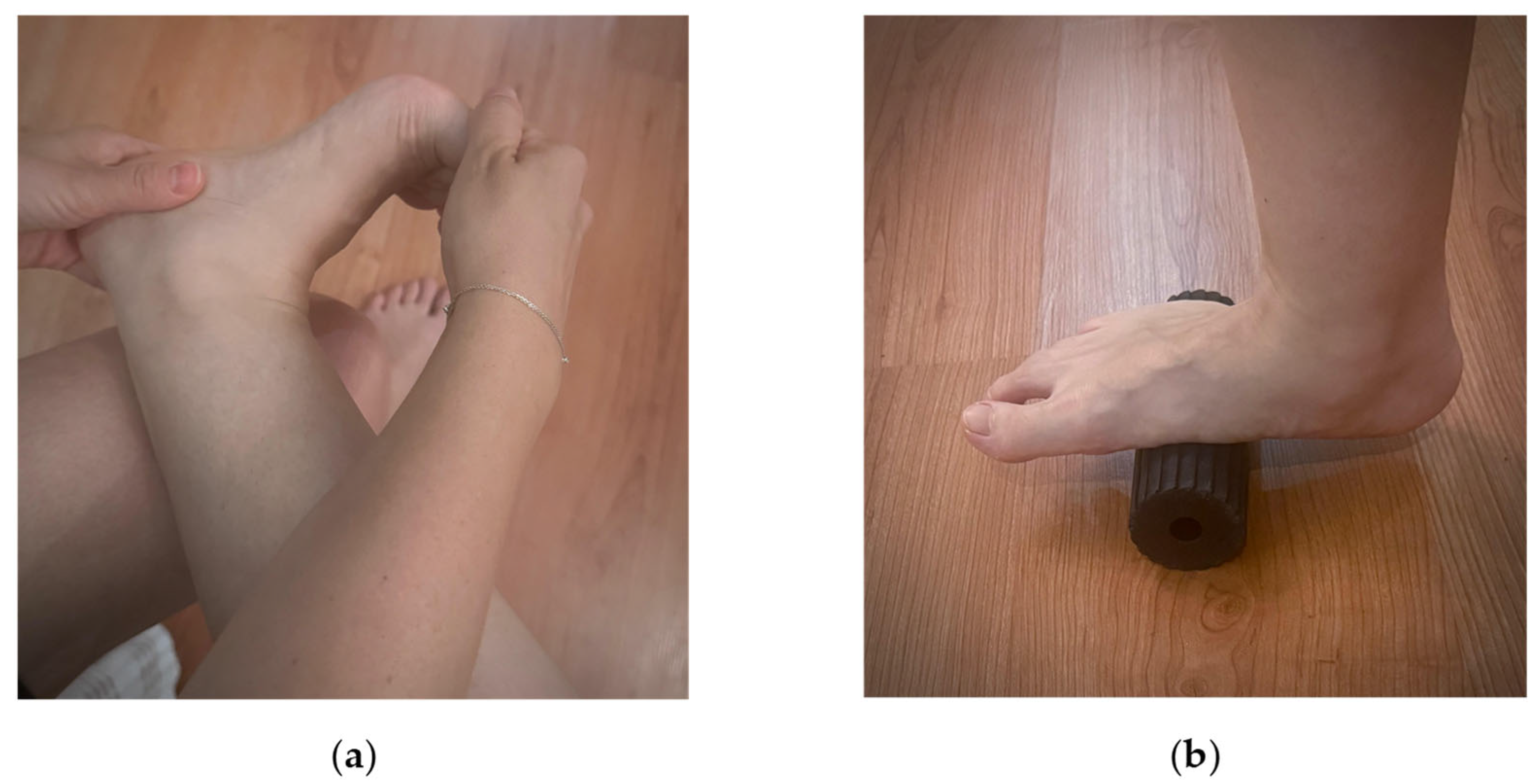
Metatarsophalangeal Joint Stretching: In a seated position, the dominant leg was crossed over the non-dominant. The heel was stabilized with one hand while passive dorsiflexion was applied to the metatarsophalangeal joints. Two sets of 30 s were performed with 10 s rest (Figure 2a) [24].
Foam roller: Participants performed a 4 min foam rolling session along the plantar fascia using a small roller (foam rolling, diameter: 5 cm; Young’s modulus: 80 MPa; Blackroll®, Bottighofen, Switzerland). Rolling was at a self-selected pace, with wall support allowed. Short rests were permitted if needed (Figure 2b) [24,26].
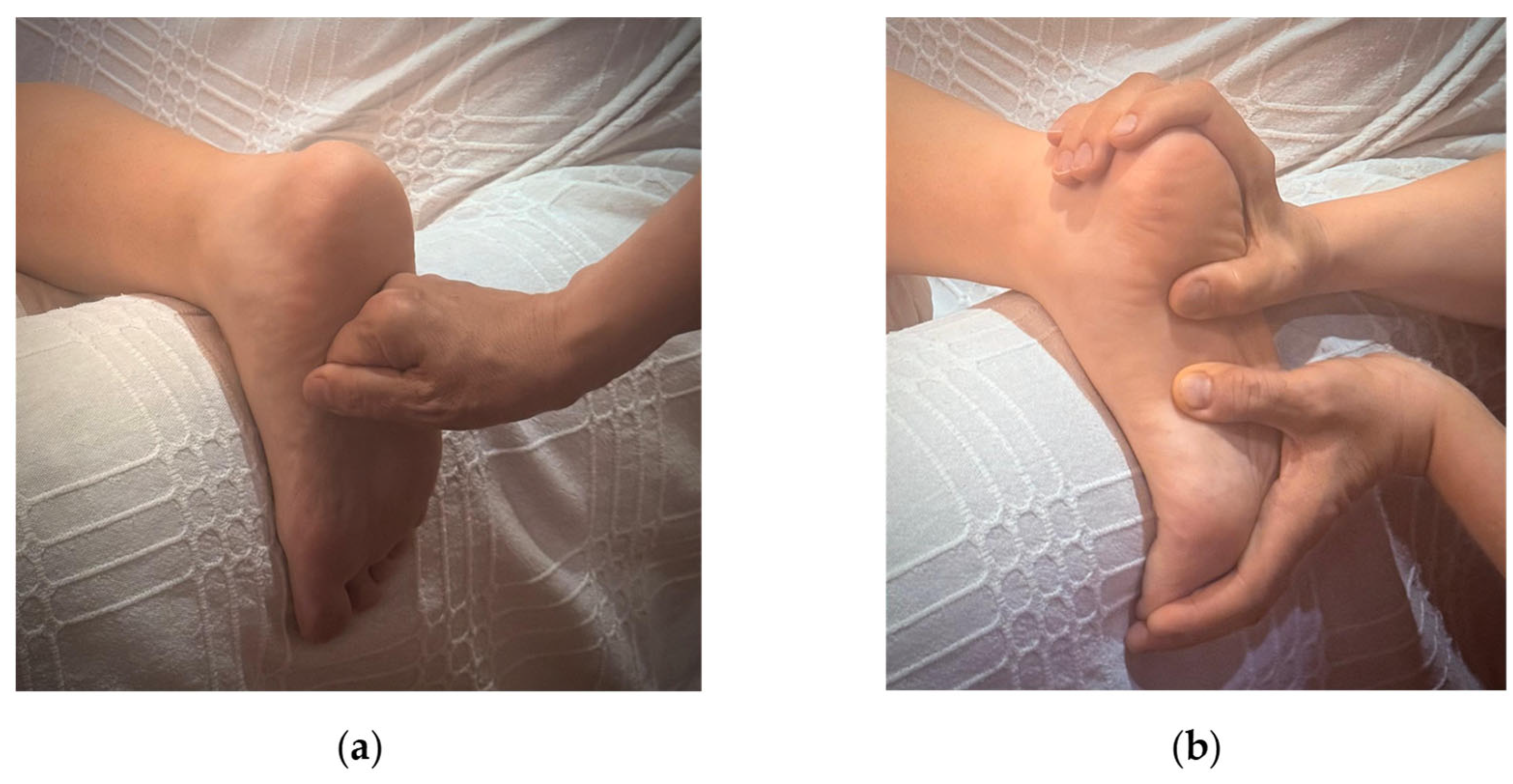
Soft tissue mobilization: Manual pressure was applied to the plantar fascia using knuckles or thumb, gliding from heel to forefoot. Two sets of 60 s with 10 s rest were performed (Figure 3a).
Gliding massage: With toes in passive hyper-dorsiflexion (focus on hallux), sustained pressure was applied with the reinforced thumb or index finger along the medial PF band from calcaneus to forefoot for 2 min (Figure 3b) [24].
Muscle flexibility, stiffness, and elasticity were objectively assessed using the MyotonPRO device (Myoton AS, Tallinn, Estonia). This portable, non-invasive tool delivers a brief mechanical impulse to the skin, eliciting oscillations in the underlying muscle, and calculates stiffness, elasticity, and tone. Each site was measured ten times; mean values were analyzed [27]. Participants lay in a prone position with a towel placed under the forehead and the arms positioned in 180° of shoulder flexion. After a 15 min rest period, a point located 5 cm below the inferior angle of the scapula was marked, and ten consecutive measurements were collected from this site. The latissimus dorsi was assessed because it is part of the Functional Back Fascial Line relevant to running mechanics. The erector spinae measurements were obtained in the same prone position. The probe was placed over the L3 level to record muscle tone and stiffness. For the hamstring assessment, participants remained prone with the knee positioned at approximately 30° of flexion. The midpoint between the ischial tuberosity and the fibular head was marked, and the probe was applied perpendicularly following a 15 min rest period. Gastrocnemius measurements were taken with the participant lying prone, the knee fully extended, and the feet relaxed. The probe was positioned over the medial head of the muscle at the midpoint between the knee joint line and the Achilles tendon [27,28].
The sit-and-reach test was used to assess hamstring extensibility. Participants sat on the floor with their legs fully extended and feet placed against the measurement box. They then reached forward as far as possible without bending their knees. The reach distance (cm) was recorded as an indicator of posterior chain flexibility [29].
Lumbar mobility was assessed using the Modified Schober test. The PSIS level was identified, and a midpoint was marked, followed by additional marks 10 cm above and 5 cm below. Participants performed maximal trunk flexion, after which the distance between the marks was remeasured. An increase of less than 5 cm was interpreted as reduced lumbar mobility [30].
To evaluate functional force generation in the superficial back line (SBL), the Bunkie Test was applied. Although validity and reliability are limited, it is widely used in clinical practice. Van Pletzen and Venter report strong comparability with sprint and jump tests, making it a practical tool for identifying chain imbalances. The Posterior Power Line test was performed with the participant in a supine position, elbows placed under the shoulders and heels positioned on a 30 cm bench while maintaining a neutral pelvic alignment. The non-dominant leg was lifted approximately 20 cm. The test was terminated if the participant experienced pain, cramping, voluntary fatigue, loss of proper posture, or upon reaching 40 s, which is considered the optimal cutoff for athletes. One posture correction was permitted; however, failure to return to the correct position resulted in test termination. After a 40 s rest, the same setup was repeated for the Posterior Stabilizing Line test, but this time with the knees flexed to 90° while the non-dominant foot was elevated approximately 20 cm. The same termination criteria—pain, cramping, voluntary fatigue, posture loss, or inability to continue—were applied [31].
The sit-and-reach test and the Modified Schober test require bilateral assessment, the intervention was applied bilaterally. Additionally, rather than randomizing the treated limb, we standardized the procedure by applying the intervention to the dominant leg due to potential differences in muscular and fascial tissue characteristics between limbs [32].

2.1. Sample Size

The sample size was calculated using G*Power 3.1. Effect size estimates were derived from previous research investigating myofascial interventions within the posterior chain. Specifically, Wilke et al. reported a large effect size (Cohen’s d = 2.16) following plantar fascia manipulation, while Cruz-Montecinos et al. demonstrated a moderate association (r = 0.449) between fascial continuity and functional outcomes. Based on these prior findings and methodological consistency with similar PFRT protocols, a large effect size (d = 0.8) was adopted for the power analysis in the present study [33]. However, given the adolescent population and the acute nature of the intervention, the assumed large effect size may have been optimistic. If the true effect is smaller, the present study may be underpowered to detect clinically relevant but subtle changes, increasing the risk of type II error. Nevertheless, the sample size was consistent with previous acute myofascial intervention studies and sufficient to detect large effects.

2.2. Statistical Analysis

Based on the power analysis, 34 adolescent long-distance runners were included, divided into an intervention group (n = 17) that received plantar fascia training and a control group (n = 17) that continued standard training. Analyses were conducted using IBM Corp (2015) IBM SPSS Statistics for Windows, Version 23.0, IBM Corp, Armonk, NY, USA. Chi-square tests were used to examine group differences in categorical variables (e.g., gender, dominant side), while Student’s t-test or Mann–Whitney U test was used to compare continuous variables depending on normality. For dependent variables, a two-way mixed ANOVA was performed to assess the effects of group, time, and group × time interaction, with Bonferroni-adjusted post hoc tests applied for multiple comparisons. Statistical significance was set at p < 0.05.
Generative AI tools (GPT-5.2, OpenAI) were used solely for minor language editing and did not contribute to content generation or data analysis.

3. Results

3.1. Demographic Characteristics

Thirty-four participants were included in the study, with 17 runners in each group. All were predominantly right-dominant (PFRT: 100%, control: 94.1%). Demographic characteristics are shown in Table 1.

3.2. Bunkie Tests: PSL, PPL

For the Posterior Stabilization Line (PSL), a significant main effect of time was observed (p = 0.016), indicating changes across the recovery period. However, no significant group (p = 0.994) or group × time interaction effects were found (p = 0.996).
For the Posterior Power Line (PPL), no significant main effects of time (p = 0.113), group (p = 0.584), or group × time interaction (p = 0.708) were detected. (Table 2).

3.3. Flexibility Tests: Modified Schober Test, Sit and Reach Test

In the sit-and-reach test, a significant time-dependent improvement was observed, with both post-exercise and post-PFRT measurements showing higher flexibility values compared to pre-exercise (p = 0.001). A significant difference was also found between the groups, with the PFRT group demonstrating greater flexibility gains (p = 0.008). However, no significant group × time interaction was detected. These findings suggest that the flexibility advantages provided by the intervention continued after the application; nevertheless, additional studies are required to more clearly determine the effectiveness of the intervention.
For the modified Schober test, a significant group × time interaction was observed (p = 0.029). In the PFRT group, the reductions in lumbar and hamstring mobility seen after exercise increased again following the recovery period. All results are shown in Table 2.

3.4. Myotonometry Assessment

Several Myoton-derived parameters differed between groups at baseline, indicating pre-existing differences that were independent of the intervention. Regarding the erector spinae muscle, a significant time-dependent decrease in mean muscle tone was detected after exercise (p = 0.018). Additionally, the PFRT group exhibited higher tone values than the control group, resulting in a significant between-group difference (p = 0.013). However, PFRT did not provide an additional recovery-enhancing effect after exercise, and no significant group × time interaction was observed. Although improvements in erector spinae elasticity (reflected by lower numeric stiffness values) were noted after exercise and intervention, PFRT did not show a distinct effect on post-exercise values. Likewise, no significant group × time interaction was found. Erector spinae stiffness values changed over time, with both post-exercise and post-PFRT measurements being lower compared to pre-exercise values (p = 0.08). However, there was no significant group × time interaction.
For hamstring muscle stiffness, a significant between-group difference was detected (p < 0.001), but no significant change was observed following recovery. Furthermore, no group × time interactions were identified for either the gastrocnemius or hamstring muscles.
For the latissimus dorsi, no significant time-dependent differences were observed between the groups, although a significant between-group difference existed (dominant LD p = 0.001; nondominant LD p = 0.001). However, this difference was not attributable to a group × time interaction following PFRT. The results are shown in Table 3 and Table 4.

4. Discussion

The present study investigated the acute effects of PFRT on flexibility, neuromuscular properties, and posterior chain performance in adolescent distance runners. Overall, the findings provide partial support for the proposed hypotheses and demonstrate that the effects of PFRT during short-term recovery are task-specific and limited in magnitude.

4.1. Flexibility and Posterior Chain Mobility

The first hypothesis was partially supported. PFRT resulted in a significant group × time interaction in the Modified Schober Test, indicating a PFRT-specific improvement in lumbar mobility during the recovery period. This finding suggests that plantar fascia-focused interventions can acutely influence spinal and proximal posterior chain mobility in adolescent runners. Improvements in sit-and-reach performance were observed over time in both groups, but without significant group × time interaction, indicating that these changes were likely due to general post-exercise recovery rather than PFRT. Taken together, these findings indicate that PFRT exerted a localized effect primarily on lumbar mobility, while most other outcomes—including general flexibility, muscle tone, stiffness, and power-oriented posterior chain performance—did not demonstrate significant PFRT-specific effects.
The observed lumbar mobility improvements may be interpreted within the framework of myofascial continuity and neurophysiological modulation. According to Myers’ fascial line model, the plantar fascia represents the distal component of the Superficial Back Line, a continuous myofascial pathway extending through the Achilles tendon, gastrocnemius, hamstrings, sacrotuberous ligament, and thoracolumbar fascia to the cranial region [34]. Mechanical stimulation applied to the plantar surface may therefore propagate along this pathway, altering tension distribution and stiffness in more proximal tissues [35]. Additionally, the plantar surface contains a high density of mechanoreceptors, and pressure-based myofascial techniques may induce transient neuromodulatory effects that reduce muscle tone and increase stretch tolerance in the hamstrings and lumbar extensors [36,37]. These combined mechanisms provide a plausible explanation for the PFRT-specific lumbar mobility gains observed in the present study.

4.2. Neuromuscular Properties (Tone, Stiffness, and Elasticity)

The second hypothesis was not supported. Although temporal changes in muscle tone, stiffness, and elasticity were observed across multiple posterior chain muscles—including the erector spinae, hamstrings, gastrocnemius, and latissimus dorsi—no significant group × time interactions were detected. These findings indicate that the observed neuromuscular changes were more likely associated with natural post-exercise recovery processes rather than with a PFRT-specific effect. While the PFRT group demonstrated slightly more favorable directional trends in some parameters, these changes did not reach statistical significance and should therefore be interpreted cautiously.
These results are consistent with previous studies reporting that self-myofascial release interventions can improve flexibility without producing immediate reductions in muscle stiffness or tone beyond those associated with recovery alone [38,39,40,41]. Proposed mechanisms such as thixotropic changes in fascial tissues, temporary reductions in hyaluronic acid viscosity, and modulation of fascial sensory receptors may contribute to short-term alterations in tissue behavior [38,42]. However, direct in vivo evidence demonstrating acute changes in hyaluronan viscosity in human fascial tissues remains limited, precluding definitive mechanistic conclusions.

4.3. Posterior Chain Performance During Recovery

The third hypothesis was partially supported. Posterior chain performance, as assessed by the Posterior Stabilization Line (PSL), demonstrated a significant main effect of time, indicating a transient preservation of isometric stabilization capacity during the recovery period. However, the absence of a significant group or group × time interaction suggests that this effect was not specific to the PFRT intervention. In contrast, did not show significant effects, indicating that PFRT did not acutely enhance power-oriented posterior chain function. Observed stabilization benefits (PSL) were time-dependent but not intervention-specific.
This selective response may be explained by the distinct neuromuscular demands of the two tasks. PSL performance primarily reflects isometric stabilization endurance and neuromuscular control, whereas PPL performance relies more heavily on force and power production. Previous research has demonstrated that plantar-surface interventions can elicit non-local effects through both mechanical continuity within fascial networks and neurophysiological modulation of sensory–motor pathways [42,43,44]. However, these mechanisms appear more likely to influence stabilization-related functions rather than power-oriented performance during the acute recovery phase.
We measured outcomes on the dominant limb only because previous research has shown that limb dominance classification methods are not associated with consistent inter-limb asymmetries during change in direction tasks, suggesting that bilateral differences are minimal and dominant-limb measurements are representative for this task [45]. The Bunkie Test and other neuromuscular measures (tone, stiffness, elasticity) did not show significant group × time interactions. Any apparent PFRT-related trends should therefore be interpreted cautiously, as they likely reflect natural recovery rather than a true intervention effect. Additionally, assessments were restricted to the dominant limb. While this approach reduces variability associated with habitual motor patterns and load distribution, improving the interpretability of intervention effects, it may overlook potential asymmetrical responses. Future studies should consider using more sensitive assessment tools and incorporating bilateral evaluations to better detect subtle changes and inter-limb differences.

4.4. Hydration, Hyaluronan, and Fascial Gliding

Hydration status differed between groups, with higher fluid intake observed in the control group; however, these differences were not associated with consistent changes in muscle tone, stiffness, or elasticity. Hyaluronan plays a key role in regulating tissue viscosity and fascial gliding by binding water molecules within the extracellular matrix [10]. Under conditions of reduced hydration, hyaluronan becomes more viscous, potentially impairing fascial sliding and increasing resistance to movement. Nevertheless, the effects of hyaluronan are generally considered local and transient, and direct in vivo evidence demonstrating acute changes in hyaluronan viscosity following manual or exercise-based interventions remains scarce. In the present study, hydration did not appear to meaningfully interact with PFRT-induced adaptations. Further research is warranted to clarify the combined influence of hydration status and myofascial recovery strategies [10,46].
Although plantar foot characteristics are known to influence lower-limb biomechanics, existing evidence suggests that modifying foot structures—particularly through interventions such as orthoses—does not consistently translate into improvements in athletic or motor performance outcomes, including sprinting, jumping, or balance [47]. These findings align with the absence of PFRT-specific effects on power-oriented posterior chain performance in the present study.
Conversely, accumulating evidence supports the role of the plantar fascia as an integral component of the global myofascial network [48,49,50]. Within this framework, distal plantar stimulation may elicit non-local adaptations along the posterior kinetic chain, primarily affecting mobility and stabilization rather than power production. The present findings are consistent with this perspective, demonstrating localized improvements in lumbar mobility and transient preservation of stabilization capacity without corresponding enhancements in power-related performance.
Although hydration status differed between groups, these differences did not appear to significantly interact with PFRT-induced adaptations. Future studies could standardize hydration or assess extracellular matrix factors to clarify their potential influence on myofascial recovery outcomes.

4.5. Practical Implications for Adolescent Distance Runners

Adolescence is characterized by rapid growth-related changes in the muscle–tendon–fascia system, which may temporarily disrupt neuromuscular coordination and load distribution. Importantly, during adolescence, increases in muscle strength may not be matched by proportional adaptations in tendon and fascial stiffness, potentially altering load distribution and increasing reliance on compensatory muscle tone [12,13,15,17]. Even subtle impairments in fascial gliding or elastic energy transfer may disproportionately affect movement efficiency and mechanical loading patterns in young endurance athletes, increasing susceptibility to overuse injuries [8,10]. Within this developmental context, recovery strategies that support mobility and stabilization may be particularly relevant. The present findings suggest that PFRT may serve as a practical adjunct recovery strategy for adolescent runners, particularly for preserving lumbar mobility and stabilization capacity during short-term recovery periods. However, PFRT should not be expected to acutely enhance neuromuscular stiffness modulation or power-oriented posterior chain performance.
Given the acute and short-term nature of the intervention, observed changes may reflect transient adaptations rather than lasting modifications in neuromuscular properties. Longitudinal studies are warranted to determine the sustained effects of PFRT in adolescent runners.

4.6. Limitations

Several methodological considerations should be acknowledged. Measurements were taken only from the dominant side, which may have overlooked potential asymmetrical responses. Given that bilateral adaptations can differ and that measurements from both sides are often correlated, future studies should incorporate bilateral assessments to enhance methodological robustness and generalizability. Foot deformities were not evaluated in the present study, which may have influenced mobility and posterior chain responses. Fluid intake was based on participants’ self-reports; objective measures such as urine specific gravity, osmolality, or bioelectrical impedance analysis were not performed, potentially limiting the accuracy of hydration assessment. Additionally, the inverse interpretation of the logarithmic decrement as an indicator of “elasticity” may complicate understanding of the mechanical properties measured. Finally, fascial flexibility was assessed without the support of imaging techniques such as ultrasound elastography or magnetic resonance imaging, which may restrict the precision of these findings. Collectively, these limitations should be considered when interpreting the acute effects of plantar fascia-focused recovery interventions in adolescent distance runners. Taken together, these methodological constraints limit causal inference and underscore the exploratory nature of the present findings. Given the acute effect of the intervention and the adolescent population, the true effect size may be smaller, potentially reducing statistical power for detecting subtle changes.

5. Conclusions

This study suggests that PFRT may provide modest acute support for flexibility and lumbar mobility during post-exercise recovery in adolescent distance runners. Given the rapid developmental changes in the adolescent muscle–tendon–fascia system, even modest improvements in lumbar and posterior thigh mobility may be particularly meaningful for supporting movement efficiency and reducing injury risk in this population. Improvements in sit-and-reach performance were time-dependent and likely reflect general recovery rather than a PFRT-specific effect. In contrast, lumbar mobility showed a clearer contribution of PFRT, indicating a potential role in supporting recovery of the dorsal kinetic chain following exercise-induced reductions. PFRT did not confer additional benefits for muscle stiffness, muscle tone, or power-oriented posterior chain performance compared with natural recovery alone. Although posterior stabilization capacity demonstrated time-dependent preservation, this effect was not intervention-specific. Overall, PFRT provided modest, task-specific support for lumbar mobility and proximal posterior chain stabilization during recovery. Most other outcomes did not demonstrate PFRT-specific effects, highlighting the limited and localized nature of acute intervention effects. Further studies with larger samples and longer intervention periods are needed to clarify the clinical relevance of PFRT in youth endurance athletes.

Author Contributions

Author Contributions: Conceptualization, K.S. and V.B.T.; methodology, K.S.; software, K.S.; validation, K.S.; formal analysis, K.S.; investigation, K.S.; resources and data curation, K.S.; writing—original draft preparation—review and editing, K.S.; visualization, V.B.T. and K.S.; project administration, V.B.T.; funding acquisition, K.S. All authors have read and agreed to the published version of the manuscript.

Funding

This research received no external funding.

Institutional Review Board Statement

Ethics Approval and Consent: The study was approved by ‘Non-invasive Clinical Research Ethics Committee of Hacettepe University’ (Project no: FTREK25_03, Evaluation date: 24 April 2025, Decision no: FTREK 25/03, Clinical Trial No: NCT07164716). An ‘Informed Consent Form’ was signed and participation approval was obtained from all participants. The standards are in accordance with the Declaration of Helsinki.

Informed Consent Statement

Informed consent was obtained from all subjects involved in the study. Written informed consent has been obtained from the individuals who will publish this paper.

Data Availability Statement

The datasets used and/or analyzed during the current study are available from the corresponding author on reasonable request.

Acknowledgments

The experiments comply with the current laws of the country in which they were performed. The authors have no conflict of interest to declare. The datasets generated during and/or analyzed during the current study are not publicly available but are available from the corresponding author, who organized the study.

Conflicts of Interest

The authors declare no conflicts of interest.

Abbreviations

The following abbreviations are used in this manuscript:
LDLatissimus Dorsi
ROMRange of motion
TLThoracolumbar
TLFThoracolumbar fascia
PPLPosterior power line
PSLPosterior stabilizing line
DKCDorsal kinetic chain
PFRTPlantar fascia recovery training
DDominant

References

  1. Masi, A.T.; Hannon, J.C. Human resting muscle tone (HRMT): Narrative introduction and modern concepts. J. Bodyw. Mov. Ther. 2008, 12, 320–332. [Google Scholar] [CrossRef] [Scilit] [PubMed]
  2. Wajswelner, H. Clinical application of neuromuscular techniques: Volume 1, the upper body. Br. J. Sports Med. 2005, 39, 183. [Google Scholar] [CrossRef] [Scilit]
  3. Cheatham, S.W.; Kolber, M.J.; Cain, M.; Lee, M. The effects of self-myofascial release using a foam roll or roller massager on joint range of motion, muscle recovery, and performance: A systematic review. Int. J. Sports Phys. Ther. 2015, 10, 827–838. [Google Scholar]
  4. Hughes, G.A.; Ramer, L.M. Duration of myofascial rolling for optimal recovery, range of motion, and performance: A systematic review. Int. J. Sports Phys. Ther. 2019, 14, 845–859. [Google Scholar] [CrossRef] [Scilit] [PubMed]
  5. Adstrum, S.; Hedley, G.; Schleip, R.; Stecco, C.; Yucesoy, C.A. Defining the fascial system. J. Bodyw. Mov. Ther. 2017, 21, 173–177. [Google Scholar] [CrossRef] [Scilit]
  6. Schleip, R.; Hedley, G.; Yucesoy, C.A. Fascial nomenclature: Update on related consensus process. Clin. Anat. 2019, 32, 929–933. [Google Scholar] [CrossRef] [Scilit]
  7. Ajimsha, M.S.; Al-Mudahka, N.R.; Al-Madzhar, J.A. Effectiveness of myofascial release: Systematic review of randomized controlled trials. J. Bodyw. Mov. Ther. 2015, 19, 102–112. [Google Scholar] [CrossRef] [Scilit]
  8. Stecco, C.; Stern, R.; Porzionato, A.; Macchi, V.; Masiero, S.; Stecco, A.; De Caro, R. Hyaluronan within fascia in the etiology of myofascial pain. Surg. Radiol. Anat. 2011, 33, 891–896. [Google Scholar] [CrossRef] [Scilit]
  9. Stecco, C.; Pirri, C.; Fede, C.; Fan, C.; Giordani, F.; Stecco, A.; De Caro, R. Fascial components of the myofascial pain syndrome. Curr. Pain Headache Rep. 2014, 18, 352. [Google Scholar] [CrossRef] [Scilit]
  10. Cowman, M.K.; Schmidt, T.A.; Raghavan, P.; Stecco, A. Viscoelastic properties of hyaluronan in physiological conditions. F1000Research 2015, 4, 622. [Google Scholar] [CrossRef] [Scilit]
  11. Schleip, R.; Müller, D.G. Training principles for fascial connective tissues: Scientific foundation and suggested practical applications. J. Bodyw. Mov. Ther. 2013, 17, 103–115. [Google Scholar] [CrossRef] [Scilit]
  12. Mersmann, F.; Bohm, S.; Arampatzis, A. Imbalances in the development of muscle and tendon as risk factor for tendinopathies in youth athletes. Scand. J. Med. Sci. Sports 2017, 27, 452–461. [Google Scholar] [CrossRef] [Scilit]
  13. Radnor, J.M.; Oliver, J.L.; Waugh, C.M.; Myer, G.D.; Moore, I.S.; Lloyd, R.S. The influence of growth and maturation on stretch–shortening cycle function in youth. Sports Med. 2018, 48, 57–71. [Google Scholar] [CrossRef] [Scilit] [PubMed]
  14. Kubo, K.; Ikebukuro, T.; Yata, H.; Tomita, M.; Okada, M. Morphological and mechanical properties of muscle and tendon in highly trained sprinters. J. Appl. Physiol. 2011, 110, 528–535. [Google Scholar] [CrossRef] [Scilit] [PubMed]
  15. van der Sluis, A.; Elferink-Gemser, M.T.; Brink, M.S.; Visscher, C. Importance of peak height velocity timing in terms of injuries in talented youth soccer players. Int. J. Sports Med. 2015, 36, 327–332. [Google Scholar] [CrossRef] [Scilit]
  16. Arampatzis, A.; Schade, F.; Walsh, M.; Brüggemann, G.-P. Influence of leg stiffness and its effect on myodynamic jumping performance. J. Electromyogr. Kinesiol. 2001, 11, 355–364. [Google Scholar] [CrossRef] [Scilit]
  17. Yucesoy, C.A. Epimuscular myofascial force transmission implies novel principles for muscular mechanics. Exerc. Sport Sci. Rev. 2010, 38, 128–134. [Google Scholar] [CrossRef] [Scilit]
  18. Welte, L.; Kelly, L.A.; Lichtwark, G.A.; Rainbow, M.J. Influence of the windlass mechanism on arch-spring mechanics during dynamic foot arch deformation. J. R. Soc. Interface 2018, 15, 20180270. [Google Scholar] [CrossRef] [Scilit]
  19. Zwirner, J.; Zhang, M.; Ondruschka, B.; Akita, K.; Hammer, N. An ossifying bridge—On the structural continuity between the Achilles tendon and the plantar fascia. Sci. Rep. 2020, 10, 14523. [Google Scholar] [CrossRef] [Scilit]
  20. Welte, L.; Kelly, L.A.; Kessler, S.E.; Lieberman, D.E.; D’Andrea, S.E.; Lichtwark, G.A.; Rainbow, M.J. The extensibility of the plantar fascia influences the windlass mechanism during human running. Proc. Biol. Sci. 2021, 288, 20202095. [Google Scholar] [CrossRef] [Scilit]
  21. Krause, F.; Wilke, J.; Vogt, L.; Banzer, W. Intermuscular force transmission along myofascial chains: A systematic review. J. Anat. 2016, 228, 910–918. [Google Scholar] [CrossRef] [Scilit]
  22. Behara, B.; Jacobson, B.H. Acute effects of deep tissue foam rolling and dynamic stretching on muscular strength, power, and flexibility in Division I linemen. J. Strength Cond. Res. 2017, 31, 888–892. [Google Scholar] [CrossRef] [Scilit] [PubMed]
  23. Monteiro, E.R.; Vigotsky, A.; Škarabot, J.; Brown, A.F.; Fiuza, A.G.F.M.; Gomes, T.M.; Halperin, I.; da Silva Novaes, J. Acute effects of different foam rolling volumes in the interset rest period on maximum repetition performance. Hong Kong Physiother. J. 2017, 36, 57–62. [Google Scholar] [CrossRef] [Scilit]
  24. Gabriel, A.; Konrad, A.; Roidl, A.; Queisser, J.; Schleip, R.; Horstmann, T.; Pohl, T. Myofascial treatment techniques on the plantar surface influence functional performance in the dorsal kinetic chain. J. Sports Sci. Med. 2022, 21, 13–22. [Google Scholar] [CrossRef] [Scilit] [PubMed]
  25. Wiewelhove, T.; Döweling, A.; Schneider, C.; Hottenrott, L.; Meyer, T.; Kellmann, M.; Pfeiffer, M.; Ferrauti, A. A meta-analysis of the effects of foam rolling on performance and recovery. Front. Physiol. 2019, 10, 376. [Google Scholar] [CrossRef] [Scilit]
  26. Lu, J.; Lv, X.; Liu, J.; Liu, H.; Huang, T. Fully Flexible Wearable Pouch Pneumatic Artificial Muscle Based on Asymmetric Expansion. Chin. J. Mech. Eng. 2026, 39, 100085. [Google Scholar]
  27. Sawilowsky, S.S. New effect size rules of thumb. J. Mod. Appl. Stat. Methods 2009, 8, 26. [Google Scholar] [CrossRef] [Scilit]
  28. Uysal, Ö.; Delioğlu, K.; Fırat, T. The effects of hamstring training methods on muscle viscoelastic properties in healthy young individuals. Scand. J. Med. Sci. Sports 2021, 31, 371–379. [Google Scholar] [CrossRef] [Scilit]
  29. Mayorga-Vega, D.; Merino-Marban, R.; Viciana, J. Criterion-related validity of sit-and-reach tests for estimating hamstring and lumbar extensibility: A meta-analysis. J. Sports Sci. 2014, 32, 1083–1096. [Google Scholar] [CrossRef] [Scilit]
  30. Wells, K.F.; Dillon, E.K. The sit and reach—A test of back and leg flexibility. Res. Q. Am. Assoc. Health Phys. Educ. Recreat. 1952, 23, 115–118. [Google Scholar] [CrossRef] [Scilit]
  31. Van Pletzen, D.; Venter, R. The relationship between the Bunkie test and physical performance in rugby union players. J. Sports Sci. Med. 2012, 11, 693–699. [Google Scholar] [CrossRef] [Scilit]
  32. van Melick, N.; Meddeler, B.M.; Hoogeboom, T.J.; Nijhuis-van der Sanden, M.W.G.; van Cingel, R.E.H. How to determine leg dominance: Agreement between self-reported and observed performance in healthy adults. PLoS ONE 2017, 12, e0189876. [Google Scholar] [CrossRef] [Scilit]
  33. Wilke, J.; Debelle, H.; Tenberg, S.; Dilley, A.; Maganaris, C. Ankle motion is associated with soft tissue displacement in the dorsal thigh: An in vivo investigation suggesting myofascial force transmission across the knee joint. Front. Physiol. 2020, 11, 180. [Google Scholar] [CrossRef] [Scilit]
  34. Myers, T.W. Anatomy Trains: Myofascial Meridians for Manual and Movement Therapists; Elsevier: London, UK, 2009. [Google Scholar]
  35. Wilke, J.; Krause, F.; Vogt, L.; Banzer, W. What is evidence-based about myofascial chains? A systematic review. Arch. Phys. Med. Rehabil. 2016, 97, 454–461. [Google Scholar] [CrossRef] [Scilit]
  36. Aboodarda, S.J.; Spence, A.J.; Button, D.C. Pain-pressure threshold of a muscle tender spot increases following local and remote myofascial rolling. J. Bodyw. Mov. Ther. 2015, 19, 450–456. [Google Scholar]
  37. Schleip, R.; Gabbiani, G.; Wilke, J.; Naylor, I.; Hinz, B.; Zorn, A.; Jäger, H.; Breul, R.; Schreiner, S.; Klingler, W. Fascia is able to actively contract and may thereby influence musculoskeletal dynamics: A histochemical and mechanographic investigation. Front. Physiol. 2019, 10, 336. [Google Scholar] [CrossRef] [Scilit]
  38. Agyapong-Badu, S.; Warner, M.; Samuel, D.; Stokes, M. Measurement of ageing effects on muscle tone and mechanical properties. Arch. Gerontol. Geriatr. 2016, 62, 59–67. [Google Scholar] [CrossRef] [Scilit] [PubMed]
  39. Wilke, J.; Kalo, K.; Niederer, D.; Vogt, L.; Banzer, W. Remote exercise effects age dependent? J. Sport Rehabil. 2019, 28, 758–763. [Google Scholar] [CrossRef] [Scilit] [PubMed]
  40. Wilke, J.; Schleip, R.; Yucesoy, C.A.; Banzer, W. Not merely a protective packing organ? A review of fascia and its force transmission capacity. J. Appl. Physiol. 2018, 124, 234–244. [Google Scholar] [CrossRef] [Scilit]
  41. Pontzer, H.; Holloway, J.H., Jr.; Raichlen, D.A.; Lieberman, D.E. Control and function of arm swing in human walking and running. J. Exp. Biol. 2009, 212, 523–534. [Google Scholar] [CrossRef] [Scilit]
  42. Grieve, R.; Goodwin, F.; Alfaki, M.; Bourton, A.J.; Jeffries, C.; Scott, H. The immediate effect of bilateral self-myofascial release on the plantar surface of the feet on hamstring and lumbar spine flexibility: A pilot randomized controlled trial. J. Bodyw. Mov. Ther. 2015, 19, 544–552. [Google Scholar] [CrossRef] [Scilit] [PubMed]
  43. Stecco, C.; Pirri, C.; Fede, C.; Fan, C.; Giordani, F.; Stecco, L.; Foti, C.; De Caro, R. Dermatome and fasciatome. Clin. Anat. 2019, 32, 896–902. [Google Scholar] [CrossRef] [Scilit]
  44. Behm, D.G.; Alizadeh, S.; Anvar, S.H.; Mahmoud, M.M.I.; Ramsay, E.; Hanlon, C.; Cheatham, S. Foam rolling prescription: A clinical commentary. J. Strength Cond. Res. 2020, 34, 3301–3308. [Google Scholar] [CrossRef] [Scilit]
  45. McFadden, C.; Daniels, K.A.J.; Strike, S. Six methods for classifying lower-limb dominance are not associated with asymmetries during a change of direction task. Scand. J. Med. Sci. Sports 2022, 32, 106–115. [Google Scholar] [CrossRef] [Scilit]
  46. MacDonald, G.Z.; Penney, M.D.H.; Mullaley, M.E.; Cuconato, A.L.; Drake, C.D.; Behm, D.G.; Button, D.C. An acute bout of self-myofascial release increases range of motion without a subsequent decrease in muscle activation or force. J. Strength Cond. Res. 2013, 27, 812–821. [Google Scholar] [CrossRef] [Scilit]
  47. Şahin, F. Examining the relationship between pes planus degree, balance, and jump performance in athletes. J. Phys. Ther. Sports Sci. 2022, 19, 11602. [Google Scholar] [CrossRef] [Scilit]
  48. Bisht, D.; Rawat, P.; Srivastava, A.; Agarwal, V.; Singh, R. Comparing the effect of self-plantar fascia release on flexibility and balance. J. Lifestyle Med. 2025, 15, 80–86. [Google Scholar] [CrossRef] [Scilit]
  49. Do, K.-S.; Kim, J.; Yim, J. Acute effect of self-myofascial release using a foam roller on flexibility and pain. Phys. Ther. Rehabil. Sci. 2018, 7, 35–40. [Google Scholar] [CrossRef] [Scilit]
  50. Lorenzo, I.; Serra-Prat, M.; Yébenes, J.C. The role of water homeostasis in muscle function and frailty: A review. Nutrients 2019, 11, 1857. [Google Scholar] [CrossRef] [Scilit] [PubMed]
Figure 1. Schematic representation of the study design. PFRT: plantar fascia recovery training; t0 = baseline measurement; t1 = post-exercise measurement; t2 = follow-up measurement. The figure illustrates the recruitment, randomisation, baseline assessment, exercise session, post-exercise measurement, recovery period, and post-recovery measurement phases.
Applsci 16 01017 g001
Figure 2. The participants applied (a) a targeted stretch for the plantar fascia; (b) foam rolling.
Applsci 16 01017 g002
Figure 3. The treatment included manual techniques such as (a) soft tissue mobilization; (b) gliding massage.
Applsci 16 01017 g003
Table 1. Demographic characteristics of the subjects, mean ± SD.
ParameterPFRT
N = 17
(X ± SD)
Control
N = 17
(X ± SD)
p
Age (year)14.65 ± 0.2114.71 ± 0.340.884
Height (m)1.71 ± 0.021.72 ± 0.020.901
Body weight (kg)55.65 ± 2.3357.29 ± 1.800.579
Number of training days per week6.94 ± 0.106.88 ± 0.080.658
Weekly training distance (km)52.35 ± 6.7134.41 ± 4.370.140
Training time (year)4.09 ± 0.463.50 ± 0.460.339
Amount of water consumed
(last 12 h) (lt)
755.88 ± 81.111120.59 ± 101.56* 0.026
BMI (kg/m2)18.83 ± 0.5119.39 ± 0.420.402
PFRT: plantar fascia recovery training, X ± SD: mean ± Standard deviation, * p < 0.05, m: meter, kg: kilogram, km: kilometer, h: hour, m: meter, BMI: Body mass index.
Table 2. Bunkie Test, PSL, PPL measurement, Sit and Reach test, Modify Schober test.
ParameterGroupTimeMean
X ± SD
(Group)
p
123GroupTimeG X T
Bunkie test
‘PSL’
PFRT37.29 ± 1.8536.77 ± 1.7839.17 ± 0.5637.74 ± 1.380.9940.0160.996
Control37.28 ± 1.8236.76 ± 1.7939.18 ± 0.5537.75 ± 1.39
Mean (Time) X ± SD37.29 ± 1.31 ab36.7 ± 1.26 b39.18 ± 0.40 a
Bunkie test
‘PPL’
PFRT32.01 ± 2.6831.24 ± 2.3034.82 ± 2.1232.69 ± 1.980.5840.1130.708
Control32.00 ± 2.6829.06 ± 2.3032.35 ± 2.1231.14 ± 1.98
Mean (Time) X ± SD32.00 ± 1.9030.15 ± 1.6333.59 ± 1.50
Modify
Schober Test
PFRT7.94 ± 0.35 ab, X7.59 ± 0.29 b8.29 ± 0.32 a, X7.94 ± 0.270.0100.1670.029
Control6.47 ± 0.35 Y7.24 ± 0.296.94 ± 0.32 Y6.88 ± 0.27
Mean (Time) X ± SD7.21 ± 0.257.41 ± 0.207.62 ± 0.23
Sit and
Reach Test
PFRT12.29 ± 1.7313.5 ± 1.6813.29 ± 1.7713.06 ± 1.68 X0.0080.0010.386
Control4.82 ± 1.737.18 ± 1.687.12 ± 1.776.37 ± 1.68 Y
Mean (Time) X ± SD8.56 ± 1.22 b10.38 ± 1.19 a10.2 ± 1.25 a
PFRT: plantar fascia recovery training, X ± SD: mean ± Standard deviation; X, Y: Indicates differences between groups (p < 0.05); a, b: Indicates differences between time points (p < 0.05); G: Group, T: time, PPL: Posterior Power Line, PSL: Posterior Stabilizing Line.
Table 3. Myoton Pro, latissimus dorsi muscles assessment.
ParameterGroupTimeMean (Group)
X ± SD
p
123GroupTime G X T
Tone LD
Dominant
PFRT15.55 ± 0.4415.51 ± 0.3615.72 ± 0.3315.59 ± 0.320.5410.6370.555
Control15.55 ± 0.4415.18 ± 0.3615.22 ± 0.3315.32 ± 0.32
Overall Mean (Time)
X ± SD
15.55 ± 0.3115.34 ± 0.2515.47 ± 0.23
Tone LD
Nondominant
PFRT15.41 ± 0.3814.88 ± 0.3015.67 ± 0.3415.32 ± 0.260.4970.1630.294
Control15.34 ± 0.3814.91 ± 0.3014.95 ± 0.3415.07 ± 0.26
Overall Mean (Time)
X ± SD
15.37 ± 0.2714.90 ± 0.2115.31 ± 0.24
Elasticity LD
Dominant
PFRT1.50 ± 0.101.35 ± 0.101.27 ± 0.071.37 ± 0.070.5500.2260.248
Control1.44 ± 0.101.41 ± 0.101.45 ± 0.071.43 ± 0.07
Overall Mean (Time)
X ± SD
1.47 ± 0.071.38 ± 0.071.36 ± 0.05
Elasticity LD
Nondominant
PFRT1.27 ± 0.071.14 ± 0.081.21 ± 0.091.21 ± 0.070.0630.3110.412
Control1.41 ± 0.071.40 ± 0.081.39 ± 0.091.40 ± 0.07
Overall Mean (Time)
X ± SD
1.34 ± 0.051.27 ± 0.061.30 ± 0.06
Stiffness LD
Dominant
PFRT256.41 ± 10.97259.82 ± 14.91255.94 ± 14.61257.39 ± 11.94 Y<0.0010.7780.931
Control327.88 ± 10.97328.82 ± 14.91322.41 ± 14.61326.37 ± 11.94 X
Overall Mean (Time)
X ± SD
292.15 ± 7.76294.32 ± 10.54289.18 ± 10.33
Stiffness LD
Nondominant
PFRT241.18 ± 9.03229.65 ± 11.24254.00 ± 15.13241.61 ± 9.32 Y<0.0010.5660.269
Control317.24 ± 9.03318.35 ± 11.24312.24 ± 15.13315.94 ± 9.32 X
Overall Mean (Time)
X ± SD
279.21 ± 6.38274.00 ± 7.95283.12 ± 10.70
PFRT: plantar fascia recovery training, X, Y: Indicates differences between groups (p < 0.05); G: Group, T: time; LD: Latissimus dorsi; X ± SD: mean ± Standard deviation.
Table 4. Myoton Pro, erector spinea, hamstring and gastrocnemius muscles assessment.
ParameterGroupTimeMean (Group)
X ± SD
p
123GroupTime G X T
Tone erector
spinea (L3)
PFRT18.54 ± 0.8616.34 ± 0.5316.50 ± 0.5517.13 ± 0.49 X0.0130.0180.305
Control15.74 ± 0.8615.06 ± 0.5315.14 ± 0.5515.31 ± 0.49 Y
Overall Mean (Time)
X ± SD
17.14 ± 0.61 a15.70 ± 0.37 b15.82 ± 0.39 ab
Elasticity erector
spinea (L3)
PFRT1.35 ± 0.071.14 ± 0.041.15 ± 0.041.21 ± 0.04 Y<0.0010.003 0.173
Control1.46 ± 0.071.39 ± 0.041.39 ± 0.041.41 ± 0.04 X
Overall Mean (Time)1.41 ± 0.05 a1.26 ± 0.03 b1.27 ± 0.03 b
Stiffness erector
spinea (L3)
PFRT313.06 ± 17.22282.00 ± 14.89276.88 ± 13.87290.65 ± 11.180.2440.0080.964
Control334.88 ± 17.22296.94 ± 14.89296.41 ± 13.87309.4 ± 11.18
Overall Mean (Time)
X ± SD
323.97 ± 12.18 a289.47 ± 10.53 b286.65 ± 9.81 b
Tone
hamstring (D)
PFRT15.22 ± 0.3615.31 ± 0.3115.45 ± 0.3115.32 ± 0.300.9920.5510.859
Control15.25 ± 0.3615.36 ± 0.3115.37 ± 0.3115.33 ± 0.30
Overall Mean (Time)
X ± SD
15.24 ± 0.2515.33 ± 0.2215.41 ± 0.22
Elasticity
Hamstring (D)
PFRT1.41 ± 0.051.36 ± 0.041.41 ± 0.041.40 ± 0.040.1730.6310.608
Control1.46 ± 0.051.47 ± 0.041.47 ± 0.041.47 ± 0.04
Overall Mean (Time)
X ± SD
1.43 ± 0.041.42 ± 0.031.44 ± 0.03
Stiffness
hamstring (D)
PFRT220.65 ± 7.76202.29 ± 6.50213.65 ± 5.95212.20 ± 5.96 Y<0.0010.0540.129
Control267.12 ± 7.76264.82 ± 6.50264.06 ± 5.95265.33 ± 5.96 X
Overall Mean (Time)
X ± SD
243.88 ± 5.49233.56 ± 4.59238.85 ± 4.21
Tone
gastrocnemius (D)
PFRT15.90 ± 0.4415.55 ± 0.3315.78 ± 0.4115.75 ± 0.350.0950.2660.706
Control15.14 ± 0.4414.81 ± 0.3314.72 ± 0.4114.89 ± 0.35
Overall Mean (Time)
X ± SD
15.52 ± 0.3115.18 ± 0.2315.25 ± 0.29
Elasticity
gastrocnemius (D)
PFRT1.42 ± 0.081.27 ± 0.051.37 ± 0.051.35 ± 0.050.0650.4550.073
Control1.45 ± 0.081.51 ± 0.051.50 ± 0.051.49 ± 0.05
Overall Mean (Time)
X ± SD
1.44 ± 0.051.39 ± 0.031.43 ± 0.04
Stiffness
gastrocnemius (D)
PFRT206.06 ± 7.26197.59 ± 5.70201.12 ± 5.64201.59 ± 5.53 Y<0.0010.0720.819
Control247.65 ± 7.26239.00 ± 5.70238.71 ± 5.64241.78 ± 5.53 X
Overall Mean (Time)
X ± SD
226.85 ± 5.13218.29 ± 4.03219.91 ± 3.99
PFRT: plantar fascia recovery training, X, Y: Indicates differences between groups (p < 0.05); G: Group, T: time; D: Dominant, a, b: Indicates differences between time points (p < 0.05); X ± SD: mean ± Standard deviation.
Disclaimer/Publisher’s Note: The statements, opinions and data contained in all publications are solely those of the individual author(s) and contributor(s) and not of MDPI and/or the editor(s). MDPI and/or the editor(s) disclaim responsibility for any injury to people or property resulting from any ideas, methods, instructions or products referred to in the content.