Abstract
Background. The aim of this study was to propose and establish the proof of concept of an approach to synchronize 3D Electromagnetic Articulography (3D-EMA) with Surface Electromyography (SEMG) based on the standard components of this equipment. Methods. The appropriate equipment and instruments were selected according to specifications stablished for this study. Once the necessary equipment was gathered, the proper conditions to synchronize the signals were created. Thus, we selected a SEMG with a switch module incorporated to be able to achieve synchronization of the signals. After the system setup was stablished, chewing tasks were recorded on a healthy volunteer, collecting a proof-of-concept database. The variability among recordings of the database were analyzed in terms of its standard deviation in order to detect possible interferences. Results. The analysis of the chewing task recordings obtained with the synchronized 3D EMA and SEMG signals in the present study did not reveal significant distortions, and all values were within those that had been given by the manufacturers of both of the systems. The method presented the advantage of using only components that are already included with the equipment employed. Conclusion. The method of analysis described in this paper is an effective tool that facilitates the investigation of mandibular movements synchronized in two domains: articulatory movements and electromyographic activity. Thus, it seems promising that it can be applied in different clinical situations to improve the analysis of the complexity of masticatory activity in addition to being able to generate new insights on this topic.
1. Introduction
For a complete understanding of the kinesiology of the mandible, it is necessary to record muscle activity and movement pattern simultaneously as a strategy to relate the phases of chewing cycles to muscular contractions [1]. Neuromuscular analyses involving surface electromyography (SEMG) of the masticatory muscles and kinematics of the mandible have been widely employed in clinical practice and research. These techniques allow quantification of disharmony in pathological conditions as well as the outcome of given therapies [2]. Some studies involved simultaneous recordings of both kinds of signals [1,3,4,5,6,7,8,9,10,11,12,13,14,15,16,17,18] but only a few achieve synchronization [1,3,4,5,15]. To synchronize signals from different acquisition systems is a challenging task, and the methodology employed by researchers is not always clearly reported.
Current advances in technology have allowed dentists precise methods to evaluate mandibular movements (MM) and muscle activity. Surface EMG is a non-invasive technique that provides information on muscle properties through electrodes located over the skin. The surface EMG is the sum of the electrical contributions of the active motor units and reflects both the muscle membrane properties and the central control strategies [19]. Many researchers apply surface EMG recordings in both basic research and clinical studies [20]. In controlled experimental conditions, surface EMG has been shown to be a powerful tool to study the jaw elevator muscles physiology and muscle impairments associated with different temporomandibular disorders (TMD), and also to detect muscle hyper- and hypo-activity, muscle imbalance, rest position, and fatigue [20].
Electromagnetic articulography (EMA) is a non-invasive technique that can be employed to analyze kinematics of the mandible [21]. It is based on the inductive measurement of distances, originally introduced to track the movement of articulators such as the tongue and lips during speech production [22]. EMA systems generate an alternating magnetic field of controlled frequency in the measurement area by the implementation of transmitter coils [22,23,24]. The magnetic field induces an alternating electric current in small receiver coils, which are also called sensors [22]. The induced current is analyzed to calculate the spatial position of the sensors [22]. Fuentes et al. employed the AG501 articulograph to assess cinematic characteristics of masticatory cycles in healthy participants with normal occlusion, tracking the position of an EMA sensor placed in the interincisal midline of the mandible [25,26,27,28,29]. Hoke et al. employed the Wave articulograph to analyse the micro-movement of dental prosthesis during mastication [30]. Compared with other methods employed to track MM, EMA systems allow more natural movements because they do not restrict the movement of body—including the head—and also achieves high spatial precision (up to 0.3 mm) and temporal precision (sample frequency of up to 1 kHz) [25,26,31].
Fusing electromagnetic articulography with surface electromyography provides new opportunities to analyze the biomechanics of the mandible. Our goal is to propose and establish the proof-of-concept of an approach to synchronize a 3D electromagnetic articulograph with a surface electromyograph based on the standard components of the equipment, showing that there is no significant interference between the systems.
2. Materials and Methods
At first, it was necessary to plan and gather the required equipment. The next step was to create the necessary conditions to register and synchronize the signals provided by the different systems. For this purpose, we selected a surface electromyograph with a switch module incorporated, which is usually employed to synchronize the EMGs’ signal with mechanical events. After the system setup was ready, a proof-of-concept database was collected for further analysis.
2.1. Equipment and System Setup
A 3D electromagnetic articulograph (AG501, Carstens Medizinelektronik, Bovenden, Germany) was selected to register MM [21]. This equipment is classified as a Low Power Communication Device Transmitter by the Federal Communications Commission of the United States (FCC ID 2AHFP-AG501). A surface electromyograph (SEMG VIII, ArtOficio, Santiago, Chile) was selected to register the electromyographic activity of the Temporal (anterior portion; right and left) and masseter (right and left) muscles. An essential accessory to achieve the proposed synchronization is the EMGs “switch”. When pressed, this accessory generates an electrical event on the SEMG signal and, at the same time, an acoustic signal that can be easily recorded using the EMA microphone. The audio recording is synchronized with the articulographic recordings. Therefore, when synchronizing SEMG and audio signals, all articulographic recordings will be synchronized, too.
The main characteristics of the equipment employed are summarized in Table 1. The setup was arranged as shown in Figure 1. The EMA microphone and the EMG switch should be close to each other.

Table 1.
Equipment characteristics.
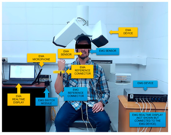
Figure 1.
System setup.
2.2. Recording Protocol
In order to conduct a simultaneous recording for posterior synchronization, the following sequence of steps (Figure 2) should be followed:
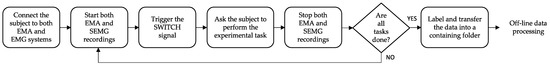
Figure 2.
Recording protocol steps.
- The subject under study should be located and connected, as shown in Figure 1.
- To register an experimental task, both systems should start recording before conducting the task.
- The EMG switch is pressed to generate a visible event in the signals of both systems.
- The experimental task is carried out.
- Both systems stop recording.
- EMA and EMG data are saved as binary files or text files.
- Steps 1 to 5 are repeated until all experimental tasks are finished.
- All resulting files are organized within a folder containing all of the recordings made within the whole session.
2.3. Off-Line Signal Processing and Synchronization
The recordings are processed off-line in order to achieve synchronization. The spikes in the audio signal and the square signal registered by the SEMG (with both events generated by the same switch) are employed to synchronize the signals of both systems, as shown in Figure 3. The switch was pressed three times at the beginning of the simultaneous recording. Three square pulses of 5 V of amplitude can be observed in the SEMG signal and six peeks can be observed in the audio recording. Both signals are synchronized when the spikes coincide with the flanks of the square pulses.
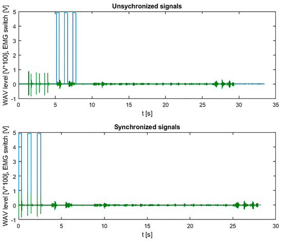
Figure 3.
Synchronization of the switch signal of SEMG system (blue) and audio signal of EMA system (green).
2.4. Proof-of-Concept Database
To prove the viability of the proposed methodology and to analyze possible interferences between the systems, the following recordings were made in one healthy volunteer (Ethics Committee Approval number 125/18, Comité Ético Científico, Universidad de La Frontera, Temuco, Chile):
- -
- Articulographic recording the chewing of 3.7 g of peanuts placing 3 active sensors on the jaw: in the interincisal midline, between first right molar and premolar, and between left molar and premolar.
- -
- Electromyographic recording of the chewing of 3.7 g of peanuts.
- -
- Simultaneous electromyographic and articulographic recording of the chewing of 3.7 g of peanuts.
Each chewing task was performed two times. First, chewing to the right side, and then to the left side. These recordings were labeled and stored, generating a proof-of-concept database.
2.5. Accuracy Analysis
The proof-of-concept database (EMA and EMG data) was processed to analyze the differences between simultaneous and individual recordings employing Matlab (R2019a, MathWorks, Natick, MA, USA). The following parameters were calculated:
- -
- Standard deviation of instantaneous measurements of Euclidean distance between pairs of sensors for each articulographic recording.
- -
- Average of the EMGrms (40 ms window) signal of each electromyographic recording.
The variation of both of the parameters between the different registration modalities (simultaneous and individual) were then analyzed.
3. Results
Regarding the analysis of the articulographic recordings of chewing, the standard deviation of the distance measurements between the sensors was 0.2 mm for the simultaneous registration of EMA with EMG and 0.3 mm for the individual registration of EMA. The values were within the limits indicated by the manufacturer. Figure 4 shows the trajectory of the mid-incisal point registered with EMA with and without connecting the SEMG device to register the muscle activity.
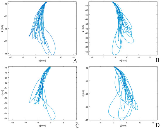
Figure 4.
EMA recordings: trajectories of the mid-incisal point. When combined with EMG, chewing to the right side (A) and to the left side (B). Without connecting the EMG device, chewing to the right side (C) and to the left side (D).
A difference between 1% and 10% was found between the means of the EMGrms (Figure 5) associated with the simultaneous EMG records with the EMA and individual EMG, respectively (Table 2).
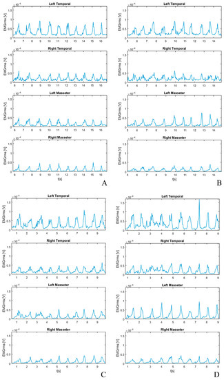
Figure 5.
EMG recordings: EMGrms of masseter and temporalis (anterior portion). When combined with EMA, chewing to the right side (A) and to the left side (B). Without connecting EMA device, chewing to the right side (C) and to the left side (D).
Table 2.
EMGrms mean.
Finally, the achieved synchronization of EMGrms and MM signals is shown in Figure 6. Only the SEMG recording of chewing from the right masseter muscle is shown. Scales were adjusted to achieve a better representation of the relation of both signals.
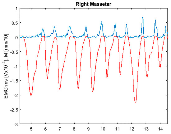
Figure 6.
EMGrms signal from the right masseter muscle (blue) and MM (red) synchronized.
4. Discussion
The present study proposed and established a proof-of-concept for the synchronization of two electromedical systems, 3D electromagnetic articulography, and surface electromyography, explaining it in detail so that it can be replicated by other research groups. No significant interferences between both systems were detected in the recordings. The implementation of the synchronization and further analysis aims to improve understanding of the complexity of mandibular functional movements, which may be applied to study different clinical situations.
The study of functional movements through analysis of kinematics and muscle activity is widely approached in various disciplines [32], including dentistry [1,2,3,5,7,8,9,11,13,14,16,33], which reveals the importance of analyzing these complex movements by various methods that complement each other.
Simultaneous measurements of different biomechanical variables of mandibular movements have been carried out for many years. Bigland measured the speed of contraction, the EMG activity, and the tension applied controlled by putting different weights [34]. More recent studies have also analyzed mandibular movements with simultaneous recordings of electromyographic activity and jaw movements in dentate individuals [8] and patients with maxillary and mandibular complete dentures [13] without synchronizing both recordings. These studies, even with clinical value, have limitations due to the lack of synchronization of their recordings or lack of more detailed explanation of the methodology employed. This limits the extrapolation of their results in the clinical understanding of mandibular movements, which reveals the need for a more complete analysis.
Other authors have developed studies demonstrating the concern to synchronize the signals recorded by different electromedical equipment related to mandibular movements with the electromyographic activity of the muscles of the stomatognathic system [6,7,9,10,11,14], but these studies did not provide enough information regarding the methods to be replicated.
Some studies have shown the synchronization between electromedical equipment applied to different analyses of the stomatognathic system, using arrangements or equipment specially developed for this purpose. Widmalm et al. designed and built a special device to combine recordings of vibration and EMG activity [35]. Nakazawa et al. performed a master-slave arrangement for palatal pressure, mandibular movement, and EMG recordings with 1 ms delay in the synchronization [36]. Some researchers used a specific synchronization module for EMG signal analysis and occlusal force distribution analyzed using the T-SCAN system [37,38,39,40], and Grigoriadis et al. synchronized EMG records of the masseter muscle and the mandibular movements using a commercial system with 2D magnetic sensors [16].
Other studies performed the synchronization of the recordings by generating an independent and identifiable event in the signals acquired by different systems. Ahlgren used a light signal to synchronize EMG and cinematography recordings [1]. A pulse generator was used to synchronize EMG activity and mandibular movements by videofluorography and cineradiography [3,4,5]. Freitas et al. synchronized the recordings of mandibular movements obtained by a Kinect sensor and EMG activity through a computational custom-made system [15]. More recently, Okawa et al. also used a synchronizing signal and post-processed the recordings to synchronize them [41].
Synchronization through the generation of a specific event was the method chosen by our research group to synchronize the EMG recordings of masticatory muscles and mandibular movements. In our case, we used a “switch” device that comes with EMG equipment and a sound recorder that comes with AG501 3D-EMA equipment. Activation of this switch generated an independent and distinguishable event in both registers, which in the case of EMG was recorded as a square electrical signal, and in the EMA was recorded as a peek in the acoustic signal that was used to synchronize both recordings.
Our recording method is a novelty in the scientific literature, and it has the advantage of the high accuracy of the movement-recording method demonstrated in previous articles from our research group [25,26,27]. In addition, this synchronization was performed using only the components that already accompany both pieces of equipment selected, a fact that besides facilitating its realization and replication, does not add costs to its realization.
In general, our results obtained during synchronized recordings showed no distortion/alteration outside the limit values informed by the manufacturers. Even though both instruments are very sensitive to electromagnetism, the EMA AG501 and the EMGs proposed in this methodology do not generate interferences between them during the simultaneous recording. This is probably because the bandwidths with which each system work do not overlap. This analysis could be improved by incrementing sample size and making a more complex experiment, e.g., determining the natural variability of EMG and EMA recordings for the application considered.
5. Conclusions
The present study was able to establish a detailed proof-of-concept of the synchronization of chewing muscle EMG activity related to mandibular movements, recorded by 3D-EMA, without generating significant distortion (within the values informed by the manufacturers), with the advantage of using components that are included with the equipment employed. The measurement system described in this paper is an effective tool that facilitates simultaneous investigation of mandibular movements in two domains: articulatory movements and electromyographic activity of muscles. Thus, it seems that is can be applied in different clinical situations in order to improve the analysis of the complexity of masticatory activity, thus generating new insights on this topic. The signal distortion analysis could be improved by incrementing sample size and making more complex experiment.
Author Contributions
Conceptualization, R.F. and M.F.L.; methodology, R.F., M.F.L., F.D., N.F.-B., C.C. and M.C.M.-C.; software, M.F.L.; validation, M.F.L. and F.D.; formal analysis, M.F.L., F.D., N.F.-B., C.C. and M.C.M.-C.; investigation, F.D., M.F.L., C.C., R.F. and N.F.-B.; resources, R.F.; writing—original draft preparation, R.F., M.F.L., F.D., N.F.-B., C.C. and M.C.M.-C.; writing—review and editing, M.F.L.; visualization, M.F.L.; supervision, R.F.; project administration, R.F.; funding acquisition, R.F. All authors have read and agreed to the published version of the manuscript.
Funding
Ramón Fuentes and his research group have received financial support from the Research Office, Universidad de La Frontera. This work is part of project DI22-0025.
Institutional Review Board Statement
The subject under study has participated voluntarily, signing informed consent. Ethics Committee Approval number 125/18, “Comité Ético Científico”, University de La Frontera, Chile.
Informed Consent Statement
The participant has given consent for publication.
Data Availability Statement
The datasets used and/or analysed during the current study are available from the corresponding author on reasonable request.
Conflicts of Interest
The authors declare no conflict of interest.
References
- Ahlgren, J. Kinesiology of the mandible an EMG study. Acta Odontol. Scand. 1967, 25, 593–612. [Google Scholar] [CrossRef]
- Campillo, B.; Martín, C.; Palma, J.C.; Fuentes, A.D.; Alarcón, J.A. Electromyographic activity of the jaw muscles and mandibular kinematics in young adults with theoretically ideal dental occlusion: Reference values. Med. Oral Patol. Oral Cir. Bucal. 2017, 22, e383. [Google Scholar] [CrossRef]
- Pancherz, H.; Winnberg, A.; Westesson, P.L. Masticatory muscle activity and hyoid bone behavior during cyclic jaw movements in man: A synchronized electromyographic and videofluorographic study. Am. J. Orthodont. 1986, 89, 122–131. [Google Scholar] [CrossRef]
- Hylander, W.L.; Johnson, K.R.; Crompton, A.W. Loading patterns and jaw movements during mastication in macaca fascicularis: A bone-strain, electromyographic, and cineradiographic analysis. Am. J. Phys. Anthropol. 1987, 72, 287–314. [Google Scholar] [CrossRef]
- Winnberg, A.; Pancherz, H.; Westesson, P.L. Head posture and hyo-mandibular function in man: A synchronized electromyographic and video fluorographic study of the open-close-clench cycle. Am. J. Orthod. Dentofac. Orthop. 1988, 94, 393–404. [Google Scholar] [CrossRef]
- Gerstner, G.E.; Goldberg, L.J. An analysis of mandibular movement trajectories and masticatory muscle EMG activity during drinking in the guinea pig. Brain Res. 1989, 479, 6–15. [Google Scholar] [CrossRef]
- Ottenhoff, F.A.; Van Der Bilt, A.; Van Der Glas, H.W.; Bosman, F. Control of elevator muscle activity during simulated chewing with varying food resistance in humans. J. Neurophysiol. 1992, 68, 933–944. [Google Scholar] [CrossRef]
- Van Eijden, T.M.G.J.; Blanksma, N.G.; Brugman, P. Amplitude and timing of EMG activity in the human masseter muscle during selected motor tasks. J. Dent. Res. 1993, 72, 599–606. [Google Scholar] [CrossRef]
- Hiraba, K.; Hibino, K.; Hiranuma, K.; Negoro, T. EMG activities of two heads of the human lateral pterygoid muscle in relation to mandibular condyle movement and biting force. J. Neurophysiol. 2000, 83, 2120–2137. [Google Scholar] [CrossRef]
- Igarashi, N.; Yamamura, K.; Yamada, Y.; Kohno, S. Head movements and neck muscle activities associated with the jaw movement during mastication in the rabbit authors. Brain Res. 2000, 871, 151–155. [Google Scholar] [CrossRef]
- Van Der Bilt, A.; Weijnen, F.G.; Bosman, F.; Van Der Glas, H.W.; Kuks, J.B. Controlled study of EMG activity of the jaw closers and openers during mastication in patients with myasthenia gravis. Eur. J. Oral Sci. 2001, 109, 160–164. [Google Scholar] [CrossRef]
- Naganuma, K.; Inoue, M.; Yamamura, K.; Hanada, K.; Yamada, Y. Tongue and jaw muscle activities during chewing and swallowing in freely behaving rabbits. Brain Res. 2001, 915, 185–194. [Google Scholar] [CrossRef]
- Piancino, M.G.; Farina, D.; Talpone, F.; Castroflorio, T.; Gassino, G.; Margarino, V.; Bracco, P. Surface EMG of jaw-elevator muscles and chewing pattern in complete denture wearers. J. Oral Rehab. 2005, 32, 863–870. [Google Scholar] [CrossRef]
- Piancino, M.G.; Bracco, P.; Vallelonga, T.; Merlo, A.; Farina, D. Effect of bolus hardness on the chewing pattern and activation of masticatory muscles in subjects with normal dental occlusion. J. Electromyogr. Kinesiol. 2008, 18, 931–937. [Google Scholar] [CrossRef]
- Freitas, J.; Teixeira, A.; Dias, M.S. Multimodal silent speech interface based on video, depth, surface electromyography and ultrasonic doppler: Data collection and first recognition results. In Speech Production in Automatic Speech Recognition; International Speech Communication Association: Grenoble, France, 2013; Available online: http://www.isca-speech.org/archive/spasr_2013 (accessed on 10 September 2018).
- Grigoriadis, A.; Johansson, R.S.; Trulsson, M. Temporal profile and amplitude of human masseter muscle activity is adapted to food properties during individual chewing cycles. J. Oral Rehab. 2014, 41, 367–373. [Google Scholar] [CrossRef]
- Gómez, J.; Lezcano, M.F.; Peldoza, V.; Fuentes, R. Surface Electromyography and Electromagnetic Articulography to Analyze Vertical Dimension. Protocol Report. Av. Odontoestomatol. 2020, 36, 35–42. [Google Scholar] [CrossRef]
- Gómez, J.; Lezcano, M.F.; Marinelli, F.; Navarro, P.; Fuentes, R. Dimensión vertical de la cara en adultos con diferentes relaciones oclusales. Int. J. Morphol. 2022, 1, 2. [Google Scholar] [CrossRef]
- Basmajian, J.V. Electromyography: Its structural and neural basis. Int. Rev. Cytol. 1967, 21, 129–140. [Google Scholar]
- Castroflorio, T.; Bracco, P.; Farina, D. Surface electromyography in the assessment of jaw elevator muscles. J. Oral Rehab. 2008, 35, 638–645. [Google Scholar] [CrossRef]
- Lezcano, M.F.; Dias, F.J.; Arias, A.; Fuentes, R. Accuracy and Reliability of AG501 Articulograph for mandibular movement analysis: A quantitative descriptive study. Sensors 2020, 20, 6324. [Google Scholar] [CrossRef]
- Hixon, T.X. An electromagnetic method for transducing jaw movements during speech. J. Acoust. Soc. Am. 1971, 49, 603–606. [Google Scholar] [CrossRef]
- Schönle, P.W.; Gräbe, K.; Wenig, P.; Höhne, J.; Schrader, J.; Conrad, B. Electromagnetic articulography: Use of alternating magnetic fields for tracking movements of multiple points inside and outside the vocal tract. Brain Lang. 1987, 31, 26–35. [Google Scholar] [CrossRef] [PubMed]
- Kaburagi, T.; Wakamiya, K.; Honda, M. Three-dimensional electromagnetic articulography: A measurement principle. J. Acoust. Soc. Am. 2005, 118, 428–443. [Google Scholar] [CrossRef] [PubMed]
- Fuentes, R.; Arias, A.; Saravia, D.; Lezcano, M.F.; Dias, F.J. An innovative method to analyse the range of border mandibular movements using 3D electromagnetic articulography (AG501) and MATLAB. Biomed. Res. 2017, 28, 4239–4247. [Google Scholar]
- Fuentes, R.; Arias, A.; Lezcano, M.F.; Saravia, D.; Kuramochi, G.; Dias, F.J. Systematic standardized and individualized assessment of masticatory cycles using electromagnetic 3D articulography and computer scripts. Biomed. Res. Int. 2017, 2017, 7134389. [Google Scholar] [CrossRef]
- Fuentes, R.; Arias, A.; Lezcano, M.F.; Saravia, D.; Kuramochi, G.; Navarro, P.; Dias, F.J. A new tridimensional insight on geometric and kinematic characteristics of masticatory cycles in participants with normal occlusion. Biomed. Res. Int. 2018, 2018, 2527463. [Google Scholar] [CrossRef]
- Lezcano, M.F.; Dias, F.J.; Chuhuaicura, P.; Navarro, P.; Fuentes, R. Symmetry of mandibular movements: A 3D electromagnetic articulography technique applied on asymptomatic participants. J. Prosthet. Dent. 2020, 125, 746–752. [Google Scholar] [CrossRef]
- Vargas, S.; Lezcano, M.F.; Álvarez, G.; Navarro, P.; Fuentes, R. Three-dimensional analysis of mandibular border movements in fully dentate participants. Int. J. Mophol. 2020, 38, 983–989. [Google Scholar]
- Hoke, P.; Tiede, M.; Grender, J.; Klukowska, M.; Peters, J.; Carr, G. Using electromagnetic articulography to measure denture micromovement during chewing with and without denture adhesive. J. Prosthodont. 2019, 28, e252–e258. [Google Scholar] [CrossRef]
- Fuentes, R.; Navarro, P.; Curiqueo, A.; Ottone, N.E. Determination of mandibular border and functional movement protocols using an electromagnetic articulograph (EMA). Int. J. Clin. Exp. Med. 2015, 8, 19905. [Google Scholar]
- De Luca, C.J. The use of surface electromyography in biomechanics. J. Appl. Biomech. 1997, 13, 135–163. [Google Scholar] [CrossRef]
- Pruzansky, S. The application of electromyography to dental research. J. Am. Dent. Assoc. 1952, 44, 49–68. [Google Scholar] [CrossRef]
- Bigland, B.; Lippold, O.C.J. The relation between force, velocity and integrated electrical activity in human muscles. J. Physiol. 1954, 123, 214–224. [Google Scholar] [CrossRef] [PubMed]
- Widmalm, S.E.; Hedegård, B. An apparatus for the synchronous registration of EMG activity in jaw muscles and of vibrations in the masticatory system. J. Oral Rehab. 1974, 1, 183–190. [Google Scholar] [CrossRef]
- Nakazawa, F.; Togashi, M. Evaluation of food texture by mastication and palatal pressure, jaw movement and electromyography. Hydrocolloids 2000, 473–483. [Google Scholar] [CrossRef]
- Kerstein, R.B. Combining technologies: A computerized occlusal analysis system synchronized with a computerized electromyography system. Cranio 2004, 22, 96–109. [Google Scholar] [CrossRef] [PubMed]
- Kerstein, R.B. Reducing chronic masseter and temporalis muscular hyperactivity with computer-guided occlusal adjustments. Compend. Contin. Educ. Dent. 2010, 31, 530–534. [Google Scholar]
- Karakis, D.; Demirdag, E.D. Adjustment of occlusal splint with synchronized T-Scan III digital occlusal analysis system and Bio-EMG III in a patient with sleep bruxism. J. Adv. Oral Res. 2021, 12, 170–175. [Google Scholar] [CrossRef]
- Chen, Y.J.; Yao, C.C.; Chang, Z.C.; Lai, H.H.; Hsu, L.F.; Hsu, T.H.; Kok, S.H. Occlusal function and electromyographic activity of masticatory muscles in skeletal Class III patients with different patterns of mandibular asymmetry. J. Oral Rehab. 2023, 50, 276–285. [Google Scholar] [CrossRef]
- Okawa, J.; Hori, K.; Yoshimoto, T.; Salazar, S.E.; Ono, T. Higher masticatory performance and higher number of chewing strokes increase retronasal aroma. Front. Nutr. 2021, 8, 623507. [Google Scholar] [CrossRef]
Disclaimer/Publisher’s Note: The statements, opinions and data contained in all publications are solely those of the individual author(s) and contributor(s) and not of MDPI and/or the editor(s). MDPI and/or the editor(s) disclaim responsibility for any injury to people or property resulting from any ideas, methods, instructions or products referred to in the content. |
© 2023 by the authors. Licensee MDPI, Basel, Switzerland. This article is an open access article distributed under the terms and conditions of the Creative Commons Attribution (CC BY) license (https://creativecommons.org/licenses/by/4.0/).