
Preliminary Report for the Development of a Multiparameter Protocol for the Identification of Sinusoidal Obstruction Syndrome including Abdominal Ultrasound before and after Allogeneic Stem Cell Transplantation

 , ,
, , 
Abstract
:1. Introduction
2. Methods
2.1. Patient Characteristics
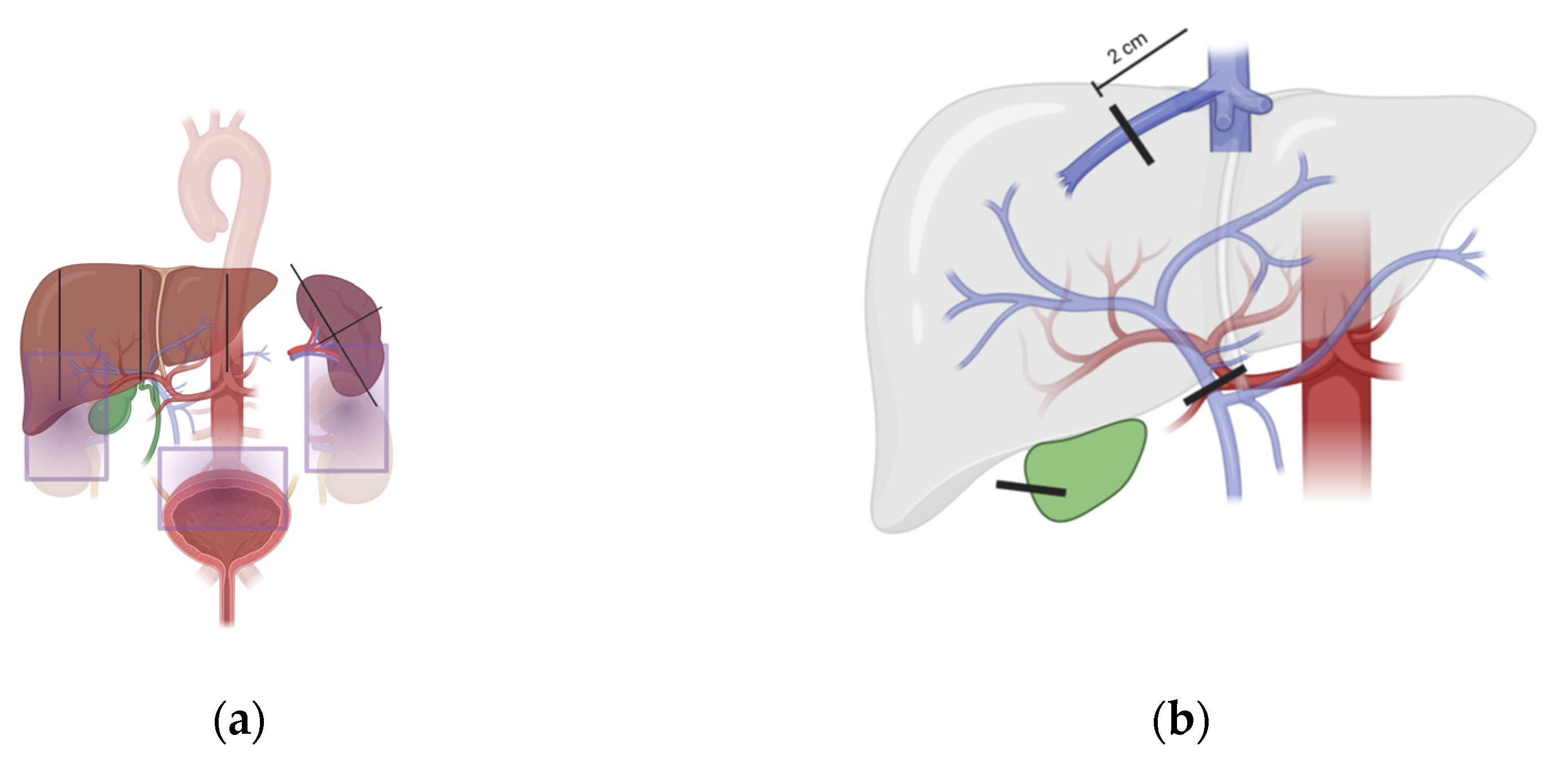
2.2. Ultrasound Examination
2.3. Inter- and Intra-Rater Reliability
2.4. Statistical Analysis
2.5. Ethics
3. Results
3.1. Patient Population
3.2. Inter-Rater and Intra-Rater Reliability Exercise
4. Discussion
Supplementary Materials
Author Contributions
Funding
Institutional Review Board Statement
Informed Consent Statement
Data Availability Statement
Conflicts of Interest
References
- Thomas, E.D.; Lochte, H.L.; Lu, W.C.; Ferrebee, J.W. Intravenous Infusion of Bone Marrow in Patients Receiving Radiation and Chemotherapy. N. Engl. J. Med. 1957, 257, 491–496. [Google Scholar] [CrossRef]
- Weiden, P.L.; Flournoy, N.; Thomas, E.D.; Prentice, R.; Fefer, A.; Buckner, C.D.; Storb, R. Antileukemic Effect of Graft-versus-Host Disease in Human Recipients of Allogeneic-Marrow Grafts. N. Engl. J. Med. 1979, 300, 1068–1073. [Google Scholar] [CrossRef] [PubMed]
- Sorror, M.L.; Maris, M.B.; Storb, R.; Baron, F.; Sandmaier, B.M.; Maloney, D.G.; Storer, B. Hematopoietic Cell Transplantation (HCT)-Specific Comorbidity Index: A New Tool for Risk Assessment before Allogeneic HCT. Blood 2005, 106, 2912–2919. [Google Scholar] [CrossRef] [PubMed] [Green Version]
- Sorror, M.L.; Sandmaier, B.M.; Storer, B.E.; Maris, M.B.; Baron, F.; Maloney, D.G.; Scott, B.L.; Deeg, H.J.; Appelbaum, F.R.; Storb, R. Comorbidity and Disease Status–Based Risk Stratification of Outcomes Among Patients With Acute Myeloid Leukemia or Myelodysplasia Receiving Allogeneic Hematopoietic Cell Transplantation. J. Clin. Oncol. 2007, 25, 4246–4254. [Google Scholar] [CrossRef]
- Luft, T.; Benner, A.; Terzer, T.; Jodele, S.; Dandoy, C.E.; Storb, R.; Kordelas, L.; Beelen, D.; Gooley, T.; Sandmaier, B.M.; et al. EASIX and Mortality after Allogeneic Stem Cell Transplantation. Bone Marrow Transplant. 2020, 55, 553–561. [Google Scholar] [CrossRef] [PubMed]
- D’Souza, A.; Fertham, C. Current Uses and Outcomes of Hematopoietic Cell Transplantation (HCT): CIBMTR Summary Slides. 2017. Available online: http://www.cibmtr.org (accessed on 23 November 2021).
- Zeiser, R.; Blazar, B.R. Acute Graft-versus-Host Disease—Biologic Process, Prevention, and Therapy. N. Engl. J. Med. 2017, 377, 2167–2179. [Google Scholar] [CrossRef] [PubMed]
- Sahin, U.; Toprak, S.K.; Atilla, P.A.; Atilla, E.; Demirer, T. An Overview of Infectious Complications after Allogeneic Hematopoietic Stem Cell Transplantation. J. Infect. Chemother. 2016, 22, 505–514. [Google Scholar] [CrossRef] [PubMed] [Green Version]
- Wingard, J.R.; Hsu, J.; Hiemenz, J.W. Hematopoietic Stem Cell Transplantation: An Overview of Infection Risks and Epidemiology. Hematol. Oncol. Clin. N. Am. 2011, 25, 101–116. [Google Scholar] [CrossRef]
- Bearman, S. The Syndrome of Hepatic Veno-Occlusive Disease after Marrow Transplantation. Blood 1995, 85, 3005–3020. [Google Scholar] [CrossRef] [PubMed] [Green Version]
- Richardson, P.G.; Murakami, C.; Jin, Z.; Warren, D.; Momtaz, P.; Hoppensteadt, D.; Elias, A.D.; Antin, J.H.; Soiffer, R.; Spitzer, T.; et al. Multi-Institutional Use of Defibrotide in 88 Patients after Stem Cell Transplantation with Severe Veno-Occlusive Disease and Multisystem Organ Failure: Response without Significant Toxicity in a High-Risk Population and Factors Predictive of Outcome. Blood 2002, 100, 4337–4343. [Google Scholar] [CrossRef] [PubMed] [Green Version]
- Richardson, P.; Guinan, E. The Pathology, Diagnosis, and Treatment of Hepatic Veno-Occlusive Disease: Current Status and Novel Approaches. Br. J. Haematol. 1999, 107, 485–493. [Google Scholar] [CrossRef]
- Coppell, J.A.; Richardson, P.G.; Soiffer, R.; Martin, P.L.; Kernan, N.A.; Chen, A.; Guinan, E.; Vogelsang, G.; Krishnan, A.; Giralt, S.; et al. Hepatic Veno-Occlusive Disease Following Stem Cell Transplantation: Incidence, Clinical Course, and Outcome. Biol. Blood Marrow Transplant. J. Am. Soc. Blood Marrow Transplant. 2010, 16, 157–168. [Google Scholar] [CrossRef] [Green Version]
- Carreras, E.; Bertz, H.; Arcese, W.; Vernant, J.-P.; Tomás, J.-F.; Hagglund, H.; Bandini, G.; Esperou, H.; Russell, J.; de la Rubia, J.; et al. Incidence and Outcome of Hepatic Veno-Occlusive Disease After Blood or Marrow Transplantation: A Prospective Cohort Study of the European Group for Blood and Marrow Transplantation. Blood 1998, 92, 3599–3604. [Google Scholar]
- Fan, C.Q.; Crawford, J.M. Sinusoidal Obstruction Syndrome (Hepatic Veno-Occlusive Disease). J. Clin. Exp. Hepatol. 2014, 4, 332–346. [Google Scholar] [CrossRef] [Green Version]
- Dalle, J.-H.; Giralt, S.A. Hepatic Veno-Occlusive Disease after Hematopoietic Stem Cell Transplantation: Risk Factors and Stratification, Prophylaxis, and Treatment. Biol. Blood Marrow Transplant. 2016, 22, 400–409. [Google Scholar] [CrossRef] [PubMed] [Green Version]
- Jones, R.J.; Lee, K.S.; Beschorner, W.E.; Vogel, V.G.; Grochow, L.B.; Braine, H.G.; Vogelsang, G.B.; Sensenbrenner, L.L.; Santos, G.W.; Saral, R. Venoocclusive Disease of the Liver Following Bone Marrow Transplantation. Transplantation 1987, 44, 778–783. [Google Scholar] [CrossRef] [PubMed]
- Shulman, H.M.; Hinterberger, W. Hepatic Veno-Occlusive Disease--Liver Toxicity Syndrome after Bone Marrow Transplantation. Bone Marrow Transplant. 1992, 10, 197–214. [Google Scholar] [PubMed]
- Corbacioglu, S.; Carreras, E.; Ansari, M.; Balduzzi, A.; Cesaro, S.; Dalle, J.-H.; Dignan, F.; Gibson, B.; Guengoer, T.; Gruhn, B.; et al. Diagnosis and Severity Criteria for Sinusoidal Obstruction Syndrome/Veno-Occlusive Disease in Pediatric Patients: A New Classification from the European Society for Blood and Marrow Transplantation. Bone Marrow Transplant. 2018, 53, 138–145. [Google Scholar] [CrossRef]
- Carreras, E.; Grañena, A.; Navasa, M.; Bruguera, M.; Marco, V.; Sierra, J.; Tassies, M.D.; García-Pagán, J.C.; Martí, J.M.; Bosch, J.; et al. On the Reliability of Clinical Criteria for the Diagnosis of Hepatic Veno-Occlusive Disease. Ann. Hematol. 1993, 66, 77–80. [Google Scholar] [CrossRef]
- Mohty, M.; Malard, F.; Abecassis, M.; Aerts, E.; Alaskar, A.S.; Aljurf, M.; Arat, M.; Bader, P.; Baron, F.; Bazarbachi, A.; et al. Revised Diagnosis and Severity Criteria for Sinusoidal Obstruction Syndrome/Veno-Occlusive Disease in Adult Patients: A New Classification from the European Society for Blood and Marrow Transplantation. Bone Marrow Transplant. 2016, 51, 906–912. [Google Scholar] [CrossRef]
- Carreras, E. How I Manage Sinusoidal Obstruction Syndrome after Haematopoietic Cell Transplantation. Br. J. Haematol. 2015, 168, 481–491. [Google Scholar] [CrossRef] [PubMed]
- Carreras, E.; Grañena, A.; Navasa, M.; Bruguera, M.; Marco, V.; Sierra, J.; Tassies, M.D.; García-Pagán, J.C.; Martí, J.M.; Bosch, J. Transjugular Liver Biopsy in BMT. Bone Marrow Transplant. 1993, 11, 21–26. [Google Scholar] [PubMed]
- Dignan, F.L.; Wynn, R.F.; Hadzic, N.; Karani, J.; Quaglia, A.; Pagliuca, A.; Veys, P.; Potter, M.N. BCSH/BSBMT Guideline: Diagnosis and Management of Veno-Occlusive Disease (Sinusoidal Obstruction Syndrome) Following Haematopoietic Stem Cell Transplantation. Br. J. Haematol. 2013, 163, 444–457. [Google Scholar] [CrossRef]
- Lassau, N.; Leclère, J.; Auperin, A.; Bourhis, J.H.; Hartmann, O.; Valteau-Couanet, D.; Benhamou, E.; Bosq, J.; Ibrahim, A.; Girinski, T.; et al. Hepatic Veno-Occlusive Disease after Myeloablative Treatment and Bone Marrow Transplantation: Value of Gray-Scale and Doppler US in 100 Patients. Radiology 1997, 204, 545–552. [Google Scholar] [CrossRef]
- Lassau, N.; Auperin, A.; Leclere, J.; Bennaceur, A.; Valteau-couanet, D.; Hartmann, O. Prognostic Value of Doppler-Ultrasonography in Hepatic Veno-Occlusive Disease: A Study of 71 Children. Transplantation 2002, 74, 60–66. [Google Scholar] [CrossRef]
- Schulz, M.; Vuong, L.G.; Müller, H.P.; Maibier, M.; Tacke, F.; Blau, I.W.; Wree, A. Shear Wave Elastography in the Detection of Sinusoidal Obstruction Syndrome in Adult Patients Undergoing Allogenic Hematopoietic Stem Cell Transplantation. Diagnostics 2021, 11, 928. [Google Scholar] [CrossRef] [PubMed]
- Trenker, C.; Burchert, A.; Schumacher, C.; Schäfer, J.A.; Dohse, M.; Timmesfeld, N.; Neubauer, A.; Sohlbach, K.; Michel, C.; Görg, C. Pathologic Hepatic Contrast-Enhanced Ultrasound Pattern in Patients Undergoing Allogeneic Stem Cell Transplantation. Ultrasound Med. Biol. 2020, 46, 1865–1871. [Google Scholar] [CrossRef]
- Nishida, M.; Kahata, K.; Hayase, E.; Shigematsu, A.; Sato, M.; Kudo, Y.; Omotehara, S.; Iwai, T.; Sugita, J.; Shibuya, H.; et al. Novel Ultrasonographic Scoring System of Sinusoidal Obstruction Syndrome after Hematopoietic Stem Cell Transplantation. Biol. Blood Marrow Transplant. 2018, 24, 1896–1900. [Google Scholar] [CrossRef] [Green Version]
- Chan, S.S.; Colecchia, A.; Duarte, R.F.; Bonifazi, F.; Ravaioli, F.; Bourhis, J.H. Imaging in Hepatic Veno-Occlusive Disease/Sinusoidal Obstruction Syndrome. Biol. Blood Marrow Transplant. 2020, 26, 1770–1779. [Google Scholar] [CrossRef]
- R Core Team—European Environment Agency. 2020. Available online: https://www.eea.europa.eu/data-and-maps/indicators/oxygen-consuming-substances-in-rivers/r-development-core-team-2006 (accessed on 23 November 2021).
- Koo, T.K.; Li, M.Y. A Guideline of Selecting and Reporting Intraclass Correlation Coefficients for Reliability Research. J. Chiropr. Med. 2016, 15, 155–163. [Google Scholar] [CrossRef] [Green Version]
- Cairo, M.S.; Cooke, K.R.; Lazarus, H.M.; Chao, N. Modified Diagnostic Criteria, Grading Classification and Newly Elucidated Pathophysiology of Hepatic SOS/VOD after Haematopoietic Cell Transplantation. Br. J. Haematol. 2020, 190, 822–836. [Google Scholar] [CrossRef]
- McDonald, G.B. Veno-Occlusive Disease of the Liver and Multiorgan Failure after Bone Marrow Transplantation: A Cohort Study of 355 Patients. Ann. Intern. Med. 1993, 118, 255. [Google Scholar] [CrossRef]
- Nishida, M.; Sugita, J.; Takahashi, S.; Iwai, T.; Sato, M.; Kudo, Y.; Omotehara, S.; Horie, T.; Sakano, R.; Shibuya, H.; et al. Refined Ultrasonographic Criteria for Sinusoidal Obstruction Syndrome after Hematopoietic Stem Cell Transplantation. Int. J. Hematol. 2021, 114, 94–101. [Google Scholar] [CrossRef]
- Ecsedi, M.; Schmohl, J.; Zeiser, R.; Drexler, B.; Halter, J.; Medinger, M.; Duyster, J.; Kanz, L.; Passweg, J.; Finke, J.; et al. Anti-Thymocyte Globulin-Induced Hyperbilirubinemia in Patients with Myelofibrosis Undergoing Allogeneic Hematopoietic Cell Transplantation. Ann. Hematol. 2016, 95, 1627–1636. [Google Scholar] [CrossRef]
- Dai, H.; Penack, O.; Radujkovic, A.; Schult, D.; Majer-Lauterbach, J.; Blau, I.W.; Bullinger, L.; Jiang, S.; Müller-Tidow, C.; Dreger, P.; et al. Early Bilirubinemia after Allogeneic Stem Cell Transplantation—An Endothelial Complication. Bone Marrow Transplant. 2021, 56, 1573–1583. [Google Scholar] [CrossRef] [PubMed]
- Franeková, J.; Sečník, P.; Lavríková, P.; Kubíček, Z.; Hošková, L.; Kieslichová, E.; Jabor, A. Serial Measurement of Presepsin, Procalcitonin, and C-Reactive Protein in the Early Postoperative Period and the Response to Antithymocyte Globulin Administration after Heart Transplantation. Clin. Transplant. 2017, 31, e12870. [Google Scholar] [CrossRef] [PubMed]
- Fein, J.A.; Shimoni, A.; Danylesko, I.; Shem-Tov, N.; Yerushalmi, R.; Chowers, G.; Cohen, Z.; Nagler, A.; Shouval, R. Early Organ Toxicity Following Allogeneic Hematopoietic Stem Cell Transplantation Differs By Conditioning Regimen. Blood 2019, 134, 4489. [Google Scholar] [CrossRef]
- Kaya, N. Grayscale and Spectral Doppler Ultrasound in the Diagnosis of Hepatic Veno-Occlusive Disease/Sinusoidal Obstruction Syndrome after Hematopoietic Stem Cell Transplantation in Children. J. Pediatr. Hematol. Oncol. 2021, 43, e1105. [Google Scholar] [CrossRef] [PubMed]

| Patient Characteristics | (N = 23) |
|---|---|
| Age | |
| Mean (SD) | 61.8 (12.8) |
| Median [Min, Max] | 64.0 [19.0, 79.0] |
| Sex | |
| Female | 8 (34.8%) |
| Male | 15 (65.2%) |
| Weight | |
| Mean (SD) | 79.9 (17.5) |
| Median [Min, Max] | 78.0 [45.4, 119] |
| Disease | |
| Leukemia | 15 (65%) |
| Multiple myeloma | 1 (4.3%) |
| Myelodysplastic syndrome | 6 (26%) |
| Myeloproliferative syndrome | 1 (4.3%) |
| Conditioning regimen | |
| Myeloablative | 1 (4.3%) |
| Non-myeloablative | 22 (95.7%) |
| AlloHSCT donor compatibility | |
| Matched unrelated donor | 15 (65.21 %) |
| Mismatch unrelated donor | 3 (13.04%) |
| Matched related donor | 2 (8.69%) |
| Haploidentical related donor | 3 (13.04%) |
| Peritransplantational findings | |
| Presence of fever | 19 (82.61%) |
| Abdominal pain | 7 (30.43%) |
| Body weight increase >2% | 9 (39.13%) |
| Pre | Post | p Value | |
|---|---|---|---|
| (N = 23) | (N = 23) | ||
| CRP (mg/L) | |||
| Mean (SD) | 22.1 (36.1) | 61.4 (79.0) | 0.0091 |
| Median [Min, Max] | 3.71 [0.340, 125] | 46.0 [0.600, 296] | |
| Bilirubin (mg/dL) | |||
| Mean (SD) | 0.641 (0.837) | 2.96 (3.86) | 0.0001 |
| Median [Min, Max] | 0.420 [0.200, 4.36] | 2.56 [0.330, 19.4] | |
| AP (U/L) | |||
| Mean (SD) | 154 (117) | 110 (61.3) | 0.36 |
| Median [Min, Max] | 99.0 [35.0, 376] | 92.0 [35.0, 297] | |
| Missing | 1 (4.3%) | 0 (0%) | |
| AST (U/L) | |||
| Mean (SD) | 34.3 (23.6) | 26.7 (26.6) | 0.048 |
| Median [Min, Max] | 29.0 [8.00, 120] | 18.0 [7.00, 138] | |
| ALT (U/L) | |||
| Mean (SD) | 53.3 (56.5) | 28.3 (24.3) | 0.0031 |
| Median [Min, Max] | 44.0 [5.00, 267] | 21.0 [5.00, 113] | |
| γGT (U/L) | |||
| Mean (SD) | 168 (186) | 114 (91.1) | 0.12 |
| Median [Min, Max] | 101 [10.0, 745] | 80.0 [0.680, 280] | |
| Thrombocytes (G/L) | |||
| Mean (SD) | 140 (124) | 45.8 (58.8) | 0.0051 |
| Median [Min, Max] | 102 [11.0, 395] | 24.0 [7.00, 260] |
| Pre | Post | |
|---|---|---|
| (N = 23) | (N = 23) | |
| Ultrasound of the liver | ||
| Vertical diameter—pre-aortic measurement | ||
| Mean (SD) | 9.55 (2.53) | 10.3 (2.94) |
| Median [Min, Max] | 9.65 [5.30, 15.5] | 10.2 [4.10, 15.0] |
| Missing | 1 (4.3%) | 1 (4.3%) |
| Vertical diameter—medio-clavicular line | ||
| Mean (SD) | 15.1 (3.68) | 16.5 (2.99) |
| Median [Min, Max] | 15.1 [7.60, 25.0] | 16.9 [10.6, 22.0] |
| Missing | 1 (4.3%) | 0 (0%) |
| Vertical diameter—pre-renal measurement | ||
| Mean (SD) | 15.1 (3.13) | 16.3 (2.78) |
| Median [Min, Max] | 15.1 [8.10, 21.0] | 17.1 [10.5, 21.7] |
| Missing | 1 (4.3%) | 1 (4.3%) |
| Maximal thickness of gallbladder | ||
| Mean (SD) | 0.256 (0.0934) | 0.314 (0.0954) |
| Median [Min, Max] | 0.250 [0, 0.400] | 0.310 [0.100, 0.430] |
| Missing | 3 (13.0%) | 5 (21.7%) |
| Ultrasound of hepatic vessels | ||
| Maximal diameter of liver veins | ||
| Mean (SD) | 0.752 (0.267) | 0.868 (0.312) |
| Median [Min, Max] | 0.700 [0.370, 1.20] | 0.850 [0.200, 1.90] |
| Missing | 0 (0%) | 0 (0%) |
| Maximal velocity of liver veins | ||
| Mean (SD) | 17.9 (7.77) | 17.9 (9.99) |
| Median [Min, Max] | 17.8 [5.00, 37.1] | 16.3 [0, 30.9] |
| Missing | 4 (17.4%) | 14 (60.9%) |
| Hepatopetal flow of liver veins | ||
| No | 23 (100%) | 23 (100%) |
| Yes | 0 (0%) | 0 (0%) |
| Missing | 0 (0%) | 0 (0%) |
| Maximal diameter of portal vein | ||
| Mean (SD) | 0.963 (0.227) | 1.01 (0.295) |
| Median [Min, Max] | 0.900 [0.600, 1.40] | 0.980 [0.500, 1.80] |
| Missing | 3 (13.0%) | 1 (4.3%) |
| Maximal velocity of portal vein | ||
| Mean (SD) | 17.8 (14.0) | 18.2 (11.7) |
| Median [Min, Max] | 16.3 [0.700, 69.6] | 15.2 [6.20, 57.0] |
| Missing | 1 (4.3%) | 3 (13.0%) |
| Hepatopetal flow of portal vein | ||
| No | 0 (0%) | 0 (0%) |
| Yes | 23 (100%) | 22 (95.7%) |
| Missing | 0 (0%) | 1 (4.3%) |
| Hepatic artery RI | ||
| Mean (SD) | 0.805 (0.186) | 0.831 (0.125) |
| Median [Min, Max] | 0.795 [0.400, 1.10] | 0.830 [0.470, 1.00] |
| Missing | 3 (13.0%) | 2 (8.7%) |
| Ultrasound of the spleen | ||
| Spleen length | ||
| Mean (SD) | 12.9 (3.58) | 12.6 (2.66) |
| Median [Min, Max] | 12.0 [8.40, 24.2] | 12.1 [7.50, 18.3] |
| Missing | 0 (0%) | 2 (8.7%) |
| Spleen width | ||
| Mean (SD) | 4.99 (1.53) | 5.39 (1.33) |
| Median [Min, Max] | 4.40 [3.60, 10.0] | 5.18 [3.40, 8.40] |
| Missing | 1 (4.3%) | 3 (13.0%) |
| Ultrasound of the abdominal cavity | ||
| Pathologic recanalization of umbilical vein | ||
| No | 23 (100%) | 22 (95.7%) |
| Yes | 0 (0%) | 0 (0%) |
| Missing | 0 (0%) | 1 (4.3%) |
| Ascites | ||
| No | 22 (95.7%) | 19 (82.6%) |
| Yes | 1 (4.3%) | 4 (17.4%) |
| Missing | 0 (0%) | 0 (0%) |
Publisher’s Note: MDPI stays neutral with regard to jurisdictional claims in published maps and institutional affiliations. |
© 2022 by the authors. Licensee MDPI, Basel, Switzerland. This article is an open access article distributed under the terms and conditions of the Creative Commons Attribution (CC BY) license (https://creativecommons.org/licenses/by/4.0/).
Share and Cite
Schlaweck, S.; Bauer, C.J.; Schmitz, F.; Brossart, P.; Holderried, T.A.W.; Schäfer, V.S. Preliminary Report for the Development of a Multiparameter Protocol for the Identification of Sinusoidal Obstruction Syndrome including Abdominal Ultrasound before and after Allogeneic Stem Cell Transplantation. Appl. Sci. 2022, 12, 829. https://doi.org/10.3390/app12020829
Schlaweck S, Bauer CJ, Schmitz F, Brossart P, Holderried TAW, Schäfer VS. Preliminary Report for the Development of a Multiparameter Protocol for the Identification of Sinusoidal Obstruction Syndrome including Abdominal Ultrasound before and after Allogeneic Stem Cell Transplantation. Applied Sciences. 2022; 12(2):829. https://doi.org/10.3390/app12020829
Chicago/Turabian StyleSchlaweck, Sebastian, Claus Juergen Bauer, Friederike Schmitz, Peter Brossart, Tobias A. W. Holderried, and Valentin Sebastian Schäfer. 2022. "Preliminary Report for the Development of a Multiparameter Protocol for the Identification of Sinusoidal Obstruction Syndrome including Abdominal Ultrasound before and after Allogeneic Stem Cell Transplantation" Applied Sciences 12, no. 2: 829. https://doi.org/10.3390/app12020829
APA StyleSchlaweck, S., Bauer, C. J., Schmitz, F., Brossart, P., Holderried, T. A. W., & Schäfer, V. S. (2022). Preliminary Report for the Development of a Multiparameter Protocol for the Identification of Sinusoidal Obstruction Syndrome including Abdominal Ultrasound before and after Allogeneic Stem Cell Transplantation. Applied Sciences, 12(2), 829. https://doi.org/10.3390/app12020829