Abstract
Entomopathogenic fungi are microbial agents of insect control in nature. They have been used as biologic strategies to manage insect invasion; however, the challenge is to maintain their shelf life and viability when exposed to high temperatures, ultraviolet radiation, and humidity. Synthesized silver nanoparticles (AgNPs) from fungal extracellular enzymes are an alternative using these microorganisms to obtain nanoparticles with insecticidal action. The present study evaluates the biomass production and the potential to synthesize silver nanoparticles using entomopathogenic fungi isolates. Sixteen isolates of entomopathogenic fungi were used in this study. The fungi pathogenicity and virulence were evaluated using the insect model Tenebrio molitor, at a concentration of 5 × 10⁶ conidia/mL. The fungal biomass was produced in a liquid medium, dried, and weighed. The synthesis of silver nanoparticles was performed with aqueous extracts of the entomopathogenic fungi and silver nitrate solution (1 mM), following characterization by a UV/vis spectrophotometer, mean size, and polydispersity index. The results showed a significant variation in pathogenicity, virulence, and biomass production among the evaluated fungi isolates; however, only one of the isolates did not have the potential to synthesize silver nanoparticles. Pearson’s correlation showed significant correlation values only between virulence × biosynthesis potential and biomass production × biosynthesis potential, both with negative values, indicating an inverse correlation. Thus, AgNPs with entomopathogenic fungus extract can produce an innovative bioinsecticide product using a green production process.
1. Introduction
Entomopathogenic fungi are eukaryotic, filamentous, heterotrophic, and saprophytic microorganisms. These fungi are essential to balance the cycle of decomposition of organic matter in the environment and show specific pathogenicity for insects without causing damage to human health [1,2].
In this way, entomopathogenic fungi are used in biological pest control strategies because they cause infection in all insect development stages with a broad spectrum of action. Their mechanism of action is associated with enzymes secreted in the extracellular medium during their infection and development in the insect’s host body, directly influencing its ability to absorb nutrients, pathogenicity, and virulence. Beauveria bassiana, Metarhizium anisopliae, and Isaria fumosorosea are widely used in biological control programs and mycoinsecticides development owing to their easy handling, knowledge about their metabolism, and interaction with insects of different orders [3,4]. The challenges of entomopathogenic fungi application to pest control include reduced shelf life and low viability when exposed to high temperatures, ultraviolet radiation, and humidity [5,6,7].
Thus, synthesizing silver nanoparticles (AgNPs) from fungal extracellular enzymes is an alternative to applying these microorganisms and obtaining nanoparticles with insecticidal action. This method consists of reducing silver metal salts using entomopathogenic fungi extracts, such as AgNPs synthesized with I. fumosorosea that showed insecticide activity in Aedes aegypti and Culex quinquefasciatus (Diptera: Culicidae) [8] and AgNPs synthesized with B. Bassiana e M. anisopliae extract with insecticidal action on the mustard aphid (Lipaphis erysimi) (Homoptera: Aphididae) and Culex pipiens (Diptera: Culicidae) [9]. Thus, AgNPs with entomopathogenic fungus extract can produce an innovative insecticidal product using a green production process.
In the synthesis of AgNPs with fungus, the formation of nanoparticles occurs by the reaction between silver ions and secondary metabolites produced by the fungi [10,11]; however, there is a difference in the potential for the synthesis of different species isolates of fungi. Not all fucane to promote the reduced action of silver ions [12]. Therefore, it is necessary to select the most promising fungi for later application to the target insect.
The present work aims to evaluate pathogenicity, virulence, biomass production, and the potential to synthesize AgNPs using isolates of entomopathogenic fungi.
2. Materials and Methods
2.1. Materials
Potato dextrose agar (PDA) and potato dextrose broth were obtained from Kasvi (São José dos Pinhais, Paraná, Brazil). Chloramphenicol was obtained from Sigma-Aldrich (St. Louis, MO, USA). Polysorbate 80 (Tween® 80) and silver nitrate (AgNO3) were purchased from Labsynth (Diadema, São Paulo, Brazil) and Neon Comercial (Suzano, São Paulo, Brazil), respectively.
2.2. Source of Fungus and Insect Breeding
Sixteen isolates of entomopathogenic fungi, 13 of Beauveria bassiana (Bals.-Criv.) Vuill. (Hypocreales: Cordycipitaceae), 2 of Metarhizium anisopliae (Metschn.) Sorokīn (Hypocreales: Clavicipitaceae), and 1 of Isaria fumosorosea Wize (Hypocreales: Cordycipitaceae), obtained from the mycology collection of the Laboratory of Biotechnological Pest Control, LCBiotec (Sergipetec—Sergipe Parque Tecnológico, São Cristovão/SE), and Laboratory of Pathology of Insects (Esalq—University of São Paulo), were used in this study. All fungi were kept in disks of 5 mm of PDA medium, packed in cryogenic tubes containing 10% glycerol solution and refrigerated at −20 °C. Tenebrio molitor larvae used in the experiments were from the Laboratory of Biotechnological Pest Control, LCBiotec (Sergipetec—Sergipe Parque Tecnológico, São Cristóvão/SE). The insects were kept in plastic trays (8 × 22 × 37.7 cm) containing wheat bran for food, at 26 ± 2 °C, 60 ± 10% relative humidity (RH), and 12:12 h (L/D) photoperiod.
2.3. Entomopathogenic Fungi Pathogenicity
Entomopathogenic fungi isolates were grown separately in petri dishes (57.7 × 12 mm) containing PDA and chloramphenicol (0.25 mg/mL). The fungi were incubated using Bio-Oxygen Demand incubator (model SL-225, Solab, Piracicaba, São Paulo, Brazil) for 7 days at 25 ± 2 °C and 12:12 h (L/D) photoperiod. The contained aerial conidia colonies’ surfaces were scraped with a metal spatula and transferred to a test tube containing 10 mL of Tween® 80 aqueous solution (0.05%). Spore counting was quantified by a Neubauer camera and an optical microscope (400×), adjusted to the concentration of 5 × 10⁶ conidia/mL. T. molitor larvae (2 cm length) were immersed in 20 mL of the fungal suspension, maintained at room temperature for 10 min until dry. Later, the insects were transferred to a glass vial (110 × 68 mm), containing a piece of carrot to feed the larvae. The flasks were closed with voil fabric and kept at 26 ± 2 °C, 60 ± 10% RH, and 12:12 h (L/D) photoperiod, for 15 days. The exterior of the dead insects was disinfected with ethyl alcohol (70%), followed by washing distilled water, and transferred to a humid chamber to favor the development of the fungus, and thus confirm the death of the insect by the action of this microorganism. The bioassay contained 17 treatments, 16 entomopathogenic fungi, and 1 negative control (Tween® 80 aqueous solution, 0.05%), with 5 repetitions and 10 T. Molitor larvae in each treatment.
2.4. Fungal Biomass Production
Discs of culture medium (PDA) (Ø 8.5 mm) containing fungus were transferred to Erlenmeyer flasks (250 mL) with 100 mL of potato dextrose broth culture medium. Erlenmeyer flasks were incubated on a rotary shaker for seven days at 25 ± 2 °C, 100 rpm. After the incubation period, the fermented broth was filtered on Whatman filter paper n°1 (Ø 90 mm) and coupled to a vacuum pump (Prismatec, model 131). The filter paper disk containing the fungus was dried in an oven (35 °C) until it reached constant weight, and its weight was quantified in an analytical balance. The evaluation was performed in triplicate, and the initial weight of the filter paper was discarded [13].
2.5. Silver Nanoparticle Synthesis
Silver nanoparticle synthesis was adapted from Amerasan et al. (2015) [14]. The wet fungal biomass (10 g) was added to 100 mL deionized water and incubated on a rotary shaker (25 ± 2 °C, 100 rpm) for three days. After that, the biomass was filtered to obtain an aqueous extract of the entomopathogenic fungus and stored under refrigeration (4 °C). AgNP synthesis was based on the homogenization of 9 mL of the aqueous extract of the fungus with 1 mL of silver nitrate aqueous solution (10 mM), in order to reach the final silver nitrate concentration of 1 mM. The reaction was protected from light and kept in a magnetic stirrer (25 ± 2 °C) for 72 h.
2.6. Silver Nanoparticle Characterization
The UV/vis spectra analysis of the freshly prepared nanoparticles was recorded with water as reference using the UV/visible spectrophotometer (Hexis, model DR 5000). The spectra were recorded in the region 300–800 nm with an interval of 2 nm in 2 nm. The absorbance values obtained were plotted on absorbance graphs by wavelength (nm) by the Origin 8 software [8]. The average diameter of the nanoparticles and the polydispersity index were determined by analysis in dynamic light scattering (DLS). For this, the synthesis reactions were diluted in proportion (1:10) and analyzed using the Zetasizer Nano Instrument (Malvern, model Nano-S) [8,10]. AgNPs were cryofuffed with liquid nitrogen; fixed in a mini sputter coater (SC 7620) by depositing a thin layer of gold of thickness of 92 Å on the surface; and characterized by scanning electron microscopy (SEM) in a JEOL, model JSM—IT200 micoscope (Tokyo, Japan).
2.7. Statistical Analysis
The results of confirmed insect mortality were corrected using the Abbott formula [15]. The daily mortality was used to estimate lethal time 50 (LT50) isolates, calculating the survival curve, using the Kaplan–Meier method (p < 0.05). The values of insect mortality and biomass production of fungi were subjected to analysis of variance (ANOVA) and Tukey’s test (p < 0.05). The result of the evaluation of the synthesis potential of silver nanoparticles was transformed into a categorical variable, converted into a binary code (0 and 1 represent absence and presence of synthesis, respectively), and use in correlation analysis together with the values of the other parameters evaluated in that study. Correlation analysis was performed by calculating Pearson’s correlation (p < 0.05). All statistical analyses were performed using SPSS software version 23.
3. Results and Discussion
All fungi used in this study had conidia with viability greater than 90%. The isolates of B. bassiana, M. anisopliae, and I. fumosorosea were pathogenic for T. molitor, with significant variation between their results (F15.79 = 25.650; p = 0.000) (Table 1).

Table 1.
Pathogenicity of isolates of entomopathogenic fungi against Tenebrio molitor larvae and fungal biomass production.
The insect average mortality treated with isolates SE104, SE106, SE107, SE109, SE111, Psi, 1260, 1258, SE201, and SE202 did not differ significantly, with the mortality rate around 98%–69.1%. The other isolates had a confirmed mortality rate of less than 65%.
The virulence of the fungi isolates against T. molitor, accessed through the lethal time 50 (LT50), also showed significant variation. The lowest time estimates for the confirmed average mortality of insects were observed by the action of isolates SE301 (I. fumosorosea), 1260 (B. bassiana), and SE202 (M. anisopliae) (LT50 = 4 days), being the most isolated virulent. Isolates 447 and SE101 (both B. bassiana) and SE201 (M. anisopliae) promoted slower mortality on T. molitor, with LT50 varying between 7 and 9 days. The insects treated with the other isolates showed an average lethal time between 5 and 6 days.
Pathogenicity and virulence evaluations are commonly used in insect pathology to analyze the pathogen–host interaction between entomopathogenic fungi in insects, observing the potential for colonization and mortality caused by the fungus. Some species of insects are considered good models for pathogenicity analysis, and T. molitor (Coleoptera: Tenebrionidae) is one of the insects most used for this owing to its easy handling and low cost for establishing crosses [16,17].
A previous study demonstrated the presence of variation in the mortality of T. molitor, which may be associated with the use of different species or isolates of entomopathogenic fungi (Table 2). In this report, B. bassiana isolates promoted a mortality rate of T. molitor larvae of 22–100%, whereas with M. anisopliae isolates, a result between 82 and 100% was observed [18]. There is no evaluation of the action of I. fumosorosea conidia directly against T. molitor, but, comparing the action of other phylogenetically approximate fungus species, we observed that Isaria cateniannulata showed high virulence against T. molitor (95%), at a concentration of 1 × 10⁸ conidia/mL, confirming the existence of the pathogen–host interaction between T. molitor and Isaria isolates [19]. At about LT50, isolates of Beauveria and Metarhizium were shown to promote LT50 in 2 days, indicating greater virulence than that observed in our isolates [20].

Table 2.
Previous results on the action of B. bassiana, M. anisopliae, and Isaria against T. molitor.
The pathogenicity and virulence of entomopathogenic fungi are directly related to their ability to penetrate and infest the body of insects, with these characteristics being mediated by the production of enzymes and mycotoxin release into the extracellular medium. Thus, the variation in the pathogenic potential of different species of entomopathogenic fungi in insects occurs as a result of the metabolic and genetic differences inherent to each isolate, mainly owing to their adaptation to their original environment. The different values observed in the mortality of T. molitor when exposed to the entomopathogenic fungi isolates tested in this study show the presence of physiological differences in the fungi, which may indicate a greater capacity for the production of enzymes mediating the infectious process and the existence of greater specificity regarding the target insect.
The production of biomass of the entomopathogenic fungi in liquid medium showed significant variation (F15,47 = 15.857; p = 0.000), and the SE201 isolate (M. anisopliae) stood out for presenting the highest average biomass production among all tested isolates (0.86 g/100 mL) (Table 1). The isolates SE104, SE107, SE109, SE111, Psi, 447, 1213, 1258, and SE202 did not present significant differences in their biomass production results, varying between 0.46 and 0.52 g/100 mL. The SE301 isolate (I. fumosorosea) had the lowest average biomass production among all tested isolates (0.28 g/100 mL). SE101, SE103, SE106, SE110, and 1260 isolates presented intermediate values of average biomass production, not forming a significantly different group (Tukey p < 0.05).
Mycelial development in culture media is the first step towards the use of entomopathogenic fungi in biological control or biological synthesis of nanoparticles. In addition, this step can be used to modulate the metabolism of the fungus, by modifying the nutrients in the culture medium, or it can show the differences in the development of fungi when compared under the same nutritional conditions. Senthamizhlselvan et al. (2010) [21] compared different isolates of B. bassiana, under the same nutritional conditions used in our study, and reported a variation in biomass production between 1.4 and 1.84 g/100 mL. Bhadauria et al. (2012) [22] observed that an isolate of B. bassiana showed biomass production of 0.635 g/100 mL for 15 days at 28 °C.
In another study, a B. bassiana maximum mycelial biomass production value of 0.52 g/100 mL was observed in 7 days, and M. anisopliae showed biomass production of 0.692 g/100 mL when incubated for 10 days at 25 °C [23]. The B. bassiana isolates used in our study showed biomass production lower than the values reported in the literature. However, the isolate of M. anisopliae SE201 had a higher production than that observed in the literature (0.86 g/100 mL), while isolate SE202 had a lower production (0.46 g/100 mL). Mascarin et al. (2010) [13] observed mycelial production of Isaria fumosorosea of 0.44 g/98 mL after 4 days of incubation, a value higher than that observed for isolate SE301 (0.28 g/100 mL).
Differences in the growth of filamentous fungi in submerged cultures are associated with metabolic variations and with the kinetic growth of fungi. Therefore, different species or isolates of the same species may vary in the nutritional requirement necessary for them to develop optimally [24]. All isolates of entomopathogenic fungi used in this study were subjected to the same conditions of cultivation and incubation, indicating that the variation observed in the biomass production of these isolates is due to their physiological characteristics.
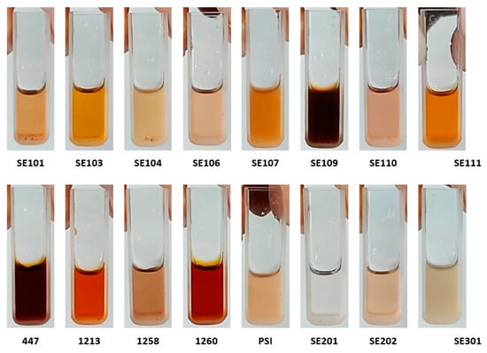
The AgNPs’ synthesis under biological conditions is described in the literature. Using this methodology, particles of nanometer size are commonly obtained. In this paper, extracts from different fungi were used for the synthesis of stable metal nanoparticles. The original fungal extracts were transparent but the addition of silver nitrate under homogenization for 72 h, turned the extract darker In both positive and negative controls, no color change was observed (Figure 1).

Figure 1.
Colors of the synthesis reaction of silver nanoparticles with aqueous extracts of different entomopathogenic fungi.
A color change during the reaction is attributed to the reduction of Ag+ ions in Ag0 and the formation of silver nanoparticles [25]. Moreover, there was a variation in the color of the synthesis reactions according to the extract used in the reduction of silver nitrate, ranging from light yellow (isolated 1258) to dark brown (isolated SE109 and 447). The plasmonic surface resonance (SPR) present in the nanoparticles promotes the color change of their synthesis reactions, being affected by the conformation of the nanoparticle inside the suspension [26].
A darker coloration in the synthesis reaction may indicate the presence of larger or aggregated nanoparticles, which tend to have peaks at a longer wavelength than smaller nanoparticles. The morphology of the nanoparticle also influences the distribution of electrons on the surface of the particle, affecting the color of its suspension. In addition, greater color intensity of the synthesis reaction is also related to the presence of a greater number of nanoparticles formed.
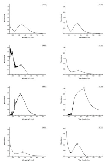
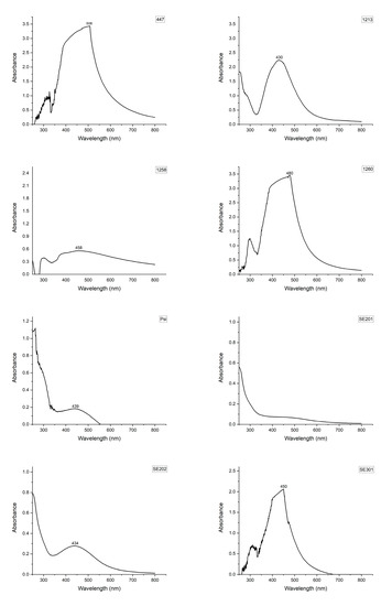
The UV/visible spectroscopy of the AgNPs showed absorbance peaks around 410–550 nm (Figure 2). According to the literature, peaks in the UV absorption spectrum between 400 and 550 nm are associated with the formation of nanoparticles [27]. The results of UV/visible spectroscopy of the synthesis reactions confirm the formation of nanoparticles by the action of tested entomopathogenic fungi extracts, except for the extract from the SE201 fungus.


Figure 2.
UV/visible absorption spectra of the synthesis reactions of silver nanoparticles with aqueous extracts of different entomopathogenic fungi. The used blank solution was the fungal extract solution.
Biomolecules (proteins, enzymes, vitamins, toxins, or other secondary metabolites) present in extracts of entomopathogenic fungi are responsible for reducing silver ions and forming AgNPs. The hydroxyl or carboxyl groups in these metabolites react with the metallic salt, promoting electron donation through an oxidation-reduction reaction, resulting in the formation of the nanoparticles.
Mean diameter and polydispersity index (PI) were also measured for AgNPs using dynamic light scattering (DLS) (Table 3).

Table 3.
Average diameter and polydispersity index of silver nanoparticles synthesized by entomopathogenic fungi extracts.
AgNPs showed a mean diameter ranging between 40.14 and 289.13 nm. Nanoparticles with a smaller size were synthesized using extracts 447 (40.14 nm), SE109 (61.14 nm), 1213 (69.36 nm), SE103 (77.08 nm), 1260 (77.56 nm), and SE110 (90.29 nm). AgNPs produced with species of entomopathogenic fungi B. bassiana, M. anisopliae, and I. fumosorosea are described in the literature. B. bassiana showed less than 100 nm [9,28] and a similar result was obtained for M. anisopliae [29] and I. fumosorosea [8]. PI varied between 0.165 and 0.627 (Table 3). This parameter assesses the homogeneity of the size of the suspended particles, and PI values up to 0.3 indicate the presence of nanoparticles with a low polydispersity [30]. Thus, among the smallest medium-sized nanoparticles, it was observed that the nanoparticles from the isolates SE110, SE103, 1260, and 1213 showed lower PI values (0.250–0.342), being considered monodisperse.
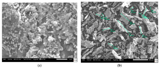
Spherical silver nanoparticles (Figure 3a) attached to the irregular fungal extract structure were observed by SEM, showing sizes between 200 and 600 nm (Figure 3b). These results suggest an irregular shape due to silver nanoparticle adherence.

Figure 3.
Scanning electron microscopy of silver nanoparticles at 3700× (a) and 50× (b).
Pearson’s correlation coefficients showed the existence of a correlation between the analyzed variables, with differences in significance and proportionality. The evaluation of the parameters evaluated in this study indicated the presence of a significant negative correlation between the potential for the synthesis of silver nanoparticles × production of fungal biomass, as well as the potential for synthesis of silver nanoparticles × LT50, suggesting that, with the increase in values for biomass production and LT50, there is a tendency to reduce the potential for the synthesis of silver nanoparticles (Table 4).

Table 4.
Coefficients of Pearson’s correlation (r) between biological parameters (pathogenicity, LT50, and biomass production) and silver nanoparticle synthesis potential with entomopathogenic fungi extracts.
The effect of Pearson’s correlation between variables is represented by the correlation coefficient (r) values that vary between −1 to +1, where zero indicates no correlation. Positive correlation coefficients represent the existence of a direct correlation between the variables, where both present the same trend (when the value of one variable increases, the value of the other variable also increases), and negative coefficients indicate the presence of an inverse correlation (when the value of one variable increases, the value of the other variable decreases). Moreover, Pearson’s coefficients can be classified by their values. Therefore, values between 0 and 0.3 (or 0 and −0.3) are considered weak and biologically negligible; values between 0.31 and 0.5 (or −0.31 and −0.5) refer to the presence of weak correlations; values between 0.51 and 0.7 (or −0.51 and −0.7) indicate moderate correlations; values between 0.71 and 0.9 (or −0.71 and −0.9) indicate strong correlations; and values >0.9 (or <−0.9) indicate the presence of very strong correlations [31].
The significance of the values calculated for Pearson’s coefficient demonstrates the presence of a strong negative correlation between the production of fungal biomass and synthesis of silver nanoparticles (−0.864, p < 0.05), indicating that an increase in the production of fungal biomass had a large influence in reducing the synthesis potential of the entomopathogenic fungi isolates. In addition, a moderate negative correlation was observed between the fungus virulence (LT50) and the potential for synthesis (−0.504, p < 0.01).
Mycelial growth, pathogenicity, and virulence of entomopathogenic fungi are biological aspects associated with the ability of the microorganism to secrete enzymes that assist in the degradation of nutrients in the environment, and subsequent absorption, and in the processes of penetration and colonization of the fungus in the insect host body. Lipases, proteases, and chitinases are the main enzymes that influence entomopathogenic fungi in their pathogen–host interaction, and their activation and regulation can promote the development of greater specificity of action between the microorganism and the target insect. Besides, entomopathogenic fungi can secrete other enzymes, such as amylases, involved in processes such as growth and absorption of nutrients in culture medium, as well as catalases and cellulases, which promote cellular protection against stress, reduced formation of reactive oxygen species (ROS) during fungus infection in the insect, and remedial action of toxic substances [32].
The formation of the silver nanoparticles in fungi-mediated biosynthesis occurs through the action of biomolecules produced by microorganisms, which promote an oxidation reaction of metal salts. However, there is no consensus as to which biomolecule is responsible for the synthesis process. It is believed that there may be variation in the enzymes involved in the biosynthesis of metallic nanoparticles depending on the species and/or fungus isolate used, making it difficult to understand the biological action in the development of these nanomaterials [33]. The presence of functional groups and amino acid residues referring to proteins in silver nanoparticles from biosynthesis mediated by B. bassiana, M. Anisopliae, and I. fumosorosea suggests the action of these compounds in the formation of nanoparticles [8,14]. Moreover, suppression of enzymes from the oxidoreductase group with the addition of potassium cyanide (KCN) to the extract of Metarhizium robertsii during the synthesis of silver nanoparticles significantly reduced the potential for the formation of nanoparticles, suggesting that the biomolecule responsible for the formation of the nanoparticle by this species of fungus may be an enzyme belonging to this group; however, the presence of KCN, which is a strong oxidizing agent, may have caused ionization of the silver atoms and interfered with the formation of the nanoparticle. Thus, the results contained in the literature do not fully clarify the mechanism of the formation of AgNPs in the biosynthesis of these entomopathogenic fungi.
Virulence (LT50) and the biomass production of the isolates showed a negative impact on the synthesis potential, suggesting that the biomolecules active in the process of the production of biomass, pathogenicity, and virulence of the studied fungi are not directly responsible for the formation of the silver nanoparticle. In our study, pathogenicity did not show any influence on the potential of entomopathogenic fungi isolates to synthesize silver nanoparticles; however, in a previous study, silver nanoparticles synthesized with aqueous extracts from fungi isolates with a high rate of pathogenicity on the target insect showed a more significant insecticidal effect [9], which can be caused by the presence of metabolites with insecticidal action in the reaction medium and their possible adhesion to the formed nanoparticle surface. Therefore, the existence of a significant correlation between the pathogenicity and potential for the synthesis of silver nanoparticles by our entomopathogenic fungi isolates was expected, but this was not observed. Thus, further evaluations are needed about the interaction of these parameters, which may serve to better understand the formation of nanoparticle by the action of the metabolites of these fungi.
4. Conclusions
The use of entomopathogenic fungi in the biological control of insects and for the synthesis of AgNPs was shown to be promising. New entomopathogenic fungi isolates have been studied to satisfy the demand for use in pest management programs and the development of biotechnological products, such as biosynthesis of AgNPs. Our study demonstrated that B. bassiana, M. anisopliae, and I. fumosorosea isolates present action against T. molitor, and differences in the production of mycelial biomass and potential of the synthesis of silver nanoparticles depending on the isolate used. It is also concluded that the increase in biomass production and virulence negatively influences the potential synthesis of silver nanoparticles.
Author Contributions
All authors have made a substantial contribution to the design, conceptualization, methodology, investigation, formal analysis, and data curation. T.S.S., E.M.d.P. and M.G.d.J.S. have contributed to the writing of the original draft, data, and literature research. E.B.S., P.S. and M.d.C.M. have contributed to the writing—review, validation, and editing. T.S.S., E.M.d.P., M.G.d.J.S., E.B.S., P.S. and M.d.C.M. have contributed to the project administration, provision of resources and software, supervision, and funding acquisition. All authors have read and agreed to the published version of the manuscript.
Funding
This work was supported by the Fundação de Amparo à Pesquisa do Estado de Sergipe (FAPITEC), Conselho Nacional de Desenvolvimento Científico e Tecnológico (CNPq, #443238/2014-6, #470388/2014-5) and Banco do Nordeste (FUNDECI/2017.0014), the Portuguese Science and Technology Foundation (FCT/MCT), and from European Funds (PRODER/COMPETE) for the project UIDB/04469/2020 (strategic fund), co-financed by FEDER, under the Partnership Agreement PT2020.
Data Availability Statement
Data are available from corresponding authors upon request.
Acknowledgments
The authors would like to thank the Coordination for the Improvement of Higher Education Personnel (Capes); Sergipe Agricultural Development Company (Emdagro); and the Industrial Biotechnology Program, University Tiradentes, for assistance during the research.
Conflicts of Interest
The authors declare no conflict of interest.
References
- Khan, M.R.; Fromm, K.M.; Rizvi, T.F.; Giese, B.; Ahamad, F.; Turner, R.J.; Füeg, M.; Marsili, E. Metal Nanoparticle–Microbe Interactions: Synthesis and Antimicrobial Effects. Part. Part. Syst. Charact. 2020, 1900419. [Google Scholar] [CrossRef]
- Moraga, E.Q. Entomopathogenic fungi as endophytes: Their broader contribution to IPM and crop production. Biocontrol Sci. Technol. 2020, 1–14. [Google Scholar] [CrossRef]
- Mora, M.A.E.; Castilho, A.M.C.; Fraga, M.E. Classification and infection mechanism of entomopathogenic fungi. Arq. Inst. Biol. 2017, 84. [Google Scholar] [CrossRef]
- Al-Ani, L.K.T.; Aguilar-Marcelino, L.; Fiorotti, J.; Sharma, V.; Sarker, M.S.; Furtado, E.L.; Wijayawardene, N.N.; Herrera-Estrella, A. Biological Control Agents and Their Importance for the Plant Health. In Microbial Services in Restoration Ecology; Elsevier: Amsterdam, The Netherlands, 2020; pp. 13–36. [Google Scholar]
- Das Chagas Bernardo, C.; Pereira-Junior, R.A.; Luz, C.; Mascarin, G.M.; Fernandes, É.K.K. Differential susceptibility of blastospores and aerial conidia of entomopathogenic fungi to heat and UV-B stresses. Fungal Biol. 2020. [Google Scholar] [CrossRef]
- Ghazanfar, M.U.; Hagenbucher, S.; Romeis, J.; Grabenweger, G.; Meissle, M. Fluctuating temperatures influence the susceptibility of pest insects to biological control agents. J. Pest Sci. 2020, 1–12. [Google Scholar] [CrossRef]
- Thaochan, N.; Benarlee, R.; Prabhakar, C.S.; Hu, Q. Impact of temperature and relative humidity on effectiveness of Metarhizium guizhouense PSUM02 against longkong bark eating caterpillar Cossus chloratus Swinhoe under laboratory and field conditions. J. Asia-Pac. Entomol. 2020, 23, 285–290. [Google Scholar] [CrossRef]
- Banu, A.N.; Balasubramanian, C. Optimization and synthesis of silver nanoparticles using Isaria fumosorosea against human vector mosquitoes. Parasitol. Res. 2014, 113, 3843–3851. [Google Scholar] [CrossRef] [PubMed]
- Kamil, D.; Prameeladevi, T.; Ganesh, S.; Prabhakaran, N.; Nareshkumar, R.; Thomas, S.P. Green Synthesis of Silver Nanoparticles by Entomopathogenic Fungus Beauveria bassiana and Their Bioefficacy Against Mustard Aphid (Lipaphis erysimi Kalt.); NISCAIR-CSIR: New Delhi, India, 2017. [Google Scholar]
- Diniz, F.R.; Maia, R.C.A.; Rannier, L.; Andrade, L.N.; VChaud, M.; Da Silva, C.F.; Corrêa, C.B.; De Albuquerque, R.L.C., Jr.; Da Costa, P.L.; Shin, S.R.; et al. Silver nanoparticles-composing alginate/gelatin hydrogel improves wound healing in vivo. Nanomaterials 2020, 10, 390. [Google Scholar] [CrossRef]
- Sánchez-López, E.; Gomes, D.; Esteruelas, G.; Bonilla, L.; Lopez-Machado, A.L.; Galindo, R.; Cano, A.; Espina, M.; Ettcheto, M.; Camins, A.; et al. Metal-Based Nanoparticles as Antimicrobial Agents: An Overview. Nanomaterials 2020, 10, 292. [Google Scholar] [CrossRef]
- Ahmad, A.; Mukherjee, P.; Senapati, S.; Mandal, D.; Khan, M.I.; Kumar, R.; Sastry, M. Extracellular biosynthesis of silver nanoparticles using the fungus Fusarium oxysporum. Colloids Surf. B Biointerfaces 2003, 28, 313–318. [Google Scholar] [CrossRef]
- Mascarin, G.M.; Alves, S.B.; Lopes, R.B. Culture media selection for mass production of Isaria fumosorosea and Isaria farinosa. Braz. Arch. Biol. Technol. 2010, 53, 753–761. [Google Scholar] [CrossRef]
- Amerasan, D.; Nataraj, T.; Murugan, K.; Panneerselvam, C.; Madhiyazhagan, M.; Nicoletti, G. Benelli, Myco-synthesis of silver nanoparticles using Metarhizium anisopliae against the rural malaria vector Anopheles culicifacies Giles (Diptera: Culicidae). J. Pest Sci. 2016, 89, 249–256. [Google Scholar] [CrossRef]
- Abbott, W.S. A method of computing the effectiveness of an insecticide. J. Econ. Entomol. 1925, 18, 265–267. [Google Scholar] [CrossRef]
- Adamski, Z.; Bufo, S.A.; Chowański, S.; Falabella, P.; Lubawy, J.; Marciniak, P.; Pacholska-Bogalska, J.; Salvia, R.; Scrano, L.; Słocińska, M.; et al. Beetles as Model Organisms in Physiological, Biomedical and Environmental Studies—A Review. Front. Physiol. 2019, 10, 319. [Google Scholar] [CrossRef]
- De Souza, P.C.; Morey, A.T.; Castanheira, G.M.; Bocate, K.P.; Panagio, L.A.; Ito, F.A.; Furlaneto, M.C.; Yamada-Ogatta, S.F.; Costa, I.N.; Mora-Montes, H.M.; et al. Tenebrio molitor (Coleoptera: Tenebrionidae) as an alternative host to study fungal infections. J. Microbiol. Methods 2015, 118, 182–186. [Google Scholar] [CrossRef] [PubMed]
- Oreste, M.; Bubici, G.; Poliseno, M.; Triggiani, O.; Tarasco, E. Pathogenicity of Beauveria bassiana (Bals.-Criv.) Vuill. and Metarhizium anisopliae (Metschn.) Sorokin against Galleria mellonella L. and Tenebrio molitor L. in laboratory assays. Redia 2012, 95, 43–48. [Google Scholar]
- Bhattarai, M.K.; Bhattarai, U.R.; Masoudi, A.; Feng, J.-N.; Wang, D. Pathogenicity and virulence of the entomopathogenic fungi depend on selective suppression of anti-oxidative and detoxification enzymes in Tenebriomolitor (Coleoptera: Tenebrionidae) larvae. Biochem. Cell Arch 2018, 1, 861–874. [Google Scholar]
- Santos, T.S.; De Freitas, A.C.; Poderoso, J.C.M.; Hernandez-Macedo, M.L.; Ribeiro, G.T.; Da Costa, L.P.; Da Costa Mendonça, M. Evaluation of isolates of entomopathogenic fungi in the genera Metarhizium, Beauveria, and Isaria, and their virulence to Thaumastocoris peregrinus (Hemiptera: Thaumastocoridae). Fla. Entomol. 2018, 101, 597–602. [Google Scholar] [CrossRef]
- Senthamizhlselvan, P.; Sujeetha, J.A.R.; Jeyalakshmi, C. Growth, sporulation and biomass production of native entomopathogenic fungal isolates on a suitable medium. J. Biopestic. 2010, 3, 466. [Google Scholar]
- Bhadauria, B.P.; Puri, S.; Singh, P. Mass production of entomopathogenic fungi using agricultural products. Bioscan 2012, 7, 229–232. [Google Scholar]
- Soundarapandian, P.; Chandra, R. Mass production of entomopathogenic fungus Metarhizium anisopliae (Deuteromycota; Hyphomycetes) in the laboratory. Res. J. Microbiol. 2007, 2, 690–705. [Google Scholar]
- El-Enshasy, H.A. Filamentous fungal cultures–Process characteristics, products, and applications. Bioprocess. Value Added Prod. Renew. Resour. 2007, 225–261. [Google Scholar] [CrossRef]
- Badi’ah, H.; Seedeh, F.; Supriyanto, G.; Zaidan, A. Synthesis of Silver Nanoparticles and the Development in Analysis Method. In IOP Conference Series: Earth and Environmental Science; IOP Publishing: Bristol, UK, 2019; p. 012005. [Google Scholar]
- Mulfinger, L.; Solomon, S.D.; Bahadory, M.; Jeyarajasingam, A.V.; Rutkowsky, S.A.; Boritz, C. Synthesis and study of silver nanoparticles. J. Chem. Educ. 2007, 84, 322. [Google Scholar] [CrossRef]
- Murei, A.; Ayinde, W.B.; Gitari, M.W.; Samie, A. Functionalization and antimicrobial evaluation of ampicillin, penicillin and vancomycin with Pyrenacantha grandiflora Baill and silver nanoparticles. Sci. Rep. 2020, 10, 11596. [Google Scholar] [CrossRef] [PubMed]
- Qamandar, M.A.; Shafeeq, M.A.A. Biosynthesis and properties of silver nanoparticles of fungus Beauveria bassiana. Int. J. Chem. Tech. Res. 2017, 10, 1073–1083. [Google Scholar]
- Peeran, M.; Deeba, K.; Lakshman, P. Extracellular myco-synthesis of silver nanoparticles from and Trichoderma virens and Metarhizzium anisopliae. J. Mycol. Plant Pathol. 2017, 47, 424–429. [Google Scholar]
- Sadeghi, R.; Etemad, S.G.; Keshavarzi, E.; Haghshenasfard, M. Investigation of alumina nanofluid stability by UV–Vis spectrum. Microfluid. Nanofluidics 2015, 18, 1023–1030. [Google Scholar] [CrossRef]
- Mukaka, M. Statistics corner: A guide to appropriate use of correlation in medical research. Malawi Med. J. 2012, 24, 69–71. [Google Scholar]
- Mondal, S.; Baksi, S.; Koris, A.; Vatai, G. Journey of enzymes in entomopathogenic fungi. Pac. Sci. Rev. A Nat. Sci. Eng. 2016, 18, 85–99. [Google Scholar] [CrossRef]
- Guilger-Casagrande, M.; Lima, R.D. Synthesis of Silver Nanoparticles Mediated by Fungi: A Review. Front. Bioeng. Biotechnol. 2019, 7. [Google Scholar] [CrossRef]
Publisher’s Note: MDPI stays neutral with regard to jurisdictional claims in published maps and institutional affiliations. |
© 2021 by the authors. Licensee MDPI, Basel, Switzerland. This article is an open access article distributed under the terms and conditions of the Creative Commons Attribution (CC BY) license (http://creativecommons.org/licenses/by/4.0/).