Authentication of an Old Violin by Multianalytical Methods

 ,
,  ,
,  and
and
Abstract
Featured Application
Abstract
1. Introduction
2. Experimental
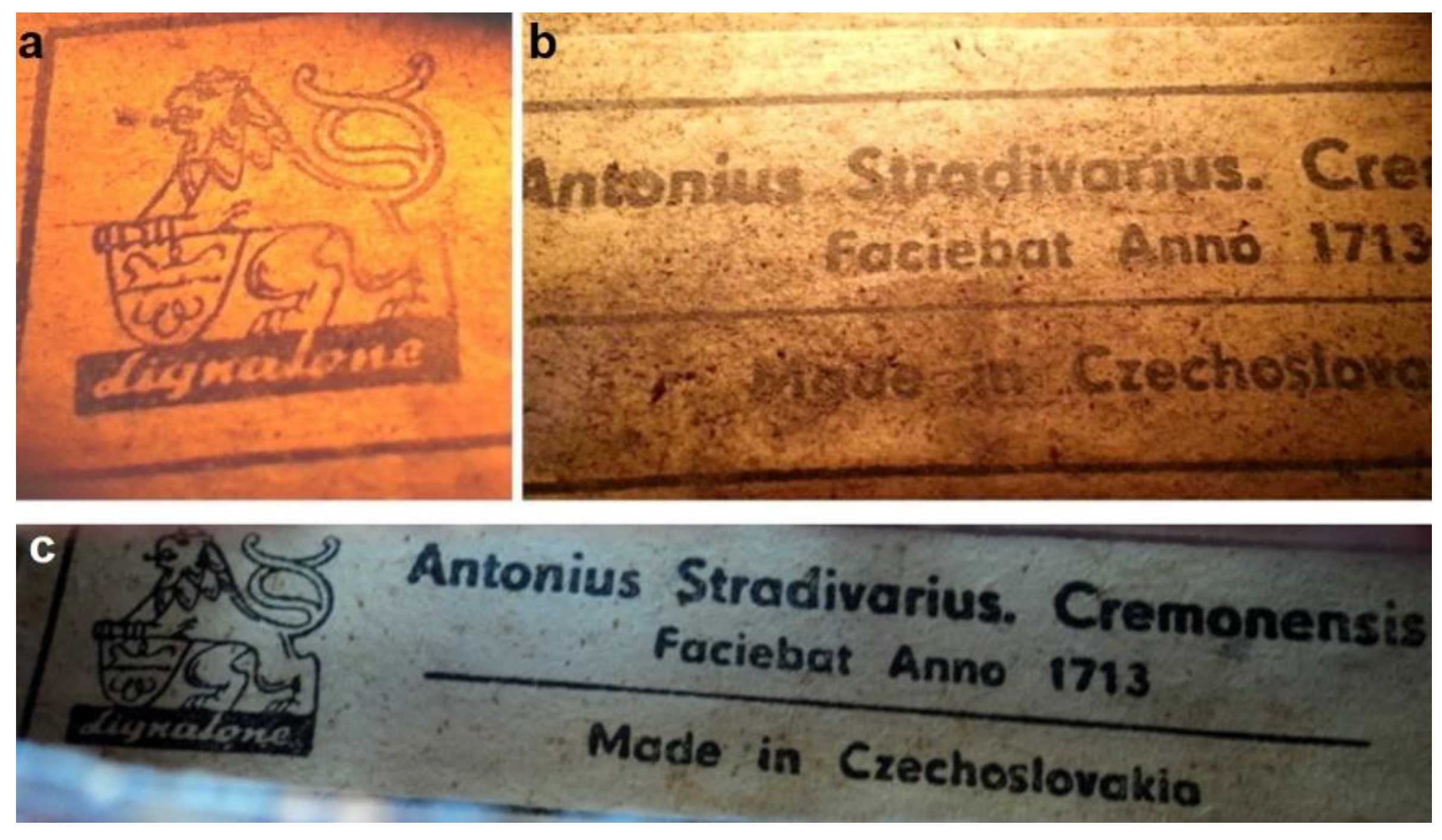
2.1. Violin History
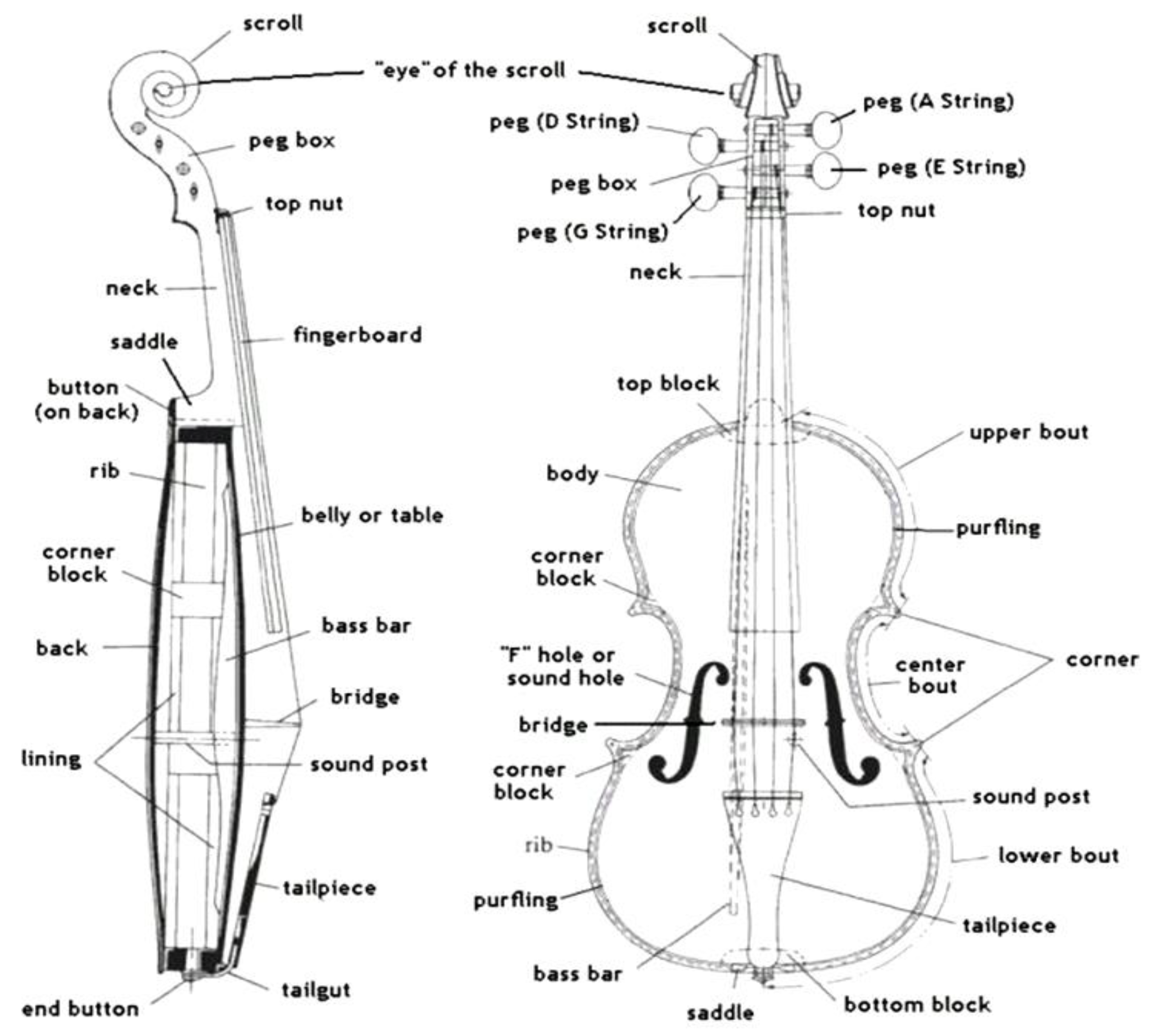
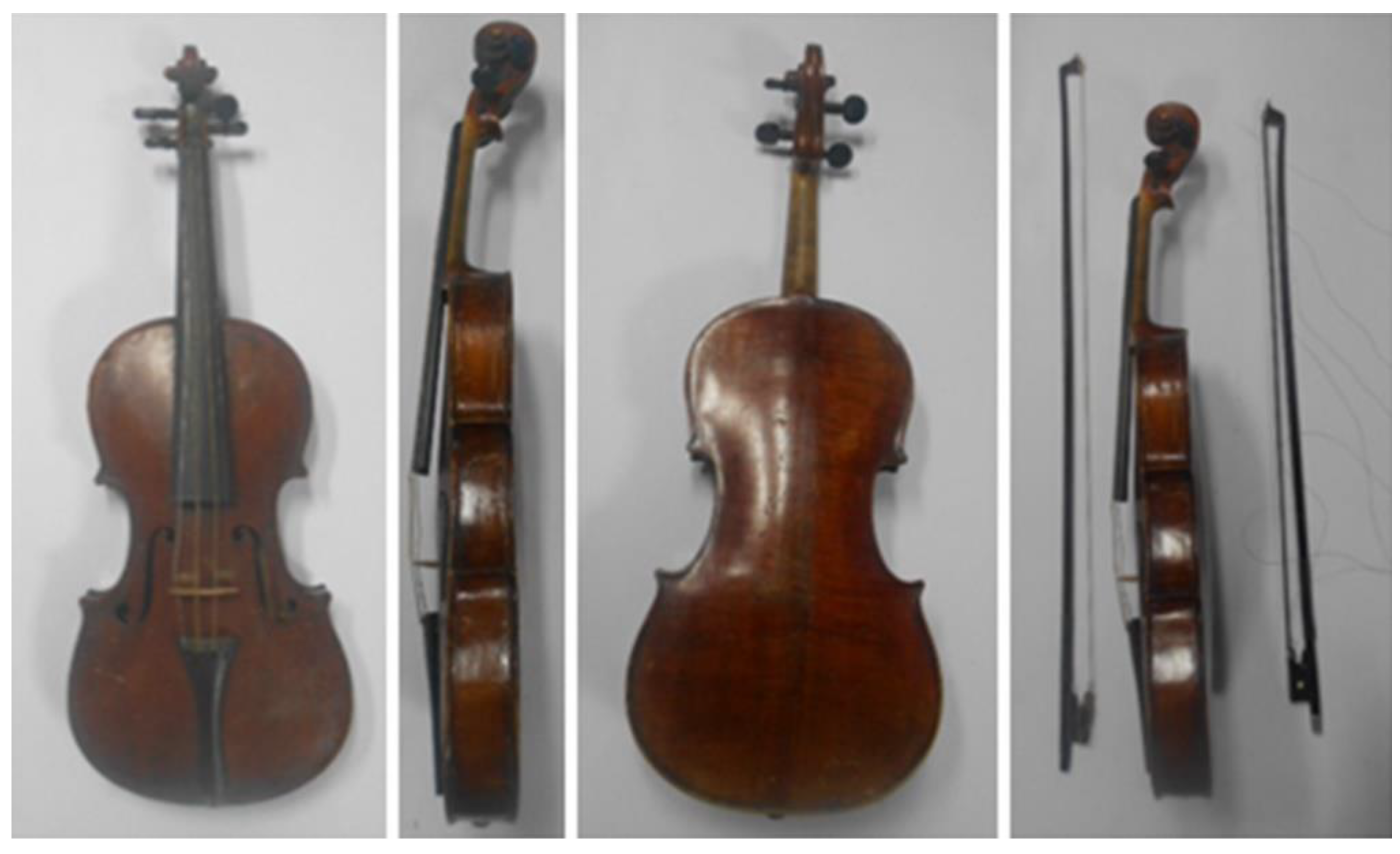
2.2. The Structural Size of the Violin
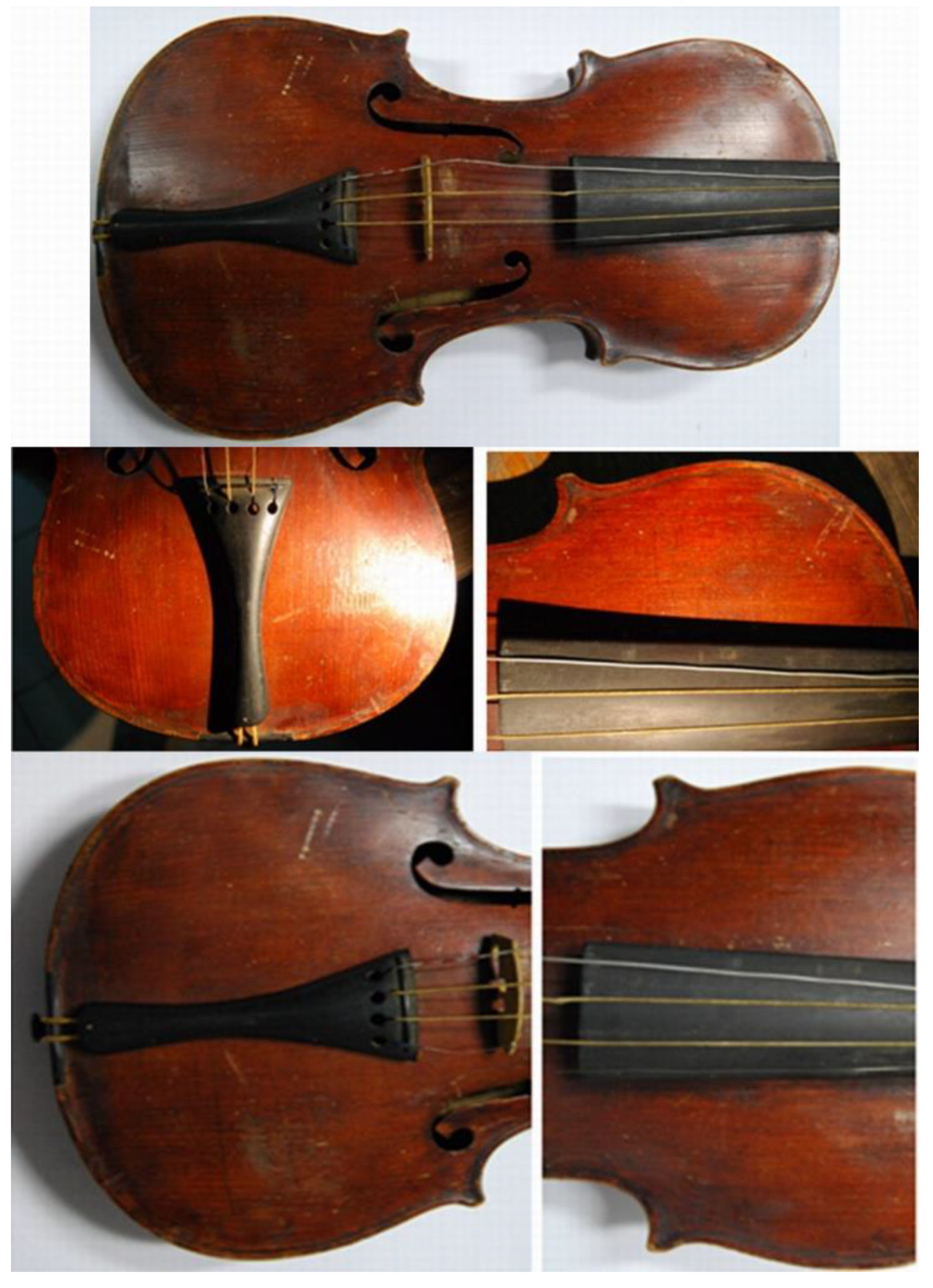
2.3. The State of Preservation
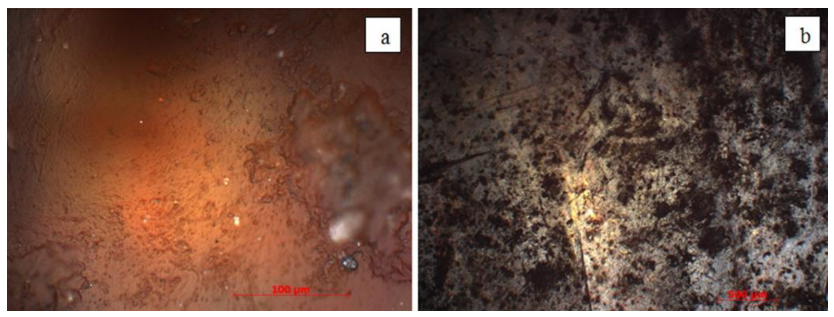
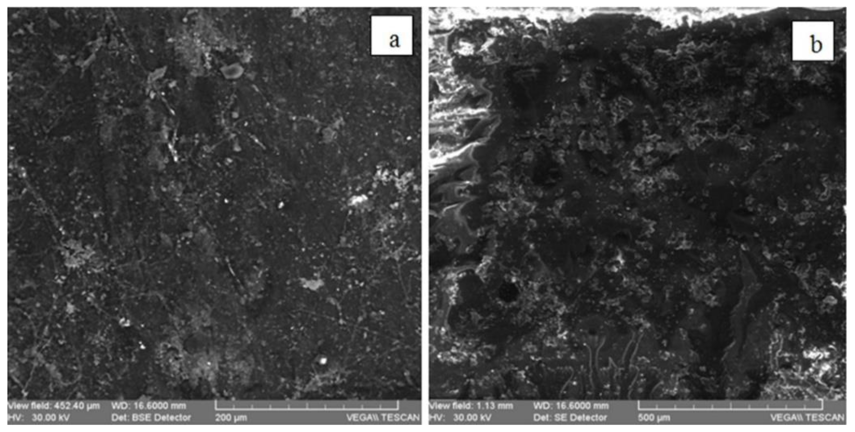
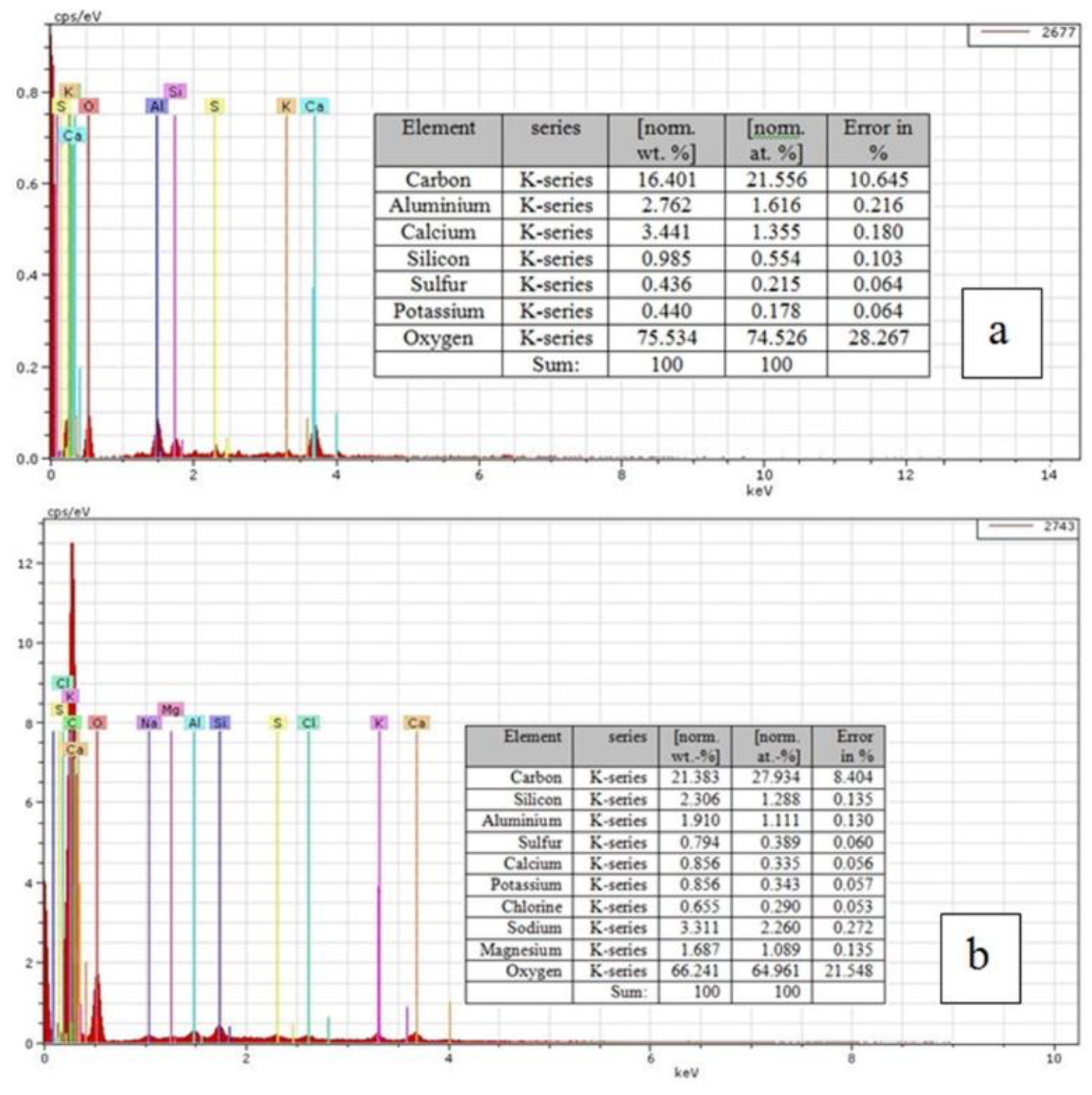
2.4. Methods Involved
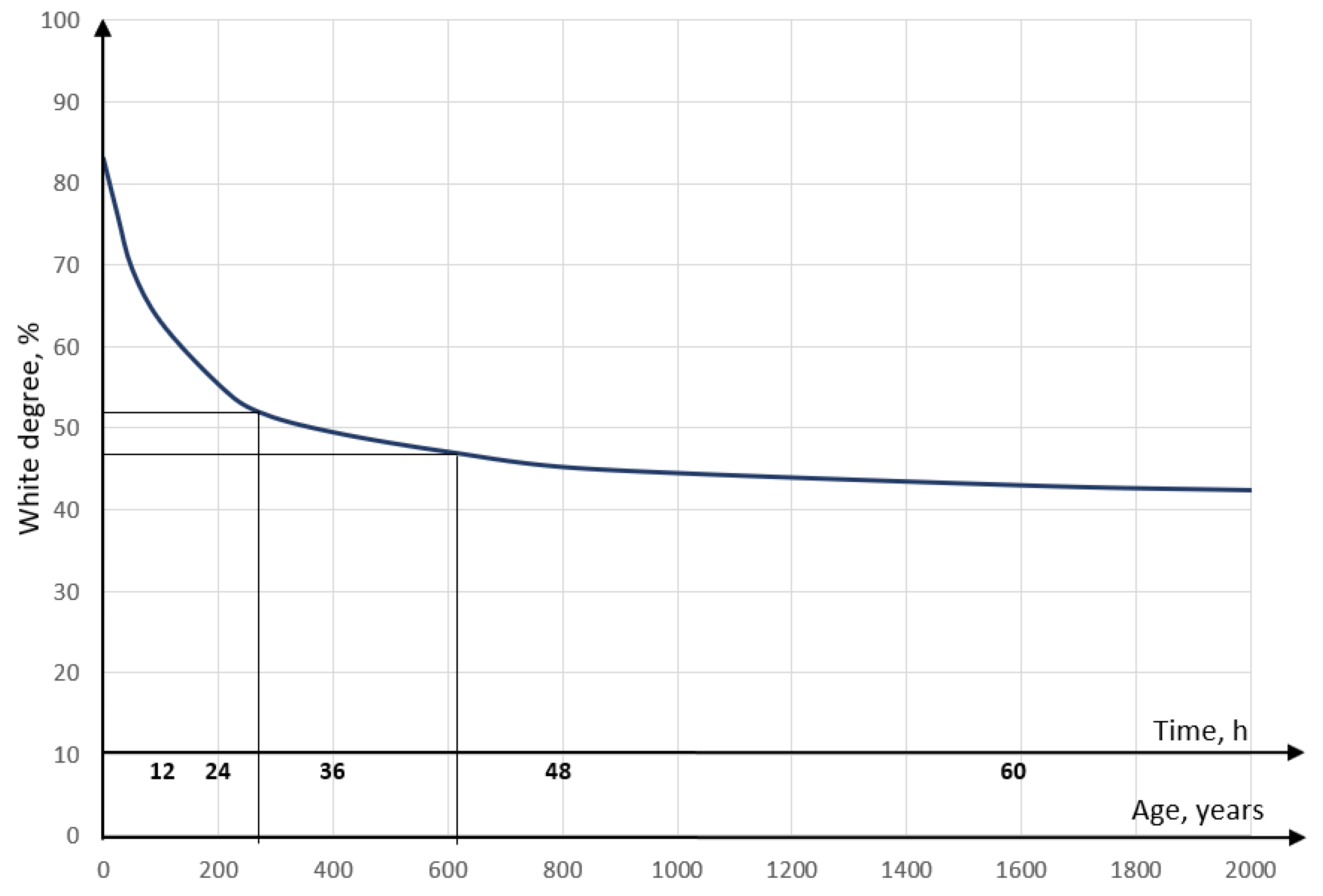
2.5. Determining the Age of the Label by White Degree
2.6. Determining the Age of the Label by Glycolysis Rate
2.7. Determination of the Archaeometric Characteristics of Varnish and Other Finishing Materials
3. Results and Discussions
3.1. Dating the Label by White Degree
3.2. Dating the Label by Glycolysis Rate
3.3. Determination of the Archaeometric Characteristics of the Varnish and Other Component Finishing Materials
4. Conclusions
Author Contributions
Funding
Conflicts of Interest
References
- Sandu, I.; Sandu, I.C.A.; van Sannen, A. Expertiza Stiințifică a Operelor de Artă; [Scientific Expertise of Works of Art]; Trinitas: Iași, Romania, 1998. [Google Scholar]
- Sandu, I. Aspecte Moderne Privind Conservarea Bunurilor Culturale, Vol. V—Identificarea Materialelor Picturale; [Modern Aspects Concerning the Conservation of Cultural Heritage. Volume V. Identification of Painting Materials]; Performantica: Iași, Romania, 2005. [Google Scholar]
- Cristache, R.A. Study of New Archaeometric Characteristics of Old Wood Artefacts. Ph.D. Thesis, Alexandru Ioan Cuza University, Iasi, Romania, 2015. [Google Scholar]
- Cortea, I.M.; Cristache, R.A.; Sandu, I. Characterization of historical violin varnishes using ATR-FTIR spectroscopy. Rom. Rep. Phys. 2016, 68, 615–622. [Google Scholar]
- Malagodi, M.; Canevari, C.; Bonizzoni, L.; Galli, A.; Maspero, F.; Martini, M. A multi-technique chemical characterization of a stradivari decorated violin top plate. Appl. Phys. A Mater. Sci. Process. 2013, 112, 225–234. [Google Scholar] [CrossRef]
- Brandmair, B.; Greiner, S.P. Stradivari Varnish, Scientific Analysis of His Finishing Technique on Selected Instruments; London, UK; Munich, Germany, 2010. [Google Scholar]
- Daher, C.; Paris, C.; Le Hô, A.S.; Bellot-Gurlet, L.; Echard, J.P. A joint use of Raman and infrared spectroscopies for the identification of natural organic media used in ancient varnishes. J. Raman Spectrosc. 2010, 41, 1494–1499. [Google Scholar] [CrossRef]
- Sacconi, S.F. The “Secrets” of Stradivari; Libreria del Convegno: Cremona, Italy, 1979. [Google Scholar]
- Weththimuni, M.L.; Canevari, C.; Legnani, A.; Licchelli, M.; Malagodi, M.; Ricca, M.; Zeffiro, A. Experimental characterization of oil-colophony varnishes, a preliminary study. Int. J. Conserv. Sci. 2016, 7, 813–826. [Google Scholar]
- Bertrand, L.; Robinet, L.; Cohen, S.X.; Sandt, C.; Le Hô, A.S.; Soulier, B.; Lattuati- Derieux, A.; Echar, J.P. Identification of the finishing technique of an early eighteenth century musical instrument using FTIR spectromicroscopy. Anal. Bioanal. Chem. 2011, 399, 3025–3032. [Google Scholar] [CrossRef] [PubMed]
- Chen, G.F. Developments in the field of colophony chemistry and its implications in coatings. Prog. Org. Coat. 1992, 20, 139–167. [Google Scholar] [CrossRef]
- Echard, J.P.; Benoit, C.; Peris-Vicente, J.; Malecki, V.; Gimeno-Adelantado, J.V.; Vaiedelich, S. Gas chromatography/mass spectrometry characterization of historical varnishes of ancient Italian lutes and violin. Anal. Chim. Acta 2007, 584, 172–180. [Google Scholar] [CrossRef]
- Echard, J.P.; Bertrand, L.; Von Bohlen, A.; Le Hô, A.-S.; Paris, C.; Bellot-Gurlet, L.; Soulier, B.; Lattuati-Derieux, A.; Thao, S.; Robinet, L.; et al. The nature of the extraordinary finish of Stradivari’s instruments. Angew. Chem. Int. Ed. 2010, 49, 197–201. [Google Scholar] [CrossRef]
- Fioravanti, M.; Goli, G.; Carlson, B. Viscoelastic and mechano-sorptive studies appliaed to the conservation of historical violins, a case study of the Guarneri “del Gesù” violin (1743) known as the “Cannone”. J. Cult. Herit. 2013, 14, 297–303. [Google Scholar] [CrossRef]
- Noguera, J.R.; Galiano, F.C.B.; Lòpez, J.M.R.; Vivas, M.A.F.; Sànchez, I.M. Study of biodegradation of diterpenic varnishes used in art painting, colophony and Venetian turpentine. Int. Biodeterior. Biodegrad. 2008, 62, 427–433. [Google Scholar] [CrossRef]
- Scalarone, D.; Lazzari, M.; Chiantore, O. Ageing behaviour and pyrolytic characterization of diterpenic resins used as art materials, colophony and Venice turpentine. J. Anal. Appl. Pyrolysis 2002, 64, 345–361. [Google Scholar] [CrossRef]
- Caruso, F.; Chillura Martino, D.F.; Saverwyns, S.; van Bos, M.; Burgio, L.; di Stefano, C.; Peschke, G.; Caponetti, E. Micro-analytical identification of the components of varnishes from South Italian historical musical instruments by PLM, ESEM–EDX, microFTIR, GC–MS, and Py–GC–MS. Microchem. J. 2014, 116, 31–40. [Google Scholar] [CrossRef]
- Echard, J.P.; Lavedrine, B. Review on the characterisation of ancient stringed musical instruments varnishes and implementation of an analytical strategy. J. Cult. Herit. 2008, 9, 420–429. [Google Scholar] [CrossRef]
- Bucur, V. Acoustics of Wood, 2nd ed.; Springer Series in Wood Sciences; Springer: Berlin, Germany, 2006; ISBN 3540261230. [Google Scholar]
- Wegst, U. Wood for sound. Am. J. Bot. 2006, 93, 1439–1448. [Google Scholar] [CrossRef] [PubMed]
- Available online: https://s-media-cache-ak0.pinimg.com/736x/fa/53/fe/fa53fe872e9b348160baea08be78782c—violin-parts-musical-instruments.jpg (accessed on 3 March 2019).
- Sandu, I.C.A.; Sandu, I.G.; Cudelcu, D.T.; Nicula, S. Method for Determining the Age of Supports Made of Cellulose Material. Patent RO116844, 29 June 2001. [Google Scholar]
- Sandu, I.C.A.; Sandu, I.; Sandu, I.G.; Sandu, A.V. Method for Determining the Age of Textile Cellulose Materials. Patent RO121151/2006-12-29.
- Sandu, I.C.A.; Luca, C.; Sandu, I. A study on the paintings cloth-supports ageing degradation. Rev. Chim. 1999, 50, 902–908. [Google Scholar]
- Sandu, I.; Sandu, I.C.A. Chimia Conservarii si Restaurarii; Chemistry of Conservation and Restauration: Corson, Romania, 2002. [Google Scholar]
- Sandu, I.C.A.; Luca, C.; Sandu, I.; Vasilache, V.; Sandu, I.G. Research concerning the evaluation of the ageing of some soft weed supports of old paintings with preparation layer. III—The thermogravimetric analysis. Rev. Chim. 2002, 53, 607–615. [Google Scholar]
- Tai, H.C.; Li, G.C.; Huang, S.J.; Jhu, C.R.; Chung, J.H.; Wang, B.Y.; Hsu, C.S.; Brandmair, B.; Chung, D.T.; Chen, H.M.; et al. Chemical distinctions between Stradivari’s maple and modern tonewood. Proc. Natl. Acad. Sci. USA 2017, 114, 27–32. [Google Scholar] [CrossRef]
- Caruso, F.; Saverwyns, S.; van Bos, M.; Chillura Martino, D.F.; Ceulemans, A.-E.; De Valck, J.; Caponetti, E. Micro-X-ray fluorescence and the old masters. Appl. Phys. A 2012, 107, 197–202. [Google Scholar] [CrossRef][Green Version]
- Budu, A.-M.; Sandu, I.; Vasilache, V.; Simionescu, A.-E.; Sandu, I.C.A. Effect of skin lipids on painting layrs of the icons. Rev. Chim. 2015, 66, 1212–1216. [Google Scholar]
- Lattuati-Derieux, A.; Gomes, S.; Tirat, S.; Thao-Hey, S.; Echard, J.-P. New insights into molecular evolution of oil/colophony varnishes, towards pyrolysis-gas chromatography/mass spectrometry based quantitation. E-Preserv. Sci. 2014, 11, 53–63. [Google Scholar]
- Baraldi, P. Manoscritto Bolognese 2861 (XV secolo); Biblioteca Universitaria di Bologna: Bologna, Italy.
- Frezzato, F.; Seccaroni, C. Segreti di Arti Diverse Nel Regno di Napoli—Manoscritto It. III 10 Della Biblioteca Marciana di Venezia; Il Prato: Saonara, Italy, 2010. [Google Scholar]
- Azemard, C.; Vieillescazes, C.; Menager, M. Effect of photodegradation on the identification of natural varnishes by FT-IR spectroscopy. Microchem. J. 2014, 112, 137–149. [Google Scholar] [CrossRef]
- Beltran, V.; Salvadó, N.; Butí, S.; Cinque, G. Micro infrared spectroscopy discrimination capability of compounds in complex matrices of thin layers in real sample coatings from artworks. Microchem. J. 2015, 118, 115–123. [Google Scholar] [CrossRef]
- Daher, C.; Pimenta, V.; Bellot-Gurlet, L. Towards a non-invasive quatitative analysis of the organic components in museum objects varnishes by vibrational spectroscopies, Methodological approach. Talanta 2014, 129, 336–345. [Google Scholar] [CrossRef][Green Version]
- Mallégol, J.; Gardette, J.L.; Jacques, L. Long-term behaviour of oil-based varnishes and paints I. Spectroscopic analysis of curing drying oils. J. Am. Oil Chem. Soc. 1999, 76, 967–976. [Google Scholar] [CrossRef]
- Mallégol, J.; Gardette, J.L.; Jacques, L. Long-term behaviour of oil-based varnishes and paints. Photo- and thermooxidation of cured linseed oil. J. Am. Oil Chem. Soc. 1999, 77, 257–263. [Google Scholar] [CrossRef]
- De la Rie, E.R. Photochemical and thermal degradation of films of dammar resin. Stud. Conserv. 1988, 33, 53–70. [Google Scholar]
- Derrick, M.R.; Stulik, D.; Landry, J.M. Infrared Spectroscopy in Conservation Science; Getty Conservation Institute: Los Angeles, CA, USA, 1999. [Google Scholar]
- Kendix, E.L. Transmission and Reflection (ATR) Far-Infrared Spectroscopy Applied in the Analysis of Cultural Heritage Materials. Ph.D. Thesis, University of Bologna, Bologna, Italy, 2009. [Google Scholar]
- Dietemann, P.; Higgitt, C.; Kälin, M.; Edelmann, M.J.; Knochenmuss, R.; Zenobi, R. Aging and yellowing of triterpenoid resin varnishes—Influence of aging conditions and resin composition. J. Cult. Herit. 2009, 10, 30–40. [Google Scholar] [CrossRef]
- Sarmiento, A.; Perez-Alonso, M.; Olivares, M.; Castro, K.; Martínez-Arkarazo, I.; Fernández, L.A.; Madariaga, J.M. Classification and identification of organic binding media in artworks by means of fourier transform infrared spectroscopy and principal component analysis. Anal. Bioanal. Chem. 2011, 399, 3601–3611. [Google Scholar] [CrossRef]
- Van der Doelen, G.A.; Van der Berg, K.L.; Boon, J.J.; Shibayama, N.; De la Rie, R.E.; Genuit, W.J.L. Analysis of fresh triterpenoid resins and aged triterpenoid varnishes by high performance liquid chromatography–Atmospheric pressure chemical ionisation (tandem) mass spectrometry. J. Chromatogr. A 1998, 809, 21–37. [Google Scholar]
- Van der Weerd, J.; Van Loon, A.; Boon, J.J. FTIR studies of the effects of pigments on the aging of oil. Stud. Conserv. 2005, 50, 3–22. [Google Scholar] [CrossRef]


| Assigned Functional Groups | Linseed Oil | Colophony | ||||
|---|---|---|---|---|---|---|
| Theor. | New | Old | Theor. | New | Old | |
| υ(OH) | 3450 | - | - | 3443 | 3455.64 | 3449.86 |
| υ(CH) in CH2=CRR’ | - | 3075 | - | - | ||
| υ(CH)as in CH2 | 2926 | - | - | 2935 | 2943.67 | 2903.31 |
| υ(CH)s in CH3/CH | - | - | - | 2870 | 2878.89 | - |
| υ(CH)s in CH2 | 2852 | 2750.63 | 2720.89 | - | - | - |
| ν(=C–H) | 3079–2650 | 2161.63 | 2138.03 | |||
| ν(COOH)/ν(C=O) | 2050–1950 | 2028.47 | 2048.97 | |||
| υ(C=O) | 1741 | 1738.30 | 1733.09 | |||
| υ(–CH=CH2) | 1650 | 1662.95 | 1657.45 | |||
| ν(CONH) ** | 1547 | 1513.28 | 1512.20 | |||
| δ(CH)as in CH3/CH2 | 1460 | 1458 | 1465.41 | |||
| δ(CONH) ** | 1430 | 1427.89 | 1429.26 | |||
| δ(CH)as in CH3 | 1374 | 1384 | 1377.58 | 1376.06 | ||
| δ(CONH) ** | 1315 | 1323.18 | 1320.84 | |||
| δ(–C=C) | 1270–1230 | 1276.25 | 1276.06 | |||
| δ(C–O) | 1164–1097 | 1123.72 | 1122.46 | 1179 | 1166.92 | 1172.84 |
| δ(C–O–C) | 1044 | 1083.19 | 1090.48 | |||
| δ(COH) | 824 | 895.47 | 897.70 | |||
| δ(C=C) carbon skeleton | 630–610 | 614.37 | 624.625 | 745–714 | 725.89 | 740.57 |
© 2019 by the authors. Licensee MDPI, Basel, Switzerland. This article is an open access article distributed under the terms and conditions of the Creative Commons Attribution (CC BY) license (http://creativecommons.org/licenses/by/4.0/).
Share and Cite
Sandu, I.; Tanasa, P.O.; Sandu, I.C.A.; Negru, I.C.; Sandu, A.V.; Vasilache, V. Authentication of an Old Violin by Multianalytical Methods. Appl. Sci. 2020, 10, 306. https://doi.org/10.3390/app10010306
Sandu I, Tanasa PO, Sandu ICA, Negru IC, Sandu AV, Vasilache V. Authentication of an Old Violin by Multianalytical Methods. Applied Sciences. 2020; 10(1):306. https://doi.org/10.3390/app10010306
Chicago/Turabian StyleSandu, Ion, Petru Ovidiu Tanasa, Irina Crina Anca Sandu, Ioan Cristinel Negru, Andrei Victor Sandu, and Viorica Vasilache. 2020. "Authentication of an Old Violin by Multianalytical Methods" Applied Sciences 10, no. 1: 306. https://doi.org/10.3390/app10010306
APA StyleSandu, I., Tanasa, P. O., Sandu, I. C. A., Negru, I. C., Sandu, A. V., & Vasilache, V. (2020). Authentication of an Old Violin by Multianalytical Methods. Applied Sciences, 10(1), 306. https://doi.org/10.3390/app10010306