

Lesion Distribution in the Metacarpophalangeal and Metatarsophalangeal Region of 341 Horses Using Standing Magnetic Resonance Imaging
 ,
,  , ,
, ,
Abstract
Simple Summary
Abstract
1. Introduction
2. Materials and Methods
2.1. Inclusion Criteria
2.2. Statistical Analysis
3. Results
3.1. Sample Population
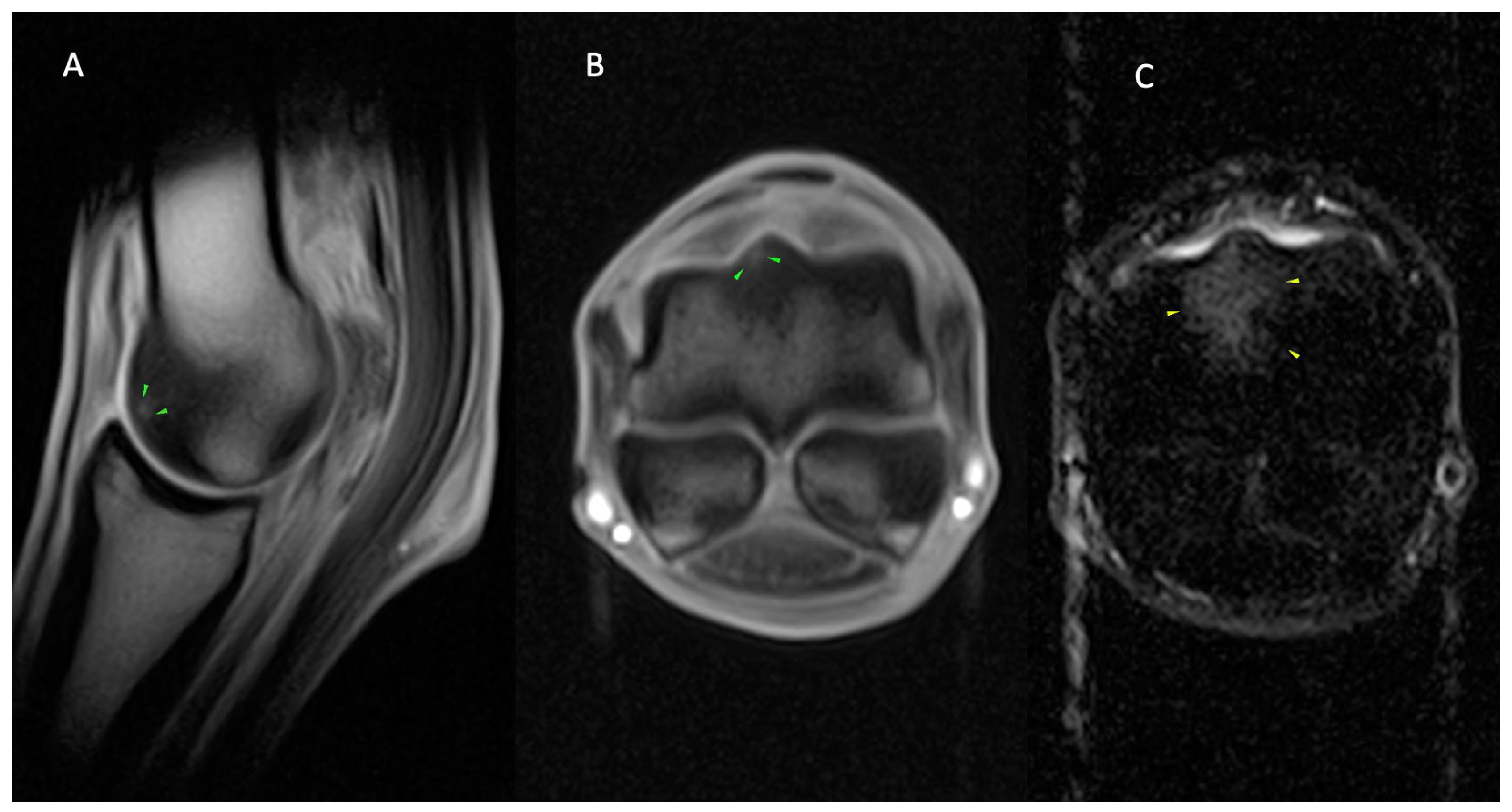
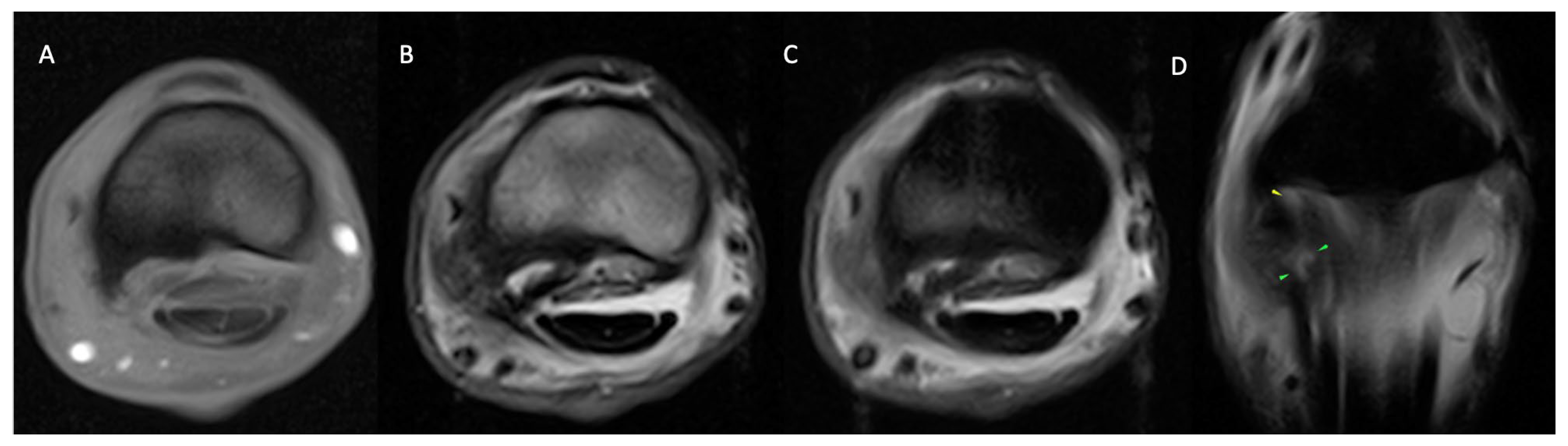
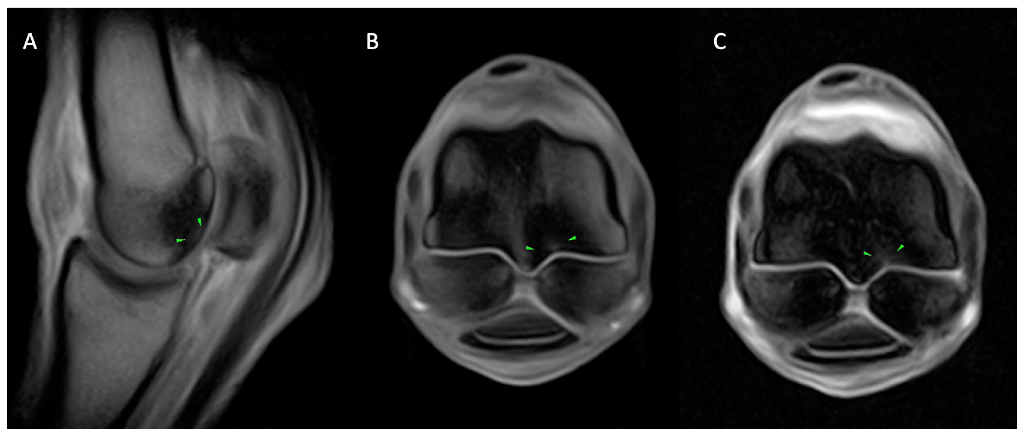
3.2. MRI Findings
3.2.1. Osseous Pathology
3.2.2. Joint Pathology
3.2.3. Soft Tissue Pathology
3.3. Comparison between Metacarpophalangeal and Metatarsophalangeal Regions
3.4. Comparison between Sports Disciplines
3.4.1. Flat Racing
3.4.2. Dressage
3.4.3. Endurance
3.4.4. Show Jumping
3.4.5. Eventing
3.4.6. Pleasure Riding
4. Discussion
5. Conclusions
Supplementary Materials
Author Contributions
Funding
Institutional Review Board Statement
Informed Consent Statement
Data Availability Statement
Acknowledgments
Conflicts of Interest
References
- Dyson, S.J.; Murray, R.C. Osseous trauma in the fetlock region of mature sports horses. In Proceedings of the American Association of Equine Practitioners Annual Convention, San Antonio, TX, USA, 2–6 December 2006; Volume 52, pp. 443–456. [Google Scholar]
- Riggs, C.; Whitehouse, G.; Boyde, A. Pathology of the distal condyles of the third metacarpal and third metatarsal bones of the horse. Equine Vet. J. 1999, 31, 140–148. [Google Scholar] [CrossRef] [PubMed]
- Riggs, C.; Whitehouse, G.; Boyde, A. Structural variation of the distal condyle of the third metacarpal and third metatarsal bones in the horse. Equine Vet. J. 1999, 31, 130–139. [Google Scholar] [CrossRef] [PubMed]
- Pool, R.R. Multidisciplinary investigation of the aetiopathogenesis of parasagittal fractures of the third metacarpal and metatarsal bones of Thoroughbreds. Equine Vet. J. 1999, 31, 96. [Google Scholar] [CrossRef] [PubMed]
- Gonzalez, L.M.; Schramme, M.C.; Robertson, I.D.; Thrall, D.E.; Redding, R.W. MRI features of metacarpo(tarso)phalangeal region lameness in 40 horses. Vet. Radiol. Ultrasound 2010, 51, 404–414. [Google Scholar] [CrossRef] [PubMed]
- Zubrod, C.J.; Schneider, R.K.; Tucker, R.L.; Gavin, P.R.; Ragle, C.A.; Farnsworth, K.D. Use of magnetic resonance imaging for identifying subchondral bone damage in horses: 11 cases (1999–2003). J. Am. Vet. Med. Assoc. 2004, 224, 411–418. [Google Scholar] [CrossRef] [PubMed]
- Sherlock, C.E.; Mair, T.S.; Braake, F. Osseous lesions in the metacarpo(tarso)phalangeal joint diagnosed using low-field magnetic resonance imaging in standing horses. Vet. Radiol. Ultrasound 2009, 50, 13–20. [Google Scholar] [CrossRef] [PubMed]
- Bachmann, G.F.; Basad, E.; Rauber, K.; Damian, M.S.; Rau, W.S. Degenerative joint disease on MRI and physical activity: A clinical study of the knee joint in 320 patients. Eur. Radiol. 1999, 9, 145–152. [Google Scholar] [CrossRef] [PubMed]
- Dyson, S.J.; Murray, R.C. Magnetic resonance imaging of the equine fetlock. Clin. Tech. Equine Pract. 2007, 6, 62–77. [Google Scholar] [CrossRef]
- Smith, M.A.; Dyson, S.J.; Rachel, C.M. The appearance of the equine metacarpophalangeal region on high-field vs. standing low-field magnetic resonance imaging. Vet. Radiol. Ultrasound 2011, 52, 61–70. [Google Scholar] [CrossRef] [PubMed]
- Auth, A.K.; Hinnigan, G.J.; Smith, M.A.; Owen, K.R. Low-Field Magnetic Resonance Imaging Findings of the Fetlock Region of Nonracehorses. J. Equine Vet. Sci. 2024, 132, 104938. [Google Scholar] [CrossRef]
- Tranquille, C.; Parkin, T.; Murray, R. Magnetic resonance imaging-detected adaptation and pathology in the distal condyles of the third metacarpus, associated with lateral condylar fracture in Thoroughbred racehorses. Equine Vet. J. 2012, 44, 699–706. [Google Scholar] [CrossRef] [PubMed]
- Tranquille, C.; Murray, R.; Parkin, T. Can we use subchondral bone thickness on high-field magnetic resonance images to identify Thoroughbred racehorses at risk of catastrophic lateral condylar fracture? Equine Vet. J. 2017, 49, 167–171. [Google Scholar] [CrossRef] [PubMed]
- Olive, J.; D’Anjou, M.A.; Alexander, K.; Laverty, S.; Theoret, C. Comparison of magnetic resonance imaging, computed tomography, and radiography for assessment of noncartilaginous changes in equine metacarpophalangeal osteoarthritis. Vet. Radiol. Ultrasound 2010, 51, 267–279. [Google Scholar] [CrossRef] [PubMed]
- O’Brien, T.; Baker, T.A.; Brounts, S.H.; Sample, S.J.; Markel, M.D.; Scollay, M.C.; Marquis, P.; Muir, P. Detection of articular pathology of the distal aspect of the third metacarpal bone in thoroughbred racehorses: Comparison of radiography, computed tomography and magnetic resonance imaging. Vet. Surg. 2011, 40, 942–951. [Google Scholar] [CrossRef] [PubMed]
- Olive, J.; D’anjou, M.A.; Girard, C.; Laverty, S.; Theoret, C. Fat-suppressed spoiled gradient-recalled imaging of equine metacarpophalangeal articular cartilage. Vet. Radiol. Ultrasound 2010, 51, 107–115. [Google Scholar] [CrossRef] [PubMed]
- Olive, J.; D’anjou, M.A.; Girard, C.; Laverty, S.; Theoret, C.L. Imaging and histological features of central subchondral osteophytes in racehorses with metacarpophalangeal joint osteoarthritis. Equine Vet. J. 2009, 41, 859–864. [Google Scholar] [CrossRef] [PubMed]
- Powell, S. Low-field standing magnetic resonance imaging findings of the metacarpo/metatarsophalangeal joint of racing Thoroughbreds with lameness localised to the region: A retrospective study of 131 horses. Equine Vet. J. 2012, 44, 169–177. [Google Scholar] [CrossRef] [PubMed]
- Gold, S.J.; Werpy, N.M.; Gutierrez-Nibeyro, S.D. Injuries of the Sagittal Groove of the Proximal Phalanx in Warmblood Horses Detected with Low-Field Magnetic Resonance Imaging: 19 Cases (2007–2016). Vet. Radiol. Ultrasound 2017, 58, 344–353. [Google Scholar] [CrossRef] [PubMed]
- Faulkner, J.E.; Joostens, Z.; Broeckx, B.J.G.; Hauspie, S.; Mariën, T.; Vanderperren, K. Follow-Up Magnetic Resonance Imaging of Sagittal Groove Disease of the Equine Proximal Phalanx Using a Classification System in 29 Non-Racing Sports Horses. Animals 2024, 14, 34. [Google Scholar] [CrossRef]
- Sampson, S.N.; Schneider, R.K.; Tucker, R.L.; Gavin, P.R.; Zubrod, C.J.; Ho, C.P. Magnetic resonance imaging features of oblique and straight distal sesamoidean desmitis in 27 horses. Vet. Radiol. Ultrasound 2007, 48, 303–311. [Google Scholar] [CrossRef]
- Smith, S.; Dyson, S.J.; Murray, C.M. Magnetic resonance Imaging of the distal sesamoidean ligament injury. Vet. Radiol. Ultrasound 2008, 49, 516–528. [Google Scholar] [CrossRef] [PubMed]
- Elemmawy, Y.M.; Senna, N.A.; Abu-Seida, A.M.; Youssef, A.F. Suspensory branch desmitis in a horse: Ultrasonography, computed tomography, magnetic resonance imaging, and gross postmortem findings. J. Equine Vet. Sci. 2019, 80, 49–55. [Google Scholar] [CrossRef] [PubMed]
- Labens, R.; Schramme, M.C.; Murray, R.C.; Bolas, N. Standing low-field MRI of the equine proximal metacarpal/metatarsal region is considered useful for diagnosing primary bone pathology and makes a positive contribution to case management: A prospective survey study. Vet. Radiol. Ultrasound 2020, 61, 197–205. [Google Scholar] [CrossRef] [PubMed]
- Murray, R.C.; Tranquille, C.A.; Walker, V.A.; Milmine, R.C.; Bak, L.; Tacey, J.B.; Bolas, N.M. Magnetic resonance imaging findings in the proximal metacarpal region of 359 horses and proximal metatarsal region of 64 horses acquired under standing sedation. J. Equine Vet. Sci. 2020, 94, 103268. [Google Scholar] [CrossRef] [PubMed]
- Murray, R.C.; Walters, J.M.; Snart, H.; Dyson, S.J.; Parkin, T.D. Identification of risk factors for lameness in dressage horses. Vet. J. 2010, 184, 27–36. [Google Scholar] [CrossRef] [PubMed]
- Murray, R.C.; Dyson, S.J.; Tranquille, C.A.; Adams, V. Association of type of sport and performance level with anatomical site of orthopaedic injury diagnosis. Equine Vet. J. 2006, 38, 411–416. [Google Scholar] [CrossRef] [PubMed]
- Egenvall, A.; Tranquille, C.A.; Lönnell, A.C.; Bitschnau, C.; Oomen, A.; Hernlund, E.; Montavon, S.; Franko, M.A.; Murray, R.C.; Weishaupto, M.A.; et al. Days-lost to training and competition in relation to workload in 263 elite show-jumping horses in four European countries. Prev. Vet. Med. 2013, 112, 387–400. [Google Scholar] [CrossRef]
- Leach, D.H. Assessment of bipedal and quadrupedal locomotion. Vet. Comp. Orthop. Traumatol. 1989, 2, 49–54. [Google Scholar]
- McDuffee, L.A.; Stover, S.M.; Coleman, K. Limb loading activity of adult horses confined to box stalls in an equine hospital barn. Am. J. Vet. Res. 2000, 61, 234–237. [Google Scholar] [CrossRef]
- Demes, B.; Larson, S.G.; Stern, J.T., Jr.; Jungers, W.L.; Biknevicius, A.R.; Schmitt, D. The kinetics of primate quadrupedalism: “hindlimb drive” reconsidered. J. Hum. Evol. 1994, 26, 353–374. [Google Scholar] [CrossRef]
- Gustås, P.; Johnston, C.; Roepstorff, L.; Drevemo, S.; Lanshammar, H. Relationships between fore-and hindlimb ground reaction force and hoof deceleration patterns in trotting horses. Equine Vet. J. 2004, 36, 737–742. [Google Scholar] [CrossRef] [PubMed]
- Stubbs, N.; Menke, E.; Back, W.; Clayton, H.M. Rehabilitation of the locomotor apparatus. In Equine Locomotion, 2nd ed.; Elsevier, Ltd.: London, UK, 2013; pp. 381–417. [Google Scholar]
- Back, W.; Schamhardt, H.C.; Hartman, W.; Barneveld, A. Kinematic differences between the distal portions of the forelimbs and hind limbs of horses at the trot. Am. J. Vet. Res. 1995, 56, 1522–1528. [Google Scholar] [CrossRef] [PubMed]
- Firth, E.C.; Schamhardt, H.C.; Hartman, W. Measurements of bone strain in foals with altered foot balance. Am. J. Vet. Res. 1988, 49, 261–265. [Google Scholar] [PubMed]
- Den Hartog, S.M.; Back, W.; Brommer, H.; Van Weeren, P.R. In vitro evaluation of metacarpophalangeal joint loading during simulated walk. Equine Vet. J. 2009, 41, 214–217. [Google Scholar] [CrossRef] [PubMed]
- Trope, G.D.; Anderson, G.A.; Whitton, R.C. Patterns of scintigraphic uptake in the fetlock joint of Thoroughbred racehorses and the effect of increased radiopharmaceutical uptake in the distal metacarpal/tarsal condyle on performance. Equine Vet. J. 2011, 43, 509–515. [Google Scholar] [CrossRef] [PubMed]
- Harrison, S.M.; Whitton, R.C.; Kawcak, C.E.; Stover, S.M.; Pandy, M.G. Evaluation of a subject-specific finite-element model of the equine metacarpophalangeal joint under physiological load. J. Biomech. 2014, 47, 65–73. [Google Scholar] [CrossRef] [PubMed]
- McCarty, C.A.; Thomason, J.J.; Gordon, K.; Hurtig, M.; Bignell, W. Effect of hoof angle on joint contact area in the equine metacarpophalangeal joint following simulated impact loading ex vivo. Equine Vet. J. 2015, 47, 715–720. [Google Scholar] [CrossRef] [PubMed]
- Alrtib, A.M.; Philip, C.J.; Abdunnabi, A.H.; Davies, H.M. Morphometrical study of bony elements of the forelimb fetlock joints in horses. Anat. Histol. Embryol. 2013, 42, 9–20. [Google Scholar] [CrossRef] [PubMed]
- Lesca, H.; Fairburn, A.; Sherlock, C.; Mair, T. The use of advanced vs. conventional imaging modalities for the diagnosis of subchondral bone injuries. Equine Vet. Educ. 2022, 34, 443–448. [Google Scholar] [CrossRef]
- Ammann, L.; Ohlerth, S.; Fürst, A.E.; Jackson, M.A. Differences of morphological attributes between 62 proximal and distal subchondral cystic lesions of the proximal phalanx as determined by radiography and computed tomography. Am. J. Vet. Res. 2022, 83, ajvr.22.04.0071. [Google Scholar] [CrossRef]
- Norrdin, R.W.; Bay, B.K.; Drews, M.J.; Martin, R.B.; Stover, S.M. Overload arthrosis: Strain patterns in the equine metacarpal condyle. J. Musculoskelet. Neuronal Interact. 2001, 1, 357–362. [Google Scholar] [PubMed]
- Firth, E.C.; Rogers, C.W. Musculoskeletal responses of 2-year-old Thoroughbred horses to early training. Conclusions. N. Z. Vet. J. 2005, 53, 377–383. [Google Scholar] [CrossRef] [PubMed]
- Reilly, G.C.; Currey, J.D.; Goodship, A.E. Exercise of young thoroughbred horses increases impact strength of the third metacarpal bone. J. Orthop. Res. 1997, 15, 862–868. [Google Scholar] [CrossRef] [PubMed]
- Brama, P.A.J.; Karssenberg, D.; Barneveld, A.; Weeren, P.R. Contact areas and pressure distribution on the proximal articular surface of the proximal phalanx under sagittal plane loading. Equine Vet. J. 2001, 33, 26–32. [Google Scholar] [CrossRef] [PubMed]
- Dyson, S.; Nagy, A.; Murray, R. Clinical and diagnostic imaging findings in horses with subchondral bone trauma of the sagittal groove of the proximal phalanx. Vet. Radiol. Ultrasound 2011, 52, 596–604. [Google Scholar] [CrossRef] [PubMed]
- Singer, E.; Garcia, T.; Stover, S. How Does Bone Strain Vary between the Third Metacarpal and the Proximal Phalangeal Bones of the Equine Distal Limb? J. Biomech. 2021, 123, 110455. [Google Scholar] [CrossRef]
- Noble, P.; Singer, E.R.; Jeffery, N.S. Does Subchondral Bone of the Equine Proximal Phalanx Adapt to Race Training? J. Anat. 2016, 229, 104–113. [Google Scholar] [CrossRef] [PubMed]
- Davidson, E.J. Pathophysiology and clinical diagnosis of cortical and subchondral bone injury. In Diagnosis and Management of Lameness in the Horse, 2nd ed.; Elsevier Health Sciences: London, UK, 2010; pp. 935–946. [Google Scholar]
- Rubio-Martínez, L.M.; Cruz, A.M.; Gordon, K.; Hurtig, M.B. Mechanical properties of subchondral bone in the distal aspect of third metacarpal bones from Thoroughbred racehorses. Am. J. Vet. Res. 2008, 69, 1423–1433. [Google Scholar] [CrossRef] [PubMed]
- Paris, A.; Beccati, F.; Pepe, M. Type, prevalence, and risk factors for the development of orthopedic injuries in endurance horses during training and competition. J. Am. Vet. Med. Assoc. 2021, 258, 1109–1118. [Google Scholar] [CrossRef]
- Nagy, A. Characteristics of Orthopaedic Problems in endurance horses. Vet. Times 2011, 41, 26–29. [Google Scholar]
- Kenkre, J.S.; Bassett, J.H.D. The bone remodelling cycle. Ann. Clin. Biochem. 2018, 55, 308–327. [Google Scholar] [CrossRef]
- Fonseca, H.; Moreira-Gonçalves, D.; Coriolano, H.J.A.; Duarte, J.A. Bone quality: The determinants of bone strength and fragility. Sports Med. 2014, 44, 37–53. [Google Scholar] [CrossRef]
- Lin, S.-T.; Foote, A.K.; Bolas, N.M.; Peter, V.G.; Pokora, R.; Patrick, H.; Sargan, D.R.; Murray, R.C. Three-Dimensional Imaging and Histopathological Features of Third Metacarpal/Tarsal Parasagittal Groove and Proximal Phalanx Sagittal Groove Fissures in Thoroughbred Horses. Animals 2023, 13, 2912. [Google Scholar] [CrossRef]
- Nagy, A.; Boros, K.; Dyson, S. Magnetic Resonance Imaging, Computed Tomographic and Radiographic Findings in the Metacarpophalangeal Joints of 40 Non-Lame Thoroughbred Yearlings. Animals 2023, 13, 3466. [Google Scholar] [CrossRef] [PubMed]
- Olive, J.; Serraud, N.; Vila, T.; Germain, J.P. Metacarpophalangeal Joint Injury Patterns on Magnetic Resonance Imaging: A Comparison in Racing Standardbreds and Thoroughbreds. Vet. Radiol. Ultrasound 2017, 58, 588–597. [Google Scholar] [CrossRef] [PubMed]
- Plevin, S.; McLellan, J. The effect of insertional suspensory branch desmitis on racing performance in juvenile Thoroughbred racehorses. Equine Vet. J. 2014, 46, 451–457. [Google Scholar] [CrossRef] [PubMed]
- Plevin, S.; McLellan, J.; O’Keeffe, T. Association between sesamoiditis, subclinical ultrasonographic suspensory ligament branch change and subsequent clinical injury in yearling Thoroughbreds. Equine Vet. J. 2016, 48, 543–547. [Google Scholar] [CrossRef]
- McLellan, J.; Plevin, S. Do radiographic signs of sesamoiditis in yearling T horoughbreds predispose the development of suspensory ligament branch injury? Equine Vet. J. 2014, 46, 446–450. [Google Scholar] [CrossRef] [PubMed]
- Misheff, M.M.; Alexander, G.R.; Hirst, G.R. Management of fractures in endurance horses. Equine Vet. Educ. 2010, 22, 623–630. [Google Scholar] [CrossRef]
- Robert, C. Veterinary aspects of training and racing endurance horses. In Equine Sports Medicine and Surgery; WB Saunders: Philadelphia, PA, USA, 2014; pp. 1083–1106. [Google Scholar]
- Nagy, A.; Dyson, S.J.; Murray, J.K. A veterinary review of endurance riding as an international competitive sport. Vet. J. 2012, 194, 288–293. [Google Scholar] [CrossRef]
- Nagy, A.; Dyson, S. Magnetic Resonance Imaging, Computed Tomographic and Radiographic Findings in the Metacarpophalangeal Joints of 31 Warmblood Showjumpers in Full Work and Competing Regularly. Animals 2024, 14, 1417. [Google Scholar] [CrossRef]
- Crevier-Denoix, N.; Munoz-Nates, F.; Camus, M.; Ravary-Plumioen, B.; Hamme, A.V.; Litaise, C.; Pourcelot, P. Kinetics and distal limb kinematics of the forelimb of 3 jumping horses at landing after a jump: Effects of the fence’s height. Comput. Meth. Biomech. Biomed. Eng. 2019, 22, S126–S128. [Google Scholar] [CrossRef]
- Smith, M.R.W.; Wright, I.M. Are There Radiologically Identifiable Prodromal Changes in Thoroughbred Racehorses with Parasagittal Fractures of the Proximal Phalanx? Equine Vet. J. 2014, 46, 88–91. [Google Scholar] [CrossRef]
- Lipreri, G.; Bladon, B.M.; Giorio, M.E.; Singer, E.R. Conservative versus Surgical Treatment of 21 Sports Horses with Osseous Trauma in the Proximal Phalangeal Sagittal Groove Diagnosed by Low-Field MRI. Vet. Surg. 2018, 47, 908–915. [Google Scholar] [CrossRef]
- King, J.N.; Zubrod, C.J.; Schneider, R.K.; Sampson, S.N.; Roberts, G. MRI findings in 232 horses with lameness localized to the metacarpo (tarso) phalangeal region and without a radiographic diagnosis. Vet. Radiol. Ultrasound 2013, 54, 36–47. [Google Scholar] [CrossRef] [PubMed]
- Routh, J.; Strang, C.; Gilligan, S.; Dyson, S.J. An investigation of the association between hindlimb conformation and suspensory desmopathy in sports horses. Equine Vet. Educ. 2020, 32, 183–192. [Google Scholar] [CrossRef]
- Dyson, S. Suspensory branch injuries in sports horses and racehorses. UK-Vet Equine 2018, 2, 90–96. [Google Scholar] [CrossRef]
- Ellis, K.L.; Barrett, M.F.; Selberg, K.T.; Frisbie, D.D. Magnetic resonance imaging and histopathological evaluation of equine oblique sesamoidean ligaments. Equine Vet. J. 2020, 52, 522–530. [Google Scholar] [CrossRef] [PubMed]
- Dyson, S. Diagnosis and management of common suspensory lesions in the forelimbs and hindlimbs of sport horses. Clin. Tech. Equine Pract. 2007, 6, 179–188. [Google Scholar] [CrossRef]
- Hawkins, A.; O’Leary, L.; Bolt, D.; Fiske-Jackson, A.; Berner, D.; Smith, R. Retrospective analysis of oblique and straight distal sesamoidean ligament desmitis in 52 horses. Equine Vet. J. 2022, 54, 312–322. [Google Scholar] [CrossRef]
- Fairburn, A.; Busschers, V.; Barr, A. Subclinical ultrasonographic abnormalities of the suspensory ligament branches in National Hunt horses. Equine Vet. J. 2017, 49, 475–479. [Google Scholar] [CrossRef] [PubMed]
- Ramzan, P.H.L.; Powell, S.E. Clinical and Imaging Features of Suspected Prodromal Fracture of the Proximal Phalanx in Three Thoroughbred Racehorses. Equine Vet. J. 2010, 42, 164–169. [Google Scholar] [CrossRef] [PubMed]
- Van Heel, M.C.V.; Barneveld, A.; Van Weeren, P.R.; Back, W. Dynamic pressure measurements for the detailed study of hoof balance: The effect of trimming. Equine Vet. J. 2004, 36, 778–782. [Google Scholar] [CrossRef] [PubMed]


| (a) Anatomical Location Bone | MRI Features | Number (N = 341) and Proportion (%) of Limbs Affected | |||
| MC/T 3 | Cortex | Location | Dorsal | 62 (26.9) | |
| Palmar/Plantar | 51 (15) | ||||
| Axial (sagittal ridge) | 37 (10.9) | ||||
| Medial | 58 (17) | ||||
| Lateral | 39 (11.4) | ||||
| Aspect | Periosteal | 57 (16.7) | |||
| Endosteal | 74 (21.7) | ||||
| All | 59 (17.3) | ||||
| Pathology type | Irregularity | 30 (8.8) | |||
| Sclerosis | 55 (16.1) | ||||
| Fluid (hyperintensity) | 50 (14.7) | ||||
| Trabecular bone | Location | Dorsal | 159 (46.6) | ||
| Palmar/Plantar | 140 (41.1) | ||||
| Axial (sagittal ridge) | 80 (23.5) | ||||
| Medial | 181 (53.1) | ||||
| Lateral | 114 (33.4) | ||||
| Pathology type | Margin irregularity | 46 (13.5) | |||
| Sclerosis | 176 (51.6) | ||||
| Fluid (hyperintensity) | 168 (49.3) | ||||
| Fracture incomplete | 13 (3.8) | ||||
| Hypervascularisation | 114 (33.4) | ||||
| P1 | Cortex | Location | Dorsal | 92 (26.9) | |
| Palmar/Plantar | 14 (4.1) | ||||
| Axial (sagittal groove) | 59 (17.3) | ||||
| Medial | 48 (14.1) | ||||
| Lateral | 20 (5.9) | ||||
| Aspect | Periosteal | 63 (18.5) | |||
| Endosteal | 104 (30.5) | ||||
| All | 60 (17.6) | ||||
| Pathology type | Irregularity | 85 (24.9) | |||
| Sclerosis | 43 (12.6) | ||||
| Fluid (hyperintensity) | 57 (16.7) | ||||
| Trabecular bone | Location | Dorsal | 84 (24.6) | ||
| Palmar/Plantar | 18 (5.3) | ||||
| Axial (sagittal groove) | 44 (12.9) | ||||
| Medial | 54 (15.8) | ||||
| Lateral | 24 (7) | ||||
| Pathology type | Margin irregularity | 45 (13.2) | |||
| Sclerosis | 47 (13.8) | ||||
| Fluid (hyperintensity) | 79 (23.2) | ||||
| PSB | Medial | Location | Axial | 56 (16.4) | |
| Abaxial | 98 (28.7) | ||||
| Proximal | 63 (18.5) | ||||
| Distal | 56 (16.4) | ||||
| Diffuse | 55 (16.1) | ||||
| Pathology type | Irregularity | 61 (17.9) | |||
| Sclerosis | 38 (11.1) | ||||
| Fluid (hyperintensity) | 68 (19.9) | ||||
| Lateral | Location | Axial | 48 (14.1) | ||
| Abaxial | 77 (22.6) | ||||
| Proximal | 49 (14.4) | ||||
| Distal | 44 (12.9) | ||||
| Diffuse | 45 (13.2) | ||||
| Pathology type | Irregularity | 42 (12.3) | |||
| Sclerosis | 28 (8.2) | ||||
| Fluid (hyperintensity) | 67 (19.7) | ||||
| (b) Anatomical Location Joint | MRI Features | Number (N = 341) and Proportion (%) of Limbs Affected | |||
| MC/MTP | Location | Dorsal P1 | 252 (73.9) | ||
| Dorsal MC/T 3 | 249 (73) | ||||
| Palmar/Plantar P1 | 60 (17.6) | ||||
| Palmar/Plantar MC/T 3 | 160 (46.9) | ||||
| Proximal P1 | 262 (76.8) | ||||
| Distal MC/T 3 | 297 (87.1) | ||||
| Medial P1 | 146 (42.8) | ||||
| Medial MC/T 3 | 220 (64.5) | ||||
| Lateral P1 | 72 (21.1) | ||||
| Lateral MC/T 3 | 128 (37.5) | ||||
| Axial P1 (sagittal groove) | 153 (44.9) | ||||
| Medial parasagittal groove | 70 (20.5) | ||||
| Lateral parasagittal groove | 69 (20.2) | ||||
| Axial MC/T3 (sagittal ridge) | 181 (53.1) | ||||
| Pathology type | Subchondral bone Irregularity | 187 (54.8) | |||
| Sclerosis | 258 (75.7) | ||||
| Fluid (hyperintensity) | 271 (79.5) | ||||
| Cartilage irregularity/fibrillation | 26 (7.6) | ||||
| Fissure | 66 (19.4) | ||||
| Osteophyte | 203 (59.5) | ||||
| POD | 16 (4.7) | ||||
| Distension | 245 (71.9) | ||||
| Chronic synovitis | 219 (64.2) | ||||
| Capsulitis | 75 (22) | ||||
| DFTS | Pathology type | Distension | 26 (7.6) | ||
| Chronic synovitis | 20 (5.9) | ||||
| (c) Anatomical Location Soft Tissue | MRI Features | Number (N = 341) and Proportion (%) of Limbs Affected | |||
| Med SB | Location | Insertion | 62 (18.2) | ||
| Body | 136 (39.9) | ||||
| Part of the branch affected | Margin/periphery | 113 (33.1) | |||
| Central/core | 91 (26.7) | ||||
| Entire branch | 23 (6.8) | ||||
| Area of the margin affected | Dorsal | 24 (7) | |||
| Palmar/plantar | 42 (12.3) | ||||
| Axial | 108 (31.7) | ||||
| Abaxial | 29 (8.5) | ||||
| Pathology type | Defect | 67 (19.7) | |||
| Fluid-based hyperintensity | 119 (34.9) | ||||
| Enlargement | 73 (21.4) | ||||
| Lat SB | Location | Insertion | 50 (14.7) | ||
| Body | 91 (26.7) | ||||
| Part of the branch affected | Margin/periphery | 73 (21.4) | |||
| Central/core | 70 (20.5) | ||||
| Entire branch | 31 (9.1) | ||||
| Area of the margin affected | Dorsal | 28 (8.2) | |||
| Palmar/plantar | 39 (11.4) | ||||
| Axial | 74 (21.7) | ||||
| Abaxial | 31 (9.1) | ||||
| Pathology type | Defect | 42 (12.3) | |||
| Fluid-based hyperintensity | 92 (27) | ||||
| Enlargement | 48 (14.1) | ||||
| MCL | Location | Dorsal | 8 (2.4) | ||
| Palmar/plantar | 7 (2.1) | ||||
| Axial | 8 (2.4) | ||||
| Abaxial | 8 (2.4) | ||||
| Entire ligament | 8 (2.4) | ||||
| Level | Superficial (long) | 8 (2.4) | |||
| Deep (short) | 9 (2.6) | ||||
| Origin | 7 (2.1) | ||||
| Body | 7 (2.1) | ||||
| Insertion | 6 (1.8) | ||||
| Entire ligament | 2 (0.6) | ||||
| Pathology type | Fluid-based hyperintensity | 8 (2.4) | |||
| Enlargement | 4 (1.2) | ||||
| LCL | Location | Dorsal | 26 (7.6) | ||
| Palmar/plantar | 23 (6.8) | ||||
| Axial | 30 (8.8) | ||||
| Abaxial | 22 (6.5) | ||||
| Entire ligament | 20 (5.9) | ||||
| Level | Superficial (long) | 27 (7.9) | |||
| Deep (short) | 29 (8.5) | ||||
| Origin | 22 (6.5) | ||||
| Body | 20 (5.9) | ||||
| Insertion | 14 (4.1) | ||||
| Entire ligament | 10 (2.9) | ||||
| Pathology type | Fluid-based hyperintensity | 28 (8.2) | |||
| Enlargement | 25 (7.3) | ||||
| SDSL | Location | Dorsal | 9 (2.6) | ||
| Palmar/plantar | 8 (2.4) | ||||
| Medial | 11 (3.2) | ||||
| Lateral | 7 (2.1) | ||||
| Entire ligament | 6 (1.8) | ||||
| Level | Bone ligament interface | 3 (0.9) | |||
| Body | 13 (3.8) | ||||
| Pathology type | Irregularity | 8 (2.4) | |||
| Enlargement | 5 (1.5) | ||||
| Fluid-based hyperintensity | 7 (2.1) | ||||
| Adhesions | 5 (1.5) | ||||
| Med ODSL | Location | Dorsal | 26 (7.6) | ||
| Palmar/plantar | 29 (8.5) | ||||
| Axial | 49 (14.4) | ||||
| Abaxial | 25 (7.3) | ||||
| Entire ligament | 25 (7.3) | ||||
| Level | Bone ligament interface | 7 (2.1) | |||
| Body | 56 (16.4) | ||||
| Pathology type | Irregularity | 33 (9.7) | |||
| Enlargement | 27 (7.9) | ||||
| Fluid-based hyperintensity | 50 (14.7) | ||||
| Adhesions | 4 (1.2) | ||||
| Lat ODSL | Location | Dorsal | 27 (7.9) | ||
| Palmar/plantar | 28 (8.2) | ||||
| Axial | 40 (11.7) | ||||
| Abaxial | 25 (7.3) | ||||
| Entire ligament | 23 (6.8) | ||||
| Level | Bone ligament interface | 4 (1.2) | |||
| Body | 41 (12) | ||||
| Pathology type | Irregularity | 24 (7) | |||
| Enlargement | 30 (8.8) | ||||
| Fluid-based hyperintensity | 37 (10.9) | ||||
| Adhesions | 3 (0.9) | ||||
| DDFT | Location | Dorsal | 4 (1.2) | ||
| Palmar/plantar | 2 (0.6) | ||||
| Medial | 1 (0.3) | ||||
| Lateral | 4 (1.2) | ||||
| Level | Proximal to the PSB | 5 (1.5) | |||
| At the level of the PSB | 3 (0.9) | ||||
| Distal to the PSB | 3 (0.9) | ||||
| Body | 3 (0.9) | ||||
| Pathology type | Irregularity | 6 (1.8) | |||
| Enlargement | 1 (0.3) | ||||
| Fluid-based hyperintensity | 2 (0.6) | ||||
| Adhesions | 4 (1.2) | ||||
| Chronic fibrosis | 4 (1.2) | ||||
| SDFT | Location | Dorsal | 8 (2.4) | ||
| Palmar/plantar | 3 (0.9) | ||||
| Medial | 9 (2.6) | ||||
| Lateral | 3 (0.9) | ||||
| Level | Proximal to the PSB | 5 (1.5) | |||
| At the level of the PSB | 12 (3.5) | ||||
| Distal to the PSB | 7 (2.1) | ||||
| Body | 5 (1.5) | ||||
| Pathology type | Irregularity | 14 (4.1) | |||
| Enlargement | 4 (1.2) | ||||
| Fluid-based hyperintensity | 12 (3.5) | ||||
| Adhesions | 7 (2.1) | ||||
| Chronic fibrosis | 2 (0.6) | ||||
| (a) Anatomical Location Bone | MRI Features | Number (N = 259) and Proportion (%) of FL Affected | Number (N = 82) and Proportion (%) of HL Affected | p value | ||
| MC/T 3 | Cortex | Location | Medial | 51 (19.7) | 7 (8.5) | 0.019 |
| Trabecular bone | Location | Axial (sagittal ridge) | 68 (26.3) | 12 (14.6) | 0.03 | |
| Medial | 151 (58.3) | 30 (36.6) | 0.001 | |||
| Pathology type | Hypervascularisation | 97 (37.5) | 17 (20.7) | 0.005 | ||
| P1 | Cortex | Location | Medial | 30 (11.6) | 18 (22) | 0.019 |
| Aspect | Endosteal | 71 (27.4) | 33 (40.2) | 0.028 | ||
| Pathology type | Irregularity | 56 (21.6) | 29 (35.4) | 0.012 | ||
| PSB | Medial | Location | Axial | 50 (19.3) | 6 (7.3) | 0.011 |
| Abaxial | 82 (31.7) | 16 (19.5) | 0.034 | |||
| Proximal | 56 (21.6) | 7 (8.5) | 0.008 | |||
| Diffuse | 50 (19.3) | 5 (6.1) | 0.005 | |||
| Pathology type | Sclerosis | 36 (13.9) | 2 (2.4) | 0.004 | ||
| (b) Anatomical Location Joint | MRI Features | Number (N = 259) and Proportion (%) of FL Affected | Number (N = 82) and Proportion (%) of HL Affected | p value | ||
| MC/MTP | Location | Medial P1 | 122 (47.1) | 24 (29.3) | 0.004 | |
| Medial MC/T 3 | 182 (70.3) | 38 (46.3) | <0.001 | |||
| Pathology type | Subchondral bone defect | 45 (28) | 17.4 (34.2) | <0.001 | ||
| Distension | 176 (68) | 69 (84.2) | 0.004 | |||
| Chronic synovitis | 158 (61) | 61 (74.4) | 0.028 | |||
| DFTS | Pathology type | Distension | 14 (5.4) | 12 (14.6) | 0.006 | |
| Chronic synovitis | 10 (3.9) | 10 (12.2) | 0.011 | |||
| (c) Anatomical Location Soft Tissue | MRI Features | Number (N = 259) and Proportion (%) of FL Affected | Number (N = 82) and Proportion (%) of HL Affected | p value | ||
| Med SB | Area of the margin affected | Abaxial | 17 (6.6) | 12 (14.6) | 0.022 | |
| Pathology type | Enlargement | 48 (18.5) | 25 (30.5) | 0.021 | ||
| Lat SB | Area of the margin affected | Abaxial | 19 (7.3) | 12 (14.6) | 0.045 | |
| Pathology type | Enlargement | 30 (11.6) | 18 (22) | 0.006 | ||
| MCL | Location | Insertion | 2 (0.8) | 4 (4.9) | 0.047 | |
| LCL | Location | Dorsal | 14 (5.4) | 12 (14.6) | 0.006 | |
| Palmar/plantar | 11 (4.3) | 12 (14.6) | 0.001 | |||
| Axial | 16 (6.2) | 14 (17.1) | 0.002 | |||
| Abaxial | 10 (3.9) | 12 (14.6) | <0.001 | |||
| Entire ligament | 10 (3.9) | 10 (12.2) | 0.011 | |||
| Level | Superficial (long) | 14 (5.4) | 13 (15.9) | 0.002 | ||
| Deep (short) | 15 (5.8) | 14 (17.1) | 0.001 | |||
| Pathology type | Fluid-based hyperintensity | 15 (5.8) | 13 (15.9) | 0.004 | ||
| Enlargement | 13 (5.2) | 12 (14.6) | 0.004 | |||
| SDSL | Location | Entire ligament | 2 (0.8) | 4 (4.9) | 0.047 | |
| Lat ODSL | Location | Palmar/plantar | 17 (6.6) | 11 (13.4) | 0.049 | |
| Axial | 25 (9.7) | 15 (18.3) | 0.034 | |||
| SDFT | Level | Distal to the PSB | 2 (0.8) | 5 (6.1) | 0.012 | |
| (a) Anatomical Location Bone | MRI Features | Dressage N = 97 | Eventing N = 27 | General Purpose N = 27 | Racing N = 88 | Show Jumping N = 47 | Endurance N = 55 | |||
| N and (%) of Limbs | N and (%) of Limbs | N and (%) of Limbs | N and (%) of Limbs | N and (%) of Limbs | N and (%) of Limbs | p Value | ||||
| MC/T 3 | Cortex | Location | Dorsal | 14 (14.4) | 8 (29.6) | 5 (18.5) | 25 (28.4) | 6 (12.8) | 4 (7.3) | 0.011 |
| Palmar/Plantar | 6 (6.2) | 5 (18.5) | 4 (14.8) | 28 (31.8) | 4 (8.5) | 4 (7.3) | <0.001 | |||
| Medial | 11 (11.3) | 8 (29.6) | 4 (14.8) | 29 (33) | 3 (6.4) | 3 (5.5) | <0.001 | |||
| Lateral | 2 (2.1) | 4 (14.8) | 3 (11.1) | 26 (29.6) | 1 (2.1) | 3 (5.5) | <0.001 | |||
| Aspect | Periosteal | 12 (12.4) | 4 (14.8) | 5 (18.5) | 29 (33) | 6 (12.8) | 1 (1.8) | <0.001 | ||
| Endosteal | 12 (12.4) | 7 (25.9) | 7 (25.9) | 35 (39.8) | 7 (14.9) | 6 (10.9) | <0.001 | |||
| All | 13 (13.4) | 5 (18.5) | 5 (18.5) | 29 (33) | 6 (12.8) | 1 (1.8) | <0.001 | |||
| Pathology type | Sclerosis | 10 (10.3) | 6 (22.2) | 5 (18.5) | 25 (28.4) | 4 (8.5) | 5 (9.1) | 0.004 | ||
| Fluid (hyperintensity) | 9 (9.3) | 3 (11.1) | 2 (7.4) | 27 (30.7) | 4 (8.5) | 5 (9.1) | <0.001 | |||
| Trabecular bone | Location | Palmar/Plantar | 19 (19.6) | 7 (25.9) | 6 (22.2) | 60 (68.2) | 8 (17) | 40 (72.7) | <0.001 | |
| Medial | 52 (53.6) | 11 (40.7) | 9 (33.3) | 57 (64.8) | 22 (46.8) | 30 (54.6) | 0.042 | |||
| Lateral | 17 (17.5) | 7 (25.9) | 5 (18.5) | 53 (60.2) | 6 (12.8) | 26 (47.3) | <0.001 | |||
| Pathology type | Sclerosis | 45 (46.4) | 10 (37) | 10 (37) | 54 (61.4) | 18 (38.3) | 39 (70.9) | <0.001 | ||
| Fluid (hyperintensity) | 45 (46.4) | 5 (18.5) | 6 (22.2) | 52 (59.1) | 18 (38.3) | 42 (76.4) | <0.001 | |||
| Fracture incomplete | - | - | 1 (3.7) | 2 (2.3) | - | 10 (18.2) | <0.001 | |||
| Hypervascularisation | 16 (16.5) | 6 (22.2) | 1 (3.7) | 59 (67.1) | 6 (12.8) | 26 (47.3) | <0.001 | |||
| P1 | Cortex | Location | Dorsal | 24 (24.7) | 5 (18.5) | 4 (14.8) | 25 (28.4) | 10 (21.3) | 24 (43.6) | 0.037 |
| Palmar/Plantar | 1 (1) | 1 (3.7) | 4 (14.8) | 5 (5.7) | 3 (6.4) | - | 0.017 | |||
| Lateral | 2 (2.1) | - | 5 (18.5) | 7 (7.9) | 2 (4.3) | 4 (7.3) | 0.022 | |||
| Aspect | Endosteal | 27 (27.8) | 5 (18.5) | 7 (25.9) | 28 (31.8) | 10 (21.3) | 27 (49.1) | 0.02 | ||
| Pathology type | Irregularity | 24 (24.7) | 5 (18.5) | 7 (25.9) | 17 (19.3) | 6 (12.8) | 26 (47.3) | 0.001 | ||
| PSB | Medial | Location | Axial | 12 (12.4) | 4 (14.8) | 2 (7.4) | 26 (29.6) | 6 (12.8) | 6 (10.9) | 0.008 |
| Abaxial | 28 (28.9) | 3 (11.1) | 2 (7.4) | 31 (35.2) | 13 (27.7) | 21 (38.2) | 0.014 | |||
| Proximal | 15 (15.5) | 4 (14.8) | 2 (7.4) | 26 (29.6) | 6 (12.8) | 6 (10.9) | 0.008 | |||
| Distal | 16 (16.5) | 1 (3.7) | 3 (11.1) | 24 (27.3) | 7 (14.9) | 5 (9.1) | 0.019 | |||
| Diffuse | 9 (9.3) | 2 (7.4) | 3 (11.1) | 30 (34.1) | 3 (6.4) | 8 (14.6) | <0.001 | |||
| Pathology type | Irregularity | 23 (23.7) | 5 (18.5) | - | 9 (10.2) | 8 (17) | 16 (29.1) | 0.006 | ||
| Sclerosis | 6 (6.2) | 1 (3.7) | 2 (7.4) | 23 (26.1) | 3 (6.4) | 3 (5.5) | <0.001 | |||
| Fluid (hyperintensity) | 17 (17.5) | 1 (3.7) | 2 (7.4) | 24 (27.3) | 7 (14.9) | 17 (30.9) | 0.009 | |||
| Lateral | Location | Abaxial | 24 (24.7) | 1 (3.7) | 2 (7.4) | 24 (27.3) | 8 (17) | 18 (32.7) | 0.013 | |
| Distal | 9 (9.3) | - | 3 (11.1) | 20 (22.7) | 7 (14.9) | 5 (9.1) | 0.018 | |||
| Diffuse | 7 (7.2) | 1 (3.7) | 2 (7.4) | 25 (28.4) | 4 (8.5) | 6 (10.9) | <0.001 | |||
| Pathology type | Irregularity | 18 (18.6) | 3 (11.1) | - | 4 (4.6) | 3 (6.4) | 14 (25.5) | <0.001 | ||
| Sclerosis | 7 (7.2) | 1 (3.7) | 1 (3.7) | 15 (17.1) | - | 4 (7.3) | 0.011 | |||
| (b) Anatomical Location Joint | MRI Features | N and (%) of Limbs DRE (N = 97) | N and (%) of Limbs EVE (N = 27) | N and (%) of Limbs GP (N = 27) | N and (%) of Limbs RAC (N = 88) | N and (%) of Limbs SJ (N = 47) | N and (%) of Limbs END (N = 55) | p | ||
| MC/MTP | Location | Dorsal P1 | 82 (84.5) | 17 (63) | 15 (55.6) | 56 (63.6) | 40 (85.1) | 42 (76.4) | 0.001 | |
| Palmar/Plantar P1 | 16 (16.5) | 5 (18.5) | 7 (25.9) | 11 (12.5) | 17 (36.2) | 4 (7.3) | 0.003 | |||
| Palmar/Plantar MC/T 3 | 21 (21.7) | 12 (44.4) | 5 (18.5) | 63 (71.6) | 16 (34) | 43 (78.2) | <0.001 | |||
| Medial P1 | 50 (51.6) | 14 (51.9) | 9 (33.3) | 31 (35.2) | 27 (57.5) | 15 (27.3) | 0.006 | |||
| Lateral MC/T3 | 21 (21.7) | 10 (37) | 7 (25.9) | 48 (54.6) | 17 (36.2) | 25 (45.5) | <0.001 | |||
| Axial P1 (sagittal groove) | 58 (59.8) | 11 (40.7) | 7 (25.9) | 23 (26.1) | 28 (59.6) | 26 (47.3) | <0.001 | |||
| Medial Parasagittal groove | 13 (13.4) | 2 (7.4) | 1 (3.7) | 32 (36.4) | 5 (10.6) | 17 (30.9) | <0.001 | |||
| Lateral Parasagittal groove | 7 (7.2) | - | 1 (3.7) | 32 (36.4) | 4 (8.5) | 25 (45.5) | <0.001 | |||
| Axial MC/T 3 (sagittal ridge) | 64 (66) | 14 (51.9) | 4 (14.8) | 36 (40.9) | 25 (53.2) | 38 (69.1) | <0.001 | |||
| Pathology type | Subchondral bone Irregularity | 51 (52.6) | 14 (51.9) | 9 (33.3) | 43 (48.9) | 27 (57.5) | 43 (78.2) | 0.002 | ||
| Subchondral bone defect | 19 (19.6) | 3 (11.1) | 1 (3.7) | 35 (39.8) | 7 (14.9) | 8 (14.6) | <0.001 | |||
| Fissure | 5 (5.2) | 1 (3.7) | - | 38 (43.2) | - | 22 (40) | <0.001 | |||
| Osteophyte | 81 (83.5) | 11 (40.7) | 14 (51.9) | 18 (20.5) | 35 (74.5) | 44 (80) | <0.001 | |||
| POD | 1 (1) | - | - | 15 (17.1) | - | - | <0.001 | |||
| Distension | 84 (86.6) | 16 (59.3) | 16 (59.3) | 36 (40.9) | 40 (85.1) | 53 (96.4) | <0.001 | |||
| Chronic synovitis | 75 (77.3) | 18 (66.7) | 16 (59.3) | 28 (31.8) | 33 (70.2) | 49 (89.1) | <0.001 | |||
| Capsulitis | 17 (17.5) | 11 (40.7) | 6 (22.2) | 21 (23.9) | 17 (36.2) | 3 (5.5) | <0.001 | |||
| DFTS | Pathology Type | Distension | 12 (12.4) | 3 (11.1) | 5 (18.5) | 2 (2.3) | 3 (6.4) | 1 (1.8) | 0.013 | |
| Chronic synovitis | 10 (10.3) | 3 (11.1) | 3 (11.1) | 1 (1.1) | 2 (4.3) | 1 (1.8) | 0.041 | |||
| (c) Anatomical Location Soft Tissue | MRI Features | N and (%) of Limbs DRE (N = 97) | N and (%) of Limbs EVE (N = 27) | N and (%) of Limbs GP (N = 27) | N and (%) of Limbs RAC (N = 88) | N and (%) of Limbs SJ (N = 47) | N and (%) of Limbs END (N = 55) | p value | ||
| Med SB | Location | Insertion | 19 (19.6) | 4 (14.8) | 3 (11.1) | 5 (5.7) | 14 (29.8) | 17 (30.9) | <0.001 | |
| Body | 45 (46.4) | 7 (25.9) | 11 (40.7) | 18 (20.5) | 21 (44.7) | 34 (61.8) | <0.001 | |||
| Part of the Branch affected | Margin/periphery | 40 (41.2) | 6 (22.2) | 5 (18.5) | 14 (15.9) | 19 (40.4) | 29 (52.7) | <0.001 | ||
| Central/core | 33 (34) | 5 (18.5) | 3 (11.1) | 13 (14.7) | 16 (34) | 21 (38.2) | 0.003 | |||
| Area of the Margin affected | Axial | 38 (39.2) | 5 (18.5) | 7 (25.9) | 15 (17.1) | 15 (31.9) | 28 (50.9) | <0.001 | ||
| Pathology type | Defect | 22 (22.7) | 3 (11.1) | 3 (11.1) | 6 (6.8) | 4 (8.5) | 29 (52.7) | <0.001 | ||
| Fluid-based hyperintensity | 40 (41.2) | 10 (37) | 9 (33.3) | 13 (14.8) | 19 (40.4) | 28 (50.9) | <0.001 | |||
| Enlargement | 21 (21.7) | 4 (14.8) | 7 (25.9) | 12 (13.6) | 18 (38.3) | 11 (20) | 0.032 | |||
| Lat SB | Location | Insertion | 23 (23.7) | 4 (14.8) | 2 (7.4) | 3 (3.4) | 8 (17) | 10 (18.2) | 0.004 | |
| Body | 32 (33) | 11 (40.7) | 6 (22.2) | 10 (11.4) | 16 (34) | 16 (29.1) | 0.005 | |||
| Part of the branch affected | Margin/periphery | 30 (30.9) | 5 (18.5) | 4 (14.8) | 8 (9.1) | 10 (21.3) | 16 (29.1) | 0.007 | ||
| Central/core | 26 (26.8) | 8 (29.6) | 3 (11.1) | 9 (10.2) | 14 (29.8) | 10 (18.2) | 0.019 | |||
| Entire branch | 12 (12.4) | 4 (14.8) | 3 (11.1) | 2 (2.3) | 8 (17) | 2 (3.6) | 0.024 | |||
| Area of the Margin affected | Axial | 31 (32) | 6 (22.2) | 5 (18.5) | 7 (8) | 10 (21.3) | 15 (27.3) | 0.005 | ||
| Pathology type | Fluid-based hyperintensity | 33 (34) | 11 (40.7) | 7 (25.9) | 10 (11.4) | 18 (38.3) | 13 (23.6) | 0.002 | ||
| Enlargement | 20 (20.1) | 4 (14.8) | 3 (11.1) | 2 (2.3) | 11 (23.4) | 8 (14.6) | 0.004 | |||
| LCL | Location | Dorsal | 9 (9.3) | 1 (3.7) | 8 (29.6) | 4 (4.6) | 3 (6.4) | 1 (1.8) | <0.001 | |
| Palmar/plantar | 9 (9.3) | - | 7 (25.9) | 4 (4.6) | 2 (4.3) | 2 (1.8) | <0.001 | |||
| Axial | 10 (10.3) | 2 (7.4) | 8 (29.6) | 4 (4.6) | 5 (10.6) | 1 (1.8) | <0.001 | |||
| Abaxial | 9 (9.3) | - | 7 (25.9) | 3 (3.4) | 2 (4.3) | 1 (1.8) | <0.001 | |||
| Entire ligament | 8 (8.3) | - | 7 (25.9) | 2 (2.3) | 2 (4.3) | 1 (1.8) | <0.001 | |||
| Level | Superficial (long) | 9 (9.3) | 1 (3.7) | 8 (29.6) | 4 (4.6) | 4 (8.5) | 1 (1.8) | <0.001 | ||
| Deep (short) | 9 (9.3) | 2 (7.4) | 8 (29.6) | 4 (4.6) | 5 (10.6) | 1 (1.8) | <0.001 | |||
| Origin | 7 (7.2) | 1 (3.7) | 6 (22.2) | 4 (4.6) | 3 (6.4) | 1 (1.8) | 0.015 | |||
| Body | 5 (5.2) | 1 (3.7) | 8 (29.6) | 2 (2.3) | 3 (6.4) | 1 (1.8) | <0.001 | |||
| Insertion | 6 (6.2) | - | 4 (14.8) | 2 (2.3) | 1 (2.1) | 1 (1.8) | 0.034 | |||
| Pathology type | Fluid-based hyperintensity | 7 (7.2) | 2 (7.4) | 9 (33.3) | 4 (4.6) | 5 (10.6) | 1 (1.8) | <0.001 | ||
| Enlargement | 8 (8.3) | 1 (3.7) | 8 (29.6) | 4 (4.6) | 3 (6.4) | 1 (1.8) | <0.001 | |||
| SDSL | Location | Dorsal | 8 (8.3) | - | - | - | - | 1 (1.8) | 0.004 | |
| Palmar/plantar | 7 (7.2) | - | - | - | - | 1 (1.8) | 0.012 | |||
| Medial | 9 (9.3) | - | 1 (3.7) | - | - | 1 (1.8) | 0.004 | |||
| Entire ligament | 6 (6.2) | - | - | - | - | - | 0.009 | |||
| Level | Body | 11 (11.3) | 1 (3.7) | - | - | - | 1 (1.8) | <0.001 | ||
| Pathology type | Irregularity | 7 (7.2) | - | - | - | - | 1 (1.8) | 0.012 | ||
| Fluid-based hyperintensity | 7 (7.2) | - | - | - | - | - | 0.003 | |||
| Med ODSL | Location | Palmar/plantar | 15 (15.5) | 3 (11.1) | 3 (11.1) | 2 (2.3) | 4 (8.5) | 2 (3.6) | 0.028 | |
| Axial | 25 (25.8) | 1 (3.7) | 3 (11.1) | 2 (2.3) | 13 (27.7) | 5 (9.1) | <0.001 | |||
| Abaxial | 16 (16.5) | 2 (7.4) | 1 (3.7) | 2 (2.3) | 2 (4.3) | 2 (3.6) | 0.004 | |||
| Entire ligament | 14 (14.4) | 5 (18.5) | 1 (3.7) | 3 (3.4) | 2 (4.3) | - | 0.001 | |||
| Level | Body | 27 (27.8) | 5 (18.5) | 4 (14.8) | 3 (3.4) | 12 (25.5) | 5 (9.1) | <0.001 | ||
| Pathology type | Irregularity | 16 (16.5) | - | 3 (11.1) | - | 10 (21.3) | 4 (7.3) | <0.001 | ||
| Enlargement | 17 (17.5) | 3 (11.1) | 2 (7.4) | 1 (1.1) | 3 (6.4) | 1 (1.8) | <0.001 | |||
| Fluid-based hyperintensity | 27 (27.8) | 5 (18.5) | 3 (11.1) | 2 (2.3) | 7 (14.9) | 6 (10.9) | <0.001 | |||
| Lat ODSL | Location | Dorsal | 15 (15.5) | 3 (11.1) | 5 (18.5) | - | 3 (6.4) | 1 (1.8) | <0.001 | |
| Palmar/plantar | 17 (17.5) | 3 (11.1) | 2 (7.4) | - | 4 (8.5) | 2 (3.6) | <0.001 | |||
| Axial | 21 (21.7) | 3 (11.1) | 6 (22.2) | - | 7 (14.9) | 3 (5.5) | <0.001 | |||
| Abaxial | 16 (16.5) | 2 (7.4) | 3 (11.1) | - | 3 (6.4) | 1 (1.8) | <0.001 | |||
| Entire ligament | 15 (15.5) | 2 (7.4) | 4 (14.8) | - | 2 (4.3) | - | <0.001 | |||
| Level | Bone ligament interface | 2 (2.1) | - | 2 (7.4) | - | - | - | 0.031 | ||
| Body | 20 (20.6) | 3 (11.1) | 6 (22.2) | - | 8 (17) | 4 (7.3) | <0.001 | |||
| Pathology type | Irregularity | 12 (12.4) | - | 5 (18.5) | - | 4 (8.5) | 3 (5.5) | 0.002 | ||
| Enlargement | 14 (14.3) | 3 (11.1) | 5 (18.5) | - | 4 (8.5) | 4 (7.3) | 0.007 | |||
| Fluid-based hyperintensity | 20 (20.6) | 3 (11.1) | 4 (14.8) | - | 6 (12.8) | 4 (7.3) | <0.001 | |||
| DDFT | Location | Lateral | - | 1 (3.7) | 2 (7.4) | - | - | 1 (1.8) | 0.019 | |
| Level | Proximal to the PSB | - | 1 (3.7) | 2 (7.4) | - | 1 (2.1) | 1 (1.8) | 0.019 | ||
| Distal to the PSB | - | 2 (7.4) | 1 (3.7) | - | - | - | 0.003 | |||
| Pathology type | Irregularity | - | 2 (7.4) | 2 (7.4) | - | 1 (2.1) | 1 (1.8) | 0.021 | ||
| Enlargement | - | 1 (3.7) | - | - | - | - | 0.04 | |||
| Adhesions | - | 2 (7.4) | 1 (3.7) | - | - | 1 (1.8) | 0.019 | |||
| Chronic fibrosis | - | - | 3 (11.1) | - | 1 (2.1) | - | <0.001 | |||
| SDFT | Level | Proximal to the PSB | 1 (1) | - | 2 (7.4) | - | 2 (4.3) | - | 0.038 | |
| Pathology type | Enlargement | 1 (1) | - | 2 (7.4) | - | 2 (4.3) | - | 0.038 | ||
Disclaimer/Publisher’s Note: The statements, opinions and data contained in all publications are solely those of the individual author(s) and contributor(s) and not of MDPI and/or the editor(s). MDPI and/or the editor(s) disclaim responsibility for any injury to people or property resulting from any ideas, methods, instructions or products referred to in the content. |
© 2024 by the authors. Licensee MDPI, Basel, Switzerland. This article is an open access article distributed under the terms and conditions of the Creative Commons Attribution (CC BY) license (https://creativecommons.org/licenses/by/4.0/).
Share and Cite
Schiavo, S.; Beccati, F.; Pokora, R.; Lin, S.T.; Milmine, R.C.; Bak, L.; Peter, V.G.; Murray, R.C. Lesion Distribution in the Metacarpophalangeal and Metatarsophalangeal Region of 341 Horses Using Standing Magnetic Resonance Imaging. Animals 2024, 14, 1866. https://doi.org/10.3390/ani14131866
Schiavo S, Beccati F, Pokora R, Lin ST, Milmine RC, Bak L, Peter VG, Murray RC. Lesion Distribution in the Metacarpophalangeal and Metatarsophalangeal Region of 341 Horses Using Standing Magnetic Resonance Imaging. Animals. 2024; 14(13):1866. https://doi.org/10.3390/ani14131866
Chicago/Turabian StyleSchiavo, Stefano, Francesca Beccati, Rachel Pokora, Szu Ting Lin, Rebecca C. Milmine, Lars Bak, Vanessa G. Peter, and Rachel C. Murray. 2024. "Lesion Distribution in the Metacarpophalangeal and Metatarsophalangeal Region of 341 Horses Using Standing Magnetic Resonance Imaging" Animals 14, no. 13: 1866. https://doi.org/10.3390/ani14131866
APA StyleSchiavo, S., Beccati, F., Pokora, R., Lin, S. T., Milmine, R. C., Bak, L., Peter, V. G., & Murray, R. C. (2024). Lesion Distribution in the Metacarpophalangeal and Metatarsophalangeal Region of 341 Horses Using Standing Magnetic Resonance Imaging. Animals, 14(13), 1866. https://doi.org/10.3390/ani14131866