
Diagnostic, Management, and Neonatal Outcomes of Colorectal Cancer during Pregnancy: Two Case Reports, Systematic Review of Literature and Metanalysis

 ,
,  , , ,
, , ,  , , and
Abstract
1. Introduction
2. Case Reports
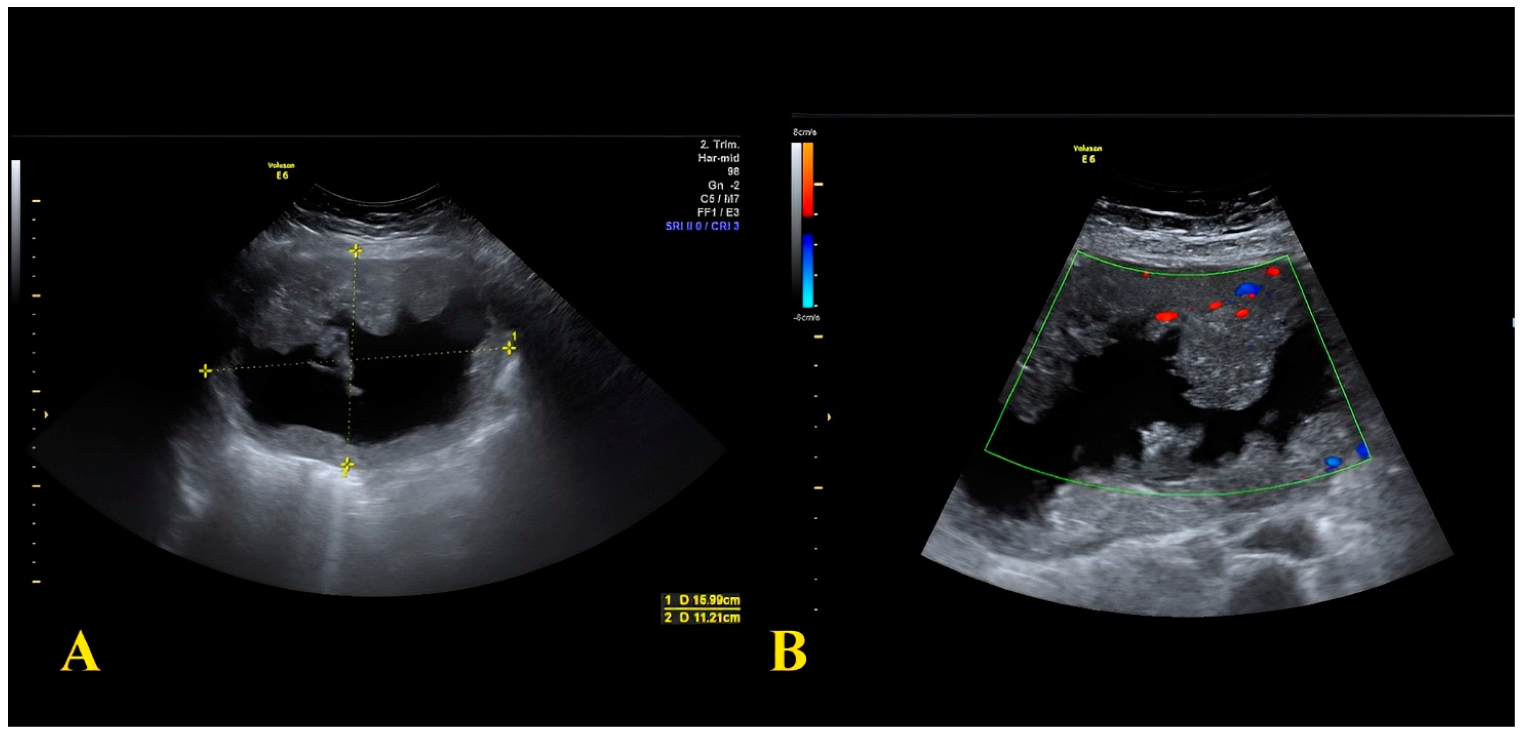
2.1. Case Report 1
2.2. Case Report 2
3. Systematic Review and Metanalysis: Methods
3.1. Inclusion and Exclusion Criteria
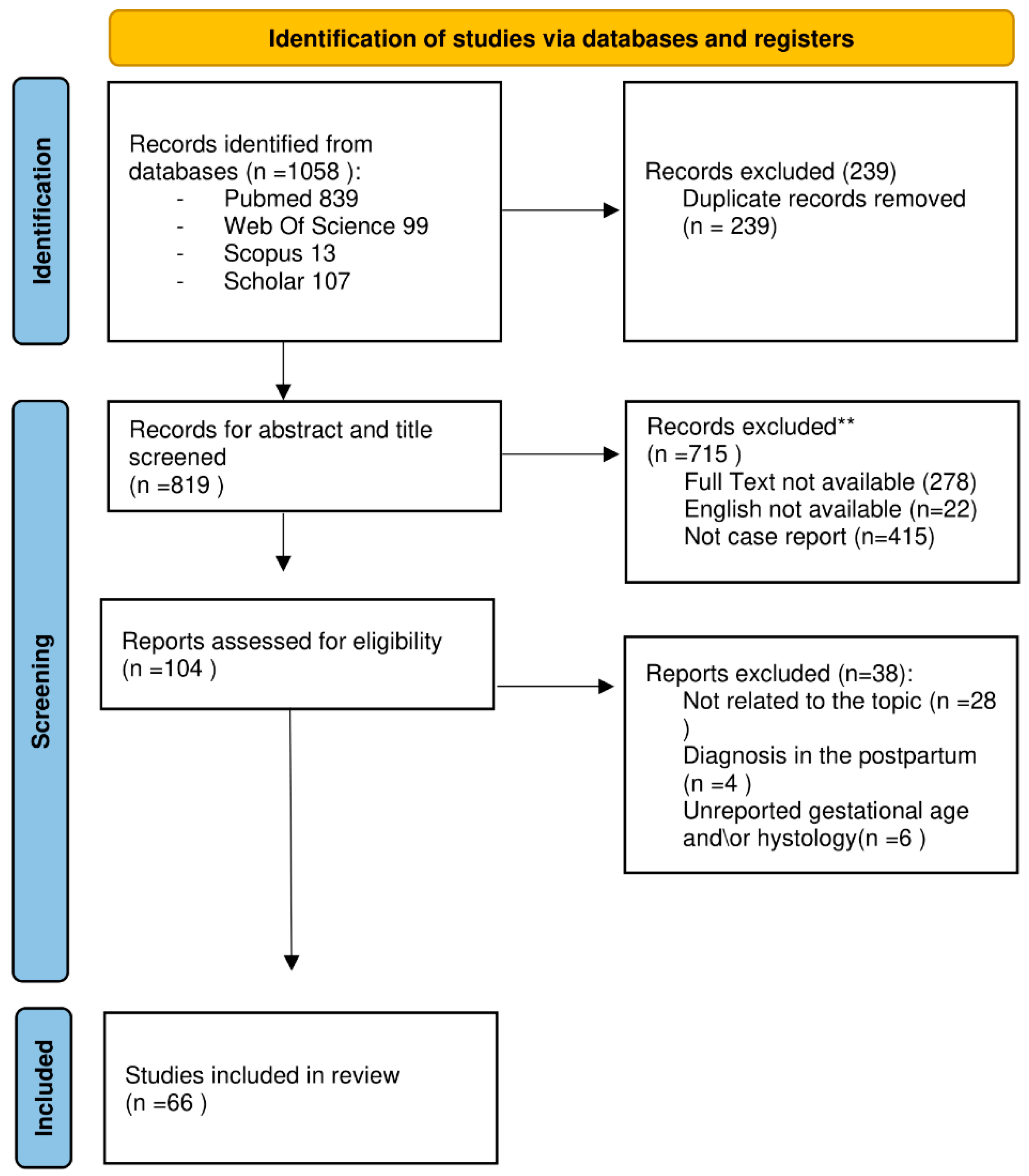
3.2. Search Strategy
3.3. Data Extraction
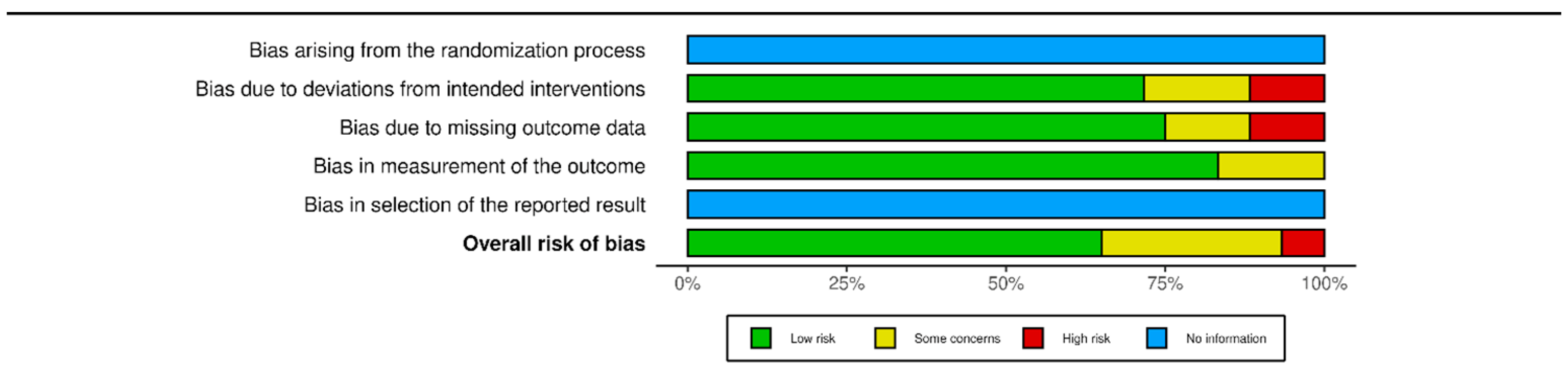
3.4. Quality of the Studies
4. Systematic Review and Metanalysis: Results
4.1. Primary Outcomes
4.2. Subgroup Analysis 1: Chemotherapy
4.3. Subgroup Analysis 2: Upfront Surgery
4.4. Subgroup Analysis 3: Acute Abdomen at Diagnosis
4.5. Subgroup Analysis 4: Surgery versus Chemotherapy
5. Discussion
5.1. Case Studies
5.2. Diagnostic Work-Up
5.3. Treatment Options: Upfront Surgery
5.4. Treatment Options: Chemotherapy
6. Conclusions
Author Contributions
Funding
Institutional Review Board Statement
Informed Consent Statement
Data Availability Statement
Conflicts of Interest
References
- Murgia, F.; Marinaccio, M.; Cormio, G.; Loizzi, V.; Cicinelli, R. Pregnancy related cancer in Apulia. A population based linkage study. Eur. J. Obstet. Gynecol. Reprod. Biol. X 2019, 3, 100025. [Google Scholar] [CrossRef] [PubMed]
- Schwab, R.; Anic, K.; Hasenburg, A. Cancer and pregnancy: A comprehensive review. Cancers 2021, 13, 3048. [Google Scholar] [CrossRef] [PubMed]
- Pavlidis, N.A. Coexistence of pregnancy and malignancy. Oncologist 2002, 7, 279–287. [Google Scholar] [CrossRef]
- Stensheim, H.; Møller, B.; van Dijk, T.; Fosså, S.D. Cause-specific survival for women diagnosed with cancer during pregnancy or lactation: A registry-based cohort study. J. Clin. Oncol. 2009, 27, 45–51. [Google Scholar] [CrossRef]
- Eibye, S.; Kjær, S.K.; Mellemkjær, L. Incidence of pregnancy-associated cancer in Denmark, 1977–2006. Obstet. Gynecol. 2013, 122, 608–617. [Google Scholar] [CrossRef]
- Amant, F.; Berveiller, P.; Boere, I.A.; Cardonick, E.; Fruscio, R.; Fumagalli, M.; Halaska, M.J.; Hasenburg, A.; Johansson, A.L.V.; Lambertini, M.; et al. Gynecologic cancers in pregnancy: Guidelines based on a third international consensus meeting. Ann. Oncol. 2019, 30, 1601–1612. [Google Scholar] [CrossRef]
- Sorouri, K.; Loren, A.W.; Amant, F.; Partridge, A.H. Patient-Centered Care in the Management of Cancer During Pregnancy. Am. Soc. Clin. Oncol. Educ. Book 2023, 43, e100037. [Google Scholar] [CrossRef]
- Peccatori, F.A.; Azim, H.A.; Orecchia, R., Jr.; Hoekstra, H.J.; Pavlidis, N.; Kesic, V.; Pentheroudakis, G.; ESMO Guidelines Working Group. Cancer, pregnancy and fertility: ESMO Clinical Practice Guidelines for diagnosis, treatment and follow-up†. Ann. Oncol. 2013, 24, vi160–vi170. [Google Scholar] [CrossRef]
- The International Agency for Research on Cancer IARC. Global Cancer Observatory. Available online: https://gco.iarc.fr/ (accessed on 23 January 2023).
- Sung, H.; Ferlay, J.; Siegel, R.; Laversanne, M.; Soerjomataram, I.; Jemal, A.; Bray, F. Global Cancer Statistics 2020: GLOBOCAN Estimates of Incidence and Mortality Worldwide for 36 Cancers in 185 Countries. CA Cancer J. Clin. 2021, 71, 209–249. [Google Scholar] [CrossRef]
- Cao, S.; Okekpe, C.C.; Dombrovsky, I.; Valenzuela, G.J.; Roloff, K. Colorectal cancer diagnosed during pregnancy with delayed treatment. Cureus 2020, 12, e8261. [Google Scholar] [CrossRef]
- National Cancer Institute Statistics. The Surveillance, Epidemiology, and End Results (SEER) Program. Available online: https://seer.cancer.gov/index.html (accessed on 8 July 2023).
- Chen, F.W.; Sundaram, V.; Chew, T.A.; Ladabaum, U. Advanced-stage colorectal cancer in persons younger than 50 years not associated with longer duration of symptoms or time to diagnosis. Clin. Gastroenterol. Hepatol. 2017, 15, 728–737.e723. [Google Scholar] [CrossRef] [PubMed]
- Cercek, A.; Chatila, W.K.; Yaeger, R.; Walch, H.; Fernandes, G.D.S.; Krishnan, A.; Palmaira, L.; Maio, A.; Kemel, Y.; Srinivasan, P.; et al. A comprehensive comparison of early-onset and average-onset colorectal cancers. J. Natl. Cancer Inst. 2021, 113, 1683–1692. [Google Scholar] [CrossRef] [PubMed]
- Abraham, A.; Habermann, E.B.; Rothenberger, D.A.; Kwaan, M.; Weinberg, A.D.; Parsons, H.M.; Gupta, P.; Al-Refaie, W.B. Adjuvant chemotherapy for stage III colon cancer in the oldest old. Cancer 2013, 119, 395–403. [Google Scholar] [CrossRef] [PubMed]
- Oken, M.; Creech, R.; Tormey, D.; Horton, J.; Davis, T.; McFadden, E.; Carbone, P. Toxicity and response criteria of the Eastern Cooperative Oncology Group. Am. J. Clin. Oncol. 1982, 5, 649–655. [Google Scholar] [CrossRef]
- Benson, A.B.; Venook, A.P.; Al-Hawary, M.M.; Azad, N.; Chen, Y.J.; Ciombor, K.K.; Cohen, S.; Cooper, H.S.; Deming, D.; Garrido-Laguna, I.; et al. Rectal Cancer, Version 2.2022, NCCN Clinical Practice Guidelines in Oncology. J. Natl. Compr. Cancer Netw. 2022, 20, 1139–1167. [Google Scholar] [CrossRef]
- Page, M.J.; McKenzie, J.E.; Bossuyt, P.M.; Boutron, I.; Hoffmann, T.C.; Mulrow, C.D.; Shamseer, L.; Tetzlaff, J.M.; Akl, E.A.; Brennan, S.E.; et al. The PRISMA 2020 statement: An updated guideline for reporting systematic reviews. BMJ 2021, 372, n71. [Google Scholar] [CrossRef]
- Sterne, J.; Savović, J.; Page, M.; Elbers, R. RoB 2: A revised tool for assessing risk of bias in randomised trials. BMJ 2019, 366, l4898. [Google Scholar] [CrossRef]
- RoB 2; ROBINS-I; ROBINS-E; ROB ME. Risk of Bias Visualization Tool. Available online: https://www.riskofbias.info/welcome/robvis-visualization-tool (accessed on 18 August 2023).
- Page, M.J.; Moher, D.; Bossuyt, P.M.; Boutron, I.; Hoffmann, T.C.; Mulrow, C.D.; Shamseer, L.; Tetzlaff, J.M.; Akl, E.A.; Brennan, S.E.; et al. PRISMA 2020 explanation and elaboration: Updated guidance and exemplars for reporting systematic reviews. BMJ 2021, 372, n160. [Google Scholar] [CrossRef]
- Morales Santana, D.A.; Czigany, Z.; Meister, F.A.; Wiltberger, G.J.; Caspers, R.; Enzensberger, C.; Stickeler, E.; Neumann, U.P.; Lambertz, A. Bowel Obstruction Due to Stenotic Sigmoid Colon Cancer in a 32-Year-Old Patient Presenting in the Third Trimester of Pregnancy: A Case Report of an Interval Surgical Approach. Am. J. Case Rep. 2022, 23, e935920. [Google Scholar] [CrossRef]
- Tarik, I.A.; Henry, F.J.; Shabery, N.A.; Omar, Z.; Ibrahim, O.E. Colonic mucinous adenocarcinoma in a pregnant woman presented as pseudo Sister Mary Joseph’s nodule: A case report. Med. J. Malays. 2022, 77, 258–260. [Google Scholar]
- Yang, H.; Han, X. Colorectal cancer in pregnancy: A case report and literature review. J. Gastrointest. Oncol. 2021, 12, 885–891. [Google Scholar] [CrossRef] [PubMed]
- Maqueira, A.J.; Rifai, A.O.; Albury, C.; Kantrales, W.A.; Rydell, D.; Breland, H. Delayed Diagnosis of Colonic Adenocarcinoma in a 30-Year-Old Postpartum Woman Misdiagnosed with HELLP Syndrome. Case Rep. Surg. 2020, 2020, 3085413. [Google Scholar] [CrossRef] [PubMed]
- Alkhamis, W.H.; Naama, T.; Arafah, M.A.; Abdulghani, S.H. Good Outcome of Early-Stage Rectal Cancer Diagnosed during Pregnancy. Am. J. Case Rep. 2020, 21, e925673. [Google Scholar] [CrossRef]
- Frydenberg, H.; Harsem, N.K.; Ofigsbø, Å.; Skoglund, H.; Brændengen, M.; Kaasa, S.; Guren, M.G. Chemotherapy during Pregnancy for Advanced Colon Cancer: A Case Report. Clin. Color. Cancer 2020, 19, 141–144. [Google Scholar] [CrossRef] [PubMed]
- Petruzzelli, P.; Zizzo, R.; Tavassoli, E.; Sutera, M.; Tin, M.C.F.; Petruzzelli, L.; Benedetto, M.; D’Anna, M.R.; De Paolis, P.; Menato, G. Colon Adenocarcinoma during Pregnancy: A Case Report and Review of the Literature. Case Rep. Obs. Gynecol. 2020, 2020, 8894722. [Google Scholar] [CrossRef]
- Sravanthi, M.V.; Suma Kumaran, S.; Palle, A.; Bojanapally, P. Adenocarcinoma of Sigmoid Colon Diagnosed in Pregnancy: A Case Report. Cureus 2020, 12, e9491. [Google Scholar] [CrossRef]
- Ibargüengoitia Ochoa, F.; Miranda Dévora, G.; Silva Lino, L.; Sepulveda Rivera, C.; González Vázquez, D.; Pérez Quintanilla, M. Colorectal Signet Ring Cell Carcinoma in a Young Pregnant Woman. Case Rep. Oncol. 2020, 13, 182–187. [Google Scholar] [CrossRef]
- Lee, S.F.; Burge, M.; Eastgate, M. Metastatic colorectal cancer during pregnancy: A tertiary center experience and review of the literature. Obs. Med. 2019, 12, 38–41. [Google Scholar] [CrossRef]
- Munteanu, O.; Voicu, D.; Voiculescu, D.I.; Negreanu, L.; Georgescu, T.A.; Sajin, M.; Berceanu, C.; Mehedinţu, C.; Brătilă, E.; Istrate-Ofiţeru, A.M.; et al. Colon cancer in pregnancy: A diagnostic and therapeutic challenge. Rom. J. Morphol. Embryol. 2019, 60, 307–317. [Google Scholar]
- Žegarac, Ž.; Duić, Ž.; Stasenko, S. Nonobstetrical Acute Abdomen during Pregnancy as a Consequence of Colorectal Carcinoma Perforation: Case Report and Review of the Literature. Case Rep. Gastrointest. Med. 2019, 2019, 3682980. [Google Scholar] [CrossRef]
- Murphy, J.E.; Shampain, K.; Riley, L.E.; Clark, J.W.; Basnet, K.M. Case 32-2018: A 36-Year-Old Pregnant Woman with Newly Diagnosed Adenocarcinoma. N. Engl. J. Med. 2018, 379, 1562–1570. [Google Scholar] [CrossRef]
- Xu, Y.; Kong, B.; Shen, K. Adenocarcinoma of the ascending colon in a 31-year-old pregnant woman: A case report. Medicine 2018, 97, e13707. [Google Scholar] [CrossRef]
- Makhijani, R.; Bhagat, V.H.; Fayek, M. Colon cancer presenting as pseudo-obstruction during pregnancy—A case report. Obs. Med. 2017, 10, 198–200. [Google Scholar] [CrossRef]
- Ye, W.; Tang, Y.; Yao, C.; Shi, J.; Xu, Y.; Jiang, J. Advanced gastrointestinal carcinoma with massive ascites and hydrothorax during pregnancy: A case report and review of the literature. Medicine 2017, 96, e9354. [Google Scholar] [CrossRef]
- Jones, A.; Povlow, M.R. Colorectal Cancer Presenting with Constipation During Pregnancy. Cureus 2017, 9, e1190. [Google Scholar] [CrossRef]
- Gabriel, I.; Olejek, A.; Drozdzowska, B. Colon cancer in pregnancy—A difficult diagnosis. Eur. J. Obs. Gynecol. Reprod. Biol. 2016, 203, 340–341. [Google Scholar] [CrossRef]
- Ossendorp, R.R.; Silvis, R.; van der Bij, G.J. Advanced colorectal cancer resulting in acute bowel obstruction during pregnancy; A case report. Ann. Med. Surg. 2016, 8, 18–20. [Google Scholar] [CrossRef]
- Makoshi, Z.; Perrott, C.; Al-Khatani, K.; Al-Mohaisen, F. Chemotherapeutic treatment of colorectal cancer in pregnancy: Case report. J. Med. Case Rep. 2015, 9, 140. [Google Scholar] [CrossRef]
- Chouaib, N.; Rafai, M.; El Bouti, A.; Belkouch, A.; El bakkali, H.; Belyamani, L. Discovery of a small bowel tumor following an acute intestinal obstruction in a pregnant woman: Report of a case. Pan Afr. Med. J. 2015, 20, 340. [Google Scholar] [CrossRef]
- Kocián, P.; Hoch, J.; Halaska, M. Sigmoid colon cancer in pregnancy—A case report. Rozhl. Chir. 2015, 94, 170–173. [Google Scholar]
- Dogan, N.U.; Tastekin, D.; Kerimoglu, O.S.; Pekin, A.; Celik, C. Rectal cancer in pregnancy: A case report and review of the literature. J. Gastrointest. Cancer 2013, 44, 354–356. [Google Scholar] [CrossRef]
- Ho, M.Y.; Cassano-Bailey, A.; Czaykowski, P. Colorectal Cancer in Pregnancy: Driven by Pregnancy-Associated Growth Factors? J. Gastrointest. Cancer 2012, 43 (Suppl. S1), S239–S242. [Google Scholar] [CrossRef]
- Jeppesen, J.B.; Østerlind, K. Successful twin pregnancy outcome after in utero exposure to FOLFOX for metastatic colon cancer: A case report and review of the literature. Clin. Color. Cancer 2011, 10, 348–352. [Google Scholar] [CrossRef]
- Gensheimer, M.; Jones, C.A.; Graves, C.R.; Merchant, N.B.; Lockhart, A.C. Administration of oxaliplatin to a pregnant woman with rectal cancer. Cancer Chemother. Pharmacol. 2009, 63, 371–373. [Google Scholar] [CrossRef]
- Kanate, A.S.; Auber, M.L.; Higa, G.M. Priorities and uncertainties of administering chemotherapy in a pregnant woman with newly diagnosed colorectal cancer. J. Oncol. Pharm. Pr. 2009, 15, 5–8. [Google Scholar] [CrossRef]
- Taylor, J.; Amanze, A.; Di Federico, E.; Verschraegen, C. Irinotecan use during pregnancy. Obs. Gynecol. 2009, 114, 451–452. [Google Scholar] [CrossRef]
- Duffy, A.; Shia, J.; Klimstra, D.; Temple, L.; O’Reilly, E.M. Collision tumor of the large bowel in the context of advanced pregnancy and ulcerative colitis. Clin. Color. Cancer 2008, 7, 402–405. [Google Scholar] [CrossRef]
- Mechery, J.; Ikhena, S.E. Cancer of the descending colon during pregnancy. J. Obstet. Gynaecol. 2007, 27, 311–312. [Google Scholar] [CrossRef]
- Lolis, E.D.; Likoudis, P.; Voiniadis, P.; Hassiakos, D.; Samanides, L. Synchronous rectal and colon cancer caused by familial polyposis coli during pregnancy. J. Obs. Gynaecol. Res. 2007, 33, 199–202. [Google Scholar] [CrossRef]
- Chêne, G.; Tardieu, A.S.; Favard, A.; Lebel, A.; Voitellier, M. Colorectal cancer discovered during pregnancy. J. Gynecol. Obs. Biol. Reprod. 2006, 35, 504–512. [Google Scholar] [CrossRef]
- Penney, D.; Ganapathy, R.; Jonas-Obichere, M.; El-Refeay, H. Intussusception: A rare cause of abdominal pain in pregnancy. Ultrasound Obs. Gynecol. 2006, 28, 723–725. [Google Scholar] [CrossRef]
- Harma, M.; Uzunkoy, A. Colorectal cancer presenting with uncommon soft tissue invasion during pregnancy. Acta Obs. Gynecol. Scand. 2005, 84, 491–493. [Google Scholar] [CrossRef]
- Minter, A.; Malik, R.; Ledbetter, L.; Winokur, T.S.; Hawn, M.T.; Saif, M.W. Colon cancer in pregnancy. Cancer Control 2005, 12, 196–202. [Google Scholar] [CrossRef]
- Papathanasiou, K.; Tolikas, A.; Savvidis, A.; Traianos, V.; Tzafettas, J. Advanced sigmoid colon cancer in pregnancy presented as an abscess. J. Obs. Gynaecol. 2004, 24, 319–320. [Google Scholar] [CrossRef]
- Kömürcü, S.; Ozet, A.; Oztürk, B.; Arpaci, F.; Altundağ, M.K.; Tezcan, Y. Colon cancer during pregnancy. A case report. J. Reprod. Med. 2001, 46, 75–78. [Google Scholar]
- Kitoh, T.; Nishimura, S.; Fukuda, S.; Hirabuki, S.; Kaganoi, J.; Tokunaga, Y.; Ohsumi, K. The incidence of colorectal cancer during pregnancy in Japan: Report of two cases and review of Japanese cases. Am. J. Perinatol. 1998, 15, 165–171. [Google Scholar] [CrossRef]
- Rojansky, N.; Shushan, A.; Livni, N.; Jurim, O.; Sulam, M.; Galun, E. Pregnancy associated with colon carcinoma overexpressing p53. Gynecol. Oncol. 1997, 64, 516–520. [Google Scholar] [CrossRef]
- Vitoratos, N.; Louridas, K.; Zourlas, P.A. Carcinoma of the colon during pregnancy. Int. J. Gynaecol. Obs. 1994, 44, 72–73. [Google Scholar] [CrossRef]
- Heres, P.; Wiltink, J.; Cuesta, M.A.; Burger, C.W.; van Groeningen, C.J.; Meijer, S. Colon carcinoma during pregnancy: A lethal coincidence. Eur. J. Obs. Gynecol. Reprod. Biol. 1993, 48, 149–152. [Google Scholar] [CrossRef]
- Shioda, Y.; Koizumi, S.; Furuya, S.; Akira, S.; Kameyama, N.; Hamano, N.; Takahashi, W.; Tokunaga, A.; Tanaka, N.; Onda, M. Intussusception caused by a carcinoma of the cecum during pregnancy: Report of a case and review of the literature. Surg. Today 1993, 23, 556–561. [Google Scholar] [CrossRef]
- Heise, R.H.; Van Winter, J.T.; Wilson, T.O.; Ogburn, P.L., Jr. Colonic cancer during pregnancy: Case report and review of the literature. Mayo Clin. Proc. 1992, 67, 1180–1184. [Google Scholar] [CrossRef]
- Gonsoulin, W.; Mason, B.; Carpenter, R.J., Jr. Colon cancer in pregnancy with elevated maternal serum alpha-fetoprotein level at presentation. Am. J. Obs. Gynecol. 1990, 163, 1172–1173. [Google Scholar] [CrossRef]
- Jaffe, R.; Schwartz, I.; Freund, U.; Cohen, I.; Rosen, D.; Fejgin, M. Perforated adenocarcinoma of the colon during pregnancy. Int. J. Gynaecol. Obs. 1989, 30, 371–373. [Google Scholar] [CrossRef]
- Tsukamoto, N.; Uchino, H.; Matsukuma, K.; Kamura, T. Carcinoma of the colon presenting as bilateral ovarian tumors during pregnancy. Gynecol. Oncol. 1986, 24, 386–391. [Google Scholar] [CrossRef]
- Nesbitt, J.C.; Moise, K.J.; Sawyers, J.L. Colorectal carcinoma in pregnancy. Arch. Surg. 1985, 120, 636–640. [Google Scholar] [CrossRef]
- Hill, J.A.; Kassam, S.H.; Talledo, O.E. Colonic cancer in pregnancy. South. Med. J. 1984, 77, 375–378. [Google Scholar] [CrossRef]
- Oduncu, F.S.; Kimmig, R.; Hepp, H.; Emmerich, B. Cancer in pregnancy: Maternal-fetal conflict. J. Cancer Res. Clin. Oncol. 2003, 129, 133–146. [Google Scholar] [CrossRef]
- Vimercati, A.; Greco, P.; Loverro, G.; Lopalco, P.; Pansini, V.; Selvaggi, L. Maternal complications after caesarean section in HIV infected women. Eur. J. Obstet. Gynecol. Reprod. Biol. 2000, 90, 73–76. [Google Scholar] [CrossRef]
- Hepner, A.; Negrini, D.; Hase, E.A.; Exman, P.; Testa, L.; Trinconi, A.F.; Filassi, J.R.; Francisco, R.P.V.; Zugaib, M.; O’Connor, T.L.; et al. Cancer during pregnancy: The oncologist overview. World J. Oncol. 2019, 10, 28–34. [Google Scholar] [CrossRef]
- Giorgione, V.; Cavoretto, P.; Cormio, G.; Valsecchi, L.; Vimercati, A.; De Gennaro, A. Prenatal Diagnosis of Twin Pregnancies with Complete Hydatidiform Mole and Coexistent Normal Fetus: A Series of 13 Cases. Gynecol. Obstet. Investig. 2017, 82, 404–409. [Google Scholar] [CrossRef]
- Lutterodt, M.C.; Kähler, P.; Kragstrup, J.; Nicolaisdottir, D.R.; Siersma, V.; Ertmann, R.K. Examining to what extent pregnancy-related physical symptoms worry women in the first trimester of pregnancy: A cross-sectional study in general practice. BJGP Open 2019, 3, bjgpopen19X101674. [Google Scholar] [CrossRef]
- de Haan, J.; Vandecaveye, V.; Han, S.N.; Van de Vijver, K.K.; Amant, F. Difficulties with diagnosis of malignancies in pregnancy. Best. Pract. Res. Clin. Obstet. Gynaecol. 2016, 33, 19–32. [Google Scholar] [CrossRef]
- Vimercati, A.; De Nola, R.; Trerotoli, P.; Metta, M.; Cazzato, G.; Resta, L.; Malvasi, A. COVID-19 Infection in Pregnancy: Obstetrical Risk Factors and Neonatal Outcomes-A Monocentric, Single-Cohort Study. Vaccines 2022, 10, 166. [Google Scholar] [CrossRef] [PubMed]
- Tremblay, E.; Thérasse, E.; Thomassin-Naggara, I.; Trop, I. Quality initiatives: Guidelines for use of medical imaging during pregnancy and lactation. RadioGraphics 2012, 32, 3. [Google Scholar] [CrossRef] [PubMed]
- Committee Opinion No. 723: Guidelines for Diagnostic Imaging During Pregnancy and Lactation. Obstet. Gynecol. 2017, 130, e210–e216. [CrossRef]
- Han, S.; Lotgerink, A.; Gziri, M.; Van Calsteren, K.; Hanssens, M.; Amant, F. Physiologic variations of serum tumor markers in gynecological malignancies during pregnancy: A systematic review. BMC Med. 2012, 10, 86. [Google Scholar] [CrossRef]
- Amant, F.; Verheecke, M.; Wlodarska, I.; Dehaspe, L.; Brady, P.; Brison, N.; Van Den Bogaert, K.; Dierickx, D.; Vandecaveye, V.; Tousseyn, T.; et al. Presymptomatic Identification of Cancers in Pregnant Women During Noninvasive Prenatal Testing. JAMA Oncol. 2015, 1, 814–819. [Google Scholar] [CrossRef]
- Heesterbeek, C.J.; Aukema, S.M.; Galjaard, R.-J.H.; Boon, E.M.J.; Srebniak, M.I.; Bouman, K.; Faas, B.H.W.; Govaerts, L.C.P.; Hoffer, M.J.V.; den Hollander, N.S.; et al. Noninvasive Prenatal Test Results Indicative of Maternal Malignancies: A Nationwide Genetic and Clinical Follow-Up Study. J. Clin. Oncol. 2022, 40, 22. [Google Scholar] [CrossRef]
- Shigemi, D.; Aso, S.; Matsui, H.; Fushimi, K.; Yasunaga, H. Safety of Laparoscopic Surgery for Benign Diseases during Pregnancy: A Nationwide Retrospective Cohort Study. J. Minim. Invasive Gynecol. 2019, 26, 501–506. [Google Scholar] [CrossRef] [PubMed]
- Obermair, A.; Asher, R.; Pareja, R.; Frumovitz, M.; Lopez, A.; Moretti-Marques, R. Incidence of adverse events in minimally invasive vs open radical hysterectomy in early cervical cancer: Results of a randomized controlled trial. Am. J. Obstet. Gynecol. 2020, 222, 249.e1–249.e10. [Google Scholar] [CrossRef]
- Haas, J. Pregnancy in association with a newly diagnosed cancer: A population-based epidemiologic assessment. Int. J. Cancer 1984, 34, 229–235. [Google Scholar] [CrossRef] [PubMed]
- Matsuo, K.; Klar, M.; Youssefzadeh, A.C.; Mandelbaum, R.S.; Roman, L.D.; Ouzounian, J.G.; Wright, J.D. Assessment of severe maternal morbidity and mortality in pregnancies complicated by cancer in the US. JAMA Oncol. 2022, 8, 1213–1216. [Google Scholar] [CrossRef] [PubMed]
- Staud, F.; Karahoda, R. Trophoblast: The central unit of fetal growth, protection and programming. Int. J. Biochem. Cell Biol. 2018, 105, 35–40. [Google Scholar] [CrossRef] [PubMed]
- Triarico, S.; Rivetti, S.; Capozza, M.A.; Romano, A.; Maurizi, P.; Mastrangelo, S.; Attinà, G.; Ruggiero, A. Transplacental passage and fetal effects of antineoplastic treatment during pregnancy. Cancers 2022, 14, 3103. [Google Scholar] [CrossRef] [PubMed]
- Fisher, S. Why is placentation abnormal in preeclampsia? Am. J. Obstet. Gynecol. 2015, 213, S115–S122. [Google Scholar] [CrossRef] [PubMed]
- Cha, J.; Sun, X.; Dey, S. Mechanisms of implantation: Strategies for successful pregnancy. Nat. Med. 2012, 18, 1754–1767. [Google Scholar] [CrossRef] [PubMed]
- Benoit, L.; Mir, O.; Vialard, F.; Berveiller, P. Cancer during Pregnancy: A Review of Preclinical and Clinical Transplacental Transfer of Anticancer Agents. Cancers 2021, 13, 1238. [Google Scholar] [CrossRef]
- Pacifici, G.M.; Nottoli, R. Placental transfer of drugs administered to the mother. Clin. Pharmacokinet. 1995, 28, 235–269. [Google Scholar] [CrossRef]
- Syme, M.; Paxton, J.; Keelan, J. Drug transfer and metabolism by the human placenta. Clin. Pharmacokinet. 2004, 43, 487–514. [Google Scholar] [CrossRef]
- Adam, M. The all-or-none phenomenon revisited. Birth Defects Res. Part A Clin. Mol. Teratol. 2012, 94, 664–669. [Google Scholar] [CrossRef]
- Ruggiero, S.; Ripetti, V.; Bianchi, A.; La Vaccara, V.; Alloni, R.; Coppola, R. A singular observation of a giant benign Brenner tumor of the ovary. Arch. Gynecol. Obs. 2011, 284, 513–516. [Google Scholar] [CrossRef]
- Glynne-Jones, R.; Wyrwicz, L.; Tiret, E.; Brown, G.; Rödel, C.; Cervantes, A.; Arnold, D. Rectal cancer: ESMO Clinical Practice Guidelines for diagnosis, treatment and follow-up. Ann. Oncol. 2017, 28 (Suppl. S4), iv22–iv40. [Google Scholar] [CrossRef] [PubMed]
- Pellino, G.; Simillis, C.; Kontovounisios, C.; Baird, D.; Nikolaou, S.; Warren, O.; Tekkis, P.; Rasheed, S. Colorectal cancer diagnosed during pregnancy: Systematic review and treatment pathways. Eur. J. Gastroenterol. Hepatol. 2017, 29, 743–753. [Google Scholar] [CrossRef] [PubMed]
- National Toxicology Program. NTP Monograph: Developmental Effects and Pregnancy Outcomes Associated with Cancer Chemotherapy Use During Pregnancy. NTP Monogr. 2013, i-214, NIH Publication No. 13-5956. [Google Scholar]
- de Haan, J.; Verheecke, M.; Van Calsteren, K.; Van Calster, B.; Shmakov, R.G.; Mhallem Gziri, M.; Halaska, M.J.; Fruscio, R.; Lok, C.A.R.; Boere, I.A. Oncological management and obstetric and neonatal outcomes for women diagnosed with cancer during pregnancy: A 20-year international cohort study of 1170 patients. Lancet Oncol. 2018, 19, 337–346. [Google Scholar] [CrossRef] [PubMed]
- Kocián, P.; de Haan, J.; Cardonick, E.H.; Uzan, C.; Lok, C.A.R.; Fruscio, R.; Halaska, M.J.; Amant, F.; On behalf of the Writing Committee of the International Network on Cancer. Infertility and Pregnancy (INCIP) on this particular manuscript. Management and outcome of colorectal cancer during pregnancy: Report of 41 cases. Acta Chir. Belg. 2019, 119, 166–175. [Google Scholar] [CrossRef]


| Author | Year | Maternal Age at Diagnosis | Hystotype | Tumour Stage | Chemotherapy |
|---|---|---|---|---|---|
| Santana et al. [22] | 2022 | 32 | Colic adk | III | Not performed |
| Tarik et al. [23] | 2022 | 41 | Colic adk | IV | Adjuvant |
| Yang et al. [24] | 2021 | 36 | Undifferentiated | IV | Adjuvant |
| Maqueira et al. [25] | 2020 | 30 | Colic adk | IV | Not performed |
| Alkhamis et al. [26] | 2020 | 30 | Colic adk (focal) | 0 | Not performed |
| Frydenberg et al. [27] | 2020 | 43 | Colic adk | IV | NeoAdjuvant |
| Petruzzelli et al. [28] | 2020 | 38 | Colic adk | III | Adjuvant |
| Sravantthi et al. [29] | 2020 | 31 | Colic adk | III | Neo + Adjuvant |
| Ochoa et al. [30] | 2020 | 24 | Signet ring | IV | Not performed |
| Lee et al. [31] | 2019 | 32 | Colic adk | IV | Not performed |
| Lee et al. [31] | 2019 | 25 | Colic adk | IV | NeoAdjuvant |
| Lee et al. [31] | 2019 | 34 | Colic adk | IV | NeoAdjuvant |
| Munteanu et al. [32] | 2019 | 36 | Colic adk | IV | Not performed |
| Žegarac et al. [33] | 2019 | 33 | Colic adk | II | Not performed |
| Zegarac et al. [33] | 2019 | 33 | Colic adk | III | Not performed |
| Muntenau et al. [32] | 2019 | 36 | Colic adk | III | Adjuvant |
| Murphy et al. [34] | 2018 | 36 | Colic adk | IV | Not performed |
| Xu et al. [35] | 2018 | 31 | Colic adk | II | Not performed |
| Xu et al. [35] | 2018 | 31 | Colic adk | IV | Adjuvant |
| Murphy et al. [34] | 2018 | 36 | Colic adk | IV | Adjuvant |
| Makhijani et al. [36] | 2017 | 29 | Colic adk | III | Adjuvant |
| Wenfeng Ye et al. [37] | 2017 | 29 | Signet ring | IV | Not performed |
| Jones et al. [38] | 2017 | 33 | Colic adk | III | Not performed |
| Jones et al. [38] | 2017 | 33 | Colic adk | III | Adjuvant |
| Makhjani et al. [36] | 2017 | 29 | Colic adk | IV | Neo + Adjuvant |
| Gabriel et al. [39] | 2016 | 31 | Colic adk | IV | Not performed |
| Ossendorp et al. [40] | 2016 | 34 | Colic adk | IV | Not performed |
| Ossendrorp et al. [40] | 2016 | 34 | Colic adk | IV | Adjuvant |
| Makoshi et al. [41] | 2015 | 33 | Colic adk | IV | Adjuvant |
| Chouaib et al. [42] | 2015 | 32 | Neuroendocrine | Not performed | |
| P Kocián et al. [43] | 2015 | 37 | Colic adk | 0 | Not performed |
| Makoshi et al. [41] | 2015 | 33 | Colic adk | III | Neo + Adjuvant |
| Dogan et al. [44] | 2013 | 38 | Colic adk | IV | NeoAdjuvant |
| HO et al. [45] | 2012 | 29 | Colic adk | III | Not performed |
| HO et al. [45] | 2012 | 29 | Colic adk | III | Adjuvant |
| Jeppesen et al. [46] | 2011 | 26 | Colic adk | IV | Adjuvant |
| Jeppesen et al. [46] | 2011 | 26 | Colic adk | III | Neo + Adjuvant |
| Gensheimer et al. [47] | 2009 | 25 | Colic adk | IV | Adjuvant |
| Kanate et al. [48] | 2009 | 40 | Colic adk | IV | NeoAdjuvant |
| Taylor et al. [49] | 2009 | 34 | Colic adk | IV | Adjuvant |
| Taylor et al. [49] | 2009 | 34 | Colic adk | III | Adjuvant |
| Duffy et al. [50] | 2008 | 33 | Signet ring | IV | Not performed |
| Duffy et al. [50] | 2008 | 33 | Mixed | IV | Adjuvant |
| Mechery et al. [51] | 2007 | 34 | Colic adk | IV | Not performed |
| Lolis et al. [52] | 2007 | 29 | Colic adk | III | Not performed |
| Lolis et al. [52] | 2007 | 29 | Colic adk | III | Adjuvant |
| Chêne et al. [53] | 2006 | 26 | Colic adk | III | Not performed |
| Penney et al. [54] | 2006 | 34 | Colic adk | IV | Not performed |
| Harma et al. [55] | 2005 | 30 | Colic adk | IV | Not performed |
| Minter et al. [56] | 2005 | 28 | Colic adk | II | Not performed |
| Papathanasiou et al. [57] | 2004 | 37 | Colic adk | IV | Not performed |
| S Kömürcü et al. [58] | 2001 | 33 | Colic adk | IV | Not performed |
| Kitoh et al. [59] | 1998 | 34 | Colic adk | IV | Not performed |
| Kitoh et al. [59] | 1998 | 35 | Colic adk | IV | Not performed |
| Rojansky et al. [60] | 1997 | 39 | Colic adk | III | Not performed |
| Vitoratos et al. [61] | 1994 | 32 | Undifferentiated | IV | Not performed |
| Heres et al. [62] | 1993 | 38 | Colic adk | IV | Not performed |
| Yoshinobu et al. [63] | 1993 | 27 | Colic adk | III | Not performed |
| Heise et al. [64] | 1992 | 29 | Colic adk | III | Adjuvant |
| Gonsoulin et al. [65] | 1990 | 33 | Colic adk | IV | Not performed |
| R Jaffe et al. [66] | 1989 | 42 | Colic adk | Not performed | |
| Tsukamoto et al. [67] | 1986 | 29 | Colic adk | IV | Not performed |
| Nesbitt et al. [68] | 1985 | 35 | Colic adk | III | Not performed |
| Nesbitt et al. [68] | 1985 | 31 | Colic adk | III | Not performed |
| Nesbitt et al. [68] | 1985 | 28 | Colic adk | IV | Not performed |
| Hill et al. [69] | 1984 | 29 | Colic adk | IV | Not performed |
| Author | Year | Gestational Age at Diagnosis (Weeks) | Mode of Birth | Gestational Age at Birth | Neonatal Outcomes |
|---|---|---|---|---|---|
| Santana et al. [22] | 2022 | 28 | CS | 32 | Liveborn |
| Tarik et al. [23] | 2022 | 24 | CS | 28 | |
| Yang et al. [24] | 2021 | 39 | CS | 39 | Liveborn |
| Maqueira et al. [25] | 2020 | 35 | VB | 35 | Liveborn |
| Alkhamis et al. [26] | 2020 | 9 | VB | 40 | Liveborn |
| Frydenberg et al. [27] | 2020 | 23 | CS | 34 | Liveborn |
| Petruzzelli et al. [28] | 2020 | 26 | VB | 37 | Liveborn |
| Sravantthi et al. [29] | 2020 | 21 | |||
| Ochoa et al. [30] | 2020 | 22 | VB | 25 | Liveborn |
| Lee et al. [31] | 2019 | 37 | VB | 37 | Liveborn |
| Lee et al. [31] | 2019 | 19 | VB | 33 | Stillbirth |
| Lee et al. [31] | 2019 | 11 | VB | 36 | Liveborn |
| Munteanu et al. [32] | 2019 | 33 | CS | 33 | Liveborn |
| Žegarac et al. [33] | 2019 | 22 | VB | 26 | Liveborn |
| Zegarac et al. [33] | 2019 | 22 | |||
| Muntenau et al. [32] | 2019 | 33 | CS | 33 | Liveborn |
| Murphy et al. [34] | 2018 | 32 | CS | 35 | Liveborn |
| Xu et al. [35] | 2018 | 33 | CS | 33 | Liveborn |
| Xu et al. [35] | 2018 | 33 | CS | 33 | Stillbirth |
| Murphy et al. [34] | 2018 | 33 | CS | 35 | Stillbirth |
| Makhijani et al. [36] | 2017 | 17 | |||
| Wenfeng Ye et al. [37] | 2017 | 27 | CS | 28 | Liveborn |
| Jones et al. [38] | 2017 | 29 | VB | 29 | Liveborn |
| Jones et al. [38] | 2017 | 29 | VB | 23 | Abortion |
| Makhjani et al. [36] | 2017 | 17 | |||
| Gabriel et al. [39] | 2016 | 30 | CS | 35 | Liveborn |
| Ossendorp et al. [40] | 2016 | 32 | CS | 33 | Liveborn |
| Ossendrorp et al. [40] | 2016 | 32 | CS | 33 | Liveborn |
| Makoshi et al. [41] | 2015 | 11 | VB | 38 | Liveborn |
| Chouaib et al. [42] | 2015 | 32 | VB | Liveborn | |
| P Kocián et al. [43] | 2015 | 27 | CS | 28 | Liveborn |
| Makoshi et al. [41] | 2015 | 11 | VB | 38 | Liveborn |
| Dogan et al. [44] | 2013 | 19 | CS | 36 | Liveborn |
| HO et al. [45] | 2012 | 21 | VB | 37 | Liveborn |
| HO et al. [45] | 2012 | 21 | VB | 37 | Liveborn |
| Jeppesen et al. [46] | 2011 | 10 | CS | 33 | Liveborn |
| Jeppesen et al. [46] | 2011 | 11 | CS | 23 | Abortion |
| Gensheimer et al. [47] | 2009 | 12 | VB | 33 | Liveborn |
| Kanate et al. [48] | 2009 | 23 | CS | 31 | Liveborn |
| Taylor et al. [49] | 2009 | 15 | VB | 37 | Liveborn |
| Taylor et al. [49] | 2009 | 15 | VB | 38 | Liveborn |
| Duffy et al. [50] | 2008 | 30 | CS | 33 | Liveborn |
| Duffy et al. [50] | 2008 | 30 | CS | 33 | Liveborn |
| Mechery et al. [51] | 2007 | 38 | VB | 38 | Liveborn |
| Lolis et al. [52] | 2007 | 27 | CS | 33 | Liveborn |
| Lolis et al. [52] | 2007 | CS | 33 | Liveborn | |
| Chêne et al. [53] | 2006 | 22 | CS | 34 | Liveborn |
| Penney et al. [54] | 2006 | 19 | |||
| Harma et al. [55] | 2005 | 37 | VB | 37 | Liveborn |
| Minter et al. [56] | 2005 | 16 | VB | 16 | Abortion |
| Papathanasiou et al. [57] | 2004 | 34 | CS | 34 | Liveborn |
| S Kömürcü et al. [58] | 2001 | 30 | CS | 34 | Liveborn |
| Kitoh et al. [59] | 1998 | 39 | CS | 39 | Liveborn |
| Kitoh et al. [59] | 1998 | 31 | CS | 35 | Liveborn |
| Rojansky et al. [60] | 1997 | 34 | VB | 34 | Liveborn |
| Vitoratos et al. [61] | 1994 | 26 | VB | 26 | Stillbirth |
| Heres et al. [62] | 1993 | 27 | CS | 32 | Liveborn |
| Yoshinobu et al. [63] | 1993 | 19 | VB | Liveborn | |
| Heise et al. [64] | 1992 | 35 | CS | Liveborn | |
| Gonsoulin et al. [65] | 1990 | 25 | CS | 25 | Stillbirth |
| R Jaffe et al. [66] | 1989 | 30 | VB | 30 | Liveborn |
| Tsukamoto et al. [67] | 1986 | 21 | 21 | Abortion | |
| Nesbitt et al. [68] | 1985 | 22 | 22 | Abortion | |
| Nesbitt et al. [68] | 1985 | 25 | CS | 32 | Liveborn |
| Nesbitt et al. [68] | 1985 | 29 | VB | 29 | Liveborn |
| Hill et al. [69] | 1984 | 26 | VB | 26 | Liveborn |
| Maternal Data | ||
| Mean age at diagnosis (years) | 32.47 ± 4.17 | |
| Mean gestational age at diagnosis (weeks) | 25 ± 8 | |
| Mean gestational age at birth (weeks) | 32 ± 5 | |
| Type of Surgery | % | n |
| Laparotomy | 47.0 | 31 |
| Laparoscopy | 0 | 0 |
| Endoscopic treatment | 1.5 | 1 |
| Not performed | 39.4 | 26 |
| Missing data | 12.1 | 8 |
| Chemotherapy during Pregnancy | % | n |
| Total | 39.9 | 27 |
| Adjuvant | 27.3 | 18 |
| Neoadjuvant | 7.6 | 5 |
| Neoadjuvant + Adjuvant | 6.1 | 4 |
| Not performed | 59.1 | 39 |
| Histology | % | n |
| Colic adenocarcinoma | 89.4 | 59 |
| Signet ring cell | 4.5 | 3 |
| Undifferentiated | 4.5 | 3 |
| Neuroendocrine | 1.5 | 1 |
| Tumour Stage | % | n |
| I | 3.0 | 2 |
| II | 4.5 | 3 |
| III | 31.8 | 21 |
| IV | 57.6 | 38 |
| missing | 3.0 | 2 |
| Mode of Delivery | % | n |
| Caesarean section | 48.5 | 32 |
| Natural delivery | 39.9 | 27 |
| Missing data | 10.6 | 7 |
| Neonatal Outcomes | % | n |
| Live births | 75.8 | 50 |
| Stillbirths | 7.6 | 5 |
| Miscarriages | 7.6 | 5 |
| Missing data | 9.0 | 6 |
Disclaimer/Publisher’s Note: The statements, opinions and data contained in all publications are solely those of the individual author(s) and contributor(s) and not of MDPI and/or the editor(s). MDPI and/or the editor(s) disclaim responsibility for any injury to people or property resulting from any ideas, methods, instructions or products referred to in the content. |
© 2024 by the authors. Licensee MDPI, Basel, Switzerland. This article is an open access article distributed under the terms and conditions of the Creative Commons Attribution (CC BY) license (https://creativecommons.org/licenses/by/4.0/).
Share and Cite
Galante, A.; Cerbone, M.; Mannavola, F.; Marinaccio, M.; Schonauer, L.M.; Dellino, M.; Damiani, G.R.; Pinto, V.; Cormio, G.; Cicinelli, E.; et al. Diagnostic, Management, and Neonatal Outcomes of Colorectal Cancer during Pregnancy: Two Case Reports, Systematic Review of Literature and Metanalysis. Diagnostics 2024, 14, 559. https://doi.org/10.3390/diagnostics14050559
Galante A, Cerbone M, Mannavola F, Marinaccio M, Schonauer LM, Dellino M, Damiani GR, Pinto V, Cormio G, Cicinelli E, et al. Diagnostic, Management, and Neonatal Outcomes of Colorectal Cancer during Pregnancy: Two Case Reports, Systematic Review of Literature and Metanalysis. Diagnostics. 2024; 14(5):559. https://doi.org/10.3390/diagnostics14050559
Chicago/Turabian StyleGalante, Arianna, Marco Cerbone, Francesco Mannavola, Marco Marinaccio, Luca Maria Schonauer, Miriam Dellino, Gianluca Raffaello Damiani, Vincenzo Pinto, Gennaro Cormio, Ettore Cicinelli, and et al. 2024. "Diagnostic, Management, and Neonatal Outcomes of Colorectal Cancer during Pregnancy: Two Case Reports, Systematic Review of Literature and Metanalysis" Diagnostics 14, no. 5: 559. https://doi.org/10.3390/diagnostics14050559
APA StyleGalante, A., Cerbone, M., Mannavola, F., Marinaccio, M., Schonauer, L. M., Dellino, M., Damiani, G. R., Pinto, V., Cormio, G., Cicinelli, E., & Vimercati, A. (2024). Diagnostic, Management, and Neonatal Outcomes of Colorectal Cancer during Pregnancy: Two Case Reports, Systematic Review of Literature and Metanalysis. Diagnostics, 14(5), 559. https://doi.org/10.3390/diagnostics14050559