
Differentiation of Benign and Malignant Breast Lesions Using ADC Values and ADC Ratio in Breast MRI

 , ,
, ,
Abstract
:1. Introduction
2. Materials and Methods
2.1. Patients
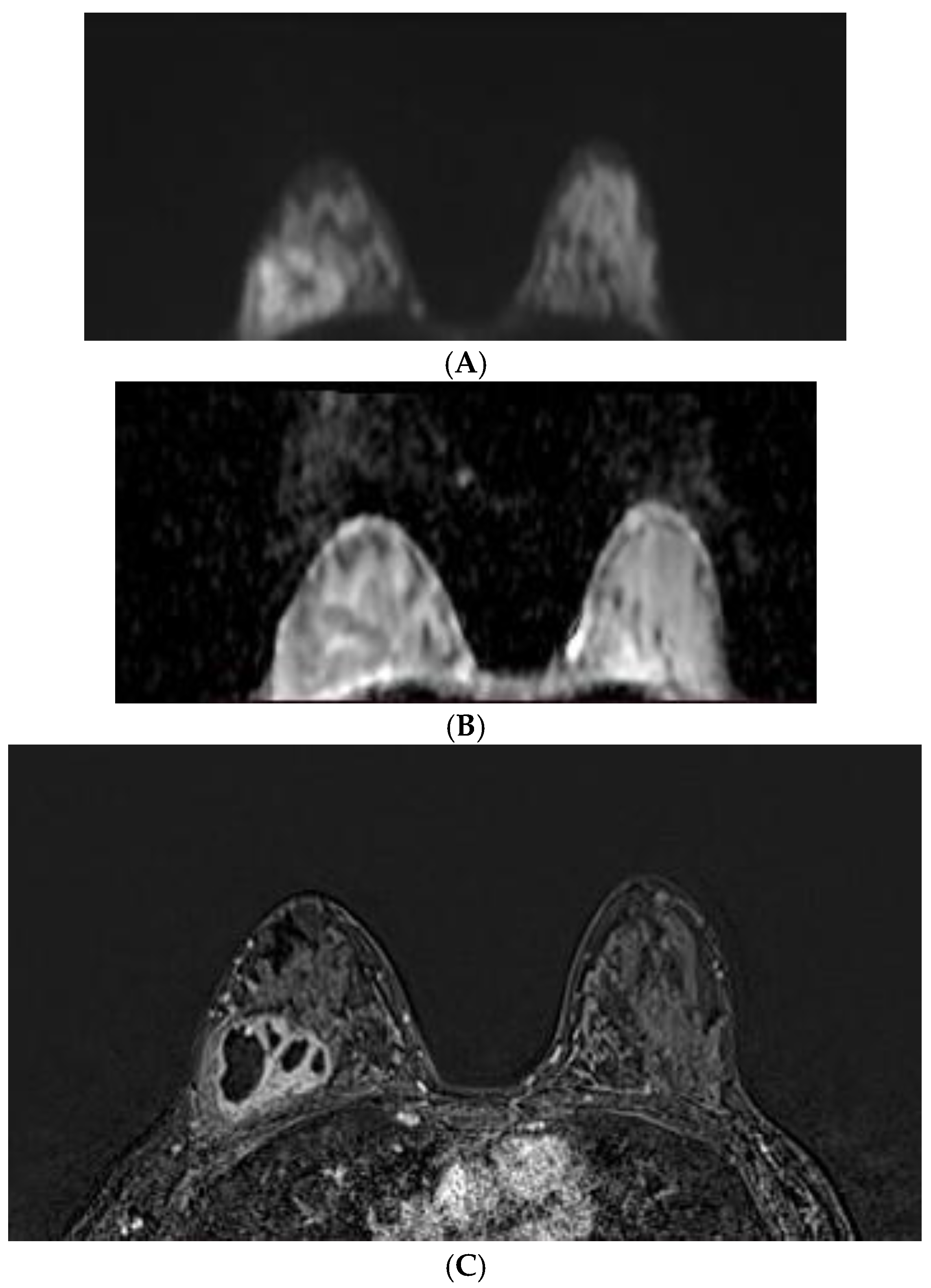
2.2. MRI Protocol
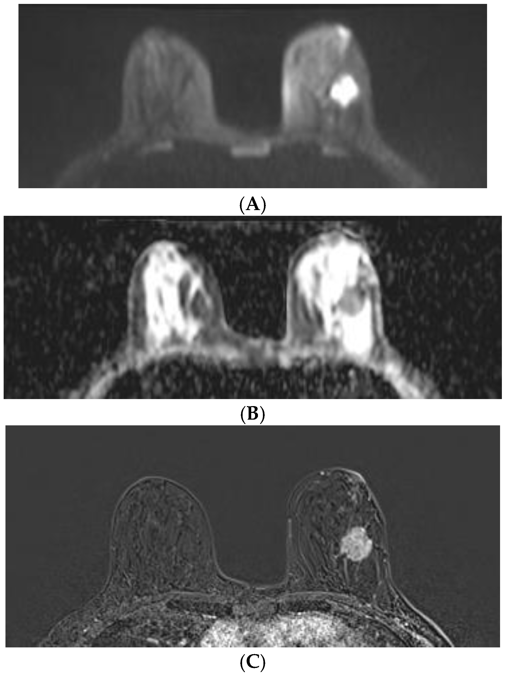
2.3. ADC Value Measurement
2.4. Pathohistological Evaluation
2.5. Statistical Analysis
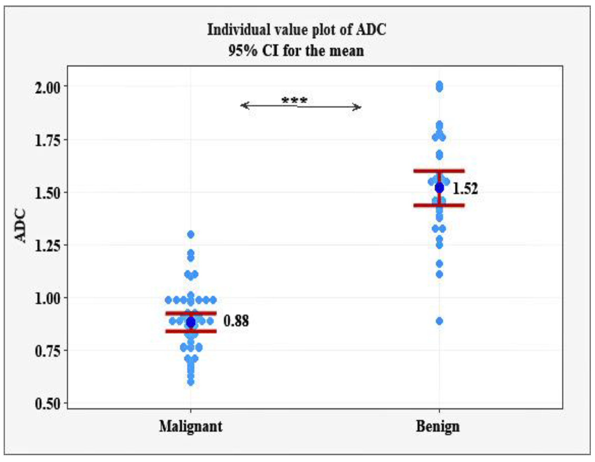
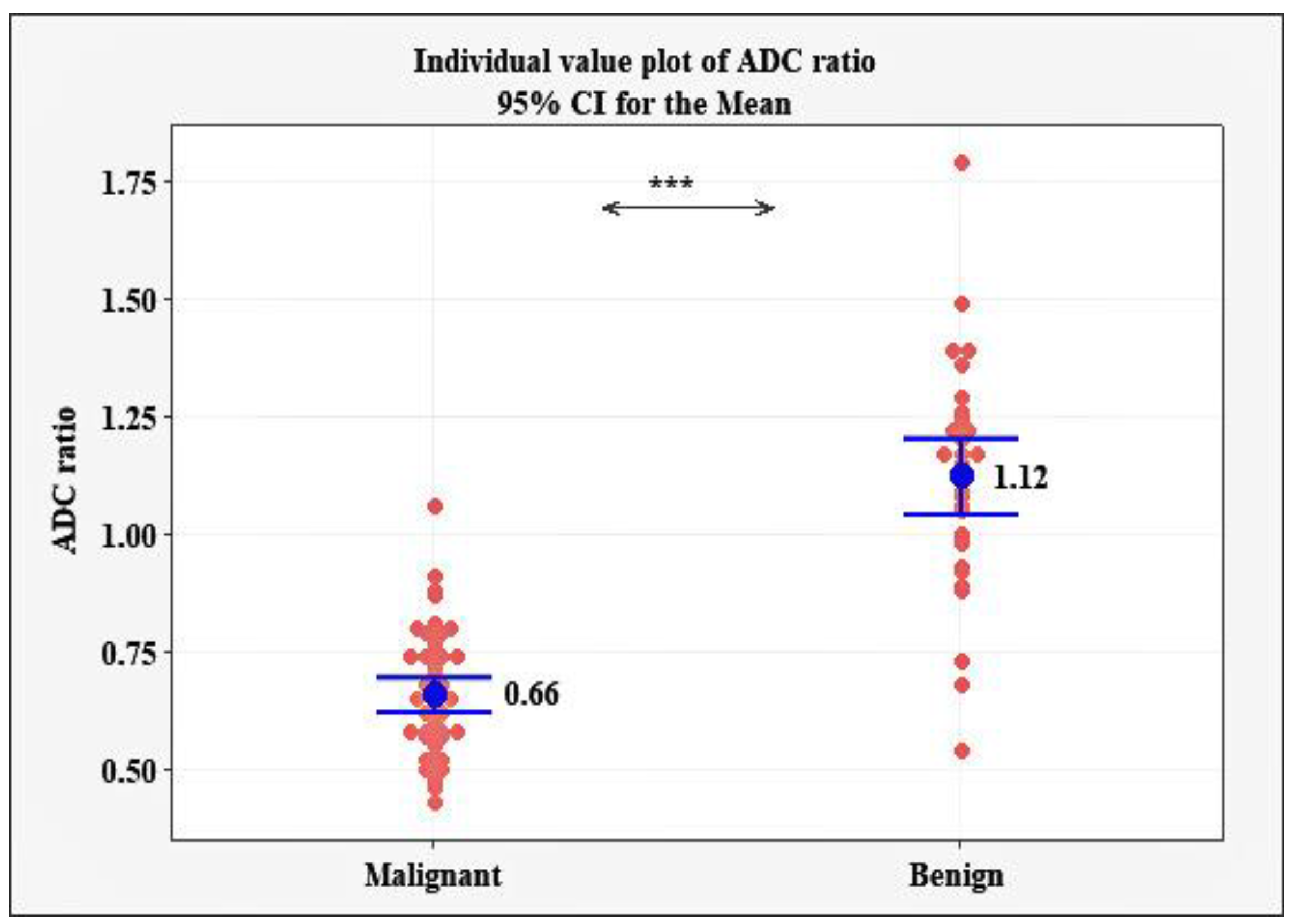
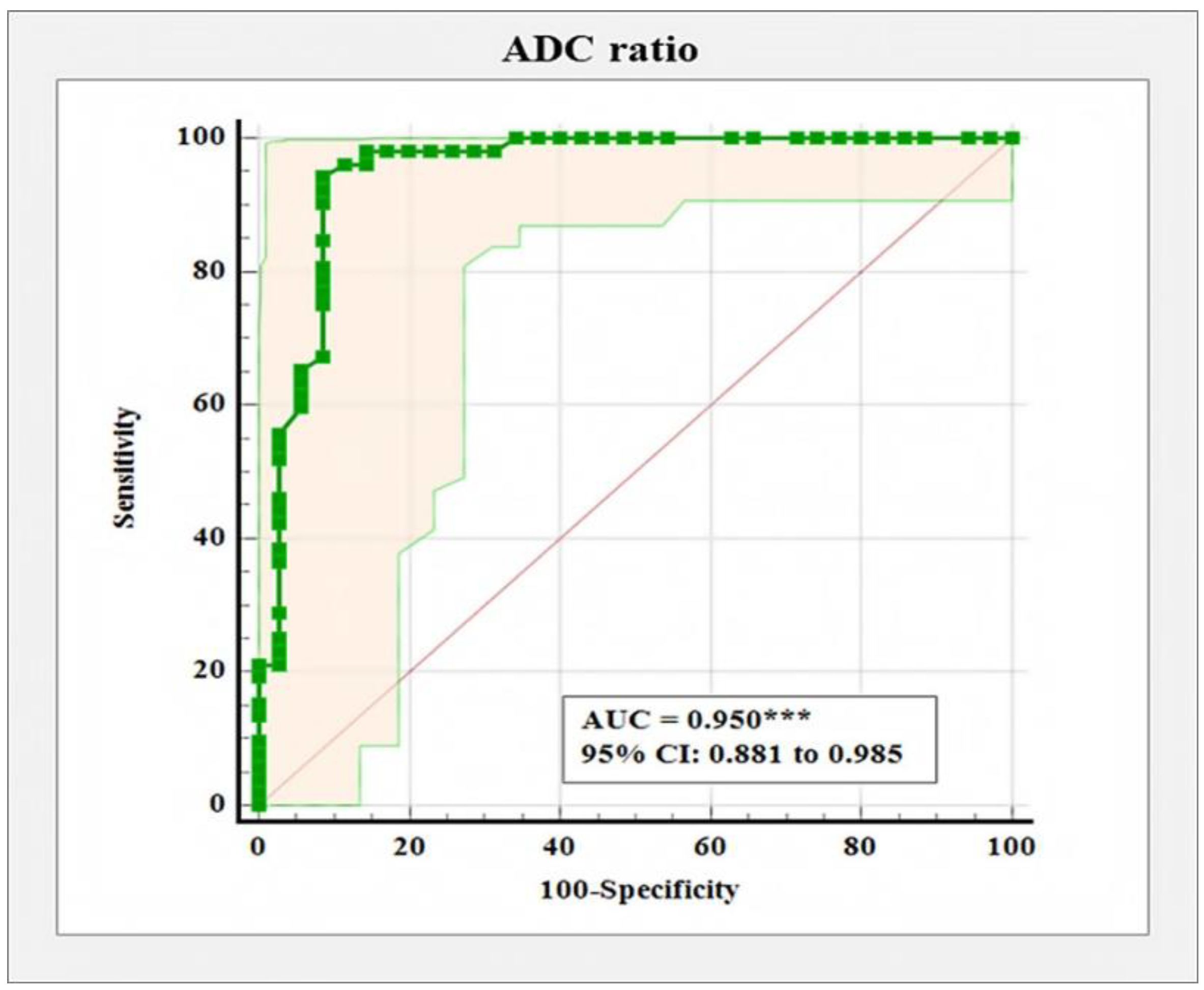
3. Results
4. Discussion
5. Conclusions
Author Contributions
Funding
Institutional Review Board Statement
Informed Consent Statement
Data Availability Statement
Conflicts of Interest
References
- Sasieni, P.D.; Shelton, J.; Ormiston-Smith, N.; Thomson, C.S.; Silcocks, P.B. What is the lifetime risk of developing cancer? The effect of adjusting for multiple primaries. Br. J. Cancer 2011, 105, 460–465. [Google Scholar] [CrossRef] [Green Version]
- Sung, H.; Ferlay, J.; Siegel, R.L.; Laversanne, M.; Soerjomataram, I.; Jemal, A.; Bray, F. Global Cancer Statistics 2020: Globocan Estimates of Incidence and Mortality Worldwide for 36 Cancers in 185 Countries. CA Cancer J. Clin. 2021, 71, 209–249. [Google Scholar] [CrossRef]
- Paepke, S.; Metz, S.; Brea Salvago, A.; Ohlinger, R. Benign Breast Tumours—Diagnosis and Management. Breast Care 2018, 13, 403–412. [Google Scholar] [CrossRef]
- Stachs, A.; Stubert, J.; Reimer, T.; Hartmann, S. Benign Breast Disease in Women. Dtsch. Arztebl. Int. 2019, 116, 565–574. [Google Scholar] [CrossRef]
- Mann, R.M.; Balleyguier, C.; Baltzer, P.A.; Bick, U.; Colin, C.; Cornford, E.; Evans, A.N.; Fallenberg, E.M.; Forrai, G.; Fuchsjäger, M.H.; et al. European Society of Breast Imaging (EUSOBI), with language review by Europa Donna–The European Breast Cancer Coalition. Breast MRI: EUSOBI recommendations for women’s information. Eur. Radiol. 2015, 25, 3669–3678. [Google Scholar] [CrossRef] [Green Version]
- Mann, R.M.; Cho, N.; Moy, L. Breast MRI: State of the Art. Radiology 2019, 292, 520–536. [Google Scholar] [CrossRef]
- Clauser, P.; Krug, B.; Bickel, H.; Dietzel, M.; Pinker, K.; Neuhaus, V.F.; Marino, M.A.; Moschetta, M.; Troiano, N.; Helbich, T.H.; et al. Diffusion-weighted Imaging Allows for Downgrading MR BI-RADS 4 Lesions in Contrast- enhanced MRI of the Breast to Avoid Unnecessary Biopsy. Clin. Cancer Res. 2021, 27, 1941–1948. [Google Scholar] [CrossRef]
- Amornsiripanitch, N.; Bickelhaupt, S.; Shin, H.J.; Dang, M.; Rahbar, H.; Pinker, K.; Partridge, S.C. Diffusion-weighted MRI for Unenhanced Breast Cancer Screening. Radiology 2019, 293, 504–520. [Google Scholar] [CrossRef]
- Amitai, Y.; Scaranelo, A.; Menes, T.S.; Fleming, R.; Kulkarni, S.; Ghai, S.; Freitas, V. Can breast MRI accurately exclude malignancy in mammographic architectural distortion? Eur. Radiol. 2020, 30, 2751–2760. [Google Scholar] [CrossRef]
- Bakker, M.F.; de Lange, S.V.; Pijnappel, R.M.; Mann, R.M.; Peeters, P.H.M.; Monninkhof, E.M.; Emaus, M.J.; Loo, C.E.; Bisschops, R.H.; Lobbes, M.B.; et al. Dense Trial Study Group. Supplemental MRI Screening for Women with Extremely Dense Breast Tissue. N. Engl. J. Med. 2019, 381, 2091–2102. [Google Scholar] [CrossRef]
- Amandeep, S.; Jasmin, P.; Kamlesh, G.; Gauaravdeep, S. Breast Lesion Characterisation with Diffusion- Weighted Imaging versus Dynamic Contrast- Enhanced- MRI: A Prospective Observational Study in a Tertiary Care Hospital. EMJ Radiol. 2021, 2, 7582. [Google Scholar] [CrossRef]
- Le Bihan, D. Diffusion MRI: What water tells us about the brain. EMBO Mol. Med. 2014, 6, 569–573. [Google Scholar] [CrossRef]
- Baliyan, V.; Das, C.J.; Sharma, R.; Gupta, A.K. Diffusion weighted imaging: Technique and applications. World J. Radiol. 2016, 8, 785–798. [Google Scholar] [CrossRef]
- Woodhams, R.; Ramadan, S.; Stanwell, P.; Sakamoto, S.; Hata, H.; Ozaki, M.; Kan, S.; Inoue, Y. Diffusion-weighted imaging of the breast: Principles and clinical applications. Radiographics 2011, 31, 1059–1084. [Google Scholar] [CrossRef] [Green Version]
- Guo, Y.; Cai, Y.Q.; Cai, Z.L.; Gao, Y.G.; An, N.Y.; Ma, L.; Mahankali, S.; Gao, J.-H. Differentiation of clinically benign and malignant breast lesions using diffusion-weighted imaging. J. Magn. Reson. Imaging 2002, 16, 172–178. [Google Scholar] [CrossRef]
- Partridge, S.C.; Nissan, N.; Rahbar, H.; Kitsch, A.E.; Sigmund, E.E. Diffusion-weighted breast MRI: Clinical applications and emerging techniques. J. Magn. Reson. Imaging 2017, 45, 337–355. [Google Scholar] [CrossRef]
- Woodhams, R.; Matsunaga, K.; Iwabuchi, K.; Kan, S.; Hata, H.; Kuranami, M.; Watanabe, M.; Hayakawa, K. Diffusion-weighted imaging of malignant breast tumors: The usefulness of apparent diffusion coefficient (ADC) value and ADC map for the detection of malignant breast tumors and evaluation of cancer extension. J. Comput. Assist. Tomogr. 2005, 29, 644–649. [Google Scholar] [CrossRef]
- Azab, E.A.; Ibrahim, M.E. Diffusion weighted (DW) MRI role in characterization of breast lesions using absolute and normalized ADC values. Egypt. J. Radiol. Nucl. Med. 2018, 47, 564–570. [Google Scholar] [CrossRef]
- Okuma, H.; Sudah, M.; Kettunen, T.; Niukkanen, A.; Sutela, A.; Masarwah, A.; Kosma, V.-M.; Auvinen, P.; Mannermaa, A.; Vanninen, R. Peritumor to tumor apparent diffusion coefficient ratio is associated with biologically more aggressive breast cancer features and correlates with the prognostication tools. PLoS ONE 2020, 15, e0235278. [Google Scholar] [CrossRef]
- Surov, A.; Clauser, P.; Chang, Y.W.; Li, L.; Martincich, L.; Partridge, S.C.; Kim, J.Y.; Meyer, H.J.; Wienke, A. Can diffusion-weighted imaging predict tumor grade and expression of Ki-67 in breast cancer? A multicenter analysis. Breast Cancer Res. 2018, 20, 58. [Google Scholar] [CrossRef]
- Baxter, G.C.; Graves, M.J.; Gilbert, F.J.; Patterson, A.J. A Meta-analysis of the Diagnostic Performance of Diffusion MRI for Breast Lesion Characterization. Radiology 2019, 291, 632–641. [Google Scholar] [CrossRef] [Green Version]
- Kim, J.Y.; Suh, H.B.; Kang, H.J.; Shin, J.K.; Choo, K.S.; Nam, K.J.; Lee, S.W.; Jung, Y.L.; Bae, Y.T. Apparent diffusion coefficient of breast cancer and normal fibroglandular tissue in diffusion-weighted imaging: The effects of menstrual cycle and menopausal status. Breast Cancer Res. Treat 2016, 157, 31–40. [Google Scholar] [CrossRef]
- Maric, J.; Boban, J.; Ivkovic-Kapicl, T.; Djilas, D.; Vucaj-Cirilovic, V.; Bogdanovic-Stojanovic, D. Differentiation of Breast Lesions and Distinguishing Their Histological Subtypes Using Diffusion-Weighted Imaging and ADC Values. Front. Oncol. 2020, 10, 332. [Google Scholar] [CrossRef]
- Rabasco, P.; Caivano, R.; Simeon, V.; Dinardo, G.; Lotumolo, A.; Gioioso, M.; Villonio, A.; Iannelli, G.; D’Antuono, F.; Zandolino, A.; et al. Can diffusion-weighted imaging and related apparent diffusion coefficient be a prognostic value in women with breast cancer? Cancer Investig. 2017, 35, 92–99. [Google Scholar] [CrossRef]
- Akın, Y.; Uğurlu, M.Ü.; Kaya, H.; Arıbal, E. Diagnostic Value of Diffusion-weighted Imaging and Apparent Diffusion Coefficient Values in the Differentiation of Breast Lesions, Histopathologic Subgroups and Correlation with Prognostic Factors using 3.0 Tesla MR. J. Breast Health 2016, 12, 123–132. [Google Scholar] [CrossRef]
- Kuhl, C.K.; Mielcareck, P.; Klaschik, S.; Leutner, C.; Wardelmann, E.; Gieseke, J.; Schild, H. Dynamic breast MR imaging: Are signal intensity time course data useful for differential diagnosis of enhancing lesions. Radiology 1999, 211, 101–110. [Google Scholar] [CrossRef]
- Baltzer, P.; Mann, R.M.; Iima, M.; Sigmund, E.E.; Clauser, P.; Gilbert, F.J.; Martincich, L.; Partridge, S.C.; Patterson, A.; Pinker, K.; et al. EUSOBI international Breast Diffusion-Weighted Imaging working group. Diffusion-weighted imaging of the breast-a consensus and mission statement from the EUSOBI International Breast Diffusion-Weighted Imaging working group. Eur. Radiol. 2020, 30, 1436–1450. [Google Scholar] [CrossRef] [Green Version]
- Bozkurt, T.B.; Koc, G.; Sezgin, G.; Altay, C.; Gelal, M.F.; Oyar, O. Value of apparent diffusion coefficient values in differentiating malignant and benign breast lesions. Balk. Med. J. 2016, 33, 294–300. [Google Scholar] [CrossRef]
- Marini, C.; Lacconi, C.; Giannelli, M.; Cilotti, A.; Moretti, M.; Bartolozzi, C. Quantitative diffusion-weighted MR imaging in the differential diagnosis of breast lesion. Eur. Radiol. 2007, 17, 2646–2655. [Google Scholar] [CrossRef]
- Partridge, S.C.; Amornsiripanitch, N. DWI in the Assessment of Breast Lesions. Top. Magn. Reson. Imaging 2017, 26, 201–209. [Google Scholar] [CrossRef]
- Lee, S.H.; Shin, H.J.; Moon, W.K. Diffusion-Weighted Magnetic Resonance Imaging of the Breast: Standardization of Image Acquisition and Interpretation. Korean J. Radiol. 2021, 22, 9–22. [Google Scholar] [CrossRef]
- Kul, S.; Cansu, A.; Alhan, E. Contribution of diffusion weighted imaging to dynamic contrast enhanced MRI in characterization of breast tumors. AJR 2011, 196, 210–217. [Google Scholar] [CrossRef]
- Rahbar, H.; Zhang, Z.; Chenevert, T.L.; Romanoff, J.; Kitsch, A.E.; Hanna, L.G.; Harvey, S.M.; Moy, L.; DeMartini, W.B.; Dogan, B.; et al. Utility of Diffusion-weighted Imaging to Decrease Unnecessary Biopsies Prompted by Breast MRI: A Trial of the ECOG-ACRIN Cancer Research Group (A6702). Clin. Cancer Res. 2019, 25, 1756–1765. [Google Scholar] [CrossRef]
- Surov, A.; Meyer, H.J.; Wienke, A. Can apparent diffusion coefficient (ADC) distinguish breast cancer from benign breast findings? A meta-analysis based on 13 847 lesions. BMC Cancer 2019, 19, 955. [Google Scholar] [CrossRef] [Green Version]
- Sahin, C.; Aribal, E. The role of apparent diffusion coefficient values in the differential diagnosis of breast lesions in diffusion-weighted MRI. Diagn. Interv. Radiol. 2013, 19, 457–462. [Google Scholar] [CrossRef] [Green Version]


| Histopathological Findings | Frequency | Percentage |
|---|---|---|
| Malignant | ||
| 36 | 41.40 |
| 5 | 5.75 |
| 3 | 3.45 |
| 2 | 2.30 |
| 2 | 2.30 |
| 1 | 1.14 |
| 1 | 1.14 |
| 1 | 1.14 |
| 1 | 1.14 |
| Total | 52 | 59.76% |
| Benign | ||
| 12 | 13.8 |
| 3 | 3.45 |
| 3 | 3.45 |
| 3 | 3.45 |
| 3 | 3.45 |
| 3 | 3.45 |
| 3 | 3.45 |
| 2 | 2.30 |
| 2 | 2.30 |
| 1 | 1.14 |
| Total | 35 | 40.24% |
Publisher’s Note: MDPI stays neutral with regard to jurisdictional claims in published maps and institutional affiliations. |
© 2022 by the authors. Licensee MDPI, Basel, Switzerland. This article is an open access article distributed under the terms and conditions of the Creative Commons Attribution (CC BY) license (https://creativecommons.org/licenses/by/4.0/).
Share and Cite
Tsvetkova, S.; Doykova, K.; Vasilska, A.; Sapunarova, K.; Doykov, D.; Andonov, V.; Uchikov, P. Differentiation of Benign and Malignant Breast Lesions Using ADC Values and ADC Ratio in Breast MRI. Diagnostics 2022, 12, 332. https://doi.org/10.3390/diagnostics12020332
Tsvetkova S, Doykova K, Vasilska A, Sapunarova K, Doykov D, Andonov V, Uchikov P. Differentiation of Benign and Malignant Breast Lesions Using ADC Values and ADC Ratio in Breast MRI. Diagnostics. 2022; 12(2):332. https://doi.org/10.3390/diagnostics12020332
Chicago/Turabian StyleTsvetkova, Silvia, Katya Doykova, Anna Vasilska, Katya Sapunarova, Daniel Doykov, Vladimir Andonov, and Petar Uchikov. 2022. "Differentiation of Benign and Malignant Breast Lesions Using ADC Values and ADC Ratio in Breast MRI" Diagnostics 12, no. 2: 332. https://doi.org/10.3390/diagnostics12020332
APA StyleTsvetkova, S., Doykova, K., Vasilska, A., Sapunarova, K., Doykov, D., Andonov, V., & Uchikov, P. (2022). Differentiation of Benign and Malignant Breast Lesions Using ADC Values and ADC Ratio in Breast MRI. Diagnostics, 12(2), 332. https://doi.org/10.3390/diagnostics12020332