Abstract
Established imaging modalities for the characterization of liver tumors are computed tomography (CT), magnetical resonance (MR) imaging, sonography, and hepatobiliary scintigraphy. In some cases, their results may be inconclusive or certain examinations not possible due to contraindications. Positron emission tomography (PET)/CT has the capability of dynamic imaging with high temporal resolution. With radiolabeled tri-alkoxysalicyl-1,4-diazepan-6-amine (TAoS-DAZA) tracers, imaging of liver perfusion and hepatobiliary function is possible in a single examination. In the presented case, the PET/CT was performed in a patient with suspected hepatocellular carcinoma and atypical CT findings. PET imaging characteristics were consistent with a hepatocellular carcinoma (HCC). PET with DAZA ligands may be a supplemental method for liver tumor characterization in difficult cases.
Characterization of tumors is a prerequisite of successful treatment, and is mostly done by biopsy and histopathological examination. A major exception is HCC, which is the most common liver cancer. In many cases, a diagnosis is established based on imaging alone, because there is a risk of extrahepatic tumor spread caused by biopsy. Several imaging criteria guidelines have been established, including non-rim arterial phase contrast media hyperenhancement and washout in the portalvenous phase. In some guidelines, the latter criterion is extended to the hepatobiliary phase [1]. However, in some clinical cases MR imaging, which is the best modality for liver tumor evaluation, is not possible due to contraindications, and other options are required (Figure 1, Figure 2, Figure 3 and Figure 4).
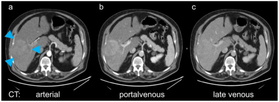
Figure 1.
In the presented patient, on computed tomography (CT) the mass in the right liver lobe (a, arrowheads) showed arterial (a) and portalvenous (b) enhancement, with a mixed appearance on the late venous phase with slightly hyper-, but also hypodense areas (c). The diagnostic dilemma was that a biopsy should be avoided, and the patient could not undergo magnetical resonance (MR) imaging because of a pacemaker. Fast dynamic hepatobiliary scintigraphy is limited to planar acquisition, and not feasible for the evaluation of focal lesions [2].
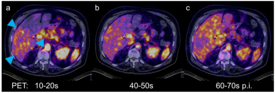
Figure 2.
Positron emission tomography (PET)/CT has dynamic imaging capabilities for the depiction of liver tumors [3]. Therefore, an examination with the hepatobiliary tracer [68Ga]Ga-tri-methoxysalicyl-(TMoS)-DAZA was performed. Similar to mebrofenin derivatives and liver-specific MR contrast agents (e.g., gadoxetic acid), this 1,4-diazepan-6-amine (DAZA) ligand is extracted from the blood by hepatocytes and secreted into the bile [4,5,6]. In this case, 155 MBq of the tracer were injected intravenously. A dynamic PET of the liver over 3 min was acquired and reconstructed in 10-s timeframes. Early arterial hyperperfusion was evident in the region of the tumor (a, arrowheads). Tumor uptake was equal to normal liver 50 s after injection (b). From 60 s after injection, uptake in the tumor was lower than in the surrounding liver tissue (c).
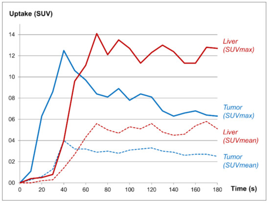
Figure 3.
Measurements of standardized uptake values (SUV) in the tumor and in a liver segment not involved by tumor showed fast tumor perfusion followed by a “washout” of tracer (blue curves), while the liver uptake remained relatively steady (red curves).
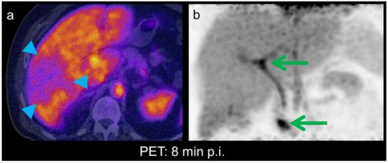
Figure 4.
Liver function in a hepatocellular carcinoma (HCC) depends on the degree of dedifferentiation, substrate uptake, processing and bile excretion still taking place in well and moderately differentiated HCCs [7]. On PET/CT acquired in the hepatobiliary phase, eight minutes after injection, tumor uptake was approx. In this case, 50% of normal liver tissue, consistent with reduced but not absent cellular function (a, arrowheads). Tracer excretion into bile ducts and duodenum can be seen on the corresponding maximum intensity projection (MIP) image (b, green arrows). The tumor itself is barely visible due to the overlying liver tissue. In summary, the features were suggestive of an HCC. The patient underwent surgery. Histopathological examination showed a moderately differentiated HCC.
The case shows that PET imaging with DAZA ligands may be evolve to be a supplemental method for liver tumor characterization in difficult cases, combining information about tissue perfusion with those of hepatobiliary function and excretion.
Author Contributions
Conceptualization and methodology, M.F. and J.G.; software, C.K.; validation, F.R. and H.-M.T.; formal analysis and investigation, R.D. and F.G.; resources, M.F.; data curation, R.D., F.G. and T.W.; writing—original draft preparation, R.D. and F.G.; writing—review and editing, all authors; visualization, M.F. and R.D.; supervision, M.F. All authors have read and agreed to the published version of the manuscript.
Funding
This research received no external funding.
Institutional Review Board Statement
The study was conducted according to the guidelines of the Declaration of Helsinki. Ethical review and approval were waived due to the retrospective presentation of a single case.
Informed Consent Statement
Informed consent was obtained from the patient.
Acknowledgments
We acknowledge support by the German Research Foundation and the Open Access Publication Fund of the Thüringer Universitäts- und Landesbibliothek Jena, project no. 433052568.
Conflicts of Interest
The authors declare no conflict of interest.
References
- Park, S.H.; Shim, Y.S.; Kim, B.; Kim, S.Y.; Kim, Y.S.; Huh, J.; Park, J.H.; Kim, K.W.; Lee, S.S. Retrospective analysis of current guidelines for hepatocellular carcinoma diagnosis on gadoxetic acid–enhanced MRI in at-risk patients. Eur. Radiol. 2021, 1–13. [Google Scholar] [CrossRef]
- Rassam, F.; Olthof, P.B.; Richardson, H.; van Gulik, T.M.; Bennink, R.J. Practical guidelines for the use of technetium-99m mebrofenin hepatobiliary scintigraphy in the quantitative assessment of liver function. Nucl. Med. Commun. 2019, 40, 297–307. [Google Scholar] [CrossRef] [PubMed]
- Schierz, J.-H.; Opfermann, T.; Steenbeck, J.; Lopatta, E.; Settmacher, U.; Stallmach, A.; Marlowe, R.J.; Freesmeyer, M. Early Dynamic 18F-FDG PET to Detect Hyperperfusion in Hepatocellular Carcinoma Liver Lesions. J. Nucl. Med. 2013, 54, 848–854. [Google Scholar] [CrossRef] [PubMed]
- Greiser, J.; Kühnel, C.; Görls, H.; Weigand, W.; Freesmeyer, M.; Kuehnel, C. N,1,4-Tri(4-alkoxy-2-hydroxybenzyl)-DAZA: Efficient one-pot synthesis and labelling with 68Ga for PET liver imaging in ovo. Dalton Trans. 2018, 47, 9000–9007. [Google Scholar] [CrossRef] [PubMed]
- Greiser, J.; Weigand, W.; Freesmeyer, M. Metal-Based Complexes as Pharmaceuticals for Molecular Imaging of the Liver. Pharmaceuticals 2019, 12, 137. [Google Scholar] [CrossRef] [PubMed]
- Freesmeyer, M.; Kuehnel, C.; Opfermann, T.; Niksch, T.; Wiegand, S.; Stolz, R.; Huonker, R.; Witte, O.W.; Winkens, T. The Use of Ostrich Eggs for In Ovo Research: Making Preclinical Imaging Research Affordable and Available. J. Nucl. Med. 2018, 59, 1901–1906. [Google Scholar] [CrossRef] [PubMed]
- Schlageter, M.; Terracciano, L.M.; D’Angelo, S.; Sorrentino, P. Histopathology of hepatocellular carcinoma. World J. Gastroenterol. 2014, 20, 15955–15964. [Google Scholar] [CrossRef] [PubMed]


Publisher’s Note: MDPI stays neutral with regard to jurisdictional claims in published maps and institutional affiliations. |
© 2021 by the authors. Licensee MDPI, Basel, Switzerland. This article is an open access article distributed under the terms and conditions of the Creative Commons Attribution (CC BY) license (https://creativecommons.org/licenses/by/4.0/).