
Dynamic PET/CT with the Hepatobiliary Tracer [68Ga]Ga-Tmos-DAZA for Characterization of a Hepatic Tumor

 , ,
, ,  ,
, 
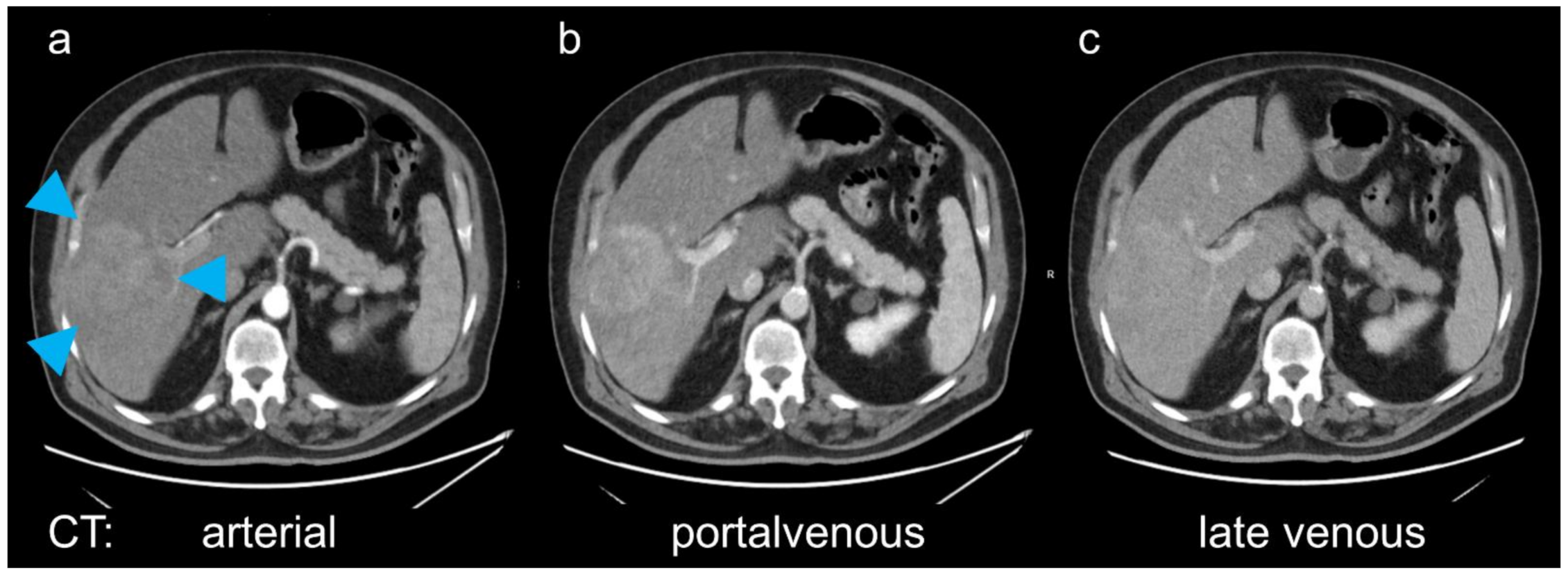
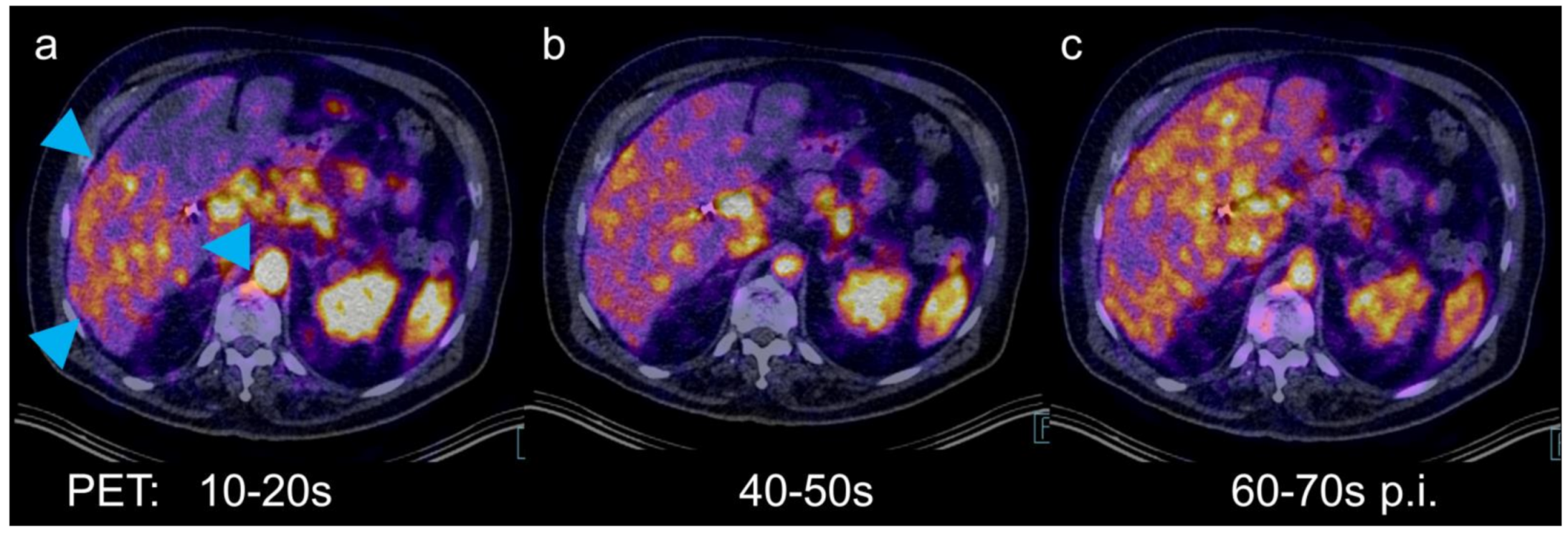
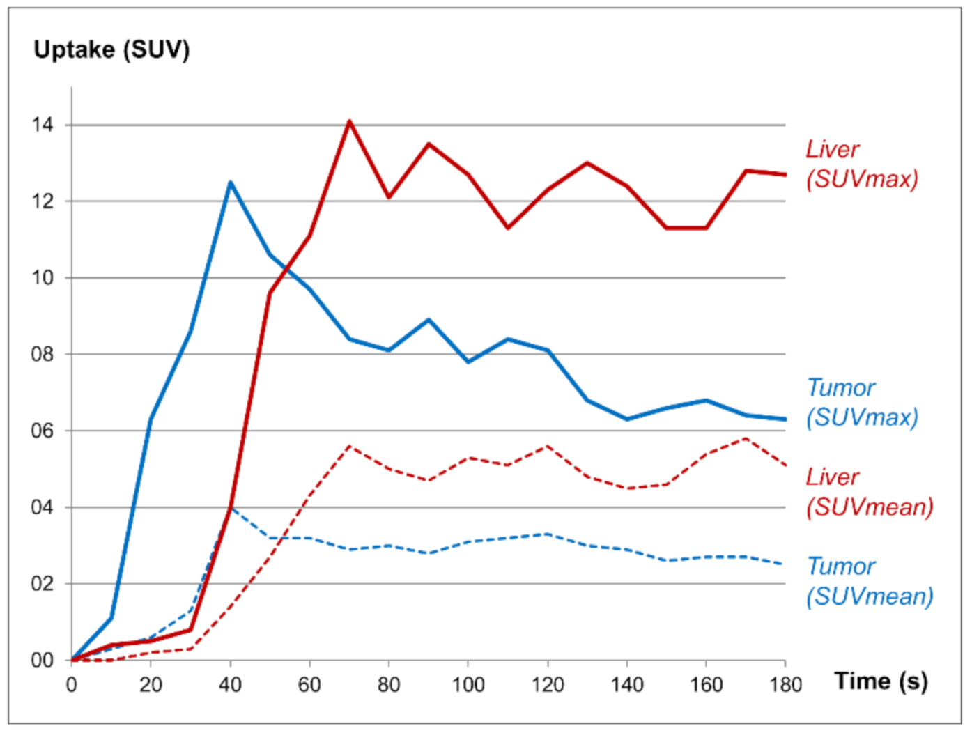
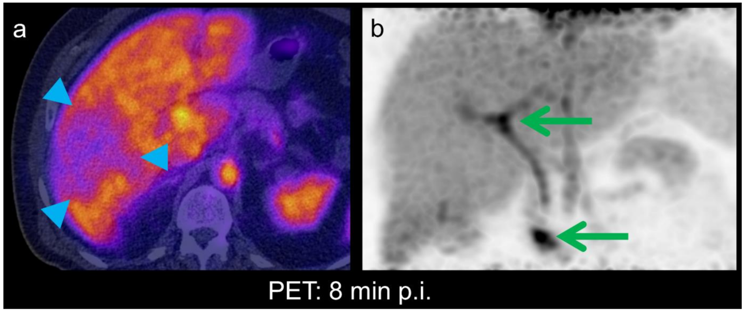
Abstract
Author Contributions
Funding
Institutional Review Board Statement
Informed Consent Statement
Acknowledgments
Conflicts of Interest
References
- Park, S.H.; Shim, Y.S.; Kim, B.; Kim, S.Y.; Kim, Y.S.; Huh, J.; Park, J.H.; Kim, K.W.; Lee, S.S. Retrospective analysis of current guidelines for hepatocellular carcinoma diagnosis on gadoxetic acid–enhanced MRI in at-risk patients. Eur. Radiol. 2021, 1–13. [Google Scholar] [CrossRef]
- Rassam, F.; Olthof, P.B.; Richardson, H.; van Gulik, T.M.; Bennink, R.J. Practical guidelines for the use of technetium-99m mebrofenin hepatobiliary scintigraphy in the quantitative assessment of liver function. Nucl. Med. Commun. 2019, 40, 297–307. [Google Scholar] [CrossRef] [PubMed]
- Schierz, J.-H.; Opfermann, T.; Steenbeck, J.; Lopatta, E.; Settmacher, U.; Stallmach, A.; Marlowe, R.J.; Freesmeyer, M. Early Dynamic 18F-FDG PET to Detect Hyperperfusion in Hepatocellular Carcinoma Liver Lesions. J. Nucl. Med. 2013, 54, 848–854. [Google Scholar] [CrossRef] [PubMed]
- Greiser, J.; Kühnel, C.; Görls, H.; Weigand, W.; Freesmeyer, M.; Kuehnel, C. N,1,4-Tri(4-alkoxy-2-hydroxybenzyl)-DAZA: Efficient one-pot synthesis and labelling with 68Ga for PET liver imaging in ovo. Dalton Trans. 2018, 47, 9000–9007. [Google Scholar] [CrossRef] [PubMed]
- Greiser, J.; Weigand, W.; Freesmeyer, M. Metal-Based Complexes as Pharmaceuticals for Molecular Imaging of the Liver. Pharmaceuticals 2019, 12, 137. [Google Scholar] [CrossRef] [PubMed]
- Freesmeyer, M.; Kuehnel, C.; Opfermann, T.; Niksch, T.; Wiegand, S.; Stolz, R.; Huonker, R.; Witte, O.W.; Winkens, T. The Use of Ostrich Eggs for In Ovo Research: Making Preclinical Imaging Research Affordable and Available. J. Nucl. Med. 2018, 59, 1901–1906. [Google Scholar] [CrossRef] [PubMed]
- Schlageter, M.; Terracciano, L.M.; D’Angelo, S.; Sorrentino, P. Histopathology of hepatocellular carcinoma. World J. Gastroenterol. 2014, 20, 15955–15964. [Google Scholar] [CrossRef] [PubMed]


Publisher’s Note: MDPI stays neutral with regard to jurisdictional claims in published maps and institutional affiliations. |
© 2021 by the authors. Licensee MDPI, Basel, Switzerland. This article is an open access article distributed under the terms and conditions of the Creative Commons Attribution (CC BY) license (https://creativecommons.org/licenses/by/4.0/).
Share and Cite
Freesmeyer, M.; Greiser, J.; Winkens, T.; Gühne, F.; Kühnel, C.; Rauchfuß, F.; Tautenhahn, H.-M.; Drescher, R. Dynamic PET/CT with the Hepatobiliary Tracer [68Ga]Ga-Tmos-DAZA for Characterization of a Hepatic Tumor. Diagnostics 2021, 11, 660. https://doi.org/10.3390/diagnostics11040660
Freesmeyer M, Greiser J, Winkens T, Gühne F, Kühnel C, Rauchfuß F, Tautenhahn H-M, Drescher R. Dynamic PET/CT with the Hepatobiliary Tracer [68Ga]Ga-Tmos-DAZA for Characterization of a Hepatic Tumor. Diagnostics. 2021; 11(4):660. https://doi.org/10.3390/diagnostics11040660
Chicago/Turabian StyleFreesmeyer, Martin, Julia Greiser, Thomas Winkens, Falk Gühne, Christian Kühnel, Falk Rauchfuß, Hans-Michael Tautenhahn, and Robert Drescher. 2021. "Dynamic PET/CT with the Hepatobiliary Tracer [68Ga]Ga-Tmos-DAZA for Characterization of a Hepatic Tumor" Diagnostics 11, no. 4: 660. https://doi.org/10.3390/diagnostics11040660
APA StyleFreesmeyer, M., Greiser, J., Winkens, T., Gühne, F., Kühnel, C., Rauchfuß, F., Tautenhahn, H.-M., & Drescher, R. (2021). Dynamic PET/CT with the Hepatobiliary Tracer [68Ga]Ga-Tmos-DAZA for Characterization of a Hepatic Tumor. Diagnostics, 11(4), 660. https://doi.org/10.3390/diagnostics11040660