Abstract
(1) Objective—Intraoperative Magnetic Resonance Imaging (IOMRI) guided surgery has revolutionized neurosurgery and has especially impacted the field of Neuro-Oncology, with randomized controlled trails demonstrating improved resection, fewer postoperative deficits and enhanced survival rates. Bibliometric analysis allows for analysing chronological trends and measuring the impact and directions of research in a particular field. To the authors’ knowledge, this is the first Bibliometric analysis conducted on IOMRI. (2) Methods—a title specific search of the Web of Science database was executed using the keywords ‘intraoperative MRI’, ‘intraoperative magnetic resonance imaging’, and “IOMRI’ on 23rd April 2021. Results—663 articles met the inclusion criteria and were included in the final analysis. In addition, the 100 most cited were analysed as well. Among these 100 articles, 76 were original research papers, while 14 others were review articles. Amongst all the authors, Ganslandt contributed the maximum number of articles, with USA being the largest single source of these articles, followed by Germany. Interestingly, a shift of trends from “Image guided surgery’ and ‘accuracy’ in the early 2000s to ‘extent of resection’, ‘impact’, and ‘survival’ in the later years was noted. (3) Conclusions—IOMRI has now become an integral part of neurosurgery, especially in neuro-oncology. Focus has now shifted from implementation to refinement of technique in the form of functional and oncological outcomes. Therefore, future research in this direction is imperative and will be of more impact that in any other sub-field related to IOMRI.
1. Introduction
Neurosurgery has always been driven by the tenets of addressing pathology while maximizing precision, accuracy and, by extension, minimizing morbidity. Neuro-navigation and the availability of image-based guidance while performing surgery has partially addressed this dictum. However, as these are reliant on preoperative images, the accuracy progressively declines during surgery, due to brain shift. From here, the surgeon must rely on his or her own visual cues and judiciously choose a further course of action. The development of the first intraoperative MRI, in 1991, addressed this challenge [1]. It was apparent that intraoperative MRI (IOMRI) images were significantly superior, as they provided accurate information on residual/resected pathology and its relationship with the brain. Although other intraoperative tools, such as ultrasonography (USG) and computed tomography (CT), are available, the image quality and interpretive ability of an MRI image is far superior [1].
A prototype intraoperative MRI unit was first installed in 1994 at the Brigham & Women’s Hospital [1]. Subsequently, the first IOMRI guided stereotactic biopsy was undertaken in June 1995, and in August 1996, the IOMRI was first used in the resection of a brain tumour [1]. In 1997 and in 1999, Black and Jolesz summarized their initial experiences with the development, implementation, and the neurosurgical applications of IOMRI in two landmark papers published in Neurosurgery. This set the stage for a more widespread use and acceptance of IOMRI in neurosurgical procedures [2,3]. This has been followed by an extensive use of IOMRI, the world over, with several landmark articles and randomised articles cementing the utility and superiority of this technique.
This article aims to identify the available literature on IOMRI, with a further emphasis on the analyses of the 100 most influential articles on IOMRI by utilizing citation and bibliometric analyses. Citation analysis is a true measure of the impact of articles with several factors influencing the same [4]. Introduced in 1969, bibliometric analysis is superior to peer reviewed eminence-based analysis [5]. It can summarize the chronology and objective citation patterns, along with areas, authors, and journals with the highest impact and the greatest number of publications in a particular topic. There have been a few bibliometric analyses conducted previously in Neurosurgery, and they have proven to be a true reflection of the topic discussed [5].
2. Methods
2.1. Search Strategy
A title specific search of the Web of Science database was executed using the keywords ‘intraoperative MRI’, ‘intraoperative magnetic resonance imaging’, and “IOMRI’ on 23rd April 2021. All the abstracts were screened for the suitable articles. The inclusion criteria were articles relevant to the use of Intraoperative MRI in neurosurgery and published in peer reviewed journals. In addition to the available literature, the 100 most cited articles were selected and reviewed by the authors. All the articles published on Intraoperative MRI and the top 100 cited articles were analysed separately. Bibliometric analysis may sometimes be superior to, or more detailed than, “human-hand search” as it is a computer base comprehensive search and has analysis trends that are not visible in a manual one.
2.2. Data
The articles were arranged in descending order according to the number of citations. The parameters assessed were title of the articles, authors, corresponding authors, country of origin, journal of publication, year of publication, citation count, and the journal impact factor.
2.3. Analysis
The statistical analysis was performed using R language v 4.0.3 (R Foundation for Statistical Computing, Vienna, Austria) employing the “bibliometrix’ package [6]. The VOSviewer software (Van Eck and Waltman, Leiden University, Leiden, The Netherlands) was also used to plot network and overlay plots [7].
3. Results
Among the articles on “Intraoperative MRI” (as per the Web of Science database) published between 1996 and 2020, 663 met the inclusion and exclusion criteria and were included in the final analysis (Table 1). The 100 most cited articles were selected using the “Times cited” feature on Web of Science and analysed separately. These articles ranged in the year they were published, from 1996 to 2016 (Table 1). Interestingly, 76 out of these 100 most cited articles constituted original research papers, while 14 others were review articles (Table 1).

Table 1.
Analysis of the major findings from the bibliometric analysis of the 100 most cited papers with intraoperative MRI.
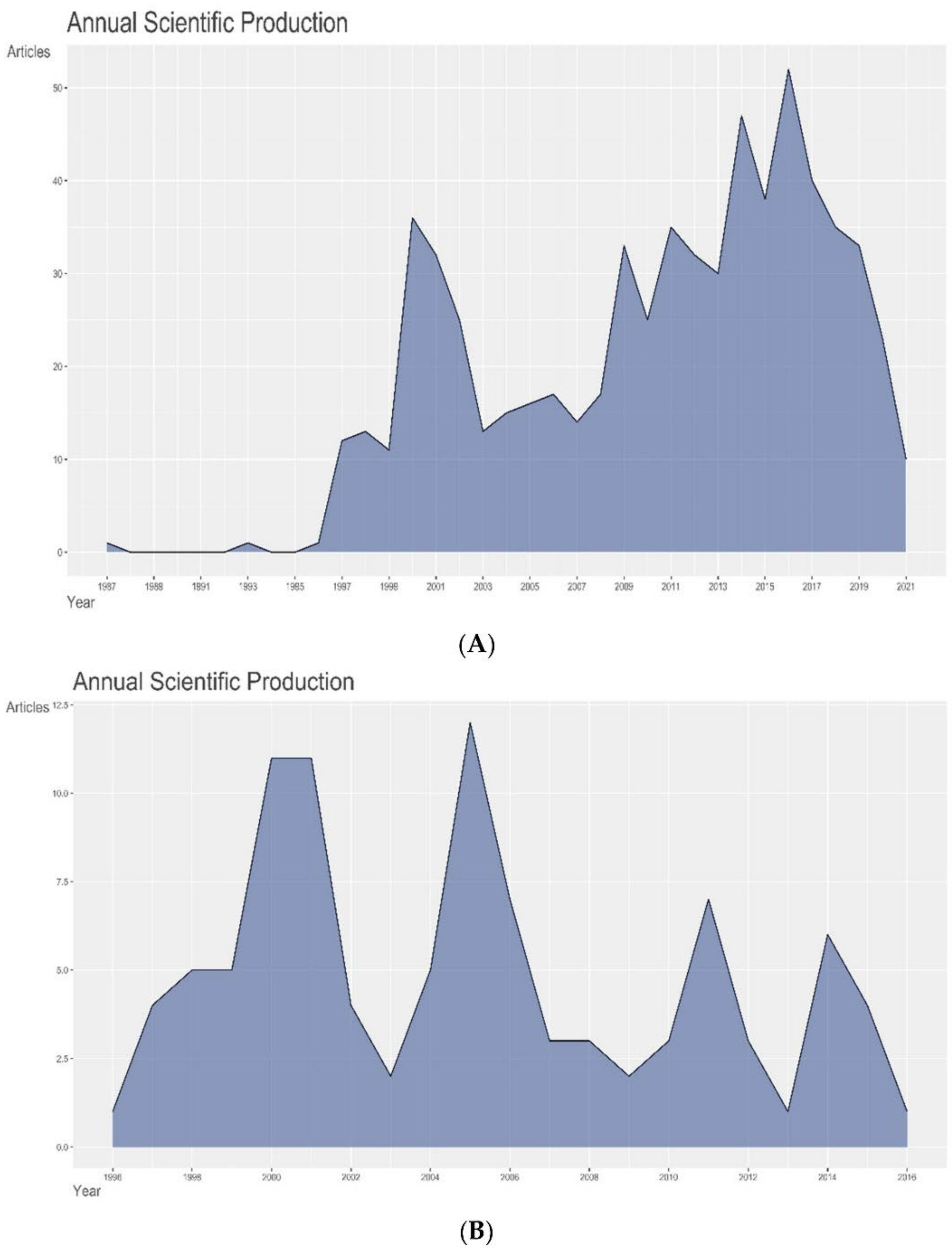
The highest number of articles on IOMRI (overall), published in a single year, were published in 2016 (n = 52) (Figure 1A), whereas the highest number of articles, among the 100 most cited articles, was published in 2005 (12%) (Figure 1B) with most of them having been published between 1996 and 2005 (67%). This may, in turn, be due to the number of citations garnered by these articles with the progression of years [8]. More importantly, the maximum number of mean citations per article and mean citations per year, for all the articles, were seen for the article published in 1996, while the articles published in 1997 received the highest number of mean citations per article and mean citations per year. These are markers of the most influential articles produced, independent of the number of citable years (Figure 1C).
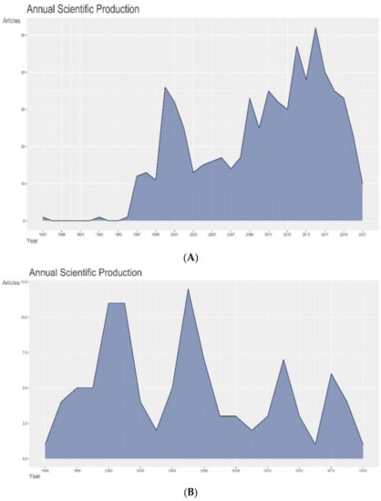

Figure 1.
Graph demonstrating (A) annual production of ALL articles on intraoperative MRI, (B) annual production of the 100 most cited articles on intraoperative MRI, and (C) average article citations per year of the 100 most cited articles on intraoperative MRI.
A point to note here is that, although Figure 1A shows reduced scientific production after 2017, the reason is that the top 100 cited articles were from 1996 to 2016, and later, citation declined due to recency of their publication. This should not be interpreted as reduced scientific interest in intraoperative MRI studies.
3.1. Authors
Among the 663 articles overall, 51 papers were single author papers, while the remaining involved multiple authors. While considering the 100 most cited articles, a similar trend was observed with only four single author papers (Table 1). While 1984 authors contributed to 663 articles, the top 100 cited articles were authored by a total of 350 authors (Table 1). An average of 6.13 co-authors were present for each article in the top 100 cited group, with a collaboration index of 3.61 (Table 1). Most of the authors (88.6%) contributed to either 1 or 2 articles.
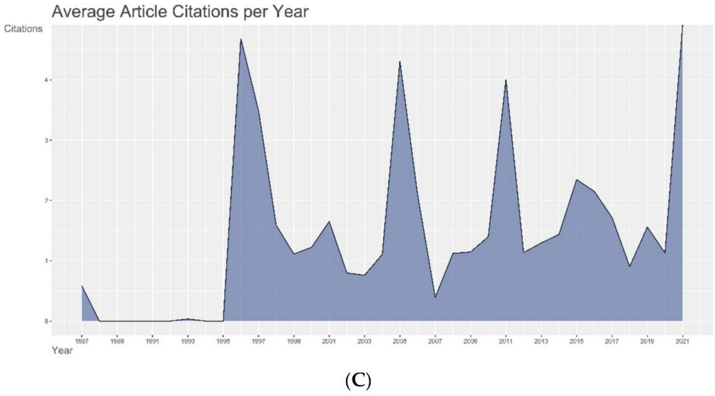
Amongst the top 100 cited articles, O Ganslandt contributed to the maximum number of articles (n = 24), followed by Nimsky C (n = 22) (Figure 2A, Supplementary Table S1). Among all the articles published on the topic, Nimsky C has had the maximum impact with a h index of 23 and, consequently, has the highest G and M index (Figure 2B, Table 2). The G-Index is wherein the top G articles have, together, received G citations. The M-Index is the H-index divided by the number of years that a scientist has been active. These indices allow for a weighted analysis of the authors’ contributions. Nimsky C also began publishing early on this topic (1998) and has accrued 2218 citations to-date on IOMRI related scientific publications (Figure 2C, Table 2). O Ganslandt and C Nimsky had the highest number of articles produced on IOMRI in a single year (n = 5), both in the year 2005 (Figure 2C). They also had the highest number of citations per year of 41.438. The average citation per article, in the top 100 cited group, was 113.5, which translated to 7.444 citations per document per year (Table 1). A total of 2089 references were used in these 100 articles.

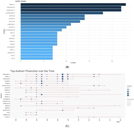
Figure 2.
Graph demonstrating (A) authors with maximum number of publications in the top 100 cited articles, (B) author impact in terms of h-index among all the articles published on IOMRI, and (C) production rate of the authors per year of the 100 most cited articles on intraoperative MRI.
Table 2.
A review of the bibliometrics for the most influential authors in intraoperative MRI related research.
3.2. Journals
Neurosurgery journal has published the maximum number of articles on IOMRI (n= 145) (Figure 3A). It was followed by Neuro-oncology (n= 51), Journal of Neurosurgery, and World Neurosurgery (n = 41 each) (Table 3). Among the top 100 cited articles, Neurosurgery (29% of articles), again, had the highest number of publications and was followed by the Journal of Neurosurgery (10% of articles), Radiology (5% of articles), and the Journal of Magnetic resonance imaging (4% of articles).
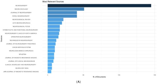
Figure 3.
Graph demonstrating (A) most relevant source journals, which published the articles on intraoperative MRI, (B) Bradford’s law related to journal sources of the 100 most cited articles on intraoperative MRI.
Table 3.
The most influential journals with intraoperative MRI related research.
Bradford’s law is a pattern, first described by Samuel C. Bradford in 1934, that estimates the exponentially diminishing results of searching for references in scientific journals [9]. Neurosurgery and Journal of Neurosurgery were in the zone 1 of Bradford’s law for the top 100 cited articles (Figure 3B). Considering all the articles published on IOMRI, Neurosurgery, Neuro-oncology, and Journal of Neurosurgery were in the zone 1 of Bradford’s law, and World Neurosurgery was in the zone 2 of Bradford’s law.
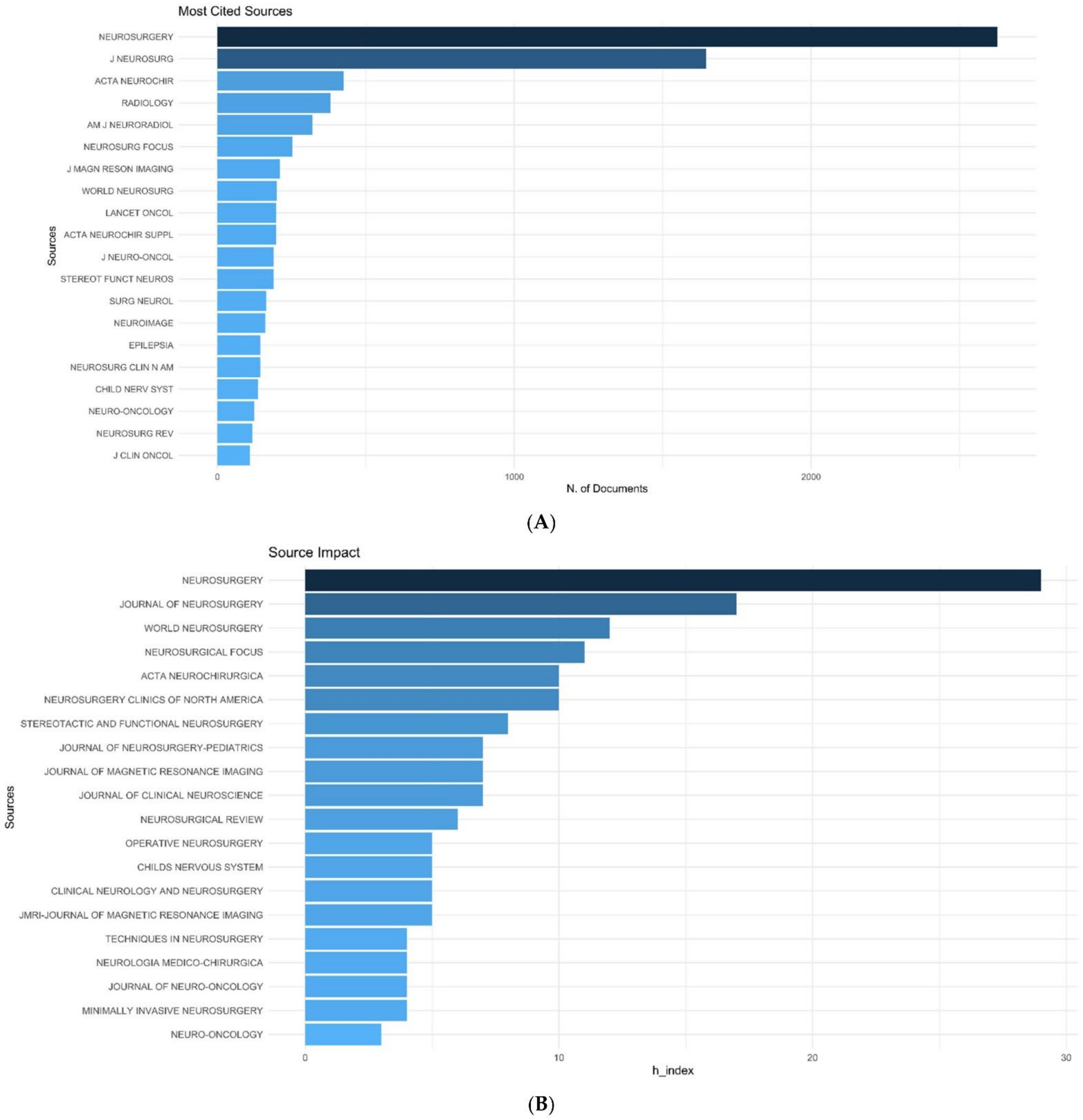
Among all the articles on IOMRI, the most cited journal was Neurosurgery, Journal of Neurosurgery, Acta Neurochirurgica, Radiology, American Journal of Neuroradiology (Figure 4A).

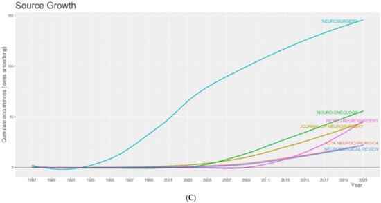
Figure 4.
Graph demonstrating (A) the most cited source journals which published the articles on intraoperative MRI, (B) source impact, in terms of h-index, among all the articles published on IOMRI, and (C) source dynamics, showing the cumulative number of publications in the different journals over time.
Neurosurgery had the highest h and g index of 29 and 64, respectively, followed by the Journal of Neurosurgery (17,28) and World Neurosurgery (12,13,18) (Figure 4B, Table 4). Total citations received by these three journals were 4189, 833, and 377, respectively. Figure 4C shows the year-wise cumulative number of publications on IOMRI in the top 6 journals, which published the maximum number of articles. The curve is steep for Neurosurgery in the 1990s and for World Neurosurgery after 2010.
Table 4.
Bibliometrics of the top impactful journals with Intraoperative MRI related research.
As expected, the most frequently used keywords were “resection”, followed by “extent” and “surgery” (Supplementary Figure S1). This, against the previous trend of “Image guided surgery” and “accuracy”, indicates an increased belief in the mechanism, use of intraoperative MRI, and focus on the potential benefit to the patients.
3.3. Countries and Institutions
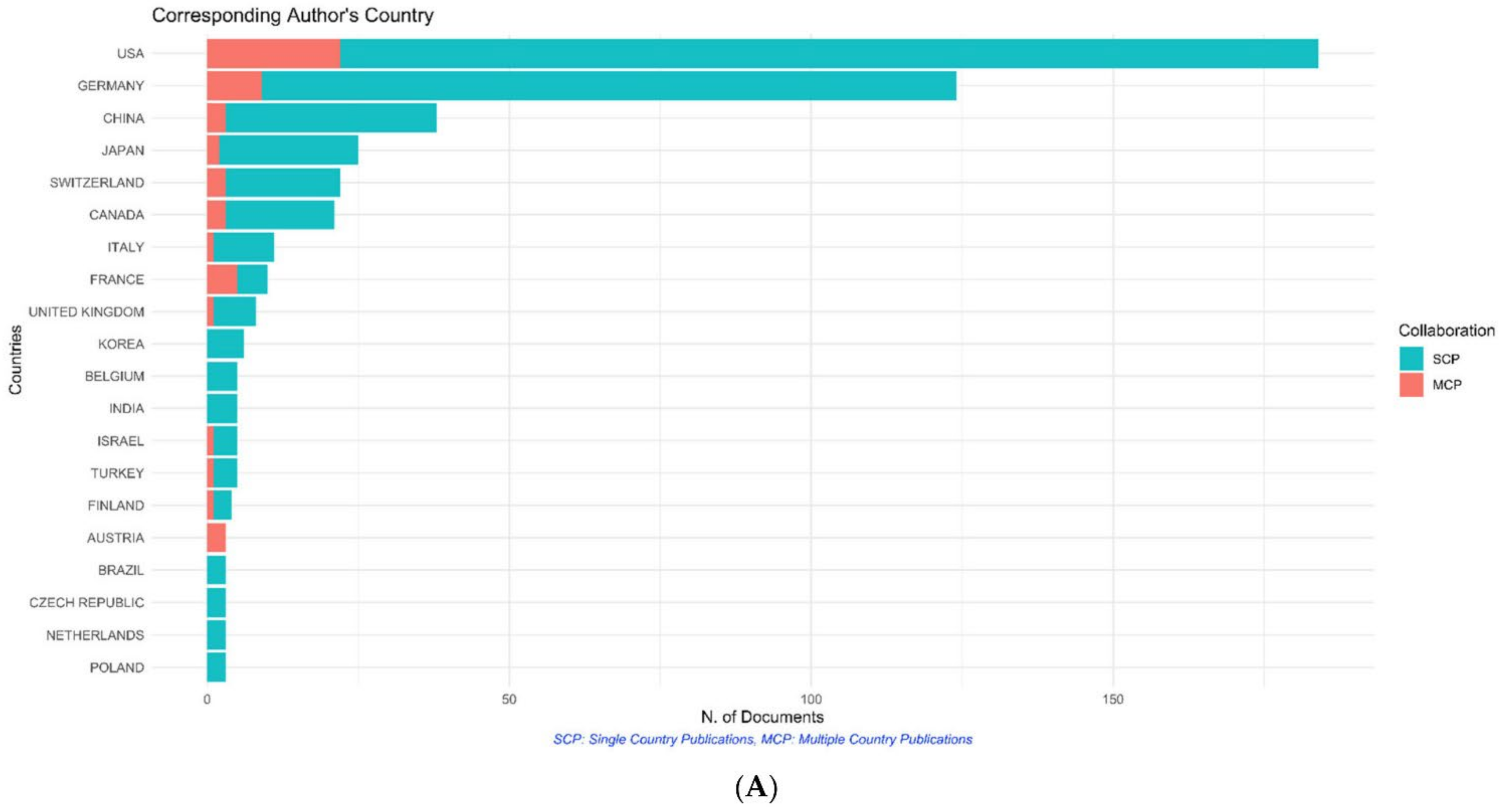
The most significant contributing countries (in terms of the country of the corresponding author) overall were the United States of America (n = 184), Germany (n = 124), and China (n = 38) (Figure 5A). The orange colour denotes the publication resulting from collaboration of authors from multiple countries. When considering the 100 most cited articles, most of the articles originated from the United States of America (USA) (n = 42), closely followed by Germany (n = 36), and then the United Kingdom (n = 4) (Figure 5B). On a similar note, the United States of America was the country that garnered the most citations per document, with a total of 5053 citations and an average of 120.3 citations for each article (Figure 5C). Interestingly, Netherlands had a higher average per article citation of 215, followed by Israel with 156. The University of Erlangen–Nuremberg was responsible for the highest number of these articles (44 articles), followed by Harvard University (39 articles) and the University of Ulm (32 articles) (Figure 5D).

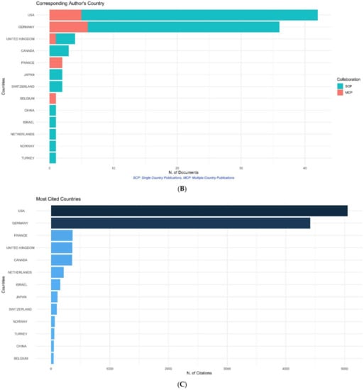
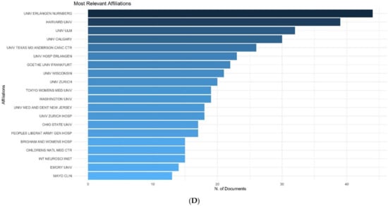
Figure 5.
Graph demonstrating the (A) origin country of the corresponding authors of all the articles published on IOMRI, (B) origin country of the corresponding authors of the top 100 cited articles published on IOMRI, (C) most cited countries, and (D) most relevant affiliations of all the articles on intraoperative MRI.
3.4. Most Cited Articles
The most cited article globally was by Zou KH et al., titled “Statistical validation of image segmentation quality based on a spatial overlap index: scientific reports” [10], with a total of 656 citations, while the article titled “Development and implementation of intraoperative magnetic resonance imaging and its neurosurgical applications”, authored by Black et al., was the most locally cited article related to IOMRI [11] (Supplementary Figures S2 and S3). This article has garnered 49 local and 577 global citations, with 8.49% of all its citations accrued locally (Supplementary Table S2). It is of note that the review article titled “Interventional MR imaging: concepts, systems, and applications in neuroradiology” by Lewin JS [12] has the highest local citation percentage of 15.69%.
4. Discussion
Bibliometric analyses can help provide insight into the status of research within a particular field, identifying strengths of research and areas of lacunae. Moreover, articles that can assist researchers, trainees, and clinicians can be highlighted [13,14]. To the best of the authors’ knowledge, this is the first instance where bibliometric analyses have been used to identify the most significant studies, individuals, institutions, and research disciplines, with respect to the use of IOMRI. By identifying the ‘Top 100′ articles in this field (Table 5), readers are directed to the most relevant articles.
Table 5.
The 100 most cited research articles related to Intraoperative MRI.
These articles were published in 138 different journals, the majority being journals of neurosurgery, (neuro)oncology, and radiology. This is explained by the uniqueness of this field, which involves the collaboration of these two branches (Figure 6A,B). This is further exemplified in the top two most cited articles. The most cited article (globally) by Zou et al. [10] elaborates on the science behind IOMRI and deals with the validation of image segmentation, based on a spatial overlap index. This index, the Dice similarity coefficient (DSC), was validated on two sets of patients, the first group being 10 consecutive patients who underwent prostatic brachytherapy and were imaged with a 1.5 T preoperative MRI and a 0.5 Tesla MRI, and a second group of patients were 9 patients with 3 types of brain tumours. DSC values were computed, and logit-transformed values were compared in the mean with the analysis of variance (ANOVA). The article concludes that the DSC value is a useful, yet simple, tool that, when used for the analyses of spatial overlap, can be used to ensure reproducibility and accuracy in image segmentation. The validity of this approach paved the way for further studies, ensuring the fidelity of MRI image analysis, which has led to an average of 38.59 citations per year since 2004.
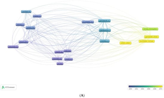
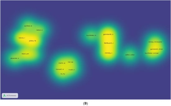
Figure 6.
A VOS (visualising of similarities) graph of bibliometric analysis, of (A) most cited authors and (B) relative influence of these authors, of the 100 most cited articles on intraoperative MRI. The density graph represents the same, with the authors with more impact highlighted.
Another article with a large number of citations per year was authored by Senft et al. This study constituted a randomised controlled trial, assessing the extent of resection in glioma surgery using IOMRI [15]. The authors reported that a higher number of patients that underwent IOMRI guided surgery had complete resection of the tumour (23 of 24 patients/96%) than patients in the control group, where conventional surgery was performed (17 of 25/68% with a p value of 0.023). Postoperative rates of new neurological deficits did not significantly or statistically differ between the two groups (3 patients (13%) in the IOMRI group and 2 (8%) patients in the control group). This article conclusively establishes the efficacy and superiority of IOMRI, explaining the number of citations. On a deeper analysis, this article emerged as having the highest number of citations per year (40.30), highlighting that, when a simple metric such as ‘total citations’ alone are utilised, high impact articles may be looked over. It also indicates that randomised controlled trials continue to have the highest impact among all study types. Additionally, the article by Black et al. [11], which describes the first IOMRI to be utilised using a 0.5 T intraoperative system, has the highest local citations and the second highest global citation rate. This is due to the pioneering nature of the article and its longevity.
An interesting aspect of this bibliometric analysis is that IOMRI is a recent and, therefore, nascent advancement in neurosurgery. Therefore, although Percival Bailey and Harvey Cushing introduced the term Glioma in 1926 [16], the articles on IOMRI primarily focussed on improved glioma resection, using IOMRI dates from 1996 to 2016. This makes the analysis unique, as most other areas in neurosurgery are far older and IOMRI is a new, growing field, and this analysis enlightens areas of relevant research [17]. Fields such as cardiology and thrombolysis, which have made rapid strides in recent years, have shown similar results [18].
These articles were predominantly published in journals dealing with three specialities: namely, Neurosciences, Radiology, and Oncology, with 95 (of 100) articles being in these three domains. Indicating the degree of collaboration required in the development, utilisation, and outcomes in IOMRI. Data garnered and analysed, in the course of preparing this list of top cited articles, also showed preponderance of articles and consistent source growth in newer journals such as World Neurosurgery (est. 2010), indicating continued research and a general increase in the focus on IOMRI. In time, it is feasible that these articles may garner additional citations, adding to their growing relevance.
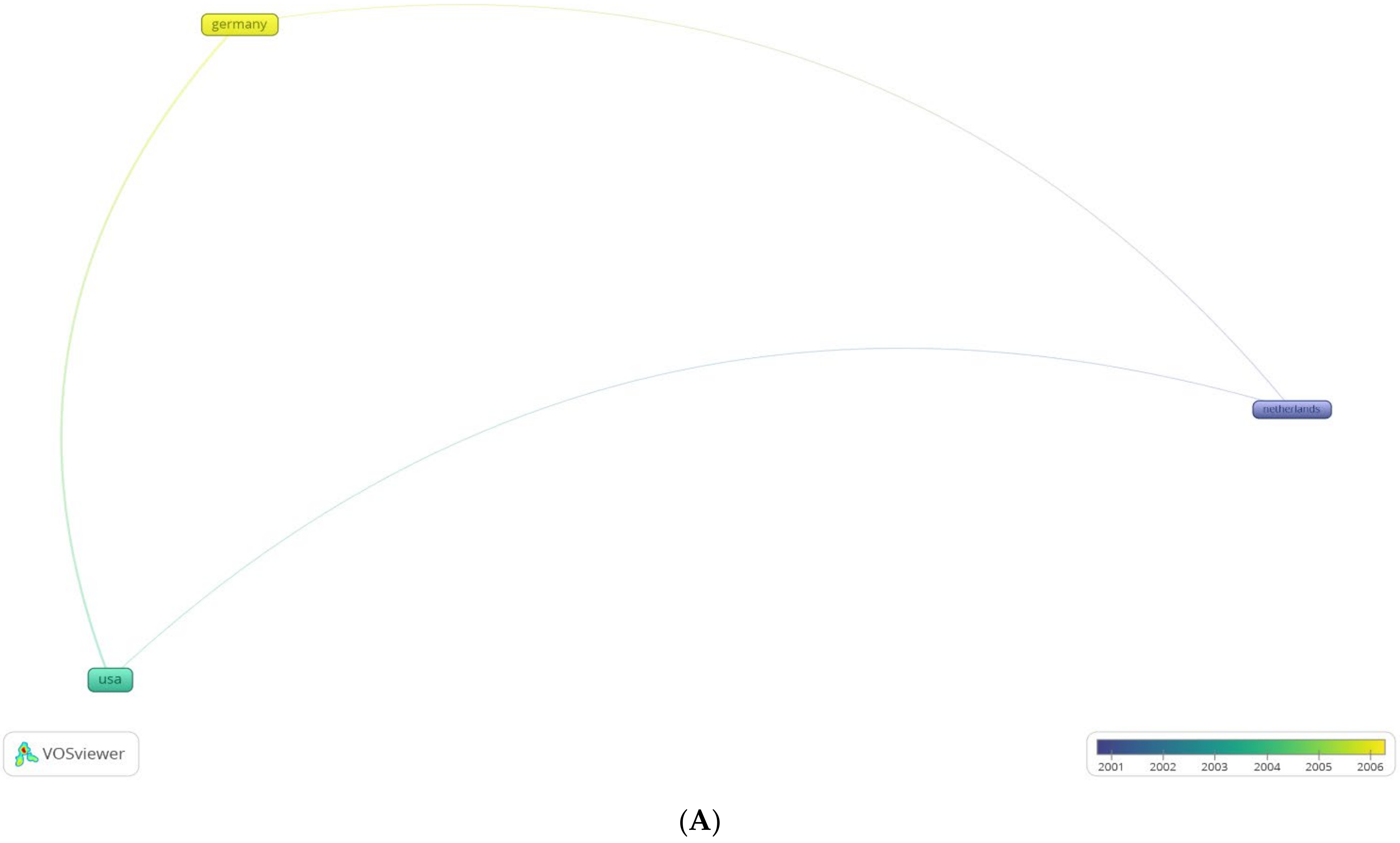
Maximum number of articles originated from the United States, followed by Germany (Figure 7A,B). This differs from other bibliometric analyses, where the United States has significantly larger impact indices than other countries. This is probably owing to the fact that the US spends nearly three times the amount of government funds as countries in Europe for research [19]. The probable reason for Germany’s impact in the field is the near simultaneous establishment of a 0.2 T MRI with a patient transportation system by the Erlangen and Heidelberg groups [20]. Prior to the establishment of IOMRI, their group demonstrated the benefits and emphasised the role of early post-operative imaging [21], which then translated to a series encompassing 47 patients with gliomas, wherein they documented additional resection of overlooked or microsurgically non identifiable tumour (Figure 8) remnants using IOMRI [22]. Owing to the degree of technological research and funding involved in the research and utilisation of IOMRI, the contribution of low-income countries has been minimal.

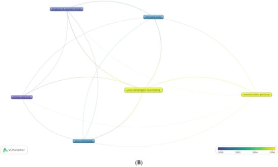
Figure 7.
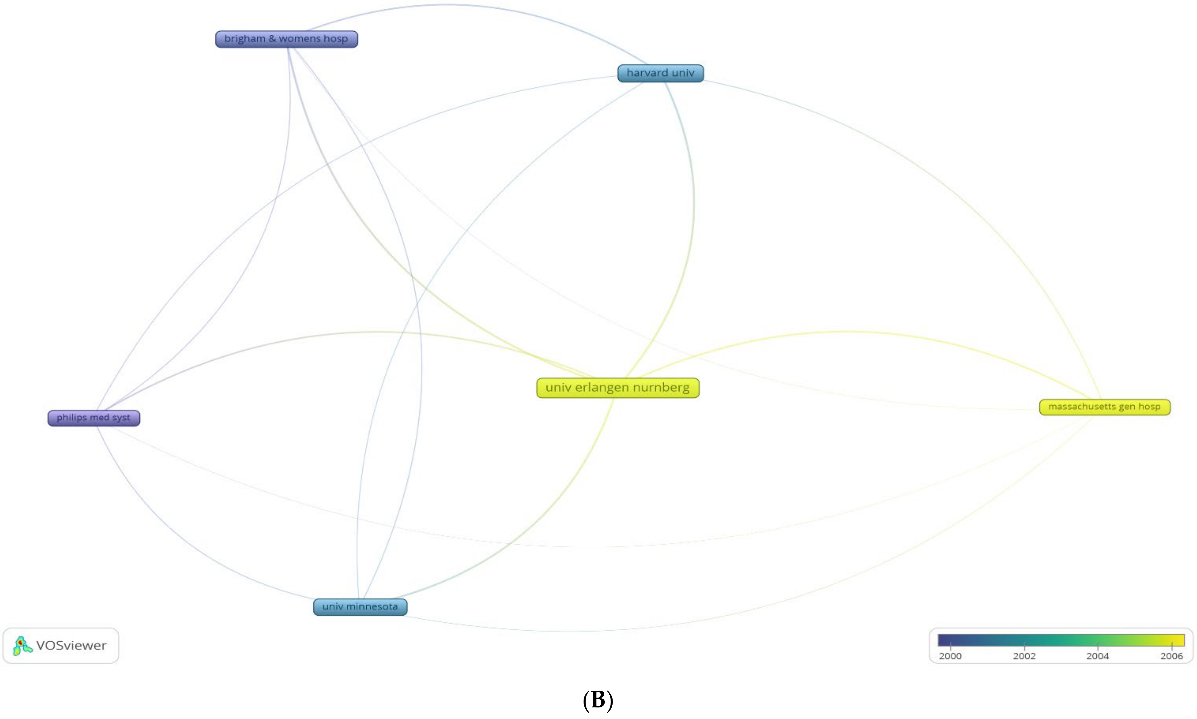
A VOS (visualising of similarities) graph of bibliometric analysis of (A) most cited nations with originating research, with overlay graph of co-citation coupling of documents, and (B) relative influence of the most cited institutions of the 100 most cited articles on intraoperative MRI.
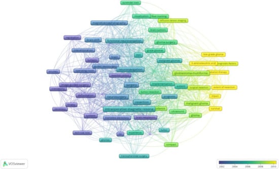
Figure 8.
A VOS (visualising of similarities) graph of bibliometric analysis of the MRI and the overlay graph of co-occurrence of keywords in the 100 most cited articles on intraoperative MRI.
The dominance of Germany is further exemplified when considering institutional affiliations (of the considered articles), where the Friedrich-Alexander-Universität (FAU) Erlangen-Nurnberg, founded in 1743, is the largest contributor for these high impact articles. In addition to the Cluster of Excellence, ‘Engineering of Advanced Materials’ (EAM), and the ‘Graduate School of Advanced Optical Technologies’ (SAOT), which was founded as part of the Excellence Initiative, FAU currently has more than 40 co-ordinated programmes funded by the German Research Foundation [23]. This is closely followed by the University of Minnesota and Harvard University, which are, again, among the top 50 NIH funded institutions [24]. Furthermore, NIH funding for brain tumours, and by extension, IOMRI, has increased over the years, which may be explained by the higher incidence of brain tumours among developed nations [25,26]. This may also be due to the fact that other diseases, which are more prevalent, such as diabetes mellitus and hypertension have well established and validated guidelines and, therefore, require lesser allocation of funds towards research (Figure 9).
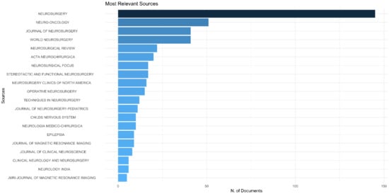
Figure 9.
Figure showing the most frequently used keywords.
It has been nearly three decades since the first IOMRI was established, and its defining role and utility for intraoperative imaging, in glioma surgery and beyond, is widely accepted and established. The permeant advancements of MR technologies, for the detection of brain function, tumour borders, and brain metabolism, ensure that this neurosurgical tool will remain superior to other technologies (such as optical imaging, ultrasound, etc.) for safe and radical tumour removal.
Bibliometric analysis is a purely quantitative tool which measures the impact as perceived by the scientific community and not a representation of the ‘real world’ impact of research. Therefore, while these articles may have a high citation rate, their role in changing patient care may be entirely different. In addition, as only a single database was used for analysis, bias may be encountered. Although certain articles exclude self-citations, they were not ruled out in this study, as the authors believe that only continued work in a particular field leads to self-citations and that this is a marker of a focussed researcher rather than one who continually changes areas of research [8].
Current Limitations and Future Areas of Interest
This article highlights avenues of research in IOMRI that may be more relevant than others. Ergonomic and economically viable methods of IOMRI will increase its availability and thereby, its use in Asian regions. The initial cost in procuring and subsequent economic factors in utilising an IOMRI have erected barriers in its widespread adoption, while ergonomic factors, the learning curve associated with the introduction and familiarisation of the operative team, have led to compliance issues among surgeons with access to IOMRI facilities. Furthermore, in an era of ever advancing technologies, failure to address ergonomic issues will lead to an ever shrinking, potentially cluttered operating room where surgical teams are forced to accommodate shrinking workspaces. Addressing these limitations will dramatically increase the number of patients treated using IOMRI and constitute a higher relevance. Additionally, increased funding of research, or for patient care via insurance companies, will increase the availability of these advanced technologies, benefitting the population overall. It is interesting to note that posterior fossa lesions, spinal tumours, and paediatric populations did not feature in these articles, indicating that these may be areas that warrant additional and future research.
One glaring limitation of bibliometric analysis is that the number of citations for each paper will increase over the years [4]. Thus, the number of citations of the articles published recently may be small despite having a greater impact. The “Average years from publication” in Table 1 also suggests this point. This means that the analysis of the top 100 most cited papers may exclude the latest-trend papers unintentionally and selectively. However, this only reiterates the need of periodic analysis to identify the trends.
5. Conclusions
This bibliometric analysis shows that most research on IOMRI originated from developed countries such as the USA and Germany. It provides insights into the evolution, utilisation, and reference points for further studies on IOMRI, highlighting ‘blind spots’ in research, such as the exclusion of paediatric populations, spinal tumours, and posterior fossa pathologies. The need for additional funding, especially in the developing world, and better ergonomics has been established. Randomised control trials and focused research on a singular topic have been shown to increase impact. We hope this analysis will lead to more areas being explored in this nascent, yet powerful, technique.
Supplementary Materials
The following supporting information can be downloaded at: https://www.mdpi.com/article/10.3390/life12020175/s1, Figure S1: Most Relevant Keywords; Figure S2: Most Local Cited References; Figure S3: Most Cited Documents Globally; Table S1: Authors with Maximum Number of Publications in The Top 100 Cited Articles; Table S2: The Most Locally Cited Articles.
Author Contributions
Conceptualization, H.D. and K.G.; methodology, K.G.; software, K.G.; validation, G.F., K.G., G.E.U.; formal analysis, K.G.; investigation, K.G.; resources, K.G.; data curation, K.G.; writing—original draft preparation, H.D.; writing—review and editing, M.D.K.N.; visualization, H.D.; supervision, K.G.; project administration, K.G.; funding acquisition, G.F. All authors have read and agreed to the published version of the manuscript.
Funding
No external funding. APC paid by Gianluca Ferini.
Institutional Review Board Statement
Not applicable.
Informed Consent Statement
Not applicable.
Data Availability Statement
All data provided in manuscript.
Conflicts of Interest
The authors declare no conflict of interest.
References
- Seifert, V. Intraoperative MRI in neurosurgery: Technical overkill or the future of brain surgery? Neurol. India 2003, 51, 329–332. [Google Scholar] [PubMed]
- Black, P.M.; Moriarty, T.; Alexander, E., 3rd; Stieg, P.; Woodard, E.J.; Gleason, P.L.; Martin, C.H.; Kikinis, R.; Schwartz, R.B.; Jolesz, F.A. Development and implementation of intraoperative magnetic resonance imaging and its neurosurgical applications. Neurosurgery 1997, 41, 831–842, discussion 842–845. [Google Scholar] [CrossRef] [PubMed]
- Black, P.M.; Alexander, E., 3rd; Martin, C.; Moriarty, T.; Nabavi, A.; Wong, T.Z.; Schwartz, R.B.; Jolesz, F. Craniotomy for tumor treatment in an intraoperative magnetic resonance imaging unit. Neurosurgery 1999, 45, 423–431, discussion 431–433. [Google Scholar] [CrossRef] [PubMed]
- Harsh, D.; Adnan, H.S.; Raees, A.P.; Manjul, T.; Anil, N. How Many Neurosurgeons Does It Take to Author an Article and What Are the Other Factors That Impact Citations? World Neurosurg. 2021, 146, e993–e1002. [Google Scholar] [CrossRef]
- Brown, N.J.; Wilson, B.; Shahrestani, S.; Choi, E.H.; Lien, B.V.; Paladugu, A.; Tran, K.; Ransom, S.C.; Tafreshi, A.R.; Ransom, R.C.; et al. The 100 Most Influential Publications on Medulloblastoma: Areas of Past, Current, and Future Focus. World Neurosurg. 2021, 146, 119–139. [Google Scholar] [CrossRef]
- Aria, M.; Cuccurullo, C. bibliometrix: An R-tool for comprehensive science mapping analysis. J. Informetr. 2017, 11, 959–975. [Google Scholar] [CrossRef]
- Synnestvedt, M.B.; Chen, C.; Holmes, J.H. CiteSpace II: Visualization and knowledge discovery in bibliographic databases. AMIA Annu. Symp. Proc. 2005, 2005, 724–728. [Google Scholar]
- Deora, H.; Tripathi, M.; Chaurasia, B.; Grotenhuis, J.A. Avoiding predatory publishing for early career neurosurgeons: What should you know before you submit? Acta Neurochir. 2021, 163, 1–8. [Google Scholar] [CrossRef]
- Bradford, S.C. Sources of Information on Specific Subjects. Engineering Illus. Wkly. J. 1934, 137, 85–86. [Google Scholar]
- Zou, K.H.; Warfield, S.K.; Bharatha, A.; Tempany, C.M.; Kaus, M.R.; Haker, S.J.; Wells, W.M., 3rd; Jolesz, F.A.; Kikinis, R. Statistical validation of image segmentation quality based on a spatial overlap index. Acad. Radiol. 2004, 11, 178–189. [Google Scholar] [CrossRef] [Green Version]
- Deora, H.; Yagnick, N.S.; Tripathi, M. Mentor-Mentee Relationship in Neurosurgery: Standing on the Shoulder of Giants. World Neurosurg. 2020, 141, 110–112. [Google Scholar] [CrossRef] [PubMed]
- Lewin, J.S. Interventional MR imaging: Concepts, systems, and applications in neuroradiology. AJNR Am. J. Neuroradiol. 1999, 20, 735–748. [Google Scholar] [PubMed]
- Steinmeier, R.; Fahlbusch, R.; Ganslandt, O.; Nimsky, C.; Buchfelder, M.; Kaus, M.; Heigl, T.; Lenz, G.; Kuth, R.; Huk, W. Intraoperative magnetic resonance imaging with the magnetom open scanner: Concepts, neurosurgical indications, and procedures: A preliminary report. Neurosurgery 1998, 43, 739–747, discussion 747–748. [Google Scholar] [CrossRef] [PubMed]
- Burak Atci, I.; Yilmaz, H.; Samanci, M.Y. The top 50 most-cited articles on low-grade glioma: A bibliometric analysis. Br. J. Neurosurg. 2019, 33, 171–175. [Google Scholar] [CrossRef] [PubMed]
- Senft, C.; Bink, A.; Franz, K.; Vatter, H.; Gasser, T.; Seifert, V. Intraoperative MRI guidance and extent of resection in glioma surgery: A randomised, controlled trial. Lancet Oncol. 2011, 12, 997–1003. [Google Scholar] [CrossRef]
- Stoyanov, G.S.; Dzhenkov, D.L. On the Concepts and History of Glioblastoma Multiforme—Morphology, Genetics and Epigenetics. Folia Med. 2018, 60, 48–66. [Google Scholar] [CrossRef] [Green Version]
- Siddiqi, T.J.; Usman, M.S.; Khan, M.S.; Fatima, K.; Norbash, A.; Qureshi, A.I.; Khan, A.R.; Khosa, F. The 100 Most Influential Papers in the Field of Thrombolytic Therapy: A Bibliometric Analysis. Am. J. Cardiovasc. Drugs 2017, 17, 319–333. [Google Scholar] [CrossRef]
- Akmal, M.; Hasnain, N.; Rehan, A.; Iqbal, U.; Hashmi, S.; Fatima, K.; Farooq, M.Z.; Khosa, F.; Siddiqi, J.; Khan, M.K. Glioblastome Multiforme: A Bibliometric Analysis. World Neurosurg. 2020, 136, 270–282. [Google Scholar] [CrossRef]
- Sobocki, P.; Lekander, I.; Berwick, S.; Olesen, J.; Jönsson, B. Resource allocation to brain research in Europe (RABRE). Eur. J. Neurosci. 2006, 24, 2691–2693. [Google Scholar] [CrossRef]
- Fahlbusch, R.; Samii, A. Intraoperative MRI. Neurosurg. Focus 2016, 40, E17. [Google Scholar] [CrossRef]
- Albert, F.K.; Forsting, M.; Sartor, K.; Adams, H.P.; Kunze, S. Early postoperative magnetic resonance imaging after resection of malignant glioma: Objective evaluation of residual tumor and its influence on regrowth and prognosis. Neurosurgery 1994, 34, 45–60, discussion 60–61. [Google Scholar] [CrossRef] [PubMed]
- Nimsky, C.; Fujita, A.; Ganslandt, O.; Von Keller, B.; Fahlbusch, R. Volumetric assessment of glioma removal by intraoperative high-field magnetic resonance imaging. Neurosurgery 2004, 55, 358–370, discussion 370–371. [Google Scholar] [CrossRef] [PubMed]
- Friedrich-Alexander-Universität Erlangen-Nürnberg. 2020. Available online: https://www.fau.eu (accessed on 4 December 2020).
- Top 50 NIH-Funded Institutions of 2018. GENe Genetic Engineering and Biotechnology News. 2019. Available online: https://www.genengnews.com/a-lists/top-50-nih-funded-institutions-of-2018/ (accessed on 4 December 2020).
- NIH Categorical Spending eNIH Research Portfolio Online Reporting Tools (RePORT). Available online: https://report.nih.gov/categorical_spending.aspx (accessed on 4 January 2021).
- Hana, T.; Tanaka, S.; Nejo, T.; Takahashi, S.; Kitagawa, Y.; Koike, T.; Nomura, M.; Takayanagi, S.; Saito, N. Mining-Guided Machine Learning Analyses Revealed the Latest Trends in Neuro-Oncology. Cancers 2019, 11, 178. [Google Scholar] [CrossRef] [PubMed] [Green Version]


Publisher’s Note: MDPI stays neutral with regard to jurisdictional claims in published maps and institutional affiliations. |
© 2022 by the authors. Licensee MDPI, Basel, Switzerland. This article is an open access article distributed under the terms and conditions of the Creative Commons Attribution (CC BY) license (https://creativecommons.org/licenses/by/4.0/).