
Natural Radionuclide Concentrations by γ-Ray Spectrometry in Granitic Rocks of the Sol Hamed Area, Southeastern Desert of Egypt, and Their Radiological Implications


Abstract
1. Introduction
2. Materials and Methods
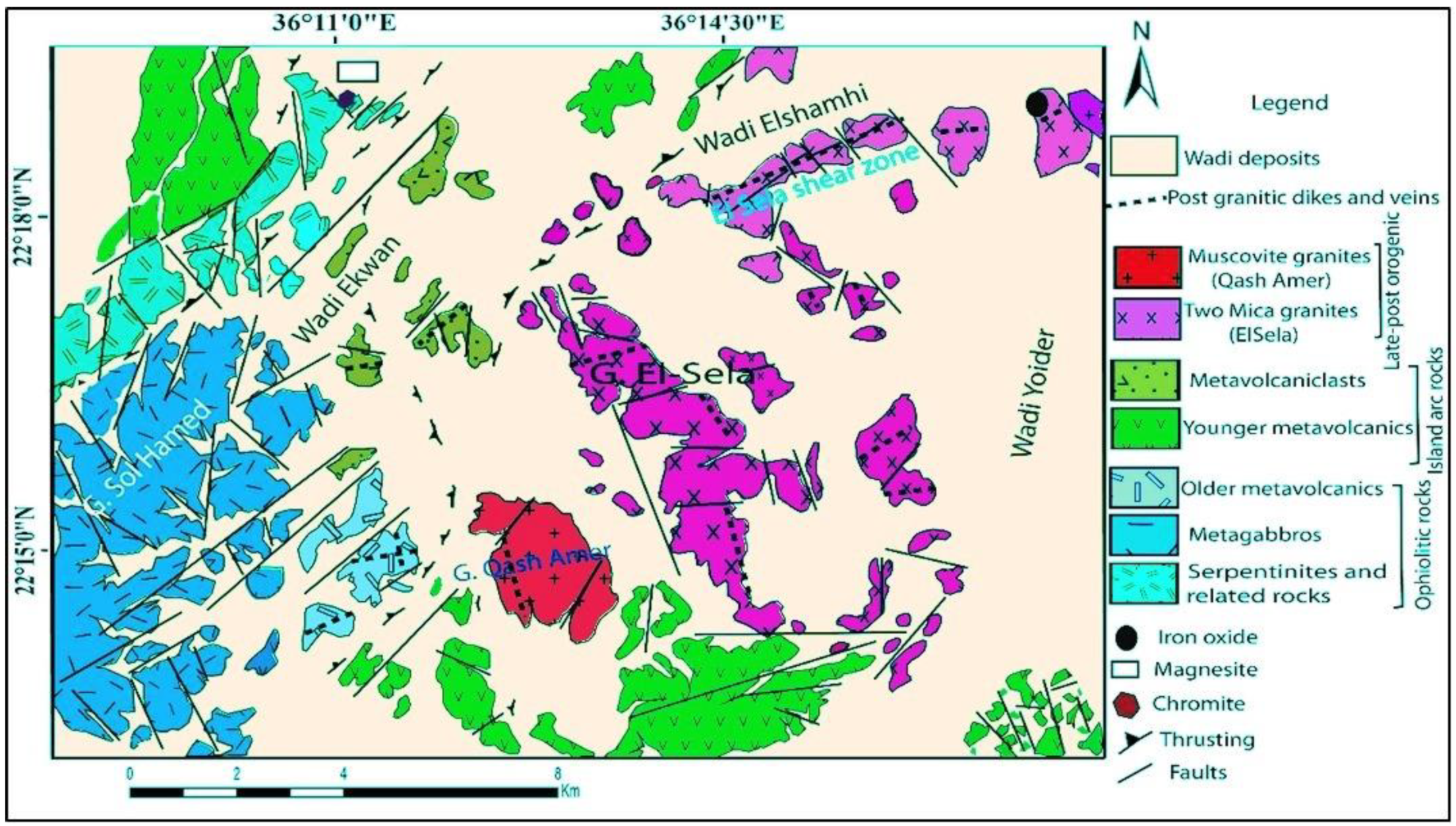
2.1. Geological Setting
2.2. Sampling and Radioactive Detection
3. Results
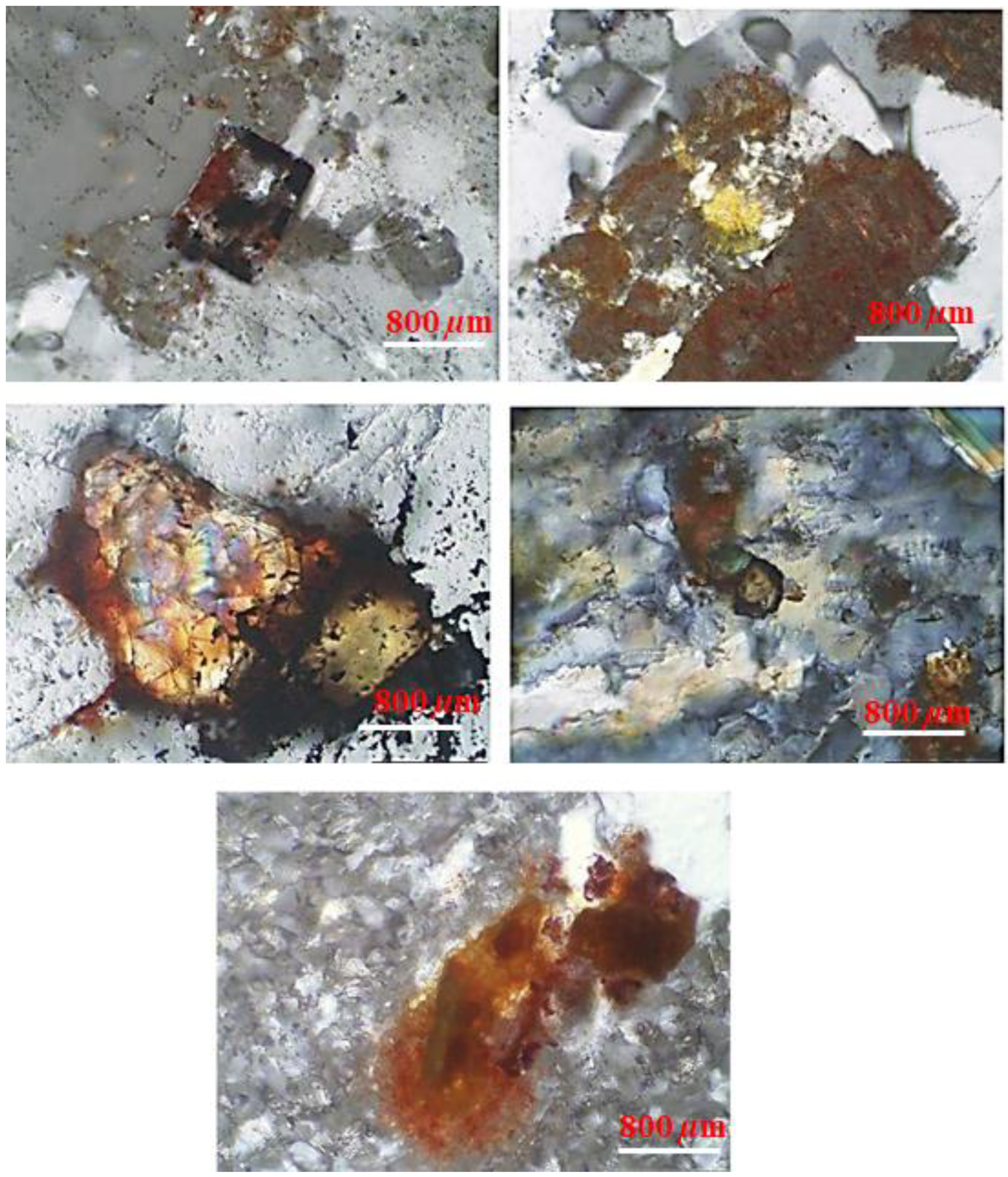
3.1. Petrographic Investigation
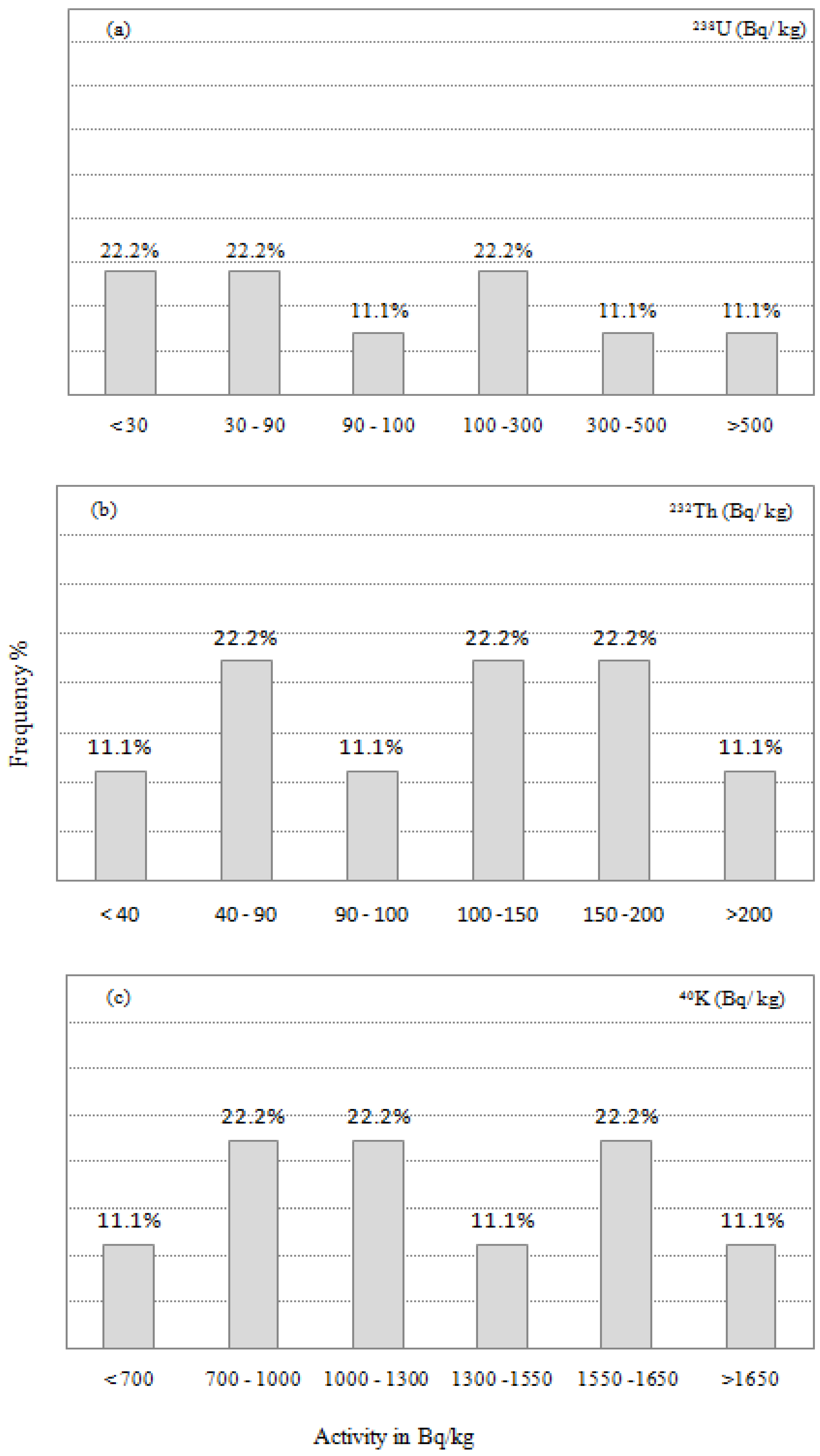
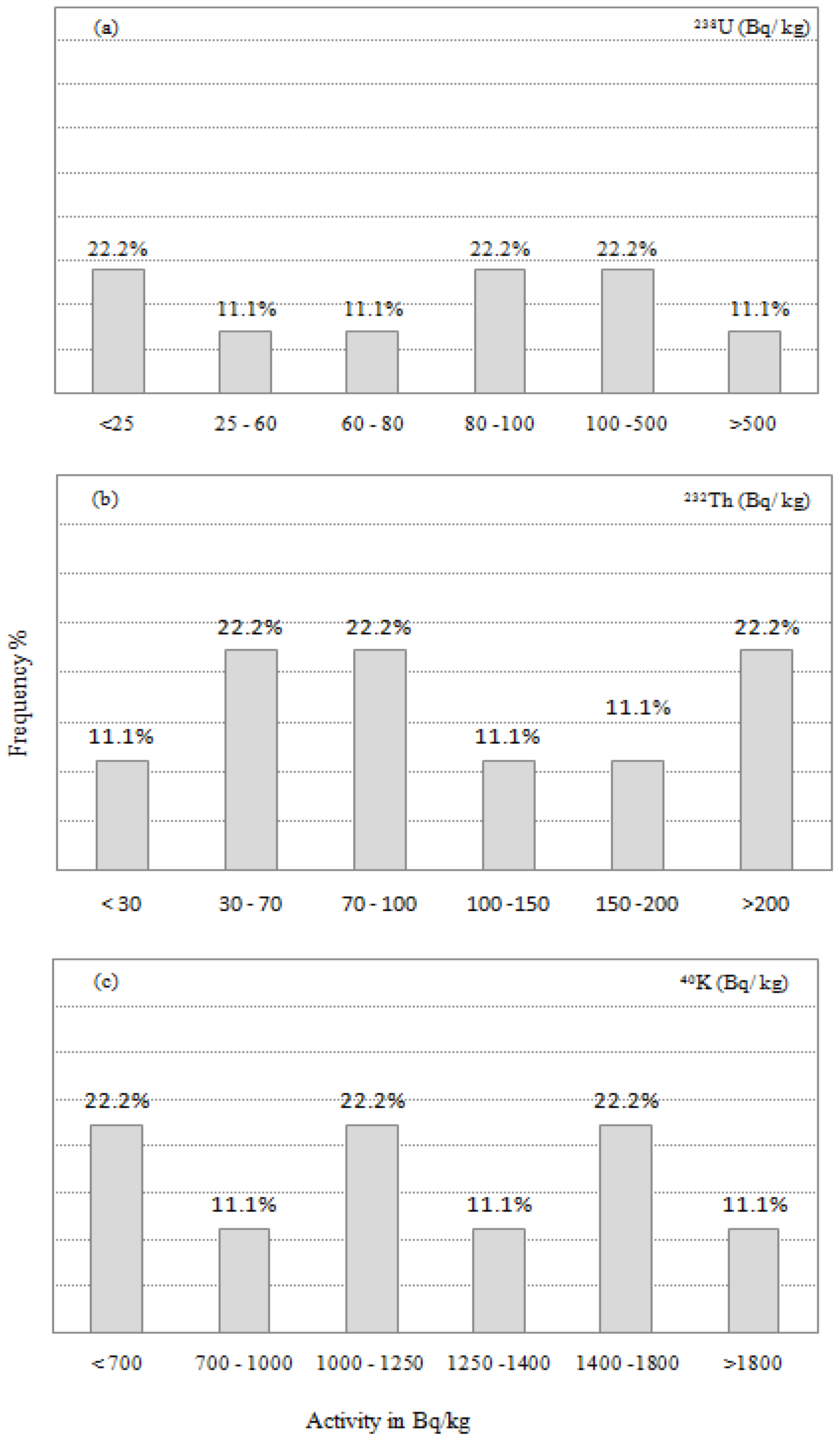
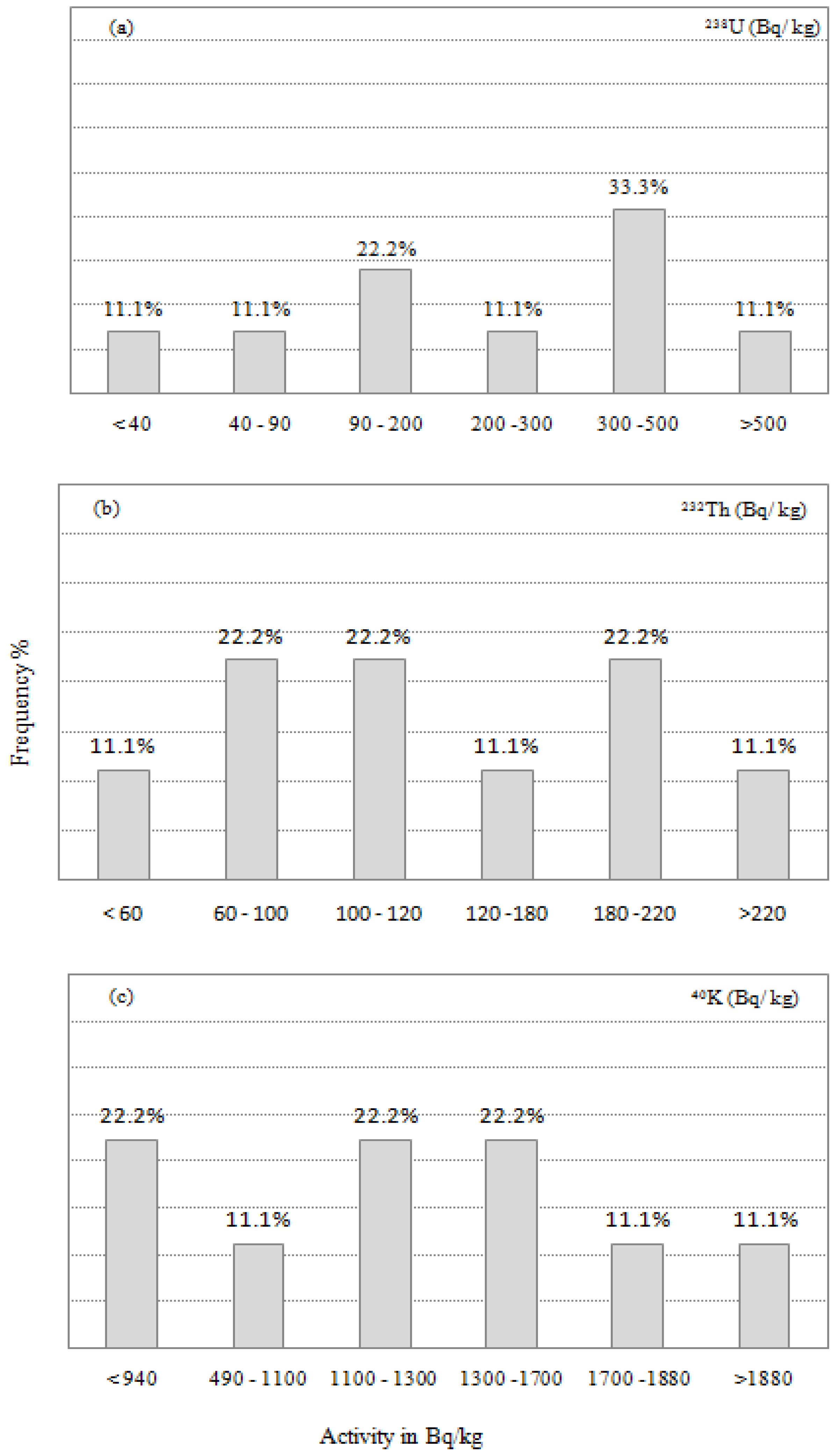
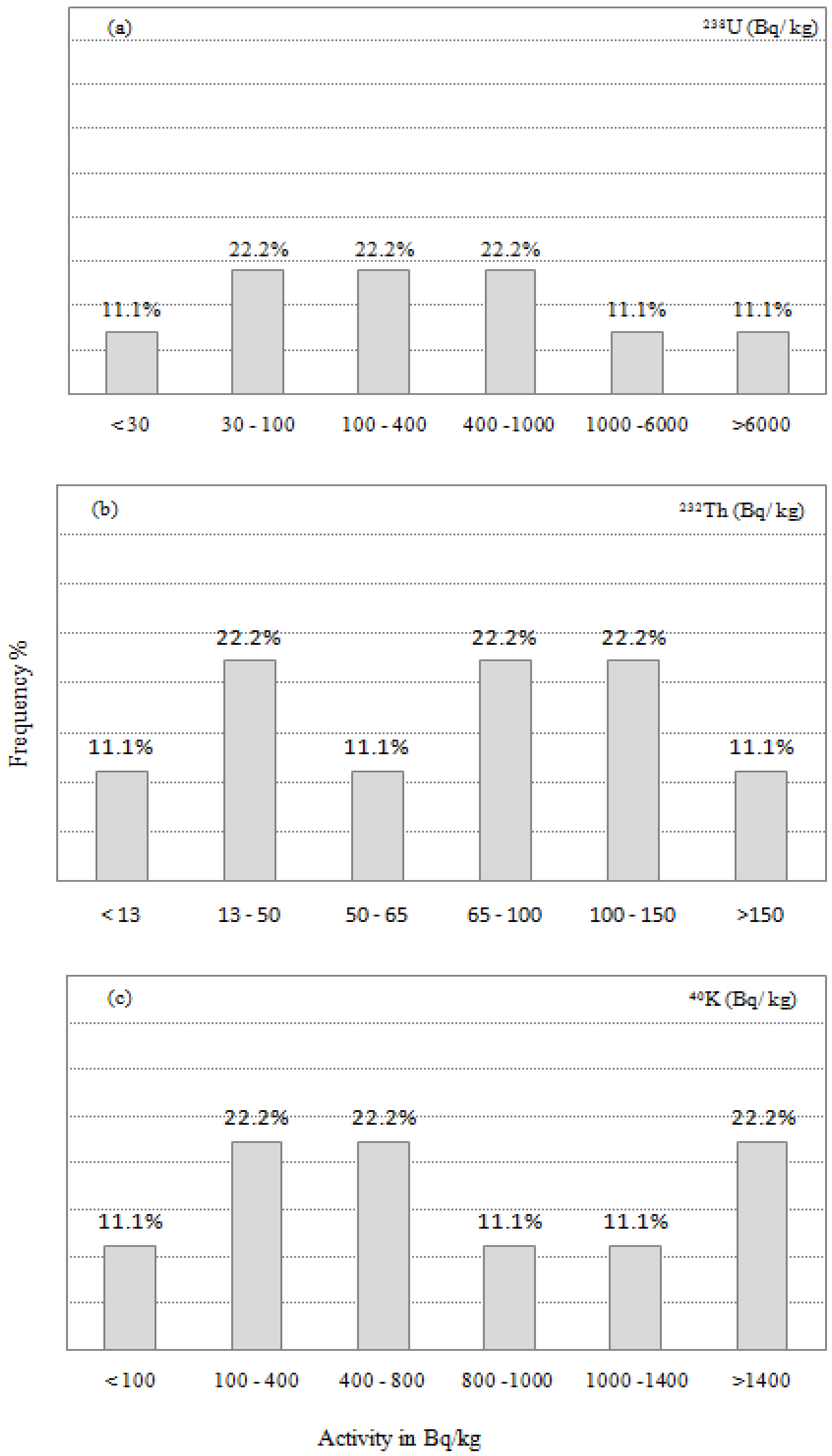
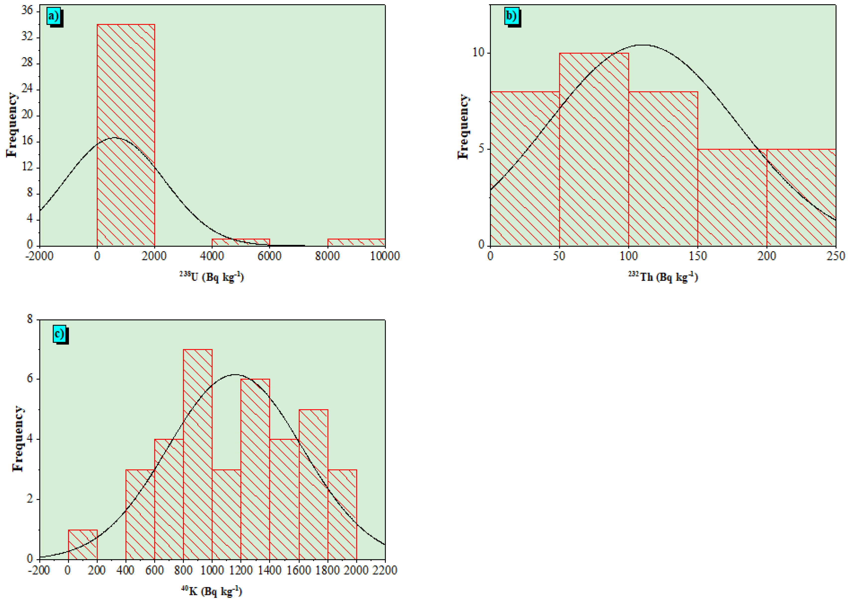
3.2. Radioactivity Analysis
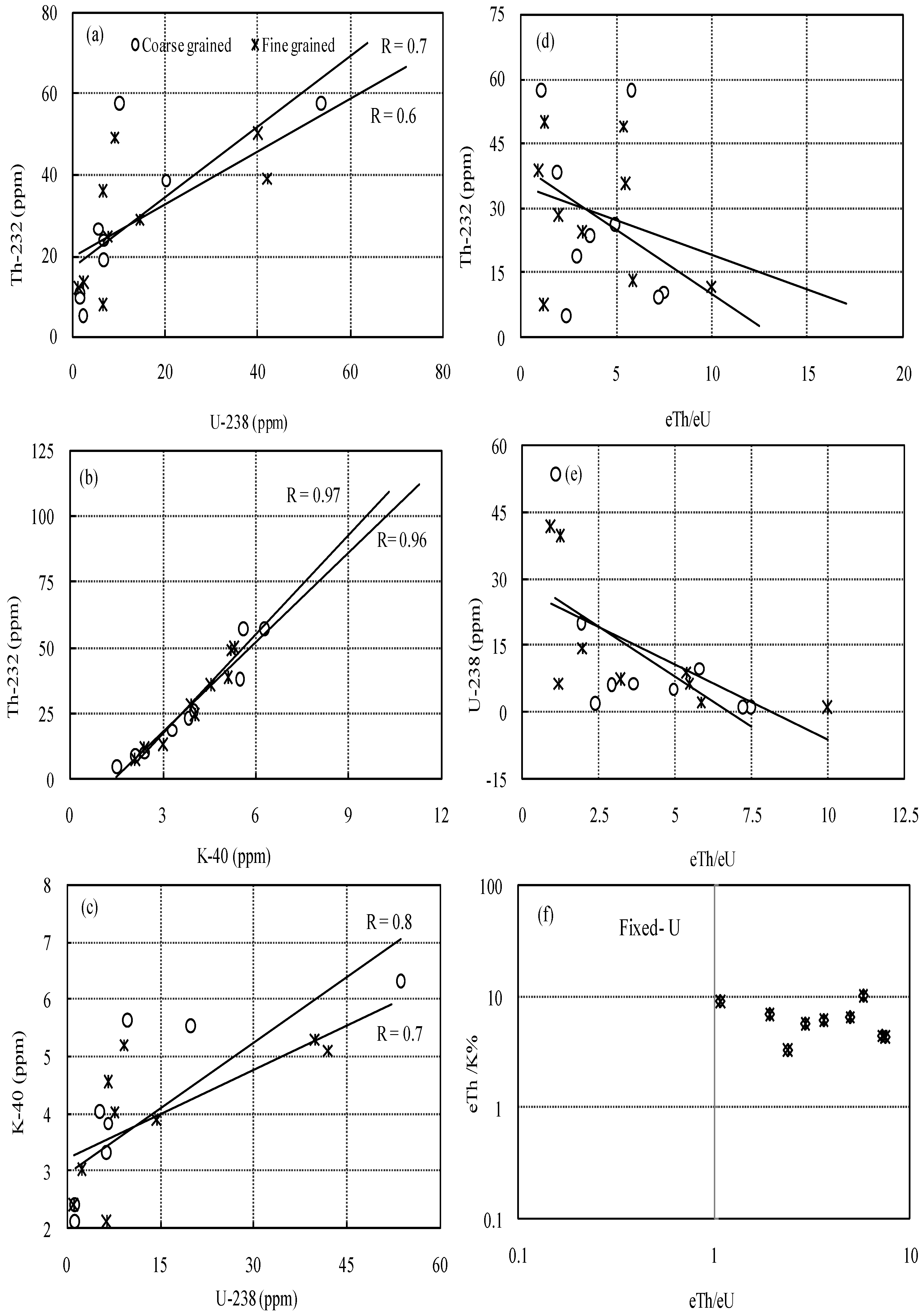
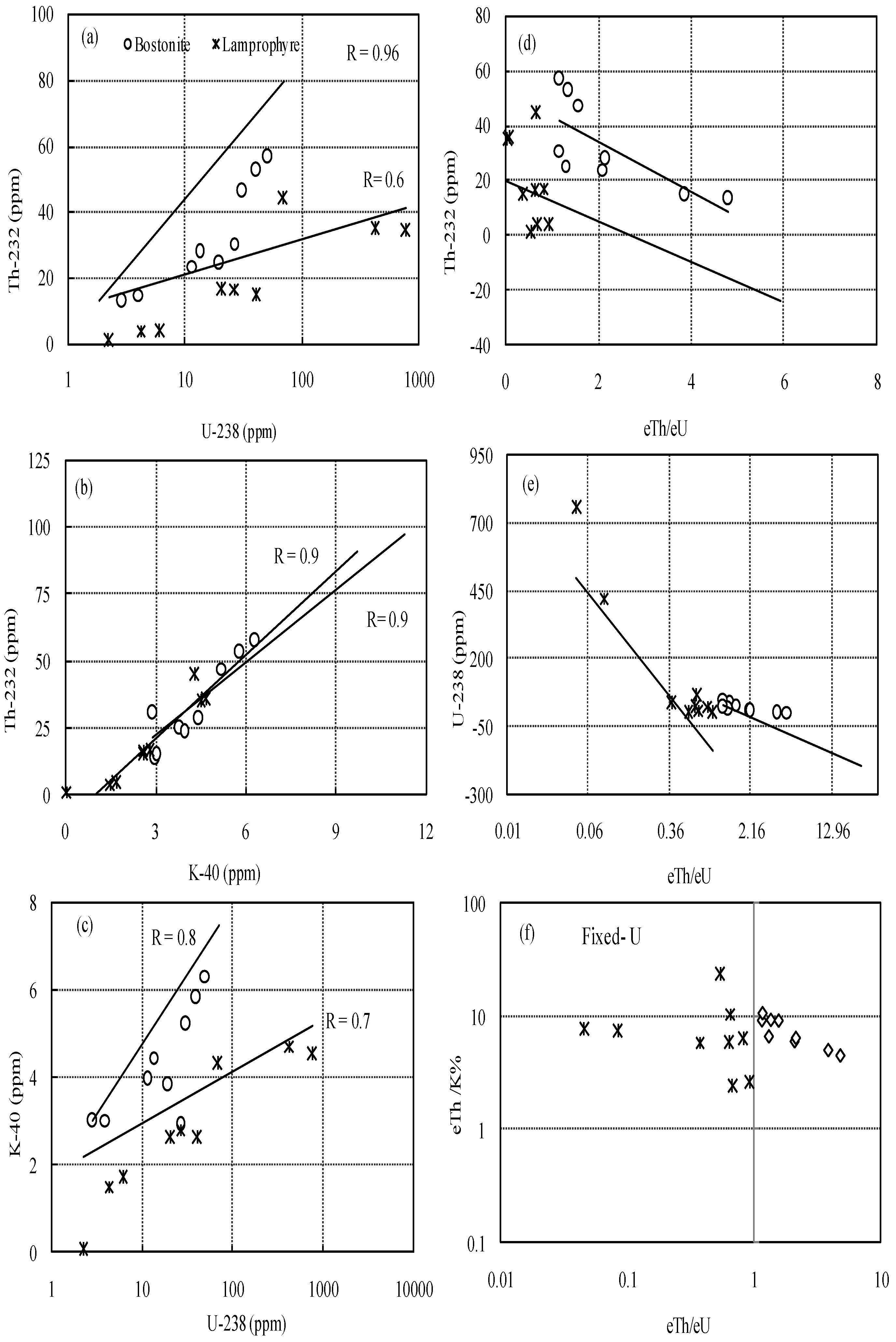
3.3. Geochemistry of U and Th in the Studied Rocks
3.4. Radioactive Concentrations in Granitic Rocks
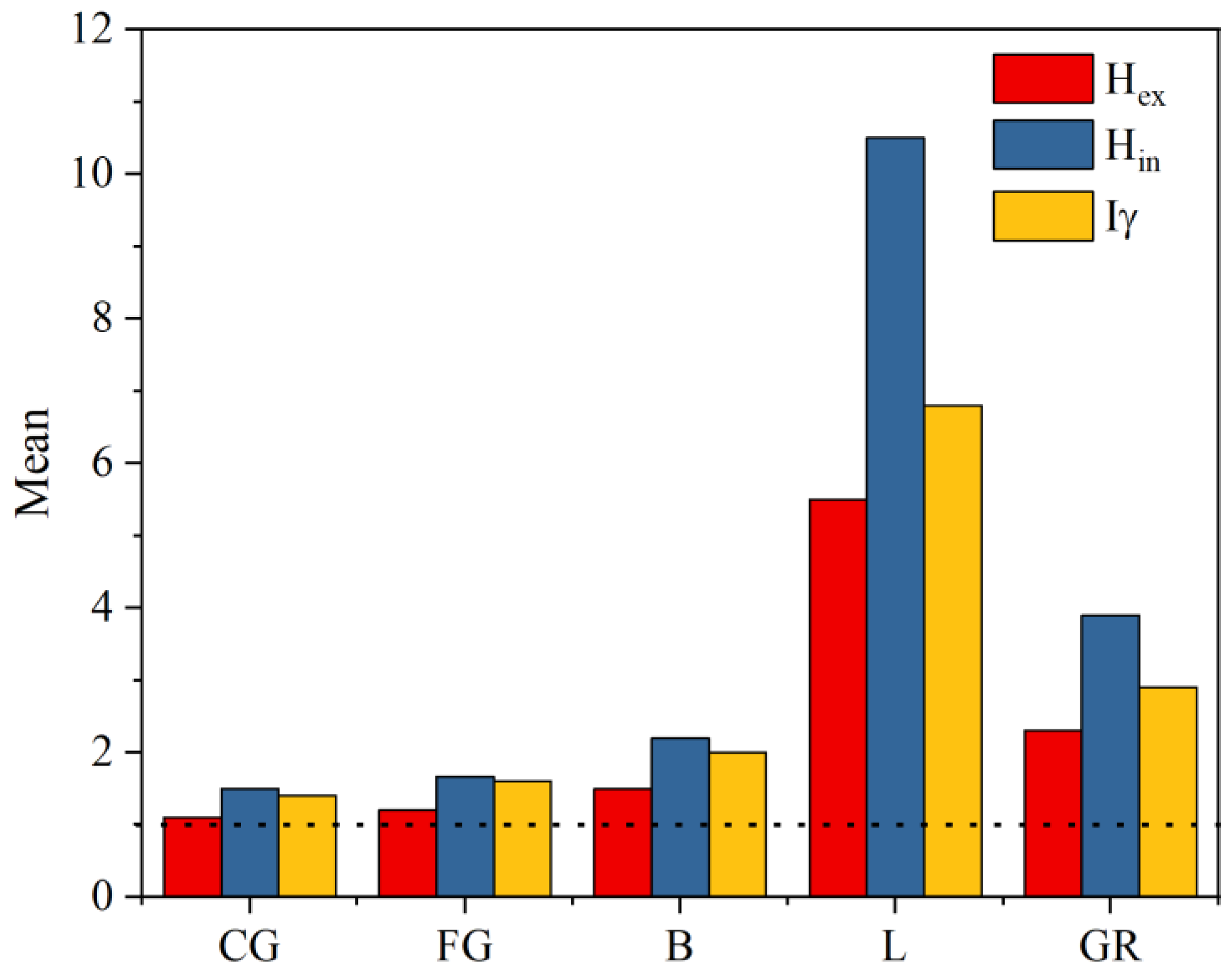
3.5. Radiological Effects
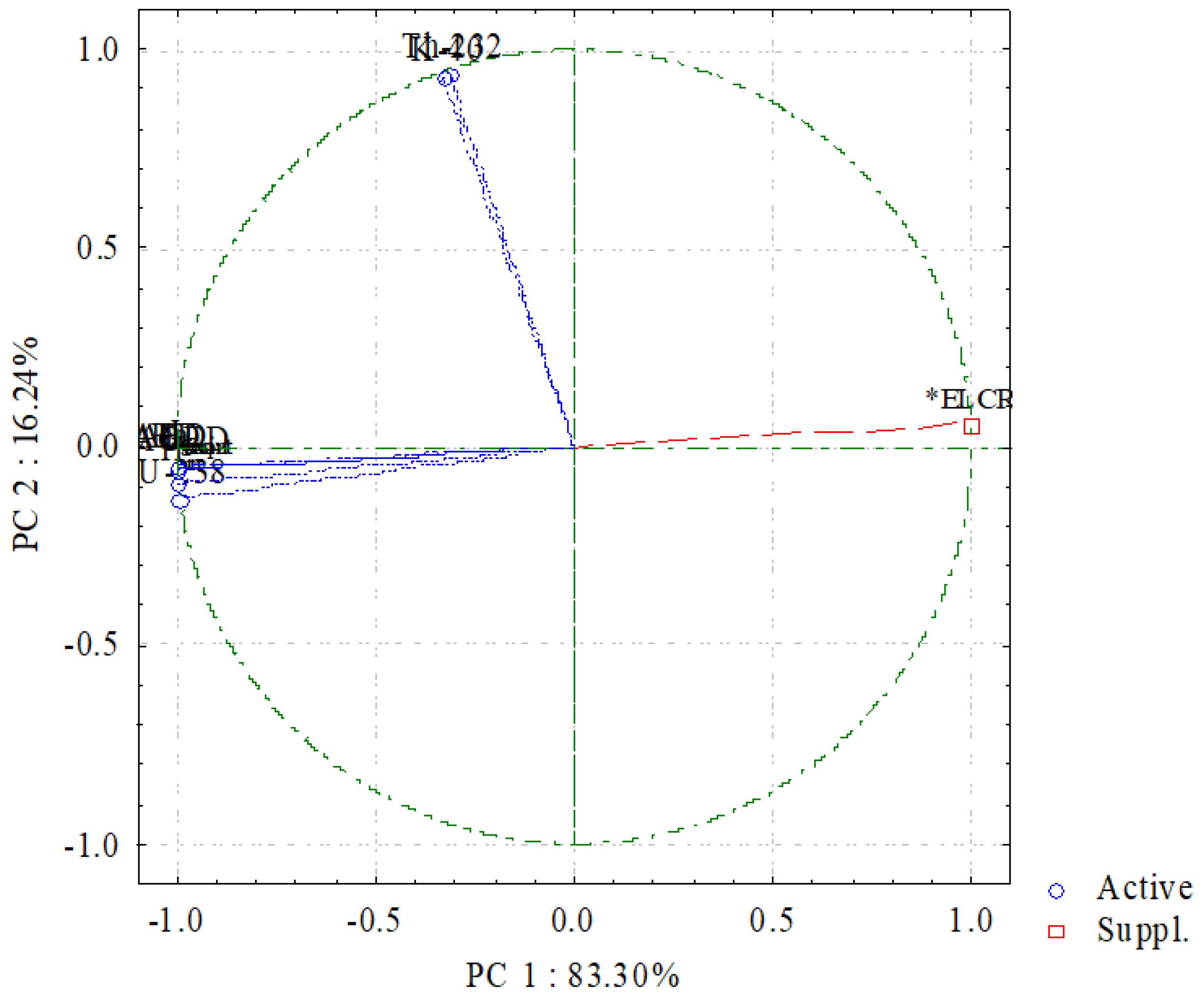
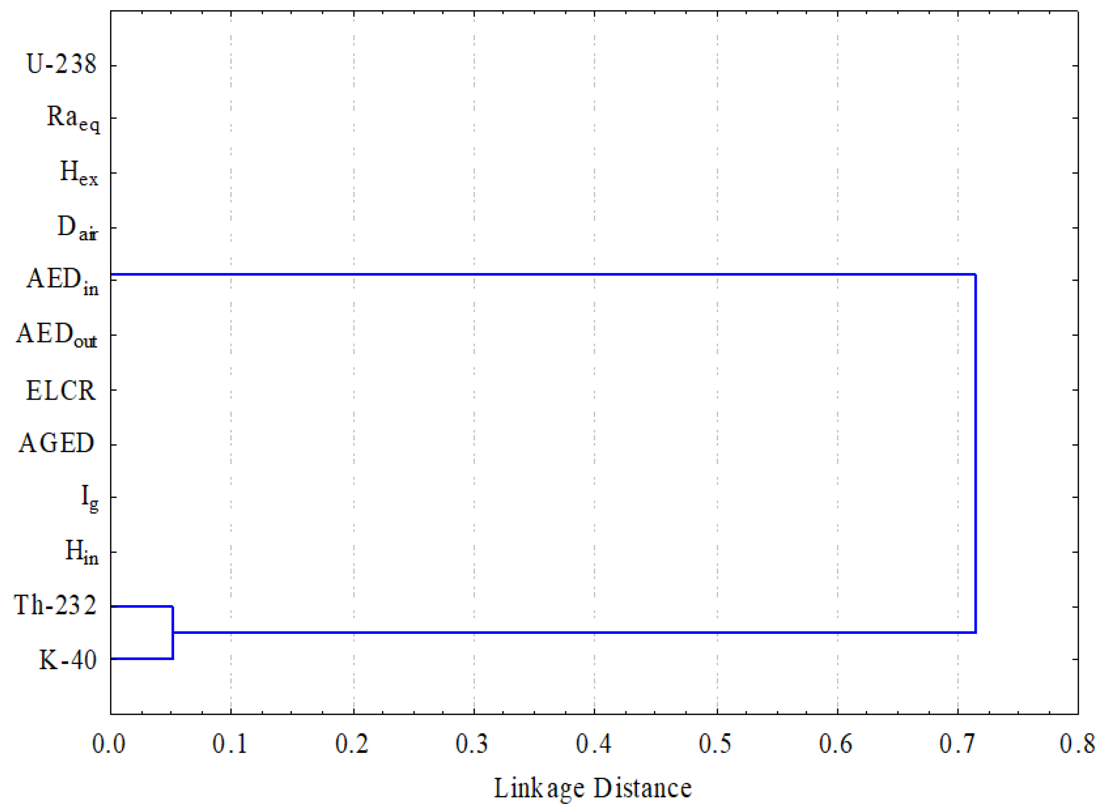
3.6. Multivariate Statistics
4. Conclusions
Author Contributions
Funding
Acknowledgments
Conflicts of Interest
References
- Hanfi, M.Y.; Yarmoshenko, V.; Seleznev, A.A.; Malinovsky, G.; Ilgasheva, E.; Zhukovsky, M.V. Beta radioactivity of urban surface–deposited sediment in three Russian cities. Environ. Sci. Pollut. Res. 2020, 27, 40309–40315. [Google Scholar] [CrossRef] [PubMed]
- Akpanowo, M.A.; Umaru, I.; Iyakwari, S.; Joshua, E.O.; Yusuf, S.; Ekong, G.B. Determination of natural radioactivity levels and radiological hazards in environmental samples from artisanal mining sites of Anka, North-West Nigeria. Sci. African 2020, 10, e00561. [Google Scholar] [CrossRef]
- Sivakumar, S.; Chandrasekaran, A.; Ravisankar, R.; Ravikumar, S.M.; Prince Prakash Jebakumar, J.; Vijayagopal, P.; Vijayalakshmi, I.; Jose, M.T. Measurement of natural radioactivity and evaluation of radiation hazards in coastal sediments of east coast of Tamilnadu using statistical approach. J. Taibah Univ. Sci. 2014, 8, 375–384. [Google Scholar] [CrossRef]
- UNSCEAR. Sources and Effects of Ionizing Radiation—Exposures of the Public and Workers from Various Sources of Radiation—UNSCEAR 2008 Report; United Nations Publication: New York, NY, USA, 2010. [Google Scholar]
- ATSDR. Toxicological Profile for Uranium; U.S. Department of Health & Human Services: Washington, WA, USA, 1999; pp. 1–145.
- ATSDR. Draft Toxicological Profile for Radon: Agency for Toxic Substances and Disease Registry; U.S. Department of Health & Human Services: Washington, WA, USA, 2012; Volume 9–11, pp. 161–167.
- ATSDR. Case Studies in Environmental Medicine; U.S. Department of Health & Human Services: Washington, WA, USA, 1992; pp. 1–28.
- Ajayi, O.S. Measurement of activity concentrations of 40K, 226Ra and 232Th for assessment of radiation hazards from soils of the southwestern region of Nigeria. Radiat. Environ. Biophys. 2009, 48, 323–332. [Google Scholar] [CrossRef]
- Khandaker, M.U.; Uwatse, O.B.; Bin Shamsul Khairi, K.A.; Faruque, M.R.I.; Bradley, D.A. Terrestrial radionuclides in surface (dam) water and concomitant dose in metropolitan Kuala Lumpur. Radiat. Prot. Dosimetry 2019, 185, 343–350. [Google Scholar] [CrossRef]
- Gaafar, I.; Hanfi, M.; El-Ahll, L.S.; Zeidan, I. Assessment of radiation hazards from phosphate rocks, Sibaiya area, central eastern desert, Egypt. Appl. Radiat. Isot. 2021, 173, 109734. [Google Scholar] [CrossRef]
- Sivakumar, S.; Chandrasekaran, A.; Senthilkumar, G.; Suresh Gandhi, M.; Ravisankar, R. Determination of radioactivity levels and associated hazards of coastal sediment from south east coast of Tamil Nadu with statistical approach. Iran. J. Sci. Technol. Trans. A Sci. 2018, 42, 601–614. [Google Scholar] [CrossRef]
- Pavlidou, S.; Koroneos, A.; Papastefanou, C. Natural radioactivity of granites used as building materials. J. Environ. Radioact. 2006, 89, 48–60. [Google Scholar] [CrossRef]
- Khandaker, U.M.; Asaduzzaman, K.; Bin Sulaiman, A.F.; Bradley, D.A.; Isinkaya, M.O. Elevated concentrations of naturally occurring radionuclides in heavy mineral-rich beach sands of Langkawi Island, Malaysia. Mar. Pollut. Bull. 2018, 127, 654–663. [Google Scholar] [CrossRef]
- Xinwei, L. Natural radioactivity in some building materials of Xi’an, China. Radiat. Meas. 2005, 40, 94–97. [Google Scholar] [CrossRef]
- Abu El-Laban, S.A. Some geological and geochemical studies in Abu Ramad Area, South Eastern Desert, Egypt. Ph.D. Thesis, Cairo University, Giza, Egypt, 2002; p. 274. [Google Scholar]
- Arunima, S.; Lekshmi, R.; Jojo, P.J.; Mayeen Uddin, K. A study on leaching of primordial radionuclides 232Th and 40K to water bodies. Radiat. Phys. Chem. 2021, 188, 109658. [Google Scholar] [CrossRef]
- Monica, S.; Jojo, P.J.; Khandaker, M.U. Radionuclide concentrations in medicinal florae and committed effective dose through Ayurvedic medicines. Int. J. Radiat. Biol. 2020, 96, 1028–1037. [Google Scholar] [CrossRef]
- Papadopoulos, A.; Christofides, G.; Koroneos, A.; Papadopoulou, L.; Papastefanou, C.; Stoulos, S. Natural radioactivity and radiation index of the major plutonic bodies in Greece. J. Environ. Radioact. 2013, 124, 227–238. [Google Scholar] [CrossRef]
- Pagel, M. The mineralogy and geochemistry of uranium, thorium, and rare-earth elements in two radioactive granites of the Vosges, France. Mineral. Mag. 1982, 46, 149–161. [Google Scholar] [CrossRef]
- Shahin, H.A.E.R.A. Zr-Y-Nb-REE mineralization associated with microgranite and basic dykes at EL Sela shear zone, South Eastern Desert, Egypt. J. Korean Phys. Soc. 2014, 3, 1–12. [Google Scholar] [CrossRef]
- Raslan, M.F.; El-Feky, M.G. Radioactivity and mineralogy of the altered granites of the Wadi Ghadir shear zone, South Eastern Desert, Egypt. Chinese J. Geochemistry 2012, 31, 30–40. [Google Scholar] [CrossRef]
- Clark, R.L.; Hickey, R.C.; Butler, J.J.; Ibanez, M.L.; Ballantyne, A.J. Thyroid cancer discovered incidentally during treatment of an unrelated head and neck cancer: Review of 16 cases. Ann. Surg. 1966, 163, 665–671. [Google Scholar] [CrossRef]
- El Mamoney, M.H.; Khater, A.E.M. Environmental characterization and radio-ecological impacts of non-nuclear industries on the Red Sea coast. J. Environ. Radioact. 2004, 73, 151–168. [Google Scholar] [CrossRef]
- Gaafar, I.; Cuney, M.; Gawad, A.A. Mineral Chemistry of Two-Mica Granite Rare Metals: Impact of Geophysics on the Distribution of Uranium Mineralization at. Open J. Geol. 2014, 4, 137–160. [Google Scholar] [CrossRef]
- Thabayneh, K.M. Measurement of natural radioactivity and radon exhalation rate in granite samples used in palestinian buildings. Arab. J. Sci. Eng. 2013, 38, 201–207. [Google Scholar] [CrossRef]
- AlZahrani, J.H.; Alharbi, W.R.; Abbady, A.G.E. Radiological impacts of natural radioactivity and heat generation by radioactive decay of phosphorite deposits from Northwestern Saudi Arabia. Aust. J. Basic Appl. 2011, 5, 683–690. [Google Scholar]
- Guillén, J.; Tejado, J.J.; Baeza, A.; Corbacho, J.A.; Muñoz, J.G. Assessment of radiological hazard of commercial granites from Extremadura (Spain). J. Environ. Radioact. 2014, 132, 81–88. [Google Scholar] [CrossRef]
- Senthilkumar, G.; Raghu, Y.; Sivakumar, S.; Chandrasekaran, A.; Prem Anand, D.; Ravisankar, R. Natural radioactivity measurement and evaluation of radiological hazards in some commercial flooring materials used in Thiruvannamalai, Tamilnadu, India. J. Radiat. Res. Appl. Sci. 2014, 7, 116–122. [Google Scholar] [CrossRef]
- Abbasi, A. Calculation of gamma radiation dose rate and radon concentration due to granites used as building materials in Iran. Radiat. Prot. Dosimetry 2013, 155, 335–342. [Google Scholar] [CrossRef]
- Aykamiş, A.Ş.; Turhan, S.; Aysun Ugur, F.; Baykan, U.N.; Kiliç, A.M. Natural radioactivity, radon exhalation rates and indoor radon concentration of some granite samples usedas construction material in Turkey. Radiat. Prot. Dosimetry 2013, 157, 105–111. [Google Scholar] [CrossRef]
- Sharaf, J.M.; Hamideen, M.S. Measurement of natural radioactivity in Jordanian building materials and their contribution to the public indoor gamma dose rate. Appl. Radiat. Isot. 2013, 80, 61–66. [Google Scholar] [CrossRef]
- Amin, R.M. Gamma radiation measurements of naturally occurring radioactive samples from commercial Egyptian granites. Environ. Earth Sci. 2012, 67, 771–775. [Google Scholar] [CrossRef]
- Sabbarese, C.; Ambrosino, F.; Onofrio, A.D.; Roca, V. Radiological characterization of natural building materials from the Campania region ( Southern Italy ). Constr. Build. Mater. 2020, 121087. [Google Scholar] [CrossRef]
- Ravisankar, R.; Chandramohan, J.; Chandrasekaran, A.; Prakash, J.P.; Vijayalakshmi, I.; Vijayagopal, P.; Venkatraman, B. Assessments of radioactivity concentration of natural radionuclides and radiological hazard indices in sediment samples from the East coast of Tamilnadu, India with statistical approach. Mar. Pollut. Bull. 2015, 97, 419–430. [Google Scholar] [CrossRef] [PubMed]
- Ravisankar, R.; Sivakumar, S.; Chandrasekaran, A.; Prince Prakash Jebakumar, J.; Vijayalakshmi, I.; Vijayagopal, P.; Venkatraman, B. Spatial distribution of gamma radioactivity levels and radiological hazard indices in the East Coastal sediments of Tamilnadu, India with statistical approach. Radiat. Phys. Chem. 2014, 103, 89–98. [Google Scholar] [CrossRef]
- USEPA EPA. Radiogenic Cancer Risk Models and Projections for the U.S. Population; EPA: Washington, DC, USA, 2011. Available online: https://www.epa.gov/radiation/epa-radiogenic-cancer-risk-models-and-projections-us-population (accessed on 25 January 2021).
- Qureshi, A.A.; Tariq, S.; Kamal, U.; Manzoor, S.; Calligaris, C.; Waheed, A. ScienceDirect Evaluation of excessive lifetime cancer risk due to natural radioactivity in the rivers sediments of Northern Pakistan. J. Radiat. Res. Appl. Sci. 2014, 7, 438–447. [Google Scholar] [CrossRef]


| Parameter | Symbol | Definition | Formula |
|---|---|---|---|
| Radium equivalent activity | Raeq | Radium equivalent activity is a weighted sum of the 226Ra, 232Th, and 40K activities according to the hypothesis that 370 Bq kg−1 of 226Ra, 259 Bq/kg of 232Th, and 4810 Bq/kg of 40K attain the same dose rates of gamma rays. | Raeq (Bq kg−1) = ARa + 1.43 ATh + 0.077 AK |
| External hazard index | Hex | The external hazard index comprises the radiological parameters applied to assess of the hazard of γ-radiation | |
| Internal hazard index | Hin | The internal hazard index is applied to the internal exposure from radon and its decay products. | |
| Radiation level index | Iγ | The other index used to estimate the level of γ-radiation hazard associated with the natural radionuclides in the samples was suggested by a group of experts due to the different combinations of specific natural activities in the sample. | |
| Absorbed dose rate | D (nGy/h) | The absorbed dose rate is the radioactive factor that was applied to detect the effect of gamma radiation at 1 m from the radiation sources in the air due to the concentrations of 238U, 232Th, and 40K | Dair (nGy h−1) = 0.430 AU + 0.666 ATh + 0.042 AK |
| Outdoor annual effective dose | AEDout | The annual effective dose is a radioactive factor utilized to detect the exposure level for radiation during a stationary duration (1 year). | AEDout (mSv/y) = Dair (nGy/h) × 0.2 × 8760 (h/y) × 0.7 (Sv/Gy) × 10−6 (mSv/nGy) |
| Indoor annual effective dose | AEDin | AEDin (mSv/y) = Dair (nGy/h) × 0.8 × 8760 (h/y) × 0.7 (Sv/Gy) × 10−6 (mSv/nGy) | |
| Annual gonadal dose equivalent | AGDE | The annual gonadal dose equivalent is the radioactive parameter used to estimate the doses absorbed by the gonads due to exposure to gamma radiation. | AGDE (mSv y−1) = 3.09 ARa + 4.18 ATh + 0.314 AK |
| Excess lifetime cancer risk | ELCR | Excess lifetime cancer risk is the radioactive factor applied to detect fatal cancer resulting from gamma radiation exposure. | ELCR = AEDout × DL × RF |
| Rock Type | eU (ppm) | eTh (ppm) | eK (%) | eTh/eU | eTh/eK | eU-eTh/3.5 |
|---|---|---|---|---|---|---|
| CG | 1.4 | 10.5 | 2.4 | 7.5 | 4.4 | −1.6 |
| 53.6 | 57.6 | 6.3 | 1.1 | 9.1 | 37.1 | |
| 6.52 | 23.7 | 3.8 | 3.63 | 6.2 | −0.3 | |
| 1.3 | 9.4 | 2.1 | 7.23 | 4.5 | −1.4 | |
| 9.9 | 57.4 | 5.6 | 5.8 | 10.3 | −6.5 | |
| 5.33 | 26.4 | 4 | 4.9 | 6.6 | −2.2 | |
| 2.1 | 5 | 1.5 | 2.2 | 3.3 | 0.7 | |
| 20 | 38.5 | 5.5 | 1.9 | 7.0 | 9 | |
| 6.5 | 19 | 3.3 | 2.9 | 5.8 | 1.1 | |
| Ave. | 11.9 | 27.5 | 3.8 | 4.2 | 6.3 | 4 |
| FG | 2.3 | 13.5 | 3 | 5.9 | 4.5 | −1.6 |
| 42.1 | 38.9 | 5.1 | 0.9 | 7.6 | 31 | |
| 7.6 | 24.6 | 4 | 3.2 | 6.1 | 0.6 | |
| 1.2 | 12 | 2.4 | 10 | 5 | −2.2 | |
| 9.1 | 49 | 5.2 | 5.4 | 9.4 | −4.9 | |
| 6.56 | 36 | 4.5 | 5.5 | 7.9 | −3.7 | |
| 6.49 | 7.8 | 2.1 | 1.2 | 3.7 | 4.3 | |
| 40 | 50.2 | 5.3 | 1.3 | 9.5 | 25.7 | |
| 14.5 | 28.7 | 3.9 | 1.9 | 7.4 | 6.3 | |
| Ave. | 14.4 | 29 | 4 | 3.9 | 6.8 | 6.2 |
| B | 2.8 | 13.4 | 3 | 4.8 | 4.5 | −1 |
| 39.1 | 53.3 | 5.8 | 1.4 | 9.2 | 23.9 | |
| 19 | 25 | 3.8 | 1.3 | 6.6 | 11.9 | |
| 11.2 | 23.5 | 3.9 | 2.1 | 6 | 4.5 | |
| 30 | 47.1 | 5.2 | 1.6 | 9.1 | 16.5 | |
| 3.9 | 15 | 3 | 3.9 | 5 | −0.4 | |
| 49.5 | 57.4 | 6.3 | 1.2 | 9.1 | 33.1 | |
| 26 | 30.5 | 2.9 | 1.2 | 10.5 | 17.3 | |
| 13.2 | 28.3 | 4.4 | 2.1 | 6.4 | 5.1 | |
| Ave. | 21.6 | 32.6 | 4.3 | 2.2 | 7.4 | 12.3 |
| L | 2.2 | 1.2 | 0.1 | 0.6 | 24 | 1.9 |
| 760 | 34.9 | 4.5 | 0.1 | 7.8 | 750 | |
| 40.3 | 15.2 | 2.6 | 0.4 | 5.8 | 35.9 | |
| 6 | 4.1 | 1.7 | 0.7 | 2.4 | 4.8 | |
| 68.2 | 44.7 | 4.3 | 0.7 | 10.4 | 55.4 | |
| 420.5 | 35.5 | 4.7 | 0.1 | 7.6 | 410.4 | |
| 26 | 16.6 | 2.8 | 0.6 | 5.9 | 21.3 | |
| 4.2 | 3.9 | 1.5 | 0.9 | 2.6 | 3.1 | |
| 20.3 | 16.8 | 2.6 | 0.8 | 6.5 | 15.5 | |
| Ave. | 149.7 | 19.2 | 2.8 | 0.5 | 8.1 | 144.3 |
| Earth’s crust | 2.9 | 10.8 | 2.7 | |||
| Safety for building | 4 | 12.3 | 1.6 |
| Radionuclides | N * | Mean | SD ** | Min | Max | Skewness | Kurtosis | CV, % |
|---|---|---|---|---|---|---|---|---|
| Bq kg−1 | Bq kg−1 | Bq kg−1 | Bq kg−1 | |||||
| Coarse-grained (CG) | ||||||||
| 238U | 9 | 146 | 206 | 16 | 662 | 2.4 | 6.1 | 141 |
| 232Th | 9 | 111 | 80 | 20 | 233 | 0.7 | −0.9 | 72 |
| 40K | 9 | 1199 | 528 | 469 | 1971 | 0.1 | −1.4 | 44 |
| Fine-grained (FG) | ||||||||
| 238U | 9 | 178 | 192 | 14 | 519 | 1.4 | 0.3 | 108 |
| 232Th | 9 | 117 | 64 | 31 | 203 | 0.0 | −1.5 | 55 |
| 40K | 9 | 1234 | 379 | 657 | 1658 | −0.4 | −1.4 | 31 |
| Bostonite (B) | ||||||||
| 238U | 9 | 267 | 196 | 34 | 611 | 0.5 | −0.6 | 74 |
| 232Th | 9 | 132 | 65 | 54 | 233 | 0.5 | −1.3 | 50 |
| 40K | 9 | 1332 | 396 | 907 | 1971 | 0.5 | −1.2 | 30 |
| Lamprophyre (L) | ||||||||
| 238U | 9 | 1849 | 3267 | 27 | 9386 | 2.0 | 3.4 | 177 |
| 232Th | 9 | 78 | 63 | 5 | 181 | 0.5 | −1.2 | 82 |
| 40K | 9 | 862 | 482 | 31 | 1471 | −0.2 | −0.6 | 56 |
| GR + L | ||||||||
| 238U | 36 | 610 | 1730 | 14 | 9386 | 4.5 | 20.8 | 284 |
| 232Th | 36 | 110 | 69 | 5 | 233 | 0.4 | −1.0 | 63 |
| 40K | 36 | 1157 | 467 | 31 | 1971 | −0.2 | −0.5 | 40 |
| Radionuclide | Kolmogorov-Smirnov * | ||
|---|---|---|---|
| DF | Statistic | p-Value | |
| 238U | 36 | 0.40 | 7.7 × 10⁻⁶ |
| 232Th | 36 | 0.12 | 0.70 |
| 40K | 36 | 0.10 | 0.96 |
| Country | 238U | 232Th | 40K | References |
|---|---|---|---|---|
| Egypt | 610.3 ± 1730.9 | 109.9 ± 68.9 | 1157.2 ± 466.8 | Present study |
| Palestine | 71 | 82 | 780 | [25] |
| Saudi Arabia | 28.82 | 34.83 | 665.08 | [26] |
| Spain | 84 | 42 | 1138 | [27] |
| India | 25.88 | 42.82 | 560.6 | [28] |
| Iran | 77.4 ± 21 | 44.5 ± 12 | 1017.2 ± 154 | [29] |
| Turkey | 80 ± 11 | 101 ± 17 | 974 ± 102 | [30] |
| Nigeria | 63.29 ± 13.87 | 226.67 ± 28.05 | 832.59 ± 241.53 | [2] |
| Egypt | 2989 ± 2757 | 460 ± 311 | 1073 ± 560 | [10] |
| Jordan | 41.52 ± 3.23 | 58.42 ± 0.44 | 897 ± 43 | [31] |
| Greek | 74 ± 51 | 85 ± 54 | 881 ± 331 | [18] |
| Egypt | 137 | 82 | 1082 | [32] |
| Granites | (Statistical parameter) | Raeq | Hin | Hex | Iγ | Dair | AEDout | AEDin | AGDE | ELCR |
|---|---|---|---|---|---|---|---|---|---|---|
| ×10−3 | ||||||||||
| CG | Range | 91–1148 | 0.3–4.9 | 0.25–3.1 | 0.3–4.0 | 43.5–528 | 0.05–0.64 | 0.21–2.5 | 0.31–3.6 | 0.18–2.27 |
| Mean ± SD | 398 ± 337 | 1.5 ± 1.4 | 1.1 ± 0.9 | 1.4 ± 1.2 | 184 ± 154 | 0.23 ± 0.18 | 0.9 ± 0.7 | 1.29 ± 1.0 | 0.8 ± 0.6 | |
| FG | Range | 142–913 | 0.4–3.8 | 0.3–2.4 | 0.5–3.2 | 67–419 | 0.08–0.5 | 0.32–2.05 | 0.48–2.89 | 0.28–1.8 |
| Mean ± SD | 441 ± 285 | 1.67 ± 1.2 | 1.2 ± 0.7 | 1.6 ± 0.9 | 204 ± 131 | 0.25 ± 0.16 | 1.0 ± 0.64 | 1.4 ± 0.8 | 0.87 ± 0.56 | |
| B | Range | 184–1096 | 0.6–4.6 | 0.5–2.9 | 0.7–3.8 | 87–504 | 0.1–0.6 | 0.4–2.5 | 0.6–3.5 | 0.37–2.1 |
| Mean ± SD | 559 ± 315 | 2.2 ± 1.4 | 1.5 ± 0.8 | 2.0 ± 1.0 | 258 ± 143 | 0.3 ± 0.17 | 1.26 ± 0.7 | 1.8 ± 0.9 | 1.1 ± 0.62 | |
| L | Range | 36–9697 | 0.17–51.5 | 0.09–26 | 0.12–32.4 | 16.7–4479 | 0.02–5.4 | 0.08–22 | 0.11–30 | 0.07–1.9 |
| Mean ± SD | 2027 ± 3349 | 10.5 ± 17.8 | 5.5 ± 9.0 | 6.8 ± 11.1 | 936 ± 1546 | 1.1 ± 1.8 | 4.5 ± 7.5 | 6.3 ± 10.3 | 4 ± 6 | |
| GR | Range | 36–9697 | 0.17–51.5 | 0.09–26 | 0.12–32.4 | 16.7–4479 | 0.02–5.4 | 0.08–22 | 0.11–30 | 0.07–19 |
| Mean ± SD | 856 ± 1762 | 3.9 ± 9.4 | 2.3 ± 4.7 | 2.9 ± 5.8 | 396 ± 814 | 0.48 ± 0.99 | 1.9 ± 3.9 | 2.7 ± 15.4 | 1.7 ± 3.4 |
| 238U | 232Th | 40K | Raeq | Hin | Hex | Iγ | Dair | AEDout | AEDin | AGDE | ELCR | |
|---|---|---|---|---|---|---|---|---|---|---|---|---|
| 238U | 1 | |||||||||||
| 232Th | 0.19 | 1 | ||||||||||
| 40K | 0.21 | 0.95 | 1 | |||||||||
| Raeq | 1.00 | 0.26 | 0.28 | 1 | ||||||||
| Hin | 1.00 | 0.23 | 0.24 | 0.99 | 1 | |||||||
| Hex | 1.00 | 0.26 | 0.28 | 1 | 0.99 | 1 | ||||||
| Iγ | 1.00 | 0.27 | 0.29 | 0.99 | 0.99 | 0.99 | 1 | |||||
| Dair | 1.00 | 0.26 | 0.28 | 1 | 0.99 | 0.99 | 0.99 | 1 | ||||
| AEDout | 1.00 | 0.26 | 0.28 | 1 | 0.99 | 0.99 | 0.99 | 1 | 1 | |||
| AED | 1.00 | 0.26 | 0.28 | 1 | 0.99 | 0.99 | 0.99 | 1 | 1 | 1 | ||
| AGDE | 1.00 | 0.27 | 0.28 | 0.99 | 0.99 | 0.99 | 0.99 | 0.99 | 0.99 | 0.99 | 1 | |
| ELCR | 1.00 | 0.26 | 0.28 | 1 | 0.99 | 0.99 | 0.99 | 1 | 1 | 1 | 0.99 | 1 |
Publisher’s Note: MDPI stays neutral with regard to jurisdictional claims in published maps and institutional affiliations. |
© 2022 by the authors. Licensee MDPI, Basel, Switzerland. This article is an open access article distributed under the terms and conditions of the Creative Commons Attribution (CC BY) license (https://creativecommons.org/licenses/by/4.0/).
Share and Cite
Adel, E.-A.H.; El-Feky, M.G.; Taha, S.H.; El Minyawi, S.M.; Sallam, H.A.; Ebyan, O.A.; Yousef, E.-S.; Hanfi, M.Y. Natural Radionuclide Concentrations by γ-Ray Spectrometry in Granitic Rocks of the Sol Hamed Area, Southeastern Desert of Egypt, and Their Radiological Implications. Minerals 2022, 12, 294. https://doi.org/10.3390/min12030294
Adel E-AH, El-Feky MG, Taha SH, El Minyawi SM, Sallam HA, Ebyan OA, Yousef E-S, Hanfi MY. Natural Radionuclide Concentrations by γ-Ray Spectrometry in Granitic Rocks of the Sol Hamed Area, Southeastern Desert of Egypt, and Their Radiological Implications. Minerals. 2022; 12(3):294. https://doi.org/10.3390/min12030294
Chicago/Turabian StyleAdel, El-Afandy H., Mohamed G. El-Feky, Samia H. Taha, Salwa M. El Minyawi, Hanaa A. Sallam, Osama A. Ebyan, El-Sayed Yousef, and Mohamed Y. Hanfi. 2022. "Natural Radionuclide Concentrations by γ-Ray Spectrometry in Granitic Rocks of the Sol Hamed Area, Southeastern Desert of Egypt, and Their Radiological Implications" Minerals 12, no. 3: 294. https://doi.org/10.3390/min12030294
APA StyleAdel, E.-A. H., El-Feky, M. G., Taha, S. H., El Minyawi, S. M., Sallam, H. A., Ebyan, O. A., Yousef, E.-S., & Hanfi, M. Y. (2022). Natural Radionuclide Concentrations by γ-Ray Spectrometry in Granitic Rocks of the Sol Hamed Area, Southeastern Desert of Egypt, and Their Radiological Implications. Minerals, 12(3), 294. https://doi.org/10.3390/min12030294