Article

Novel Haplotype in the HHEX Gene Promoter Associated with Body Length in Pigs

1
Department of Animal Genetics and Breeding, National Engineering Laboratory for Animal Breeding, MOA Laboratory of Animal Genetics and Breeding, Beijing Key Laboratory for Animal Genetic Improvement, College of Animal Science and Technology, China Agricultural University, Beijing 100193, China
2
Jiang Xi Province Key Lab of Genetic Improvement of Indigenous Chicken Breeds, Institution of Biological Technology, Nanchang Normal University, Nanchang 330029, China
3
Institute of Animal Science and Veterinary Medicine, Hainan Academy of Agricultural Science, Haikou 571100, China
4
Sanya Institute of China Agricultural University, Sanya 572025, China
*
Author to whom correspondence should be addressed.
Genes 2023, 14(2), 511; https://doi.org/10.3390/genes14020511
Submission received: 4 January 2023 / Revised: 2 February 2023 / Accepted: 13 February 2023 / Published: 17 February 2023
(This article belongs to the Special Issue Genetics and Genomics of Pig Breeding)

Abstract

:
The screening of important candidate genes and the identification of genetic markers are important for molecular selection in the pig industry. The hematopoietically expressed homeobox (HHEX) gene plays an important role in embryonic development and organogenesis; however, the genetic variation and expression pattern of the porcine HHEX gene remains to be clarified. In this study, semiquantitative RT-PCR and immunohistochemistry results showed the specific expression of the HHEX gene in porcine cartilage tissues. A novel haplotype consisting of two SNPs rs80901185 (T > C) and rs80934526 (A > G) was detected in the promoter region of the HHEX gene. The expression of the HHEX gene was significantly higher in Yorkshire pigs (TA haplotype) than in Wuzhishan pigs (CG haplotype), and a population analysis showed that this haplotype was significantly associated with body length. An analysis subsequently revealed that the –586 to –1 bp region of the HHEX gene promoter showed the highest activity. Furthermore, we found that the activity of the TA haplotype was significantly higher than that of the CG haplotype by changing the potential binding of transcription factors YY1 and HDAC2. In summary, we conclude that the porcine HHEX gene may contribute to the breeding of pigs for body length traits.

1. Introduction

Through long-term artificial and natural selection, domestic pig breeds have been significantly differentiated from wild boar in terms of body shape, appearance, and growth performance [1,2,3,4]. As one of the main centers of domestic pig domestication [5,6], China has formed many distinctive indigenous breeds with high reproductive efficiency [7], strong disease resistance [8], and good meat quality [9]; however, compared with foreign commercial pigs, they generally present the characteristics of small size, with the featuring of small pigs such as Bama Fragrant and Wuzhishan pigs. Body length, as one of the most common indicators of pig size, is closely related to pork production, with a correlation coefficient of 0.94 [10]. Therefore, analyzing the pig body length trait and increasing the body length of Chinese indigenous pigs is of great significance for pork production, and it has been the goal of Chinese indigenous pig breeding [11,12].
With the assembly and deep analysis of the pig genome, research has revealed that the formation and evolution of different breed characteristics of pigs are associated with the selective mutations of genes [13]. Body length in pigs is a quantitative trait that is controlled by multiple genes. Studies have shown that the heritability of body length is 0.16–0.32, which is above medium heritability [14,15]. There is substantial evidence that polymorphisms in genes such as BMP2, SOX-6, and VRTN have been found to be associated with pig body length traits in pigs. The rs320706814 SNP located 123 kb upstream of the BMP2 gene is associated with the length of individual thoracic vertebrae and the total length of all thoracic vertebrae in Duroc × (Landrace × Yorkshire) pigs, and it can regulate BMP2 gene expression by affecting the binding affinity of transcription factors [16]. The SNP rs81358375 in SOX-6 is associated with body length [17]. Mutations in the VRTN gene have been shown to be associated with the quantitative variation in the thoracic vertebrae between Chinese indigenous and European pig breeds [18]. Therefore, mining and identifying important candidate genes and genomic variants associated with body length is important for modern molecular breeding and early selection.
As vertebrates, the body length of pigs is directly related to the longitudinal elongation of the spine driven by endochondral ossification [19]. Hematopoietically expressed homeobox (HHEX), a homeodomain protein, is a transcription factor that participates in cell proliferation, differentiation, and migration. HHEX has been shown to have important roles in embryonic patterning and organogenesis and is involved in many different biological processes [20,21,22,23,24,25]. A study in mouse chondrocytes showed that HHEX protein expression dramatically increased during ATDC5 differentiation, and its subcellular localization was associated with chondrocyte maturation [26,27], which suggests that HHEX may play an important role in chondrocyte-mediated vertebral and body length development. However, to date, there are no relevant studies showing that the porcine HHEX gene is expressed in cartilage tissue and is associated with body length.
In this study, we aimed to investigate the expression pattern of the HHEX gene in different tissues of pigs, identify the polymorphism of the HHEX gene, and explore its relationship with body length trait. These results provide new insights for the body length of pigs.

2. Materials and Methods

2.1. Animals and Sampling

The investigated samples were collected from Yorkshire and Wuzhishan pig populations, which included three individuals at 1 month and 4 months of age, respectively. The ear marginal tissues of individuals were collected for DNA isolation; the tissues were sampled after the animals were slaughtered. Tissue samples were frozen in liquid nitrogen and stored at −80 °C before RNA isolation. The DNA samples used for the population analysis were obtained from the National Engineering Laboratory for Animal Breeding.

2.2. DNA and RNA Extraction

A TIANamp Genomic DNA Kit (Tiangen Biotech, Beijing, China) was used to extract genomic DNA from ear tissue and was checked using agarose gel electrophoresis. The concentration of genomic DNA was determined by a NanoDrop 2000 spectrophotometer (Thermo Fisher Scientific, Waltham, MA, United States), diluted to 50 ng/μL, and stored in 1.5 mL centrifuge tubes. TRIzol reagent (Invitrogen) was used to extract the total RNA from samples (1 sample of each tissue for the semiquantitative RT-PCR and 3 samples of each group for the quantitative RT-PCR), and FastKing gDNA Dispelling RT SuperMix (Tiangen Biotech, Beijing, China) was used for cDNA synthesizing.

2.3. Semiquantitative Reverse Transcription PCR (SqRT-PCR) and Quantitative Real-Time PCR (qRT-PCR)

SqRT-PCR amplification was performed using specific primer pairs for the HHEX gene (Table S1). The cycling parameters used for SqRT-PCR amplification were as follows: initial heat denaturation at 95 °C for 5 min, 28 cycles at 95 °C for 30 s, 60 °C for 30 s, and 72 °C for 30 s, and a final extension at 72 °C for 5 min. The PCR products were analyzed by electrophoresis on a 2% agarose gel and were photographed. The qRT-PCR amplification was performed using primer pairs specific to the HHEX gene. The cycling parameters used for quantitative PCR amplification were as follows: initial heat denaturation at 95 °C for 15 min, 40 cycles at 95 °C for 30 s, 60 °C for 30 s, and 72 °C for 30 s, and final extension at 72 °C for 5 min. A melting curve analysis was performed to exclude genomic DNA contamination and to confirm primer specificities. Relative mRNA levels were calculated using the 2−ΔΔCT method (where CT is the threshold cycle). β-actin was used as an endogenous control for normalization.

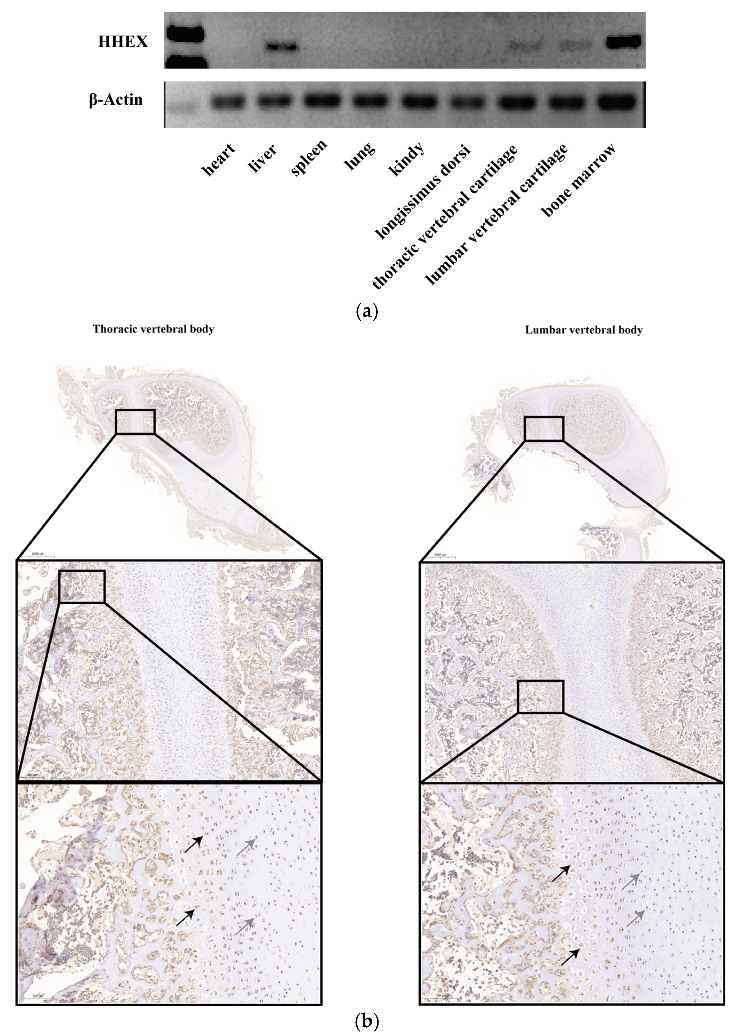
2.4. Immunohistochemistry (IHC)

Immunohistochemical staining was performed as follows: briefly, the slides were deparaffinized, and antigen retrieval was then performed in a steam cooker for 1.5 min in 1 mM of EDTA, pH 9.0 (Maixin Biological Technology Co., Ltd., Fuzhou, China). The samples were immersed in 3% H2O2, incubated at room temperature for 15 min, and kept in the dark. Objective tissues were blocked with 10% normal rabbit serum (for the case of primary antibodies that originated from goat) or 3% BSA (for the case of primary antibodies that originated from other species) at room temperature for 30 min. The slides were then incubated with a rabbit polyclonal primary antibody against HHEX (PB0675, Boster, Beijing, China) (diluted with PBS at 1:150) overnight at 4 °C and were placed in a wet box containing a small amount of water. The objective tissue was then incubated with goat anti-rabbit secondary antibody (GB23303) labeled with HRP (Servicebio, Wuhan, China) (diluted with PBS at a ratio of 1:200) at room temperature for 50 min. The sections were slightly dried, and freshly prepared DAB chromogenic reagent was added to the marked tissue. The nuclei were counterstained with hematoxylin staining solution for 3 min. The sections were successively dehydrated in gradient ethanol solutions of 75%, 85%, and 2 changes of pure ethanol for 6 min each. The sections were then cleared in xylene for 5 min and mounted with resin mounting medium.

2.5. Rapid Amplification of cDNA Ends (RACE)

Specific primers for amplification of the 3′ and 5′ ends were designed using Primer Premier 6.0 software (Premier Biosoft International, Palo Alto, CA, USA) (Table S2). The SMARTer® RACE 5′/3′ Kit (Clontech, Mountain View, CA, USA) was used to perform 3′ and 5′ RACE reactions. First-strand cDNA synthesis and PCR amplification were performed according to the instructions of the SMARTer® RACminE 5′/3′ Kit. The amplification reaction procedure was as follows: 25 cycles of 94 °C for 30 s, 72°C for 30 s, and 72 °C for 3 min. The PCR products were purified and cloned into linearized pRACE vectors, which were sequenced by Sangon Biotech (Shanghai, China). We assembled and analyzed the RACE sequences.

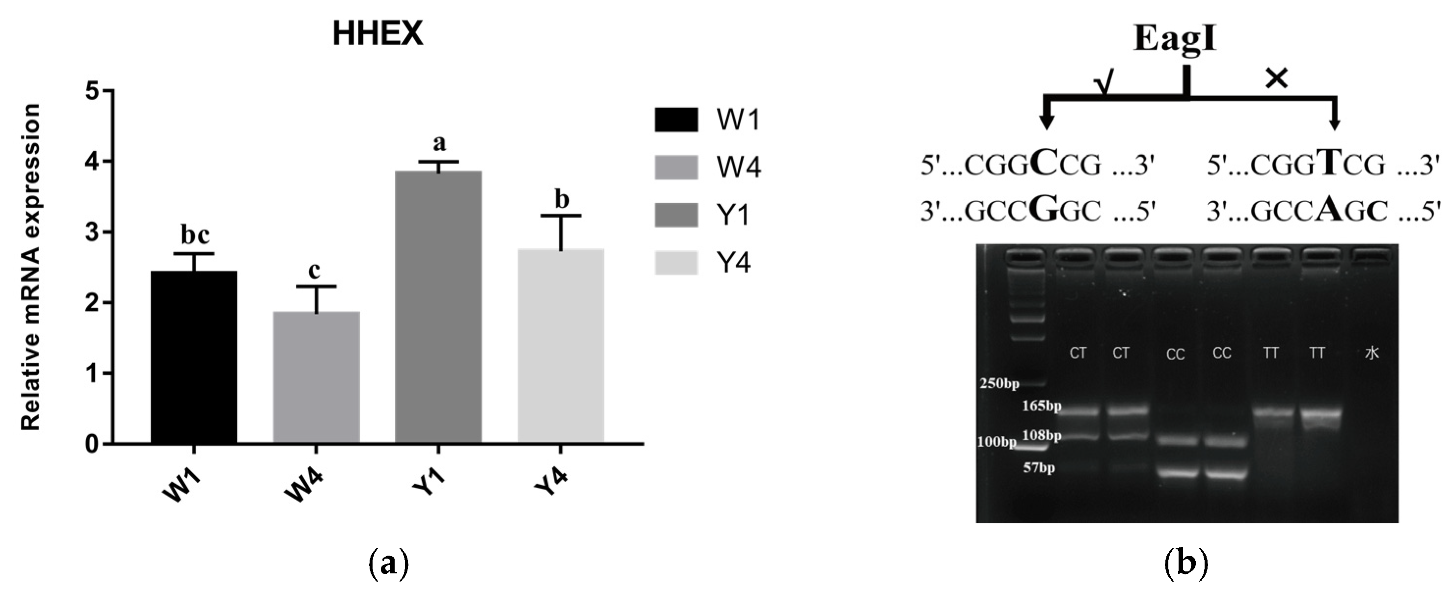
2.6. PCR Amplification, Sequencing and Genotyping

DNA sequencing of mixed pools was used to identify HHEX gene polymorphisms. The mixing pools were constructed by DNA from 10 Yorkshire pigs and 10 Wuzhishan pigs, respectively. Primers were used to amplify all exons, and the promoter regions of the HHEX gene (approximately 1000 bp upstream of the transcription start site) are shown in Table S2. Finally, amplification products were sequenced, and DNAMAN 9.0 and Chromas 2.6.5 software (Technelysium Pty Ltd., South Brisbane, Australia) were used to analyze the results. Linkage disequilibrium (LD) between the tested SNPs was estimated by using Haploview version 4.2 software (September 2009, Broad Institute, Cambridge, MA, USA).

2.7. Reporter Plasmid Construction

The promoter region sequences were amplified from pig genomic DNA (with different haplotypes) by PCR using the PCR process described in Section 2.6. Plasmids with different lengths of truncated porcine HHEX promoters (pGL4-P1: −1809/−1, pGL4-P2: −1424/−1, pGL4-P3: −1093/−1, pGL4-P4: −586/−1) were separately amplified using different forward primers and a common reverse primer (Supplementary Table S1). The amplified fragments were inserted between the Nhe I and Kpn I sites of the pGL4.10 [luc2] vector. TA and CG haplotype promoters were amplified using TA and CG haplotype DNA as templates, and TG and CA haplotype promoters were amplified using pGL4-CA-R and pGL4-TG-F primers using CG haplotype DNA as the template, respectively.

2.8. Cell Culture and Luciferase Assay

Transfection of 293T cells (ATCC ACS-4004) was performed with Lipofectamine 2000 (Invitrogen, Carlsbad, CA, USA). After 293T cells reached 80% confluence, the HHEX promoter-luciferase plasmid and the pRL-TK plasmid-expressing Renilla luciferase were co-transfected (Promega, Madison, WI, USA). After 36 h, cells were collected and analyzed for fluorescence activity using the Promega Dual Luciferase assay system (Promega, Madison, WI, USA).

2.9. Prediction of the Changes in Transcription Factor-Binding Sites (TFBSs)

We selected the 30 bp sequences before and after SNPs g.104194487G > A and g.104194685G > C to predict whether the SNPs in the promoter region of the HHEX gene impacted the TFBSs by using animalTFDB3.0 (AnimalTFDB3, hust.edu.cn, accessed on 1 August 2022).

2.10. Statistical Analysis

The distribution of genotype frequencies within a population and frequency differences between groups were tested using a chi-square test. Linear regression analyses of the SNP allele frequency and phenotype were performed using the R software package (ggplot2, v3.2.0). Gene expression in terms of mRNA level and luciferase assays was analyzed using a one-way analysis of variance followed by Duncan’s multiple range test using SPSS 24.0 software (SPSS Inc., Chicago, IL, USA). Graphs were prepared using GraphPad Prism 7 (GraphPad Software, La Jolla, CA, USA). p < 0.05 was considered statistically significant, and all results are presented as the mean ± standard error.

3. Results

3.1. Porcine HHEX Gene Expression Pattern

Pig body length traits are closely related to vertebral body development. To investigate whether the HHEX gene is associated with porcine vertebral body development, the HHEX expression patterns were first determined by an SqRT-PCR test in the heart, liver, spleen, lung, kidney, thoracic spine cartilage, lumbar spine cartilage, and bone marrow tissues. HHEX expression levels were high in the liver, bone marrow, thoracic vertebral cartilage, and lumbar vertebral cartilage tissue, but were not detected in other tissues (Figure 1a). In addition, porcine thoracic vertebral body and lumbar vertebral body tissue IHC results also showed that HHEX was expressed in round chondrocytes and hypertrophic chondrocytes (Figure 1b). These results indicated that the HHEX gene may play an important role in porcine vertebral body development.

3.2. Polymorphism of the Porcine HHEX Gene

Although a porcine HHEX transcript has been published in the NCBI database (NM_001244579.1), it is unclear whether alternate transcripts of the HHEX gene exist in the thoracic vertebral cartilage tissues of Wuzhishan and Yorkshire pigs. To analyze the genetic polymorphism of the porcine HHEX gene, we first cloned the HHEX gene based on cloning and RACE strategies, from which four exons located in chromosome fourteen were obtained. Four alternative splicing patterns were identified in the 5′UTR, and four splicing variants shared a coding region and the 3′UTR (Figure 2a). To detect polymorphisms in the porcine HHEX gene, we performed pooled sequencing on 10 Yorkshire pigs and 10 Wuzhishan pigs. After sequencing four exons, the SNP rs325806865 (G > A) and a novel SNP g.104194685 (G > C) were identified, which were both synonymous mutations (Figure 2b). In addition, we sequenced the promoter region approximately 1000 bp upstream of HHEX mRNA, and a novel haplotype consisting of two SNPs rs80901185 and rs80934526 (Figure 2c) in complete linkage disequilibrium was identified (Figure 2d, Table S3).

3.3. Comparison of Haplotype Frequencies of the HHEX Gene among Pig Breeds

The haplotypes showed significantly different distributions between Yorkshire pigs and Wuzhishan pigs, as shown in Table S3. The CG haplotype was the dominant haplotype in the Wuzhishan pigs (frequency = 0.81), while in contrast, the Yorkshire pig population was all TA haplotypes. Furthermore, to detect whether the HHEX gene was associated with body length traits in pigs, we analyzed the mRNA expression levels of the HHEX gene in Wuzhishan and Yorkshire pigs, which had significant differences in body length. The results showed that HHEX mRNA expression differed between Yorkshire (Y, TA haplotype) and Wuzhishan (W, CG haplotype) pigs in thoracic vertebral cartilage tissue. The expression of HHEX at 1 month of age was higher than that at 4 months of age in both Wuzhishan and Yorkshire pigs. Furthermore, at 1 month of age, HHEX expression in the Y group was significantly higher than in the W group (p < 0.01, Figure 3a). This suggests that the HHEX gene may be positively associated with body length traits in pigs.
To further investigate whether this haplotype is associated with body length traits in pigs, we first analyzed the differences in haplotype frequency of the Y and W groups through Sanger sequencing. Because these two SNPS were in complete linkage disequilibrium, we further investigated the rs80901185 (rs80901185T for the TA haplotype, rs80901185C for the CG haplotype) frequencies in nine other Chinese indigenous pig breeds and in Landrace pigs. The results showed that the rs80901185 frequencies were significantly different between Chinese indigenous pigs and European pigs (p < 0.001). The dominant haplotype was TA (with a haplotype frequency of 97%) in foreign pig breeds; however, it was 20% in Chinese indigenous pig breeds (Table 1). The linear regression analysis for body length trait phenotypes (China National Commission of Animal Genetic Resources, 2011) and gene frequency showed that rs80901185 frequency was significantly associated with body length (p < 0.05, Figure S1). Moreover, to facilitate the use of this haplotype in pig molecular selection and breeding, we targeted rs80901185 and developed a genotyping strategy based on PCR-RFLP. The three genotypes CC, CT, and TT were identified by differential digestion with the restriction endonuclease EagI (Figure 3b).

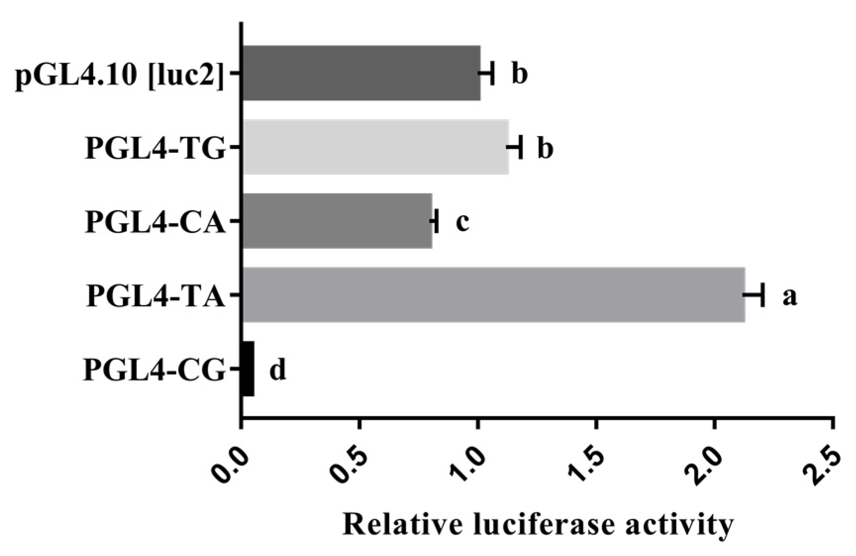
3.4. Promoter Activity Analyses of the Porcine HHEX Gene

It is known that the transcriptional activity of gene promoter regions can affect gene expression. Four reporter plasmids with different lengths of 5′ flanking sequences of the porcine HHEX gene inserted were constructed for determining the essential promoter region of the HHEX gene. The luciferase reporter plasmids pGL4-P1 (−1809/−1), pGL4-P2 (−1424/−1), pGL4-P3 (−1093/−1), and pGL4-P4 (−586/−1) (Figure 4) were transfected into 293T cells. The luciferase activities of the four promoter fragments were higher than that of the negative control pGL4.10 [luc2] groups (p < 0.01, Figure 4), revealing that the functional promoter is within the −1809/−1 region of the HHEX gene. The luciferase activities of pGL4-P1 (−1809/−1) and pGL4-P3 (−1093/−1) were significantly decreased, indicating that the −1425/−1809 and −587/−1093 regions of the porcine HHEX gene promoter may contain suppressors. Notably, the luciferase activity of pGL4-P4 was greater than that of the other three groups (p < 0.01, Figure 4), suggesting that the essential promoter region of the HHEX gene is located between −1 and −586 bp.

3.5. Effect of Haplotype on the Promoter Activity of HHEX

It is worth noting that the novel haplotype consisting of the g.104194045T > C SNP and g.104194173A > G SNP is also located in the essential promoter region. A shorter promoter region reporter vector only containing the haplotype was constructed to eliminate the interference of redundant sequences as well as the single mutation reporter vector to analyze the effect of each SNP on promoter activity. The promoter activities of the haplotype in the porcine HHEX gene were analyzed using a dual luciferase reporter assay system. As shown in Figure 5, the luciferase activities were significantly higher in plasmids of the TA haplotype, which had high frequencies in European commercial pigs, than in the plasmids of the CG haplotype (p < 0.01). In addition, the plasmids with the TA haplotype had higher (p < 0.01) luciferase activities than the plasmids with the TG and CA haplotypes, and all three haplotype luciferase activities were higher than that of the CG haplotype (Figure 5). These results suggest that the mutations g.104194045T > C and g.104194173A > G decreased the HHEX promoter activities.

3.6. The TA > CG Haplotype Adds a Transcription Suppressor Factor-Binding Site

Transcription factors are implicated in the processes of eukaryotic gene transcription and regulation by binding with cis-elements most residing in the promoter of genes. We compared the potential transcription factor-binding sites of g.104194045T and g.104194045C and g.104194173A and g.104194173G by AnimalTFDB3. The results showed that g.104194045 T > C increased multiple binding sites of YY1, TAF1, and POLR2A transcription factors, whereas g.104194173 A > G increased multiple binding sites for HDAC2, FOXA2, and RELA transcription factors (Tables S4 and S5).

4. Discussion

The body length trait is an important index for pig breeding selection. In this study, the effect of the HHEX gene and the body length trait in pigs was revealed for the first time. Semiquantitative results showed that the porcine HHEX gene was specifically expressed in liver tissue, bone marrow, and thoracic and lumbar cartilage tissues. Previous studies have shown that the loss of HHEX gene expression resulted in embryonic lethality at day 18 in pigs, indicating that HHEX plays an important role in liver organogenesis and development [22], which also explains why HHEX mRNA bands were detected in pig liver tissues. As vertebrate animals, the spinal bones of pigs are developed by the endochondral ossification of chondrocytes, including a series of processes such as the formation of the cartilage template, proliferation of chondrocytes, hypertrophic differentiation, apoptosis of hypertrophic chondrocytes, vascular invasion, and osteoblast formation [28]. Previous studies have shown that the expression of HHEX is drastically increased during chondrocyte differentiation [26,27]. Semiquantitative and immunohistochemical results showed that HHEX was specifically expressed in the thoracic vertebral cartilage, lumbar cartilage and bone marrow tissues of pigs, suggesting that HHEX is likely to play an important role in the development of pig cartilage and bone formation.
Human height, which is equivalent to body length in pigs, is a highly heritable and classic polygenic trait. Hundreds of variants clustered in genomic loci and biological pathways were found to affect human height [29], and studies have shown that almost 45% of the variance can be explained by considering all SNPs simultaneously [30]. Empirical evidence indicates that there are some common mutations with moderate to large effects on body size in livestock, e.g., in dogs and cattle [31]. Thus, we explored the internal mechanism of the HHEX gene’s differential expression between Yorkshire and Wuzhishan pigs. We first detected the mRNA splicing patterns and genome polymorphisms of HHEX and detected two SNPs in the CDS region of the first exon, both of which were synonymous mutations. Synonymous mutations are often considered silent mutations because they do not change the protein’s amino acid sequence, so these SNPs were not further investigated. Accurate gene expression is controlled by a strict transcriptional regulatory program, and studies have shown that gene promoter regions play an important role in gene transcription by regulating gene initiation transcription by binding to RNA polymerase II and transcription factors [32]. A polymorphism analysis of the HHEX promoter region revealed two linked SNP haplotypes in the HHEX promoter region. Haplotypes are the product of genetic introgression or selection during the evolution of pigs. We analyzed the frequency detection of this haplotype in Chinese indigenous breeds and in foreign pig breeds with significant differences in body length traits, and the results showed that there were significant differences in the frequency of this haplotype between Chinese indigenous breeds and foreign pig breeds. Further experiments showed that the HHEX mRNA expression level in the thoracic vertebral cartilage of Yorkshire pigs was significantly higher than that of Wuzhishan pigs. This suggests that the higher expression of HHEX in Yorkshire pigs, which promotes chondrogenesis, may be an important reason why Yorkshire pigs have longer body lengths than Wuzhishan pigs. Thus, a linear regression analysis for body length trait phenotype and gene frequency was performed, and the result showed that the g.104194045T > C frequency was significantly associated with the body length trait. Therefore, we speculated that this haplotype might be an important mutation that causes the difference in body length between Chinese indigenous and foreign pig breeds.
Furthermore, through the activity analysis of the promoter region of the HHEX gene, we confirmed that −586/−1 was the core promoter region of the HHEX gene. Notably, a haplotype consisting of the two SNPs rs80901185 and rs80934526, which were in complete linkage disequilibrium, was located in the essential promoter region, and the dominant haplotype promoter activity in Yorkshire pigs (TA haplotype) was significantly higher than that of Wuzhishan pigs (CG haplotype). These results suggested that this haplotype variation might be the main reason for the HHEX gene expression in Big White pigs being significantly higher than in Wuzhishan pigs. Mutations in the core sequences of the binding sites might alter the affinity of transcription factors for DNA and might result in changes in transcription activities [33,34,35]. To confirm that the haplotype variation changed the regulatory mechanism of the promoter activity, we built two single-mutation fluorescein plasmids. The results showed that when compared with the TA haplotype, the CA and TG single mutation haplotype promoter activities were significantly lower, but the TA, CA, and TG haplotype activities were all significantly higher than the CG haplotype promoter activity. This suggests that T > C and A > G mutations are likely to generate new binding sites for transcription repressors. The prediction results of the binding sites of transcription factors showed that T > C and A > G mutations generate three potential YY1 transcription factor-binding sites and three potential HDAC2 transcription factor-binding sites, respectively. Yin Yang 1 (YY1) is a ubiquitous and multifunctional zinc-finger transcription factor. Studies have shown that YY1 can reduce promoter activity by competing with transcriptional activators for binding sites, leading to transcriptional inhibition [36,37]. Studies on chondrogenesis and bone formation have shown that YY1 can inhibit the chondrogenic differentiation of mesenchymal stem cells by binding to target genes [38,39]; YY1 can also inhibit the cell differentiation induced by transforming growth factor β and bone morphogenetic protein, thus regulating bone formation [40]. Histone deacetylase 2 (HDAC2) is a member of the HDAC family and inhibits gene transcription by removing acetyl groups [41]. Studies have shown that HDAC2 can inhibit cartilage-specific gene expression and induce osteoarthritis [42,43]. This evidence further suggests that the differences in HHEX gene haplotype promoter activity are likely caused by changes in the binding sites of the YY1 and HDAC2 transcription factors. YY1 establishes and maintains transcriptional silencing by recruiting histone deacetylase (HDAC) [37,44]. Studies have shown that mammalian HDAC2 can repress transcription through a natural YY1 binding site and that transcriptional repression by YY1 requires interaction with the mammalian HDAC2 protein [45]. This may partly explain why the two SNPs were in complete linkage disequilibrium.

5. Conclusions

In this study, our data showed that the porcine HHEX gene was specifically expressed in the liver, bone marrow, thoracic vertebral cartilage, and lumbar vertebral cartilage tissue. The RACE results showed that four mRNA alternative splicing patterns were identified in the 5′UTR, and four splicing variants shared a coding region and 3′UTR; the two SNPs in the CDS region of the first exon were detected, both of which were synonymous mutations. Moreover, we found a novel haplotype, consisting of the two SNPs rs80901185 (T > C) and rs80934526 (A > G), which was detected in the promoter region of the porcine HHEX; the expression of the HHEX gene was significantly higher in Yorkshire pigs (TA haplotype) than in Wuzhishan pigs (CG haplotype), and the population analysis showed that this haplotype was significantly associated with body length. A subsequent analysis suggested that the promoter region of HHEX gene from −586 to −1 bp exhibited the highest activity. We also found that the activity of TA haplotype was significantly higher than that of the CG haplotype by changing the potential binding of transcription factors YY1 and HDAC2. Therefore, the porcine HHEX gene may be potentially effective genetic markers for improving body length in pigs.

Supplementary Materials

The following supporting information can be downloaded at https://www.mdpi.com/article/10.3390/genes14020511/s1: Figure S1: Linear regression analysis of the body length trait phenotype and gene frequency of the SNP rs80901185T; Table S1: Primer information of the SNP identification and RACE for the porcine HHEX gene; Table S2: Primer information of the plasmid construction and RT-qPCR for the porcine HHEX gene; Table S3: Genotype and allele frequencies of the pig HHEX gene rs80901185 and rs80934526 polymorphisms in Yorkshire and Wuzhishan pigs; Table S4: Analysis of the transcription factor-binding sites of the SNP rs80901185T > C in the promoter sequence of the porcine HHEX gene; Table S5: Analysis of the transcription factor-binding sites of the SNP rs80934526A > G in the promoter sequence of the porcine HHEX gene.

Author Contributions

Y.L. contributed to the study, the analysis of the data, and the writing of the manuscript. Q.X. and M.X. contributed to the analysis of the data and conducted the experiments. Q.T., Y.W. and Z.C. contributed to the analysis of the data. F.W., R.S., S.C. and X.Y. contributed to the collection and collation of the sample. M.F. contributed to the design of the study, interpretation of the data, and modification of the manuscript. All authors have read and agreed to the published version of the manuscript.

Funding

This research was supported by the Zaozhuang Talent Program Funding to MYF, the National Natural Science Foundation of China (32072700 and U22A20508), the 2020 Research Program of Sanya Yazhou Bay Science and Technology City (Grant No. SKJC-2020-02-007), and the China Agriculture Research System of MOF and MARA (CARS-35).

Institutional Review Board Statement

Animal care was conducted in strict accordance with the Guide for the Care and Use of Laboratory Animals in China. All experiments were approved by the Committee on the Ethics of Animal Experiments of China Agricultural University (permit number: AW32202202-2-1).

Informed Consent Statement

Not applicable.

Data Availability Statement

The data presented in this study are available on request from the corresponding authors.

Acknowledgments

We acknowledge Ran Cui for collecting the samples and Shuaihan He for his guidance on data analysis.

Conflicts of Interest

The authors declare no conflict of interest.

References

  1. Yang, T.; Lin, J. Variation of heart size and its correlation with growth performance and vascular space in domestic pigs. Anim. Sci. 1997, 64, 523–528. [Google Scholar] [CrossRef]
  2. Bogucka, J.; Kapelanski, W.; Elminowska-Wenda, G.; Walasik, K.; Lewandowska, K.L. Comparison of microstructural traits of Musculus longissimus lumborum in wild boars, domestic pigs and wild boar/domestic pig hybrids. Arch. Anim. Breed. 2008, 51, 359–365. [Google Scholar] [CrossRef] [Green Version]
  3. Ruusunen, M.; Puolanne, E. Histochemical properties of fibre types in muscles of wild and domestic pigs and the effect of growth rate on muscle fibre properties. Meat Sci. 2004, 67, 533–539. [Google Scholar] [CrossRef] [Green Version]
  4. Castillo-Contreras, R.; Mentaberre, G.; Aguilar, X.F.; Conejero, C.; Colom-Cadena, A.; Ráez-Bravo, A.; González-Crespo, C.; Espunyes, J.; Lavín, S.; López-Olvera, J.R. Wild boar in the city: Phenotypic responses to urbanisation. Sci. Total Environ. 2021, 773, 145593. [Google Scholar] [CrossRef] [PubMed]
  5. Larson, G.; Dobney, K.; Albarella, U.; Fang, M.; Matisoo-Smith, E.; Robins, J.; Lowden, S.; Finlayson, H.; Brand, T.; Willerslev, E.; et al. Worldwide phylogeography of wild boar reveals multiple centers of pig domestication. Science 2005, 307, 1618–1621. [Google Scholar] [CrossRef] [Green Version]
  6. Wang, J.; Liu, C.; Chen, J.; Bai, Y.; Wang, K.; Wang, Y.; Fang, M. Genome-Wide Analysis Reveals Human-Mediated Introgression from Western Pigs to Indigenous Chinese Breeds. Genes 2020, 11, 275. [Google Scholar] [CrossRef] [Green Version]
  7. Xinhong, L.; Zhen, L.; Fu, J.; Wang, L.; Yang, Q.; Li, P.; Li, Y. Quantitative proteomic profiling indicates the difference in reproductive efficiency between Meishan and Duroc boar spermatozoa. Theriogenology 2018, 116, 71–82. [Google Scholar] [CrossRef]
  8. Clapperton, M.; Bishop, S.C.; Glass, E.J. Innate immune traits differ between Meishan and Large White pigs. Vet. Immunol. Immunopathol. 2005, 104, 131–144. [Google Scholar] [CrossRef] [PubMed]
  9. Dai, F.; Feng, D.; Cao, Q.; Ye, H.; Zhang, C.; Xia, W.; Zuo, J. Developmental differences in carcass, meat quality and muscle fibre characteristics between the Landrace and a Chinese native pig. S. Afr. J. Anim. Sci. 2009, 39, 4. [Google Scholar] [CrossRef]
  10. Panda, S.; Gaur, G.K.; Chauhan, A.; Kar, J.; Mehrotra, A. Accurate assessment of body weights using morphometric measurements in Landlly pigs. Trop. Anim. Health Prod. 2021, 53, 362. [Google Scholar] [CrossRef] [PubMed]
  11. Li, L.Y.; Xiao, S.J.; Tu, J.M.; Zhang, Z.K.; Zheng, H.; Huang, L.B.; Huang, Z.Y.; Yan, M.; Liu, X.D.; Guo, Y.M. A further survey of the quantitative trait loci affecting swine body size and carcass traits in five related pig populations. Anim. Genet. 2021, 52, 621–632. [Google Scholar] [CrossRef]
  12. Liu, H.; Song, H.; Jiang, Y.; Jiang, Y.; Zhang, F.; Liu, Y.; Shi, Y.; Ding, X.; Wang, C. A Single-Step Genome Wide Association Study on Body Size Traits Using Imputation-Based Whole-Genome Sequence Data in Yorkshire Pigs. Front. Genet. 2021, 12, 629049. [Google Scholar] [CrossRef]
  13. Groenen, M.A.; Archibald, A.L.; Uenishi, H.; Tuggle, C.K.; Takeuchi, Y.; Rothschild, M.F.; Rogel-Gaillard, C.; Park, C.; Milan, D.; Megens, H.J.; et al. Analyses of pig genomes provide insight into porcine demography and evolution. Nature 2012, 491, 393–398. [Google Scholar] [CrossRef] [PubMed] [Green Version]
  14. Hong, Y.; Ye, J.; Dong, L.; Li, Y.; Yan, L.; Cai, G.; Liu, D.; Tan, C.; Wu, Z. Genome-Wide Association Study for Body Length, Body Height, and Total Teat Number in Large White Pigs. Front. Genet. 2021, 12, 650370. [Google Scholar] [CrossRef]
  15. Johnson, Z.; Nugent, R., III. Heritability of body length and measures of body density and their relationship to backfat thickness and loin muscle area in swine. J. Anim. Sci. 2003, 81, 1943–1949. [Google Scholar] [CrossRef]
  16. Li, J.; Peng, S.; Zhong, L.; Zhou, L.; Yan, G.; Xiao, S.; Ma, J.; Huang, L. Identification and validation of a regulatory mutation upstream of the BMP2 gene associated with carcass length in pigs. Genet. Sel. Evol. 2021, 53, 94. [Google Scholar] [CrossRef] [PubMed]
  17. Zhang, R.; Grosse-Brinkhaus, C.; Heidt, H.; Uddin, M.J.; Cinar, M.U.; Tesfaye, D.; Tholen, E.; Looft, C.; Schellander, K.; Neuhoff, C. Polymorphisms and expression analysis of SOX-6 in relation to porcine growth, carcass, and meat quality traits. Meat Sci. 2015, 107, 26–32. [Google Scholar] [CrossRef] [PubMed]
  18. Yang, J.; Huang, L.; Yang, M.; Fan, Y.; Li, L.; Fang, S.; Deng, W.; Cui, L.; Zhang, Z.; Ai, H.; et al. Possible introgression of the VRTN mutation increasing vertebral number, carcass length and teat number from Chinese pigs into European pigs. Sci. Rep. 2016, 6, 19240. [Google Scholar] [CrossRef] [Green Version]
  19. Hartmann, C. Transcriptional networks controlling skeletal development. Curr. Opin Genet. Dev. 2009, 19, 437–443. [Google Scholar] [CrossRef]
  20. Goodings, C.; Smith, E.; Mathias, E.; Elliott, N.; Cleveland, S.M.; Tripathi, R.M.; Layer, J.H.; Chen, X.; Guo, Y.; Shyr, Y.; et al. Hhex is Required at Multiple Stages of Adult Hematopoietic Stem and Progenitor Cell Differentiation. Stem Cells 2015, 33, 2628–2641. [Google Scholar] [CrossRef] [Green Version]
  21. Paz, H.; Lynch, M.R.; Bogue, C.W.; Gasson, J.C. The homeobox gene Hhex regulates the earliest stages of definitive hematopoiesis. Blood 2010, 116, 1254–1262. [Google Scholar] [CrossRef] [Green Version]
  22. Ruiz-Estevez, M.; Crane, A.T.; Rodriguez-Villamil, P.; Ongaratto, F.L.; You, Y.; Steevens, A.R.; Hill, C.; Goldsmith, T.; Webster, D.A.; Sherry, L.; et al. Liver development is restored by blastocyst complementation of HHEX knockout in mice and pigs. Stem Cell Res. 2021, 12, 292. [Google Scholar] [CrossRef] [PubMed]
  23. Jackson, J.T.; Nasa, C.; Shi, W.; Huntington, N.D.; Bogue, C.W.; Alexander, W.S.; McCormack, M.P. A crucial role for the homeodomain transcription factor Hhex in lymphopoiesis. Blood 2015, 125, 803–814. [Google Scholar] [CrossRef] [PubMed] [Green Version]
  24. Hunter, M.P.; Wilson, C.M.; Jiang, X.; Cong, R.; Vasavada, H.; Kaestner, K.H.; Bogue, C.W. The homeobox gene Hhex is essential for proper hepatoblast differentiation and bile duct morphogenesis. Dev. Biol. 2007, 308, 355–367. [Google Scholar] [CrossRef] [PubMed] [Green Version]
  25. Gauvrit, S.; Villasenor, A.; Strilic, B.; Kitchen, P.; Collins, M.M.; Marin-Juez, R.; Guenther, S.; Maischein, H.M.; Fukuda, N.; Canham, M.A.; et al. HHEX is a transcriptional regulator of the VEGFC/FLT4/PROX1 signaling axis during vascular development. Nat. Commun. 2018, 9, 2704. [Google Scholar] [CrossRef] [Green Version]
  26. Morimoto, R.; Yamamoto, A.; Akimoto, Y.; Obinata, A. Homeoprotein Hex is expressed in mouse developing chondrocytes. J. Biochem. 2011, 150, 61–71. [Google Scholar] [CrossRef] [PubMed]
  27. Morimoto, R.; Obinata, A. Overexpression of Hematopoietically Expressed Homeoprotein Induces Nonapoptotic Cell Death in Mouse Prechondrogenic ATDC5 Cells. Biol. Pharm. Bull. 2011, 34, 1589–1595. [Google Scholar] [CrossRef] [Green Version]
  28. Aghajanian, P.; Mohan, S. The art of building bone: Emerging role of chondrocyte-to-osteoblast transdifferentiation in endochondral ossification. Bone Res. 2018, 6, 19. [Google Scholar] [CrossRef] [Green Version]
  29. Allen, H.L.; Estrada, K.; Lettre, G.; Berndt, S.; Weedon, M.N.; Rivadeneira, F. Hundreds of variants influence human height and cluster within genomic loci and biological pathways. In The Role of Common Genetic Variation in Model Polygenic Monogenic Traits; University of Exeter: Exeter, UK, 2010; p. 94. [Google Scholar]
  30. Yang, J.; Benyamin, B.; McEvoy, B.P.; Gordon, S.; Henders, A.K.; Nyholt, D.R.; Madden, P.A.; Heath, A.C.; Martin, N.G.; Montgomery, G.W. Common SNPs explain a large proportion of the heritability for human height. Nat. Genet. 2010, 42, 565–569. [Google Scholar] [CrossRef] [Green Version]
  31. Kemper, K.E.; Visscher, P.M.; Goddard, M.E. Genetic architecture of body size in mammals. Genome Biol. 2012, 13, 244. [Google Scholar] [CrossRef] [Green Version]
  32. Struhl, K. Promoters, activator proteins, and the mechanism of transcriptional initiation in yeast. Cell 1987, 49, 295–297. [Google Scholar] [CrossRef] [PubMed]
  33. Hua, J.T.; Ahmed, M.; Guo, H.; Zhang, Y.; Chen, S.; Soares, F.; Lu, J.; Zhou, S.; Wang, M.; Li, H. Risk SNP-mediated promoter-enhancer switching drives prostate cancer through lncRNA PCAT19. Cell 2018, 174, 564–575.e18. [Google Scholar] [CrossRef] [PubMed] [Green Version]
  34. Bečanović, K.; Nørremølle, A.; Neal, S.J.; Kay, C.; Collins, J.A.; Arenillas, D.; Lilja, T.; Gaudenzi, G.; Manoharan, S.; Doty, C.N. A SNP in the HTT promoter alters NF-κB binding and is a bidirectional genetic modifier of Huntington disease. Nat. Neurosci. 2015, 18, 807–816. [Google Scholar] [CrossRef] [PubMed]
  35. De Gobbi, M.; Viprakasit, V.; Hughes, J.R.; Fisher, C.; Buckle, V.J.; Ayyub, H.; Gibbons, R.J.; Vernimmen, D.; Yoshinaga, Y.; De Jong, P. A regulatory SNP causes a human genetic disease by creating a new transcriptional promoter. Science 2006, 312, 1215–1217. [Google Scholar] [CrossRef] [Green Version]
  36. Shi, Y.; Lee, J.-S.; Galvin, K.M. Everything you have ever wanted to know about Yin Yang 1. Biochim. Biophys. Acta 1997, 1332, F49–F66. [Google Scholar] [CrossRef]
  37. Gordon, S.; Akopyan, G.; Garban, H.; Bonavida, B. Transcription factor YY1: Structure, function, and therapeutic implications in cancer biology. Oncogene 2006, 25, 1125–1142. [Google Scholar] [CrossRef] [Green Version]
  38. Aoyama, T.; Okamoto, T.; Fukiage, K.; Otsuka, S.; Furu, M.; Ito, K.; Jin, Y.; Ueda, M.; Nagayama, S.; Nakayama, T. Histone modifiers, YY1 and p300, regulate the expression of cartilage-specific gene, chondromodulin-I, in mesenchymal stem cells. J. Biol. Chem. 2010, 285, 29842–29850. [Google Scholar] [CrossRef] [Green Version]
  39. Guérit, D.; Brondello, J.-M.; Chuchana, P.; Philipot, D.; Toupet, K.; Bony, C.; Jorgensen, C.; Noël, D. FOXO3A regulation by miRNA-29a Controls chondrogenic differentiation of mesenchymal stem cells and cartilage formation. Stem Cells Dev. 2014, 23, 1195–1205. [Google Scholar] [CrossRef]
  40. Kurisaki, K.; Kurisaki, A.; Valcourt, U.; Terentiev, A.A.; Pardali, K.; Ten Dijke, P.; Heldin, C.-H.; Ericsson, J.; Moustakas, A. Nuclear factor YY1 inhibits transforming growth factor β-and bone morphogenetic protein-induced cell differentiation. Mol. Cell. Biol. 2003, 23, 4494–4510. [Google Scholar] [CrossRef] [Green Version]
  41. Ruijter, A.J.D.; Gennip, A.H.V.; Caron, H.N.; Kemp, S.; Kuilenburg, A.B.V. Histone deacetylases (HDACs): Characterization of the classical HDAC family. Biochem. J. 2003, 370, 737–749. [Google Scholar] [CrossRef] [Green Version]
  42. Hong, S.; Derfoul, A.; Pereira-Mouries, L.; Hall, D.J. A novel domain in histone deacetylase 1 and 2 mediates repression of cartilage—Specific genes in human chondrocytes. FASEB J. 2009, 23, 3539–3552. [Google Scholar] [CrossRef] [PubMed] [Green Version]
  43. Mao, G.; Zhang, Z.; Huang, Z.; Chen, W.; Huang, G.; Meng, F.; Kang, Y. MicroRNA-92a-3p regulates the expression of cartilage-specific genes by directly targeting histone deacetylase 2 in chondrogenesis and degradation. Osteoarthr. Cartil. 2017, 25, 521–532. [Google Scholar] [CrossRef] [PubMed] [Green Version]
  44. Yang, W.-M.; Yao, Y.-L.; Sun, J.-M.; Davie, J.R.; Seto, E. Isolation and characterization of cDNAs corresponding to an additional member of the human histone deacetylase gene family. J. Biol. Chem. 1997, 272, 28001–28007. [Google Scholar] [CrossRef] [PubMed] [Green Version]
  45. Yang, W.-M.; Inouye, C.; Zeng, Y.; Bearss, D.; Seto, E. Transcriptional repression by YY1 is mediated by interaction with a mammalian homolog of the yeast global regulator RPD3. Proc. Natl. Acad. Sci. USA 1996, 93, 12845–12850. [Google Scholar] [CrossRef] [Green Version]
Figure 1. Porcine HHEX expression pattern. (a) HHEX mRNA expression in Yorkshire pig tissues (n = 1), which were grouped based on the electrophoresis of PCR products for HHEX and β-actin. (b) Immunohistochemical observation of the expression of HHEX in porcine thoracic vertebral body and lumbar vertebral body tissue at 4 days of age. The arrow marks HHEX-positive cells; gray arrows indicate round chondrocytes, and black arrows indicate hypertrophic chondrocytes.
Genes 14 00511 g001
Figure 2. Polymorphism of the porcine HHEX gene. (a) HHEX gene structure diagram. The open reading frame is marked with a black block, the 5′ UTR with a gray block, the 3′ UTR with a white block, and the introns with line segments. The two SNPs located in the open reading frame of the first exon are marked with arrows. (b) Part of the open reading frame nucleotide sequence and the predicted amino acid sequence in the first exon of HHEX are shown, and the blue letters mark the SNPs (g.104194487G > A and g.104194685G > C) and the predicted amino acid changes, respectively; both were synonymous mutations. (c) HHEX gene promoter SNP (g.104194045T > C and g.104194173A > G) genome distribution diagram and Sanger sequencing map. (d) Linkage disequilibrium analysis for two polymorphisms in the porcine HHEX promoter region in Yorkshire and Wuzhishan pigs. The numbers in the boxes represent the r2 values between SNP pairs. The boxes without numbers indicate r2  =  1. The boxes are colored according to the standard Haploview color scheme: LD  >  2 and D′  =  1, red.
Genes 14 00511 g002
Figure 3. (a) HHEX mRNA expression levels in the thoracic vertebral cartilage tissues of Y1, Y4, W1, and W4. Values shown are the mean ± SE. Different letters indicate significant differences (p < 0.05); the same letters indicate no significant difference. Y1, 1-month-old Yorkshire pigs (n = 3); Y4, 4-month-old Yorkshire pigs (n = 3); W1, 1-month-old Wuzhishan pigs (n = 3); W4, 4-month-old Wuzhishan pigs (n = 3). (b) rs80901185 PCR-RFLP diagram.
Genes 14 00511 g003
Figure 4. Porcine HHEX promoter transcriptional activity analyses. The left image indicates the length and structure of the fragments, and the right image indicates the corresponding relative luciferase activity. The luciferase activity was normalized, and the relative values were expressed as the fold of induction relative to the pGL4.10[luc2] vector activity. The relative luciferase activity values represent the mean ± SEM of three independent experiments. A one-way ANOVA test was used to assess the differences in luciferase activity. Different letters (a, b, and c) indicate that the difference is significant (p < 0.05).
Genes 14 00511 g004
Figure 5. Luciferase reporter gene assays of the haplotypes containing the HHEX gene promoter SNPs rs80901185 and rs80934526. pGL4-TG represents rs80901185T and rs80934526G, pGL4-CA represents rs80901185C and rs80934526A, pGL4-TA represents rs80901185T and rs80934526A, and pGL4-CG represents rs80901185C and rs80934526G. The relative luciferase activity values represent the mean ± SEM of three independent experiments. A one-way ANOVA test was used to assess differences in luciferase activity. Different letters (a, b, and c) indicate that the difference is significant (p < 0.05).
Genes 14 00511 g005
Table 1. Genotype and allele frequencies of the pig HHEX gene g.104194045T > C polymorphisms in different pig breeds.
BreedTotal NumberGenotype, n, FrequencyAllele, n, FrequencyBody Length, cmχ2, df, p Value a
CCCTTTCT
Wuzhishan pig24159039967.08 ± 1.19297.459, 2, p < 0.0001
0.630.380.000.810.19
Bama
Pig
22220044074.25 ± 0.25
1.000.000.001.000.00
Tibetan
pig
2413110371172.9 ± 0.69
0.540.460.000.770.23
Banna mini-pig342473551354.6 ± 0.80
0.710.210.090.810.19
Jiangkou radish pig4730152751969
0.640.320.040.800.20
Longlin pig3722132571796.5 ± 3.80
0.590.350.050.770.23
Min
pig
36162005220124.5 ± 0.47
0.440.560.000.720.28
Yanan
pig
141220262124
0.860.140.000.930.07
Qianbei black pig3521140561493.45
0.600.400.000.800.20
Saba
pig
35171815220134.38 ± 0.7
0.470.500.030.740.29
Yorkshire pig290029058165 ± 2.47
0.000.001.000.001.00
Landrace pig300426456160.1 ± 0.73
0.000.130.870.070.93
a Square test was calculated based on the genotypes of the Chinese indigenous and commercial pig populations.
Disclaimer/Publisher’s Note: The statements, opinions and data contained in all publications are solely those of the individual author(s) and contributor(s) and not of MDPI and/or the editor(s). MDPI and/or the editor(s) disclaim responsibility for any injury to people or property resulting from any ideas, methods, instructions or products referred to in the content.

