Article

Biocontrol of Macrophomina phaseolina Using Bacillus amyloliquefaciens Strains in Cowpea (Vigna unguiculata L.)

by
Edelweiss Airam Rangel-Montoya
1,
Carmen Sanjuana Delgado-Ramírez
1,
Edgardo Sepulveda
2 and
Rufina Hernández-Martínez
1,*
1
Department of Microbiology, Centro de Investigación Científica y de Educación Superior de Ensenada, CICESE, Carr.Tijuana-Ensenada 3918, Zona Playitas, Ensenada 22860, Mexico
2
CONACYT-Department of Microbiology, Centro de Investigación Científica y de Educación Superior de Ensenada, CICESE, Carr.Tijuana-Ensenada 3918, Zona Playitas, Ensenada 22860, Mexico
*
Author to whom correspondence should be addressed.
Agronomy 2022, 12(3), 676; https://doi.org/10.3390/agronomy12030676
Submission received: 10 January 2022 / Revised: 28 February 2022 / Accepted: 2 March 2022 / Published: 11 March 2022

Abstract

:
The fungus Macrophomina phaseolina is the causal agent of charcoal rot in many crops, such as strawberries and beans. Symptoms include stem and root rot and chlorotic foliage. This disease’s management is complicated because the pathogen forms resistant microsclerotia. This work aimed to obtain bacterial isolates for the biocontrol of M. phaseolina in arid regions. Two strains that grew well under low pH and high salinity, named BsA3MX and BsC11MX, were isolated and identified as B. amyloliquefaciens, based on their morphology and analysis of the 16S ribosomal RNA. Both strains inhibited M. phaseolina up to 66.8% in vitro through the combined action of volatile and diffusible compounds. Furthermore, they produce siderophores and indole-3-acetic acid (IAA), have ACC-deaminase activity, solubilize phosphate and zinc, and decrease microsclerotia germination. Moreover, in greenhouse assays using cowpea plants (Vigna unguiculata L.), strain BsA3MX reduced lesions caused by M. phaseolina and induced a significant increase in foliage and root biomass. Overall, these results suggest B. amyloliquefaciens BsA3MX and BsC11MX can be used as biological control agents against M. phaseolina in arid zones.

1. Introduction

Macrophomina phaseolina is an Ascomycete from the Botryosphaeriaceae family, primarily soil-inhabitant, and widely distributed in warm areas [1,2]. This organism is the causal agent of charcoal-rot and root-rot diseases in more than 500 cultivated and wild plant species [3,4]; several of them are economically relevant crops such as maize, sorghum, strawberry, cotton, soybean, cowpea, sesame, and sunflower [5,6,7,8]. Symptoms in plants infected with M. phaseolina include stem and root rot, chlorotic foliage, and senescence of leaves. Disease incidence is favored by hot and dry weather, or when plants face adverse environmental conditions [9]. This fungus produces resistance structures called microsclerotia, which can survive in soil, crop residues, and seeds for 2–15 years and can be detected in root and stem tissues in advanced stages of the disease [10,11]. The microsclerotia germinate under high temperature (30–35 °C) and low soil moisture (below 60%) on the root surface [2], producing appressoria and penetrating epidermic cell walls through natural openings, which affects the vascular system. Besides, during seed emergence, this pathogen infects through cotyledons or via root surface injuries, and infects legume seeds during storage, resulting in substantial losses [12].
The management of M. phaseolina is complicated because microsclerotia provide resistance to fungicide application and even solarization [13]. Some fungicides such as carbendazim, difenoconazole, benomyl, azoxystrobin, and dazom have been evaluated against M. phaseolina; however, there are no systemic fungicides, and the use of chemicals applied in large quantities and repeatedly is harmful to humans and the environment [2,14]. Moreover, the pathogen is a competitive saprophyte, capable of circumventing cultural practices such as soil tillage and crop rotation, securing its prevalence in soils [15,16]. Given these characteristics, the use of biological control agents prevailing in soil and in plants that suppress the pathogen at different stages of its life cycle is an alternative to counteract the environmental resilience of M. phaseolina.
Soil microorganisms are considered an essential factor for fertility and plant health. Plant growth-promoting rhizobacteria (PGPR) are free-living soil bacteria that, when applied to seeds or roots, benefit crops by stimulating plant growth or by reducing the damage caused by soil-borne pathogens [17]. Bacillus is the most widely distributed genus of bacteria in agro-systems, and one of its main applications is the control of phytopathogenic fungi in crops [18]. Species from this genus are Gram-positive, rod-shaped bacteria of the Bacillaceae family that produce endospores [19]. Bacillus spp. synthesize a great variety of antibiotics, toxins, siderophores, and lytic enzymes, and induce systemic resistance and plant growth [20,21]. Lipopeptides from iturin, surfactin, and fengycin families are the most studied antibiotic compounds synthesized by Bacillus spp. Such as B. subtilis, B. amyloliquefaciens, B. licheniformis, B. pumilus, and B. coagulans. These molecules interact at the target cells’ membrane level, affecting their structure and permeability by inducing disruption or formation of ion channels and pores [22].
Worldwide, M. phaseolina causes high economic yield losses and reduces seeds quality [12,23,24]; for example, in charcoal rot of soybean, losses have been estimated at around 1.9–2.0 million tonnes [25]. In Mexico, M. phaseolina has been reported in maize, common beans, sesame, sorghum, soybeans, eggplant, sugar cane, thyme, and peanut in arid, subtropical, and tropical regions [26,27,28,29]. In strawberries, M. phaseolina has not been reported, but plants with disease symptoms have been observed recently in Baja California. Worldwide, Mexico ranks sixth in sorghum, seventh for common beans, and third in strawberry. Baja California is the state with the second-highest production of strawberries and the seventh in the production of sorghum [30]. Yield losses caused by M. phaseolina have been estimated at around 30%, 60%, and 20% for sorghum, common beans, and strawberries, respectively [31,32,33]. This highlights the potential economic problem of this fungus for Baja California. Due to the difficulty in controlling diseases caused by M. phaseolina, this study aimed to obtain bacteria from the Baja California region with potential for the biocontrol of M. phaseolina.

2. Materials and Methods

2.1. Isolation and Morphological Characterization of Microorganisms

M. phaseolina was isolated from strawberry plants with charcoal-rot symptoms. Twenty plants were collected from a farm located in San Quintin, Ensenada, Baja California. Small pieces of symptomatic plant tissue were surface-sterilized by flaming and then incubated on potato dextrose agar plates (PDA Difco) at 30 °C until fungal or bacterial growth was observed. Plates were screened for bacterial isolates growing in contact with fungi and showing visual evidence of mycelial growth inhibition. Only bacteria showing strong inhibition were recovered. These fungi and bacteria were subsequently subcultured to purity on PDA and trypticase soy agar (TSA) plates, respectively, and stored at −4 °C in 20% glycerol.

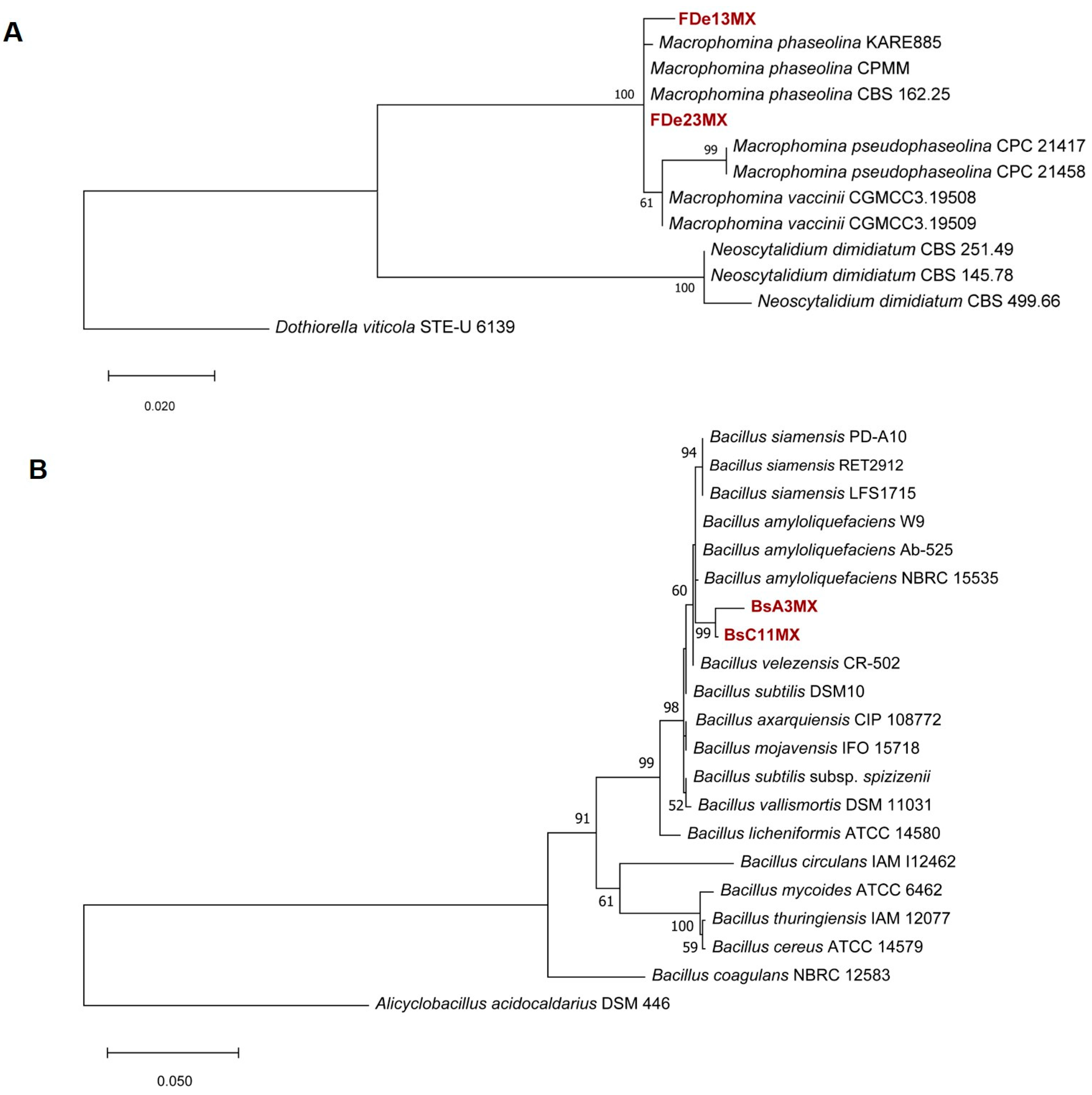
2.2. DNA Extraction and Phylogenetic Analysis

The fungal isolates were grown in potato dextrose broth (PDB) media for 5 days at 30 °C with shaking at 120 revolutions per minute (rpm), and mycelia were recovered using a sterile wood stick. Bacterial strains were grown on trypticase soy broth (TSB) at 30 °C, 120 rpm for 48 h, and cells were recovered by centrifugation. Genomic DNA was extracted using cetyltrimethylammonium bromide (CTAB) as described previously [34], and adjusted to 30 ng/µL. For fungal isolates, primers ITS1 and ITS4 were used to amplify the Internal transcribed spacer (ITS) region of the nuclear ribosomal DNA [35], and EF1-728F and EF1-986R to amplify part of the translation elongation-factor (tef-1α) gene [36]. For bacteria characterization, primers 27F and 1492R [37] were used to amplify the 16Sr RNA gene region. Each PCR reaction was carried out in a total volume of 25 µL, containing 1 µL of genomic DNA (30 ng/µL), 2.5 µL of Taq Buffer 10x, 0.5 µL of dNTP mix (20 mM), 0.625 µL of each primer (10µM), and 0.125 µL of Taq DNA polymerase (GoTaq® DNA polymerase, Promega, Madison, WI, USA, 5 units/µL), and adjusted to the final volume with ultrapure water.
Amplification reactions were carried out in a Bio-Rad T-100 thermal cycler (Bio-Rad, Hercules, CA, USA) set to the following conditions. For ITS region, an initial step of 94 °C for 2 min, followed by 35 cycles of 94 °C for 1 min, 58 °C for 1 min, and 72 °C for 1.5 min; for tef-1α, an initial step of 95 °C for 3 min, followed by 35 cycles of 95 °C for 30 s, 55 °C for 30 s, and 72 °C for 1 min; for 16S rRNA region, an initial step of 95 °C for 2 min, followed by 35 cycles of 95 °C for 30 s, 55 °C for 1 min, and 72 °C for 1.5 min. All programs ended with a final cycle of 72 °C for 10 min. PCR reactions were purified using a GeneJet PCR purification kit (Thermo Scientific, Waltham, MA, USA) and sequenced. The sequences were analyzed using BioEdit Sequence Alignment Editor and deposited in the National Center for Biotechnollogy Information GenBankFor bacterial phylogenetic analysis, a nucleotide Basic Local Alignment Search Tool (BLASTn) was performed against the GenBank 16S Ribosomal RNA sequences database, and those with the highest similarity for each phylogenetic marker were used (Tables S1 and S2) to construct the alignment using ClustalW. In the case of fungal isolates, the analysis was performed using a concatenated sequence alignment of ITS and tef-1α. Maximum Likelihood (ML) and Maximum Parsimony (MP) analyses were performed using Molecular Evolutionary Genetics Analysis (MEGA-X) software with Bootstrap values based on 1000 replicates. Gaps were treated as missing data. The tree was visualized in MX: Tree Explorer [38].

2.3. Characterization of Growth Conditions for Bacterial Isolates

For bacterial isolates, cultural, physiological, and biochemical characteristics were determined as follows. Bacterial colonies were grown on TSA (g·L−1: 15.0 tryptone, 5.0 soytone, 5.0 NaCl, 15.0 agar, pH was adjusted to 7.0) at 30 °C for two days then examined for Gram reaction and endospore formation. Activities for hemolysis, oxidase, catalase, protease, and urease, and the use of citrate and motility, were evaluated as described elsewhere [39]. Acid production from sugars (glucose, dextrose, arabinose, mannitol, xylose, lactose, trehalose, amylose, and methyl-α-D-glucoside) was evaluated following standard procedures [39]. Then, isolates were grown in trypticase soy broth (TSB) (g·L−1: 15.0 tryptone, 5.0 soytone, 5.0 NaCl, pH 7.0) varying salt concentration (0–15% NaCl), incubation temperature (4–55 °C), and pH (4–10).
For biofilm formation, B. amyloliquefaciens isolates were grown in 96-well polystyrene microplates in TSB medium. The plates were incubated at 30 °C and 50 rpm for 48 h. Biofilm formation was evaluated by adding 75 μL of 10% crystal violet to each dish well, incubating at room temperature for 15 min, and then removing the excess dye by rinsing with distilled water. The formation of a violet ring on the well indicated biofilm formation [40].

2.4. Evaluation of Plant Growth-Promoting Activities

Siderophore production was evaluated using the Chrome azurol S (CAS) agar method [41]. Isolates were streaked on the CAS agar plates and incubated at 30 °C for 48 h. A yellow halo around the bacterial colonies indicated a positive result. Siderophore production was also quantified using 1 × 10⁶ CFU·mL−1 of the bacterial isolates inoculated on MM9 medium (g·L−1: 10.0 glucose 5.0 ammonium acetate, 1.5 KH2PO4, 2.5 NaCl, pH 5.8.) at 30 °C and 120 rpm for 48 h. Cultures were filtered using a 0.22 µm-pore syringe filter, and 100 μL of supernatant plus 100 μL of Fe-CAS solution [(7.5 mL 2 mM CAS, 1.5 mL 1 mM FeCl3·6H2O in 10 mM HCl, 21.9 mg Hexadecyltrimethylammonium (HDTMA) and 9.76 g 4-Morpholineethanesulfonic acid, 2-(N-Morpholino)ethanesulfonic acid (MES) in 100 mL distilled water, pH 5.6)] were added in a 96-well microplate, and absorbance was measured at 630 nm in a Thermo ScientificTM multiskan sky microplate spectrophotometer. The percentages of siderophore units produced by each strain were determined using the formula: % siderophore units = ((Ar − As)/Ar) × 100, where Ar is the reference absorbance, and As is the sample absorbance [42].
Indole-3-acetic-acid (IAA) production was evaluated by growing bacteria in TSB medium supplemented with 500 μg·mL−1 tryptophan [43], at 30 °C and 120 rpm for 48 h. Bacterial cells were harvested at 10,000 rpm for 3 min, and 200 μL of the free-cell supernatant was placed in microplates of 96 wells, followed by the addition of 100 μL of Salkovsky reagent (50 mL distilled water, 30 mL H2SO4, 1 mL 0.5 M FeCl3) [44]. Appearance after 25 min of a pink–orange color indicated IAA synthesis. To quantify the production of IAA, bacteria were inoculated at 1 × 10⁶ CFU·mL−1 in TSB medium supplemented with 500 μg·mL−1 tryptophan, and incubated at 30 °C, 110 rpm for 48 h. Supernatants were filtered using a 0.22 µm-pore syringe filter, and 200 μL of supernatant, plus 100 μL of Salkovsky reagent, were added in a 96-well microplate. Absorbance was measured at 540 nm in a Thermo ScientificTM multiskan sky microplate spectrophotometer. The IAA concentrations were calculated according to a calibration curve of IAA (0, 5, 10, 20, 50, 100 mg·mL−1).
The production of hydrogen cyanide (HCN) was analyzed using TSA medium in 96-well microplates. Bacteria were inoculated, and a filter paper soaked in a solution of 0.5% sodium carbonate in 0.5% of Picric acid was placed on top. Microplates were sealed with parafilm and incubated for 48 h at 30 °C. The development of orange–red color in the filter paper indicated HCN production [43].
The production of 1-Aminocyclopropane-1-carboxylate (ACC)-deaminase was evaluated using minimum medium DF [(g·L−1: 4.0 KH2PO4, 6.0 Na2HPO4, 0.2 MgSO4·7H2O, 2.0 glucose, 2.0 gluconic acid, 2.0 citric acid), 1.0 mL trace element solution (mg·100 mL−1: 1.0 FeSO4·7H2O, 10.0 H3BO3, 11.19 MnSO4·H2O, 124.6 ZnSO4·7H2O, 78.22 CuSO4·5H2O, 10.0 MoO3) 15.0 agar, pH was adjusted to 7.2)] supplemented with a 0.5 M ACC solution [45]. DF medium supplemented with (NH4)2SO4 (2 g·L−1) was used as the positive control and DF medium as the negative control [46]. Bacteria were plated on each medium and incubated at 30 °C for 4 days. Uniform growth of the isolates on the DF medium supplemented with ACC was considered a positive result [47].
Nitrogen fixation was evaluated according to Baldani et al. (2014) [48]. Solution A (950 mL: 5 g malic acid, 0.5 g K2HPO4, 0.5 g FeSO4, 0.010 g MnSO4, 0.2 g MgSO4, 0.1 g NaCl, 0.002 g Na2MoO4, 0.02 g CaCl2, 0.002 g bromothymol blue, 1.75 g agar) and Solution B (4 g KOH in 50.0 mL) were sterilized separately and mixed when they reached a temperature between 45 °C and 50 °C. The medium supplemented with NH4Cl (2.5 g·L−1) as a nitrogen source was used as the positive control. Plates containing each medium were inoculated with 10 μL of bacterial culture of 24 h, and incubated at 30 °C for 8 days. A change of color of the medium from yellow to green was considered positive for nitrogen fixation.
Phosphorus solubilization was determined using Pikovskaya medium (1 L: 5 g yeast extract, 10 g glucose, 5 g Ca3(PO4), 0.5 g (NH4)2SO4, 0.2 g KCl, 0.1 g MgSO4, 0.0001 g MnSO4, 0.0001 g FeSO4, 0.01 g of bromocresol purple, 15 g agar; pH 7.2) [49,50]. Plates containing the medium were inoculated with 10 μL of bacterial culture of 24 h, incubated at 30 °C for 72 h. A change of color from purple to yellow was considered a positive result.
Potassium solubilization was determined using a modified Pikovskaya medium (5 g yeast extract, 10 g glucose, 5 g KNO3, 0.5 g (NH4)2SO4, 0.2 g KCl, 0.1 g MgSO4, 0.0001 g MnSO4, 0.0001 g FeSO4, 0.002 g bromocresol green, 15 g agar; pH 7.2) [49,51]. Plates containing the medium were inoculated with 10 μL of bacterial culture of 24 h, and incubated at 30 °C for 72 h. A change from blue to yellow was considered a positive result.
Zinc solubilization was determined using modified Pikovskaya medium (5 g yeast extract, 10 g glucose, 5 g KNO3, 0.5 g (NH4)2SO4, 0.02 g KCl, 0.1 g MgSO4, 0.0001 g MnSO4, 0.0001 g FeSO4, 12 g ZnO, 0.25 g bromothymol blue, 15 g agar; pH 7.0) [49,52,53]. Plates containing the medium were inoculated with 10 μL of bacterial culture of 24 h, and incubated at 30 °C for 48 h. A change in the medium from blue to yellow was considered a positive result.

2.5. Production of Cell Wall-Degrading Enzymes

For chitinase production, bacterial isolates were grown on a basal medium supplemented with colloidal chitin (1 L: 0.3 g MgSO4·7H2O, 3 g (NH4)2SO4, 2 g KH2PO4, 1 g citric acid monohydrate, 200 μL Tween-80, 4.5 g colloidal chitin, 0.15 g bromocresol purple, and 15 g agar; pH 4.7) [54]. Plates were incubated at 30 °C for 48 h. A change in the medium from yellow to purple was considered a positive result.
For cellulase production, bacterial isolates were grown on minimal medium (MM) supplemented with carboxymethyl-cellulose (CMC) (1 L: 1 g glucose, 2.5 g yeast extract, 1% CMC, 15 g agar) [55]. Plates were incubated at 30 °C for 48 h and stained with Congo red dye. The formation of a yellow halo (8 mm or more) against red background was positive for cellulose production.
For pectinase activity, bacterial isolates were grown on pectin agar (1 L: 1 g NaNO3, 1 g KCl, 1 g K2HPO4, 0.5 g MgSO4, 0.5 g yeast extract, 10 g Pectin, 15 g agar; pH 7.0) [56]. Plates were incubated at 30 °C for 48 h. Results were positive when observing clear zones around the colonies after 5 min of adding Lugol’s iodine solution.
For amylase activity, bacterial isolates were grown on starch agar (g·L−1: 10.0 peptone, 5.0 yeast extract, 5.0 NaCl, and 2.0 starch, 15.0 agar; pH 7.0) [57]. Plates were incubated at 30 °C for 48 h and flooded with Lugol’s iodine solution. A positive result was the observation of a clear zone around the colony after 5 min.

2.6. In Vitro Fungal Antagonism

A loop of each bacterial strain was inoculated on PDA, approximately 2 cm from the plate’s edge in a straight line of around 4 cm. Next, one fungal plug of 5 mmø was placed in the center. Control plates were inoculated with the bacterial strains and the pathogen separately. The plates were incubated at 30 °C until the fungal pathogen covered the control plates. Mycelial growth was monitored every 24 h by marking the colony’s edge, and phenotypic characteristics such as pigmentation and microsclerotia formation were recorded. The percentage of inhibition of mycelial growth was calculated as described before [58] using the formula: Inhibition percentage (%) = ((R1 − R2)/R1) × 100, where R1 is the radial growth of M. phaseolina in control plates, and R2 is the radial growth of M. phaseolina in the presence of bacterial isolates.

2.7. Fungal Growth Inhibition by Bacterial Volatile Organic and Diffusible Compounds

The effect of volatile organic compounds (VOCs) on fungal growth was evaluated through the two-sealed-base-plates method. Briefly, each bacterial isolate was spread on a base plate containing PDA and incubated until the formation of a bacterial lawn. Then, the lid was replaced by another PDA plate with an M. phaseolina mycelial plug of 5 mmø at the center. The two plates were sealed with tape and incubated at 30 °C for five days, registering mycelium growth every 24 h. As a control, a non-inoculated PDA plate was used as a cover.
The production of bacterial diffusible compounds was evaluated by growing the strains in TSB media at 30 °C with shaking at 120 rpm for 72 h. Afterward, cultures were centrifuged, and the supernatant was filtered through a 0.22 μm-pore syringe filter. Volumes of 0.1, 0.5, and 1 mL of the resulting cell-free supernatant from each bacterium were spread on PDA plates and left to dry. Then, a mycelial plug of 5 mmø of M. phaseolina was inoculated at the plates’ center. The plates were incubated at 30 °C for 5 days, and mycelial growth observed every 24 h.

2.8. Effect of Bacteria Isolates on the Germination of M. phaseolina Microsclerotia

Microsclerotia of M. phaseolina were collected under a stereoscopic microscope from a 5-day-old culture growing onto cellophane over MM9 agar [59]. The inhibition of microsclerotia germination by the bacteria was evaluated by spreading 0.1 mL of a 24 h culture of each bacterium onto PDA plates. The inhibition of microsclerotia germination by bacterial diffusible compounds was evaluated using cell-free supernatant of the bacterial isolates obtained from a 48 h culture grown in TSB at 30 °C with shaking (120 rpm); subsequently, the cultures were centrifuged, and the supernatant filtered through a 0.22 µm-pore syringe filter, then PDA media was supplemented with 30% of cell-free supernatant of each bacterium. Thirty microsclerotia were inoculated in each treatment by triplicate. Plates were incubated at 30 °C, and the percentage of germinated microsclerotia was evaluated after 24 h under a light microscope (Nikon Eclipse E200, Minato, Japan). Images of microsclerotia were taken with a camera AxioCam HRc from Zeiss (Oberkochen, Germany) and analyzed using AxioVision 4.8.2.

2.9. Biocontrol Assay in Planta

Bacterial antagonistic effect against M. phaseolina was evaluated in planta using cowpea plants (Vigna unguiculata L.). Cowpea seeds were germinated in a tray for three weeks, and then the seedlings were inoculated with the bacterial isolates by immersing the root for 2 h in a bacterial suspension of 1 × 10⁶ CFU and then transferred to 1 L pots. Macrophomina phaseolina was inoculated by mixing the substrate with two grams of rice colonized with the pathogen’s microsclerotia. Ten plants were inoculated for each treatment, and pots were arranged in a completely randomized design and kept in greenhouse conditions. After two months, plants were removed from pots, and the roots were washed with tap water to remove the substrate. Charcoal-rot symptoms were evaluated, and root and stem length and root and dry foliage weight were measured. No external fertilization was provided to the plants during the experiment process.

2.10. Statistical Analysis

All in vitro experiments were carried out in triplicate. The plant experiment was conducted twice using ten replicates per treatment. The assumption of normality was confirmed using Kolmogorov–Smirnov test (p value ≥ 0.05), then a one-way ANOVA followed by a post hoc Fisher LSD analysis, with an α < 0.05 for significance, was performed for each variable using STATISTICA 8.0.

3. Results and Discussion

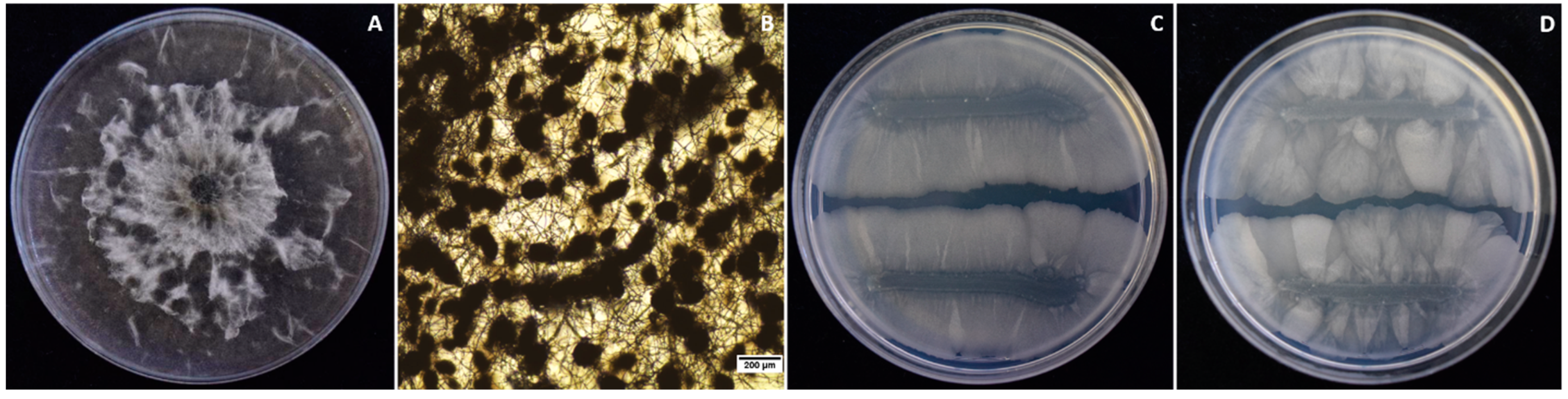
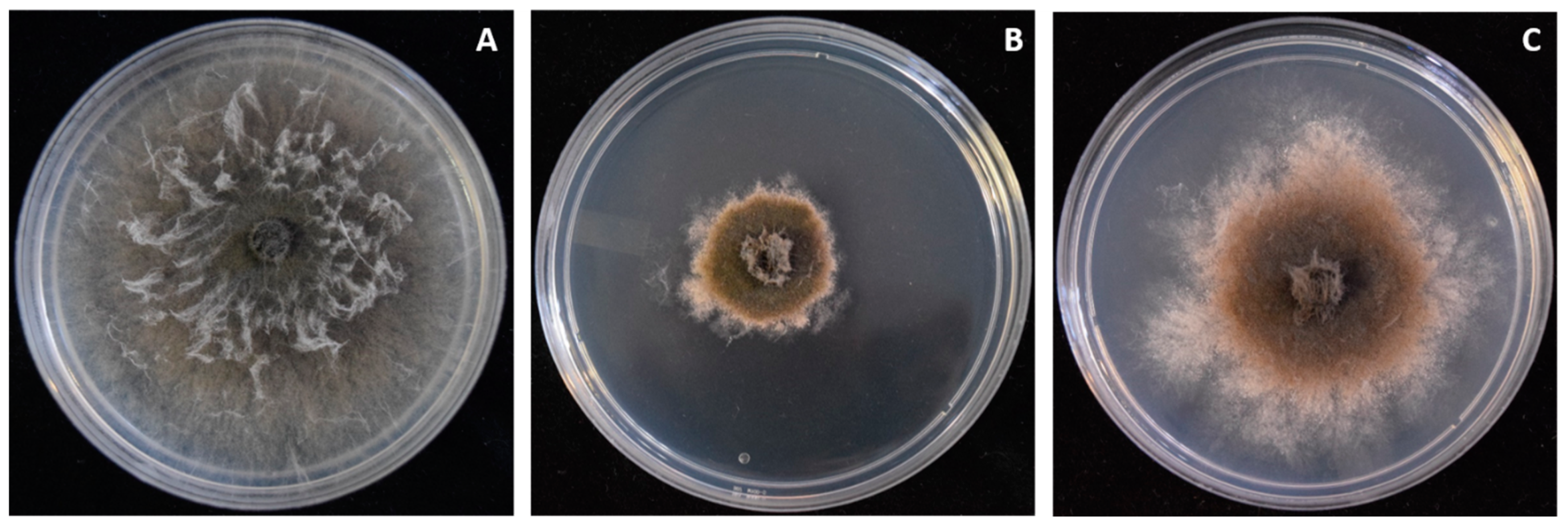
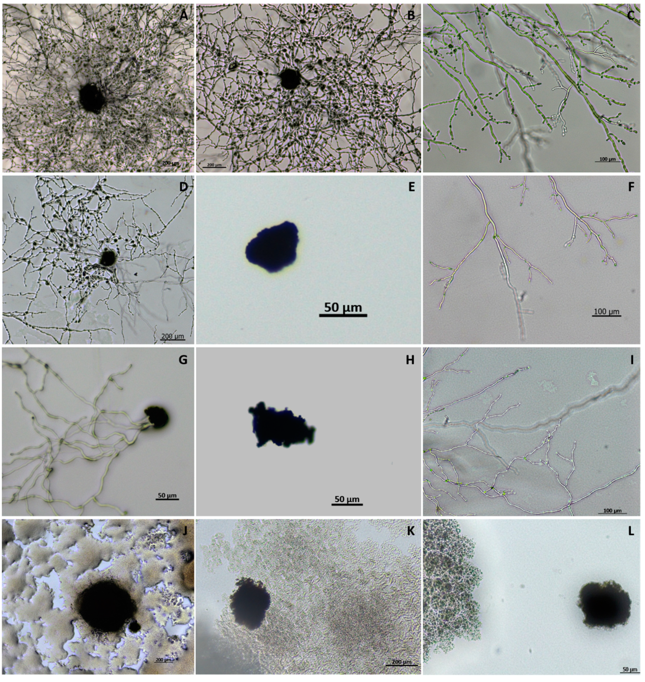
3.1. Isolation and Identification of Macrophomina phaseolina from Strawberry Crown Rot

From twenty samples, fungal isolates with morphological characteristics well-matched with Macrophomina were obtained from the crown of strawberry plants with charcoal-rot symptoms; two of them, named FDe13MX and FDe23MX, were characterized (Figure 1). Isolates were initially gray on PDA plates and became black with microsclerotia production (Figure 1A,B). Production of pycnidia in the fungal colonies was not observed. Fungal isolates were identified based on their ITS region and tef-1α locus; obtained sequences were approximately 522 and 249 bp, respectively. The combined dataset comprised 696 characters, including gaps, after alignment (439 corresponded to ITS gene and 257 to tef-1α gene), and 15 taxa. Dothiorella viticola (STE-U 6139) was used as the outgroup. Maximum likelihood analysis using Kimura’s two-parameter model resulted in a tree with a log-likelihood value of −1440.94. The phylogenetic analysis of the ITS region and tef-1α revealed that both isolates belong to M. phaseolina (Figure 2A).
Macrophomina phaseolina has been reported as a pathogen of strawberry plants in several countries including Israel [6], Spain [60], Argentina [61], Chile [62], and Australia [63]. In California, USA, which neighbors Baja California, Mexico, this fungus has also been reported as the causal agent of dieback and crown rot in strawberries [64,65]. In Mexico, M. phaseolina is an important pathogen of several crops including common bean, sorghum, sugar cane, and maize [27], but this is the first report where it has been identified in strawberries showing charcoal-rot disease. With a production above 200,000 tonnes per year [30], the state of Baja California is the second-largest producer of strawberries in Mexico. The losses of charcoal-rot disease have not been evaluated, but affected plants can be often found in organic commercial fields.

3.2. Bacterial Isolate Identification

From twenty strawberry samples, several bacterial colonies grew on the PDA plates together with M. phaseolina. Two attracted our attention due to the formation of an inhibition halo against M. phaseolina mycelium. Both colonies, found in independent plates, were picked and isolated to purity (Figure 1C,D). The phylogenetic analysis of their 16S ribosomal RNA (approximately 1168 bp) clustered the two bacteria within the B. amyloliquefaciens clade (Figure 2B). Bacillus amyloliquefaciens is part of the Bacillus subtilis complex commonly isolated from soil. It was reported as a novel Bacillus species in 1987 and is recognized for producing α-amylase and protease [66]. This bacterium is closely related to the plant-associated B. siamensis and B. velezensis, forming a monophyletic group [67].

3.3. Characterization of Bacillus amyloliquefaciens Isolates

Bacterial isolates BsA3MX and BsC11 of B. amyloliquefaciens are Gram-positive, motile, and endospore-forming rods. They formed irregular, flat, dry, dull creamy–white, with rough surface colonies on TSA plates, and a thin biofilm at the surface on TSB after 24 h of incubation at 30 °C. Both isolates were halotolerant since they grew in the presence of NaCl up to 10% (w/v), and in a pH range of 5–9. In addition, they were able to grow up to 50 °C (Table 1). The ability of both isolates to survive and even thrive in harsh environmental conditions could be explained by their isolation from a field located in the arid region of Baja California, often exposed to drought and high temperatures and being irrigated with water from wells with high salinity levels. While the ability to form endospores makes them resistant to different types of stress [18], the application of endospore-forming bacterial also has a beneficial effect on plant growth [68].
Tests for hemolytic activity, and urease and citrate utilization, were negative in both isolates, while oxidase and catalase activities and indole use were positive. The preferential utilization of carbohydrates was different for each isolate (Table 1). BsA3MX preferred glucose, trehalose, dextrose glycogen, followed by xylose, mannitol, lactose, arabinose, and amylose, while BsC11MX preferred glucose, arabinose, dextrose, mannitol, and glycogen, followed by lactose, xylose, amylose, and trehalose. The use of methyl-α-D-glucoside was negative for both bacteria isolates.

3.4. Characterization of B. amyloliquefaciens as Plant Growth Promoters

Both Bacillus amyloliquefaciens isolates were positive for siderophore and indole-3-acetic acid (IAA) production, and ACC-deaminase activity, and were able to solubilize phosphate and zinc (Table 2). Siderophores are Fe3+-chelating compounds, and their production by beneficial bacteria facilitates the uptake of iron for plants [69]. The auxin IAA is produced by several microorganisms through L-tryptophan metabolism, and in plants it facilitates cell and tissue differentiation, the production of longer roots, and nutrient uptake [70,71]. Although the strains did not fix nitrogen, produce cyanohydrin acid, or solubilize potassium, the other characteristics make them suitable for their use as plant growth-promoting agents.

3.5. Production of Hydrolytic Enzymes

Strains BsA3MX and BsC11MX were positive for the production of proteases and amylases (Table 2) but negative for chitinase. These results suggest that the antagonistic properties of these isolates must be related to the production of antifungal compounds, either volatile or diffusible, instead of hydrolytic enzyme activity. Importantly, these strains tested negative for the production of pectinases and cellulases, since pectin and cellulose are essential components of the cell wall of plants. Proteases participate in several processes in plants such as development, defense response to phytopathogens, and photosynthesis, while amylase-producing bacteria enable plants to use starch more efficiently [72].

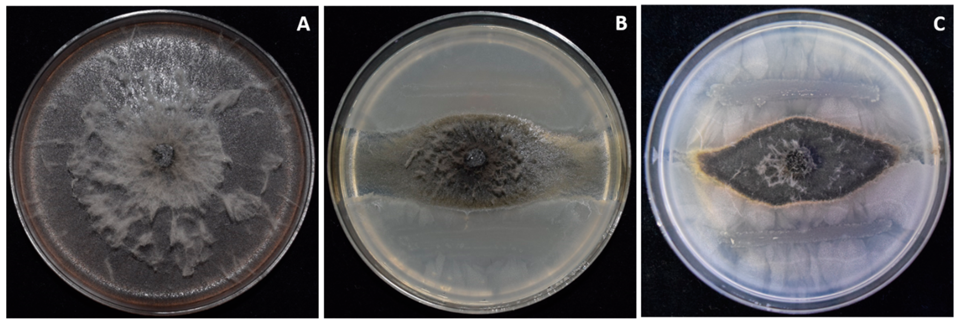
3.6. Two Bacillus amyloliquefaciens Isolates Inhibit Fungal Growth of Macrophomina In Vitro

The two B. amyloliquefaciens strains showed antagonism against M. phaseolina by inhibiting mycelial growth in vitro. Isolate BsA3MX showed 66.8% growth inhibition at 5 days of incubation on PDA, while BsC11MX showed 62.8% (Figure 3).
The antagonistic activity of PGPR against M. phaseolina has been reported before. Isolates from B. subtilis [73,74,75], Bacillus spp. [76], B. altitudinis, Pseudomonas spp., Brevibacterium antiquum, and Acinetobacter tandoii [77], have an inhibitory effect on this pathogen as well. B. amyloliquefaciens isolate PGPBacCA1 showed above 50% inhibitory activity against M. phaseolina [78] and the isolate B14 between 60 and 66% [17]; these results are comparable to those reported in the present study. Bacillus amyloliquefaciens has been studied as a biocontrol agent against other phytopathogenic fungi such as Fusarium [79], Sclerotinia sclerotiorum [80,81], B. cinerea, Colletotrichum orbiculare [81,82], Botrytis pelargonii, and Alternaria alternata [83].

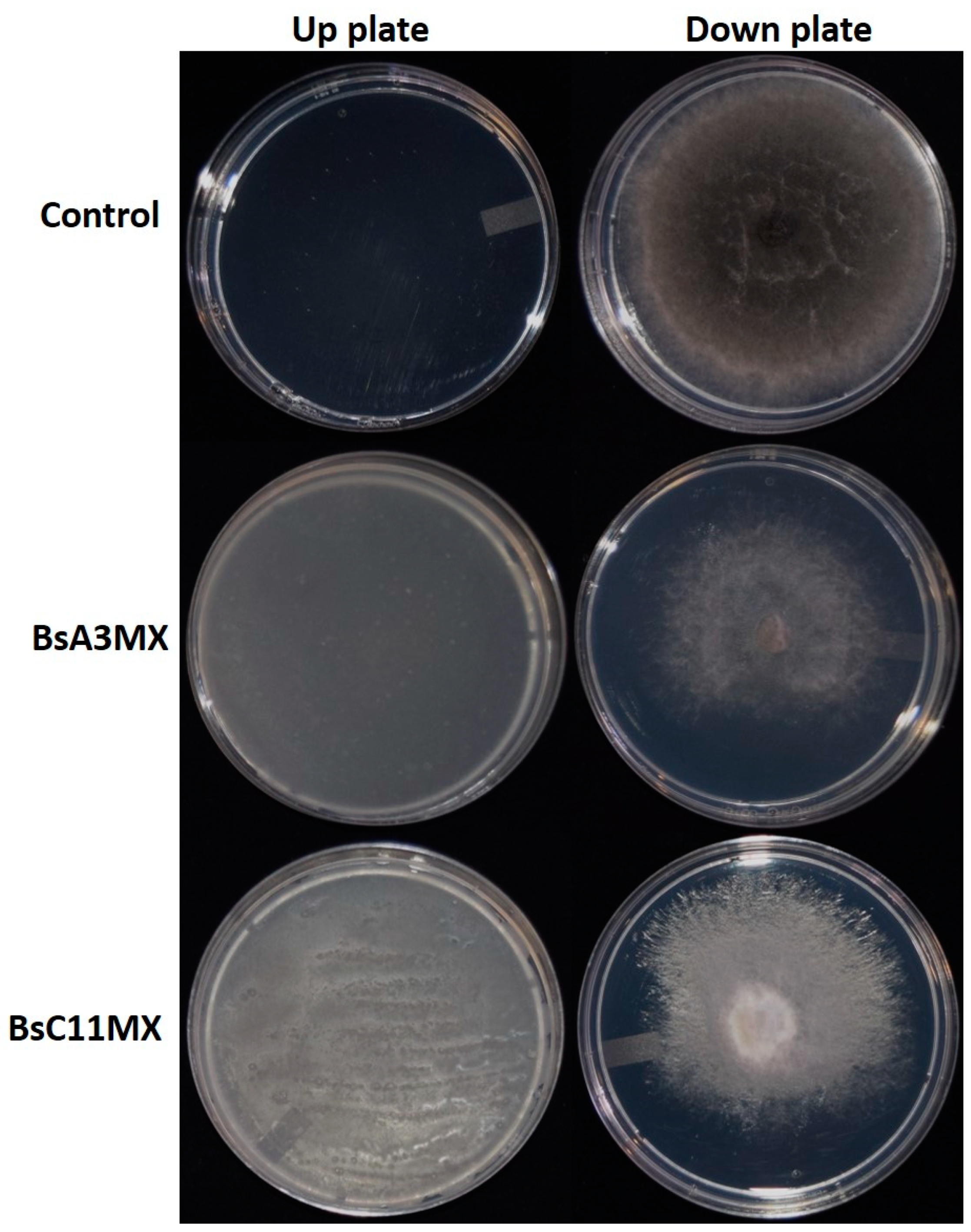
3.7. B. amyloliquefaciens BsA3MX and BsC11MX Inhibit M. phaseolina by the Combined Action of Volatile Organic Compounds and Diffusible Compounds

The effect of volatile compounds and cell-free supernatant on M. phaseolina was evaluated to determine the mechanisms used by B. amyloliquefaciens strains. Volatile compounds from BsA3MX and BsC11MX inhibited both the mycelial growth of M. phaseolina (Figure 4) and the production of microsclerotia in comparison to the controls, where the fungal pathogen covered the entire plate after five days of incubation (Figure 4). On the other hand, the cell-free supernatants of BsA3MX and BsC11MX not only inhibited the growth of M. phaseolina but also induced morphological changes (Figure 5). M. phaseolina grew irregularly in the presence of cell-free supernatant, presenting a brown color in the center with white-cream edges and reduced production of microsclerotia (Figure 5B,C).
Several secondary metabolites have been reported against plant pathogens from different isolates of B. amyloliquefaciens. The production of volatile organic compounds such as benzenes, alkyls, alcohols, ketones, and aldehyde compounds by different B. amyloliquefaciens isolates inhibits the growth of Fusarium oxysporum [84,85], Rhizoctonia solanacearum [86], Botrytis cinerea, Sclerotinia sclerotiorum, Verticillum longisporum [87,88], Monilinia laxa, M. fructicola, and V. longisporum [84,86,87,88,89]. Among the diffusible compounds produced by B. amyloliquefaciens, lipopeptides such as bacillomycin D, macrolactin, iturin A, surfactin, and fengycin have shown high antagonistic activity against M. phaseolina [78], F. oxysporum, R. solanacearum [79], B. cinerea, and Colletotrichum orbiculare [82]. These secondary metabolites have antifungal, antibacterial, and nematocidal activity; therefore, B. amyloliquefaciens is considered a potential biocontrol agent and plant growth promoter [21,90,91].

3.8. Effect of B. amyloliquefaciens on the Germination of M. phaseolina Microsclerotia

Next, the effect of cell-free supernatant on microsclerotia germination was evaluated. The use of 30% cell-free bacterial supernatant affected the development of M. phaseolina microsclerotia. While 100% of microsclerotia inoculated in PDA medium germinated after 24 h of incubation, when exposed to the BsA3MX and BsC11MX cell-free supernatants, only 79% and 71% germinated, respectively. Moreover, significant differences were observed in the germination of microsclerotia treated with B. amyloliquefaciens cell-free extract. The control treatment showed higher mycelium density (Figure 6A,B) and more branched hyphae (Figure 6C). In contrast, the germinated microsclerotia exposed to the cell-free supernatants displayed stunted growth, reduced mycelium density (Figure 6D,G), and less branched hyphae (Figure 6F,I). Additionally, non-germinated microsclerotia were observed in the treatments with both bacteria’s cell-free supernatant (Figure 6E,H). Notably, when bacteria were present in the culture medium, the germination of M. phaseolina microsclerotia was inhibited entirely (Figure 6J–L).
The microsclerotia of M. phaseolina are the primary source of inoculum since they can survive in the soil for up to 15 years [2,11] and attach to the roots’ surface. Then, microsclerotia germinate repeatedly during the crop-growing season, producing a mass of hyphal thread colonizing the root’s intercellular spaces [4,92]; consequently, the intensity of the disease on a crop is related to the amount of viable microsclerotia in soil [93]. Therefore, the inhibition of microsclerotia production and germination is important in searching for control strategies for this pathogen, increasing the value of B. amyloliquefaciens strains isolated here as biological control agents.

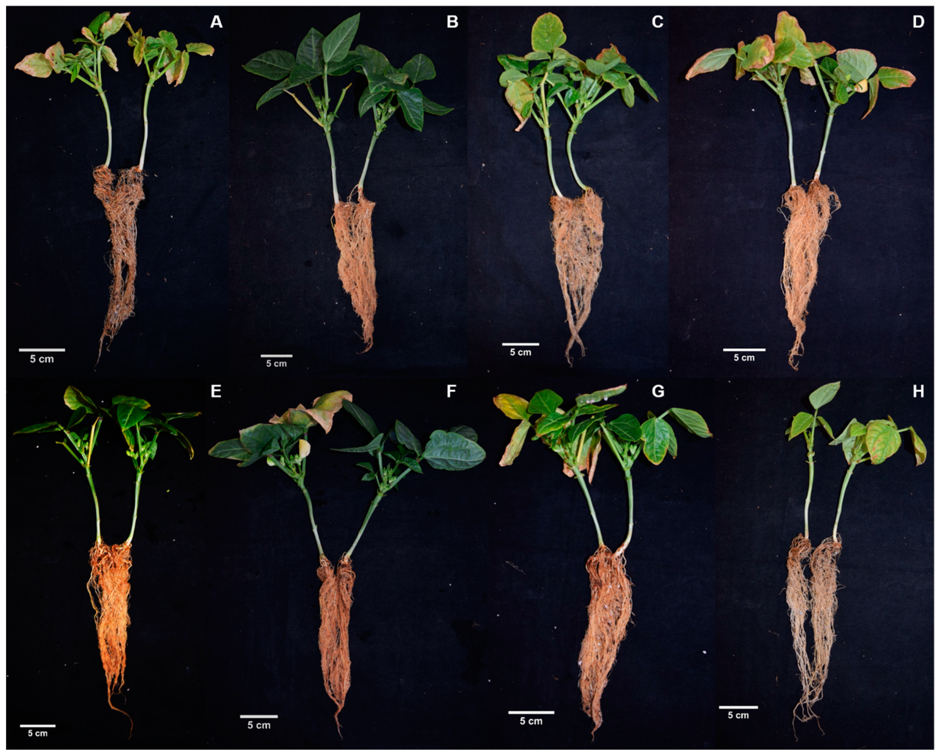
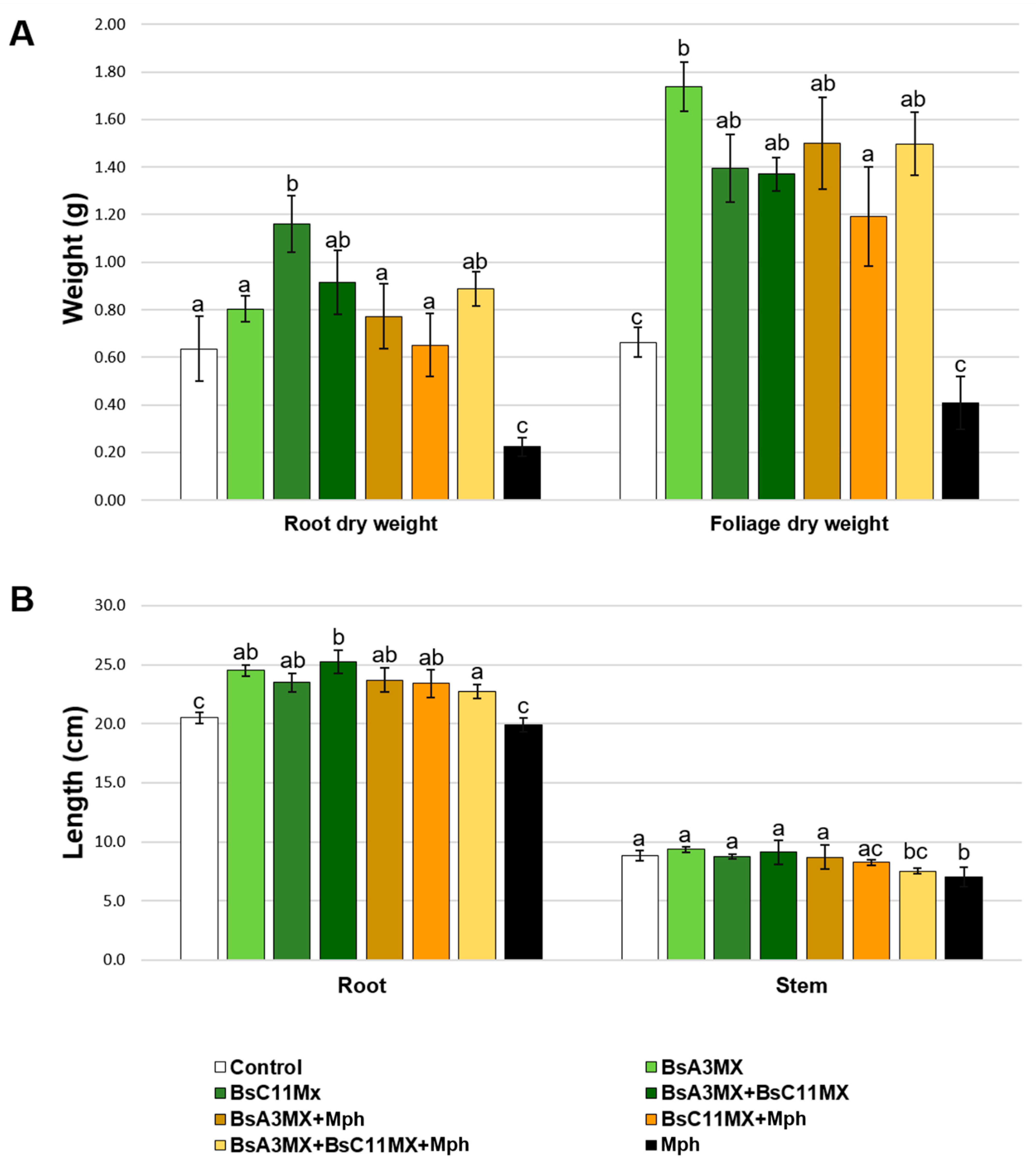
3.9. Bacillus amyloliquefaciens Isolates Promote Plant Growth and Suppress M. phaseolina in Planta

Finally, assays to evaluate the ability of isolates BsA3MX and BsC11MX to reduce the damage on cowpea plants elicited by M. phaseolina were performed. Plants inoculated with M. phaseolina FDe13MX had lower foliage and root production when compared to the negative control (Figure 7A,H), with a decrease of 64.2% and 38.3% in dry weight of roots and foliage, respectively (Figure 8). Thus, this confirmed that the M. phaseolina isolate is pathogenic.
On the contrary, plants treated with bacterial isolates, BsA3MX, BsC11MX, or both, and inoculated with M. phaseolina, presented a 39.8% and 126.2% increase in root and foliage dry weight, respectively, as well as up to a 15.6% increase in root length (Figure 7E–H and Figure 8). Moreover, in the plants inoculated only with M. phaseolina, the presence of microsclerotia and root and collar necrosis was evident. Treatments with bacterial isolates also reduced the incidence of lesions. No lesions were observed in the control plants, as well as in those inoculated only with bacteria.
Bacillus amyloliquefaciens has been reported as a biological agent against M. phaseolina in common beans in Argentina, decreasing the incidence of charcoal root rot on seeds [17].
Bacillus spp. synthesize a wide variety of compounds such as growth hormones and solubilize nutrients that help improve plant growth and are better adapted to crop plants than organisms isolated from other sources [77]. The Bacillus genus is recognized as a potential PGPR for the biocontrol of different phytopathogens due to the production of a wide array of antimicrobials. Besides, the ability to form endospores allows them to withstand adverse environmental conditions [17,20,94].
Results also show that both bacterial isolates have plant growth-promotion activity, since cowpea plants inoculated with BsA3MX, BsC11MX, or with both, but not inoculated with M. phaseolina, showed higher foliage and root production than the un-inoculated control (Figure 7A–D), and an 82.7% and 162.5% increase in root and foliage dry weights, respectively, as well as up to a 23.1% increase in root length (Figure 8). Both bacteria and the fungal strain were reisolated from all the inoculated plants on PDA plates.
The observed increase in plant growth may be due to the production of siderophores, indole-3-acetic acid (IAA), biofilm formation, and the solubilization of phosphate and zinc. The ability to form biofilm in B. amyloliquefaciens has been correlated with plant root colonization and drought-stress tolerance [95]. Baja California is bordered by the Pacific Ocean and the Gulf of California; main crops are tomato, strawberry, cotton, grapes, and wheat. The valley of San Quintin is located to the south of Ensenada along the Pacific Ocean coast, where the climate becomes desert; thus, the vegetation is scarce, and the temperature is very high during the summer. Due to the arid conditions, there is water scarcity. Besides, the region has experienced harmful heat waves in recent years, increasing maximum temperatures up to 50 °C [96]. The low rainfall and high evaporation gradually increase the salinity of the soils, making agriculture in this area very difficult. Previously, other isolates from arid zones with these characteristics have been shown to promote plant growth in different crops and constitute a feasible alternative for the replacement of organic fertilizers using these or similar PGPR [97,98].
Strains of B. amyloliquefaciens BsA3MX and BsC11MX also produce ACC-deaminase, reported to impart drought tolerance [99] due to the up-regulation ACC-deaminase gene (acdS), which cleaves the precursor of ethylene (ACC). The lower ACC levels lead to a decrease in endogenous ethylene content and its effects on plants during stress conditions [100,101,102]. Although stress tolerance was not tested here, the ability to produce ACC-deaminase and to grow in low pH and high salinity indicates that both strains can potentially improve plant growth in water-stress conditions. Agriculture in Baja California is seriously affected by climate change, increasing temperature, and diminishing water availability is common in the region. Since higher temperatures increase the survival and spread of drought pathogens such as M. phaseolina, causing significant losses in production and crop yields [12,103], it is important to continue searching for sustainable alternatives to improve control strategies for these pathogens.

4. Conclusions

In conclusion, isolates BsA3MX and BsC11MX, identified as B. amyloliquefaciens, produce volatile and diffusible compounds that inhibited the mycelial growth of M. phaseolina and decreased the production of microsclerotia. Biocontrol assays in planta showed that BsA3MX and BsC11MX induce plant growth of V. unguiculata L. Moreover, the isolates grow in conditions of low pH and high salinity that are often found in the agricultural regions of Baja California. Overall, these results suggest that these isolates of B. amyloliquefaciens can be used as biological control agents against M. phaseolina in arid zones. Future studies will aim to determine the efficacy of the selected isolates in the field.

Supplementary Materials

The following files are online at https://www.mdpi.com/article/10.3390/agronomy12030676/s1, Table S1. List of GenBank and culture accession numbers of Macrophomina used in this study for phylogenetic analysis, Table S2. List of GenBank and culture accession numbers of Bacillus used in this study for phylogenetic analysis.

Author Contributions

E.A.R.-M.: investigation, methodology, formal analysis, writing—original draft, writing—review and editing. C.S.D.-R.: investigation, methodology, writing—review and editing. E.S.: formal analysis, investigation writing—review and editing. R.H.-M.: conceptualization, supervision, methodology, writing—review and editing. All authors have read and agreed to the published version of the manuscript.

Funding

This research received no external funding.

Data Availability Statement

Not applicable.

Acknowledgments

EARM was supported during the doctoral program (Programa de Posgrado en Ciencias de la Vida, CICESE) by scholarship 339210 from Consejo Nacional de Ciencia y Tecnología (CONACYT-México).

Conflicts of Interest

The authors declare no conflict of interest.

References

  1. Dhingra, O.D.; Sinclair, J.B. Biology and Pathology of Macrophomina phaseolina; Univeridade Feederal de Vicosa: Viçosa, Brazil, 1978; p. 166. [Google Scholar]
  2. Marquez, N.; Giachero, M.L.; Declerck, S.; Ducasse, D.A. Macrophomina phaseolina: General Characteristics of Pathogenicity and Methods of Control. Front. Plant Sci. 2021, 12, 634397. [Google Scholar] [CrossRef] [PubMed]
  3. Su, G.; Suh, S.-O.; Schneider, R.W.; Russin, J.S. Host Specialization in the Charcoal Rot Fungus, Macrophomina phaseolina. Phytopathology 2001, 91, 120–126. [Google Scholar] [CrossRef] [PubMed] [Green Version]
  4. Gupta, G.K.; Sharma, S.K.; Ramteke, R. Biology, Epidemiology and Management of the Pathogenic Fungus Macrophomina phaseolina (Tassi) Goid with Special Reference to Charcoal Rot of Soybean (Glycine max (L.) Merrill). J. Phytopathol. 2012, 160, 167–180. [Google Scholar] [CrossRef]
  5. Kunwar, I.K.; Singh, T.; Machado, C.C.; Sinclair, J.B. Histopathology of soybean seed and seedling infection by Macrophomina phaseolina. Phytopathology 1986, 76, 532–535. [Google Scholar] [CrossRef]
  6. Zveibil, A.; Freeman, S. First Report of Crown and Root Rot in Strawberry Caused by Macrophomina phaseolina in Israel. Plant Dis. 2005, 89, 1014. [Google Scholar] [CrossRef]
  7. Islam, S.; Haque, S.; Islam, M.M.; Emdad, E.M.; Halim, A.; Hossen, Q.M.M.; Hossain, Z.; Ahmed, B.; Rahim, S.; Rahman, S.; et al. Tools to kill: Genome of one of the most destructive plant pathogenic fungi Macrophomina phaseolina. BMC Genom. 2012, 13, 493. [Google Scholar] [CrossRef] [Green Version]
  8. Ijaz, S.; Sadaqat, H.A.; Khan, M.N. A review of the impact of charcoal rot (Macrophomina phaseolina) on sunflower. J. Agric. Sci. 2013, 151, 222–227. [Google Scholar] [CrossRef]
  9. Salahlou, R.; Safaie, N.; Shams-Bakhsh, M. Genetic diversity of Macrophomina phaseolina populations, the causal agent of sesame charcoal rot using inter-simple sequence repeat markers. J. Agric. Sci. 2016, 18, 277–287. [Google Scholar]
  10. Vibha. Macrophomina phaseolina: The Most Destructive Soybean Fungal Pathogen of Global Concern. In Current Trends in Plant Disease Diagnostics and Management Practices; Kumar, P., Gupta, V., Tiwari, A., Kamle, M., Eds.; Fungal Biology Series; Springer: Cham, Switzerland, 2016; pp. 193–205. [Google Scholar]
  11. Meyer, W.A.; Sinclair, J.B.; Khare, M.N. Factors affecting charcoal rot of soybean seedlings. Phytopathology 1974, 64, 845–849. [Google Scholar] [CrossRef]
  12. Pandey, A.K.; Basandrai, A.K. Will Macrophomina phaseolina spread in legumes due to climate change? A critical review of current knowledge. J. Plant Dis. Prot. 2021, 128, 9–18. [Google Scholar] [CrossRef]
  13. Kanaan, H.; Medina, S.; Krassnovsky, A.; Raviv, M. Survival of Macrophomina phaseolina s.l. and Verticillium dahliae during solarization as affected by composts of various maturities. Crop Prot. 2015, 76, 108–113. [Google Scholar] [CrossRef]
  14. Lokesh, R.; Rakholiya, K.B.; Thesiya, M.R. Evaluation of Different Fungicides against Macrophomina phaseolina (Tassi) Goid. Causing Dry Root Rot of Chickpea (Cicer arietinum L.) invitro. Int. J. Curr. Microbiol. Appl. Sci. 2020, 9, 901–911. [Google Scholar] [CrossRef]
  15. Almeida, Á.M.; Sosa-Gomez, D.R.; Binneck, E.; Marin, S.R.; Zucchi, M.I.; Abdelnoor, R.V.; Souto, E.R. Effect of crop rotation on specialization and genetic diversity of Macrophomina phaseolina. Trop. Plant Pathol. 2008, 33, 257–264. [Google Scholar] [CrossRef]
  16. Adrees, H.; Haider, M.S.; Anjum, T.; Akram, W. Inducing systemic resistance in cotton plants against charcoal root rot pathogen using indigenous rhizospheric bacterial strains and chemical elicitors. Crop Prot. 2019, 115, 75–83. [Google Scholar] [CrossRef]
  17. Sabaté, D.C.; Brandan, C.P.; Petroselli, G.; Erra-Balsells, R.; Audisio, M.C. Decrease in the incidence of charcoal root rot in common bean (Phaseolus vulgaris L.) by Bacillus amyloliquefaciens B14, a strain with PGPR properties. Biol. Control 2017, 113, 1–8. [Google Scholar] [CrossRef]
  18. Villarreal-Delgado, M.F.; Villa-Rodríguez, E.D.; Cira-Chávez, L.A.; Estrada-Alvarado, M.I.; Parra-Cota, F.I.; de los Santos-Villalobos, S. The genus Bacillus as a biological control agent and its implications in the agricultural biosecurity. Mex. J. Phytopathol. 2018, 36, 95–130. [Google Scholar] [CrossRef]
  19. Maughan, H.; Van der Auwera, G. Bacillus taxonomy in the genomic era finds phenotypes to be essential though often misleading. Infect. Genet. Evol. 2011, 11, 789–797. [Google Scholar] [CrossRef]
  20. Fira, D.; Dimkić, I.; Berić, T.; Lozo, J.; Stanković, S. Biological control of plant pathogens by Bacillus species. J. Biotechnol. 2018, 285, 44–55. [Google Scholar] [CrossRef]
  21. Khan, M.; Salman, M.; Jan, S.A.; Shinwari, Z.K. Biological control of fungal phytopathogens: A comprehensive review based on Bacillus species. MOJ Biol. Med. 2021, 6, 90–92. [Google Scholar] [CrossRef]
  22. Miljaković, D.; Marinković, J.; Balešević-Tubić, S. The Significance of Bacillus spp. in Disease Suppression and Growth Promotion of Field and Vegetable Crops. Microorganisms 2020, 8, 1037. [Google Scholar] [CrossRef]
  23. Martínez-Salgado, S.J.; Andrade-Hoyos, P.; Parraguirre Lezama, C.; Rivera-Tapia, A.; Luna-Cruz, A.; Romero-Arenas, O. Biological control of charcoal rot in peanut crop through strains of Trichoderma spp., in Puebla, Mexico. Plants 2021, 10, 2630. [Google Scholar] [CrossRef] [PubMed]
  24. Khambhati, V.H.; Abbas, H.K.; Sulyok, M.; Tomaso-Peterson, M.; Shier, W.T. First report of the production of mycotoxins and other secondary metabolites by Macrophomina phaseolina (Tassi) Goid. Isolates from soybeans (Glycine max L.) symptomatic with charcoal rot disease. J. Fungi 2020, 6, 332. [Google Scholar] [CrossRef] [PubMed]
  25. Luna, M.P.R.; Mueller, D.; Mengistu, A.; Singh, A.K.; Hartman, G.L.; Wise, K.A. Advancing Our Understanding of Charcoal Rot in Soybeans. J. Integr. Pest Manag. 2017, 8, 8. [Google Scholar] [CrossRef] [Green Version]
  26. Reyes-Franco, M.C.; Hernández-Delgado, S.; Beas-Fernández, R.; Medina-Fernández, M.; Simpson, J.; Mayek-Pérez, N. Pathogenic and genetic variability within Macrophomina phaseolina from Mexico and other countries. J. Phytopathol. 2006, 154, 447–453. [Google Scholar] [CrossRef]
  27. Leyva-Mir, S.G.; Velázquez-Martínez, G.C.; Tlapal-Bolaños, B.; Tovar-Pedraza, J.M.; Rosas-Saito, G.H.; Alvarado-Gómez, O.G. Caracterización morfológica y molecular de aislados de Macrophomina phaseolina asociados a caña de azúcar en México. Rev. Argent. Microbiol. 2015, 47, 143–147. [Google Scholar] [CrossRef] [Green Version]
  28. Ayala-Armenta, Q.A.; Tovar-Pedraza, J.M.; Apodaca-Sánchez, M.A.; Correia, K.C.; Sauceda-Acosta, C.P.; Camacho-Tapia, M.; Beltrán-Peña, H. Phylogeny and pathogenicity of soilborne fungi associated with wilt disease complex of tomatillo (Physalis ixocarpa) in northern Sinaloa, Mexico. Eur. J. Plant Pathol. 2020, 157, 733–749. [Google Scholar] [CrossRef]
  29. Martínez-Salgado, S.J.; Romero-Arenas, O.; Morales-Mora, L.A.; Luna-Cruz, A.; Rivera-Tapia, J.A.; Silva-Rojas, H.V.V.; Andrade-Hoyos, P. First Report of Macrophomina phaseolina Causing Charcoal Rot of Peanut (Arachis hypogaea) in Mexico. Plant Dis. 2021, 105, 2722. [Google Scholar] [CrossRef]
  30. SIAP. Servicio de Información y Estadística Agroalimentaria y Pesquera; Ministerio de Agricultura de Mexico, Secretaría de Agricultura, Ganadería, Desarrollo Rural, Pesca y Alimentación (SAGARPA): Mexico City, Mexico, 2020. [Google Scholar]
  31. Williams-Alanís, H.; Pecina-Quintero, V.; Montes-García, N.; Zavala-García, F.; Arcos-Cavazos, G.; Gámez-Vázquez, A.J. Evaluation of sorghum [Sorghum bicolor (L.) Moench.] hybrids for resistance to charcoal rot [Macrophomina phaseolina (Tassi) Goid.] in Tamaulipas, Mexico. Revista Mex. Fitopatol. 2009, 27, 69–72. [Google Scholar]
  32. Mayek-Pérez, N.; López-Salinas, E.; Cumpián-Gutiérrez, J.; Acosta-Gallegos, J.A. Inheritance of field resistance to Macrophomina phaseolina (Tassi) Goid. in recombinant endogamic lines of common bean (Phaseolus vulgaris L.). Revista Mex. Fitopatol. 2009, 27, 1–10. [Google Scholar]
  33. Pickel, B.; Dai, N.; Maymon, M.; Elazar, M.; Tanami, Z.; Frenkel, O.; Toamy, M.A.; Mor, N.; Freeman, S. Development of a reliable screening technique for determining tolerance to Macrophomina phaseolina in strawberry. Eur. J. Plant Pathol. 2020, 157, 707–718. [Google Scholar] [CrossRef]
  34. Lee, S.B.; Milgroom, M.G.; Taylor, J.W. A rapid, high yield mini-prep method for isolation of total genomic DNA from fungi. Fungal Genet. Rep. 1988, 35, 11. [Google Scholar] [CrossRef] [Green Version]
  35. White, T.J.; Bruns, T.; Lee, S.; Taylor, J.W. Amplification and direct sequencing of fungal ribosomal RNA genes for phylogenetics. In PCR Protocols: A Guide to Methods and Applications; Innis, M.A., Gelfand, D.H., Sninsky, J.J., White, T.J., Eds.; Academic Press: New York, NY, USA, 1990; Volume 18, pp. 315–322. [Google Scholar]
  36. Carbone, I.; Kohn, L. A method for designing primer sets for speciation studies in filamentous Ascomycetes. Mycologia 1999, 91, 553–556. [Google Scholar] [CrossRef]
  37. Lane, D.J. 16S/23S rRNA sequencing. In Nucleic Acid Techniques in Bacterial Systematics; Stackebrandt, E., Goodfellow, M., Eds.; Wiley: New York, NY, USA, 1991; pp. 115–175. [Google Scholar]
  38. Kumar, S.; Stecher, G.; Li, M.; Knyaz, C.; Tamura, K. MEGA X: Molecular Evolutionary Genetics Analysis across Computing Platforms. Mol. Biol. Evol. 2018, 35, 1547–1549. [Google Scholar] [CrossRef]
  39. Cowan, S.T. Cowan and Steel’s Manual for the Identification of Medical Bacteria; Cambridge University Press: Cambridge, UK, 2004; p. 352. [Google Scholar]
  40. O’Toole, G.A.; Pratt, L.A.; Watnick, P.I.; Newman, D.K.; Weaver, V.B.; Kolter, R. [6] Genetic approaches to study of biofilms. Methods Enzymol. 1999, 310, 91–109. [Google Scholar] [CrossRef] [PubMed]
  41. Schwyn, B.; Neilands, J. Universal chemical assay for the detection and determination of siderophores. Anal. Biochem. 1987, 160, 47–56. [Google Scholar] [CrossRef]
  42. Sayyed, R.Z.; Badgujar, M.D.; Sonawane, H.M.; Mhaske, M.M.; Chincholkar, S.B. Production of microbial iron chelators (siderophores) by fluorescent Pseudomonas. Indian J. Biotechnol. 2005, 4, 484–490. [Google Scholar]
  43. Ahmad, F.; Ahmad, I.; Khan, M.S. Screening of free-living rhizosferic bacteria for their multiple plant growth promoting activities. Microbiol. Res. 2008, 163, 173–181. [Google Scholar] [CrossRef]
  44. Gordon, S.A.; Weber, R.P. Colorimetric Estimation of Indoleacetic Acid. Plant Physiol. 1951, 26, 192–195. [Google Scholar] [CrossRef] [Green Version]
  45. Dworkin, M.; Foster, J.W. Experiments with Some Microorganisms which Utilize Ethane and Hydrogen. J. Bacteriol. 1958, 75, 592–603. [Google Scholar] [CrossRef] [Green Version]
  46. Penrose, D.M.; Glick, B.R. Methods for isolating and characterizing ACC deaminase-containing plant growth-promoting rhizobacteria. Physiol. Plant. 2003, 118, 10–15. [Google Scholar] [CrossRef] [Green Version]
  47. Govindasamy, V.; Senthilkumar, M.; Mageshwaran, V.; Annapurna, K. Detection and Characterization of ACC Deaminase in Plant Growth Promoting Rhizobacteria. J. Plant Biochem. Biotechnol. 2009, 18, 71–76. [Google Scholar] [CrossRef]
  48. Baldani, J.I.; Reis, V.M.; Videira, S.S.; Boddey, L.H.; Baldani, V.L.D. The art of isolating nitrogen-fixing bacteria from non-leguminous plants using N-free semi-solid media: A practical guide for microbiologists. Plant Soil 2014, 384, 413–431. [Google Scholar] [CrossRef]
  49. Pikovskaya, R.I. Mobilization of phosphorus in soil in connection with the vital activity of some microbial species. Mikrobiologiya 1948, 17, 362–370. [Google Scholar]
  50. Corrales Ramírez, L.C.; Sánchez Leal, L.C.; Arevalo Galvez, Z.Y.; Moreno Burbano, V.E. Bacillus: Género bacteriano que demuestra ser un importante solubilizador de fosfato. Nova 2014, 12, 165–178. [Google Scholar] [CrossRef] [Green Version]
  51. Franco-Correa, M. Evaluación de Caracteres PGPR en Actinomicetos e Interacciones de Estas Rizobacterias con Hongos Formadores de Micorriza. Ph.D. Thesis, Facultad de Ciencias, Universidad de Granada,, Granada, Spain, 2008. [Google Scholar]
  52. Komal, R.; Kalavati, P. Isolation and screening of zinc mobilizing microorganisms from rhizospheric soil. Paripex Indian J. Res. 2018, 7, 58–60. [Google Scholar] [CrossRef]
  53. Tagele, S.B.; Kim, S.W.; Lee, H.G.; Lee, Y.S. Potential of novel sequence type of Burkoldera cenocepacia for biological control of root rot maize (Zea mays L.) cause by Fusarium temperatum. Int. J. Mol. Sci. 2019, 20, 1005. [Google Scholar] [CrossRef] [Green Version]
  54. Agrawal, T.; Kotashane, S.A. Chitinolytic assay of indigenous Thrichoderma isolates collected from diferente geographical locations of Chhattisgarh in Central India. SpringerPlus 2012, 1, 1–10. [Google Scholar] [CrossRef] [Green Version]
  55. Mazzucotelli, C.A.; Ponce, A.G.; Kotlar, C.E.; Moreira, M.D.R. Isolation and characterization of bacterial strains with a hydrolytic profile with potential use in bioconversion of agroindustial by-products and waste. Food Sci. Technol. 2013, 33, 295–303. [Google Scholar] [CrossRef] [Green Version]
  56. Kumar, A.; Sharma, R. Production of alkaline pectinase by bacteria (Cocci sps.) isolated from decomposing fruit materials. J. Phytol. 2012, 4, 1–5. [Google Scholar]
  57. Hankin, L.; Anagnostakis, S.L. The use of solid media for detection of enzyme production by fungi. Mycologia 1975, 67, 597–607. [Google Scholar] [CrossRef]
  58. Fokkema, N.J. Antagonism between fungal saprophytes and pathogens on aerial plant surfaces. In Microbiology of Aerial Plant Surfaces; Dickinson, C.H., Preece, T.F., Eds.; Academic Press: London, UK, 1976; pp. 487–505. [Google Scholar]
  59. Ammon, V.; Wyllie, T.D.; Brown, M.F. Investigation of the infection process of Macrophomina phaseolina on the surface of soybean roots using scanning electron microscopy. Mycopathologia 1975, 55, 77–81. [Google Scholar] [CrossRef]
  60. Aviles, M.; Castillo, S.; Bascon, J.; Zea-Bonilla, T.; Martin-Sanchez, P.M.; Pérez-Jiménez, R.M. First report of Macrophomina phaseolina causing crown and root rot of strawberry in Spain. Plant Pathol. 2008, 57, 382. [Google Scholar] [CrossRef]
  61. Baino, O.M.; Salazar, S.M.; Ramallo, A.C.; Kirschbaum, D.S. First Report of Macrophomina phaseolina Causing Strawberry Crown and Root Rot in Northwestern Argentina. Plant Dis. 2011, 95, 1477. [Google Scholar] [CrossRef] [PubMed]
  62. Sánchez, S.; Gambardella, M.; Henríquez, J.L.; Diaz, I. First Report of Crown Rot of Strawberry Caused by Macrophomina phaseolina in Chile. Plant Dis. 2013, 97, 996. [Google Scholar] [CrossRef]
  63. Hutton, D.G.; Gomez, A.O.; Mattner, S.W. Macrophomina phaseolina and its association with strawberry crown rot in Australia. Int. J. Fruit Sci. 2013, 13, 149–155. [Google Scholar] [CrossRef]
  64. Koike, S.T. Crown Rot of Strawberry Caused by Macrophomina phaseolina in California. Plant Dis. 2008, 92, 1253. [Google Scholar] [CrossRef]
  65. Koike, S.T.; Arias, R.S.; Hogan, C.S.; Martin, F.N.; Gordon, T.R. Status of Macrophomina phaseolina on strawberry in California and preliminary characterization of the pathogen. Int. J. Fruit Sci. 2016, 16 (Suppl. 1), 148–159. [Google Scholar] [CrossRef]
  66. Priest, F.G.; Goodfellow, M.; Shute, L.A.; Berkeley, R.C.W. Bacillus amyloliquefaciens sp. nov., nom. rev. Int. J. Syst. Evol. Microbiol. 1987, 37, 69–71. [Google Scholar] [CrossRef]
  67. Fan, B.; Blom, J.; Klenk, H.P.; Borriss, R. Bacillus amyloliquefaciens, Bacillus velezensis, and Bacillus siamensis form an “operational group B. amyloliquefaciens” within the B. subtilis species complex. Front. Microbiol. 2017, 8, 22. [Google Scholar] [CrossRef] [Green Version]
  68. Chavera, G.F.S.; Macuer, M.A.; Torres, P.M. Endospore-Forming Bacteria Present in a Commercial Stabilized Poultry Manure Determines the Fusarium Biocontrol and the Tomato Growth Promotion. Agronomy 2020, 10, 1636. [Google Scholar] [CrossRef]
  69. Ferreira, M.J.; Silva, H.; Cunha, A. Siderophore-Producing Rhizobacteria as a Promising Tool for Empowering Plants to Cope with Iron Limitation in Saline Soils: A Review. Pedosphere 2019, 29, 409–420. [Google Scholar] [CrossRef]
  70. Backer, R.; Rokem, J.S.; Ilangumaran, G.; Lamont, J.; Praslickova, D.; Ricci, E.; Subramanian, S.; Smith, D.L. Plant Growth-Promoting Rhizobacteria: Context, Mechanisms of Action, and Roadmap to Commercialization of Biostimulants for Sustainable Agriculture. Front. Plant Sci. 2018, 9, 1473. [Google Scholar] [CrossRef] [PubMed] [Green Version]
  71. Bhutani, N.; Maheshwari, R.; Negi, M.; Suneja, P. Optimization of IAA production by endophytic Bacillus spp. from Vigna radiate for their potential use as plant growth promoters. Isr. J. Plant Sci. 2018, 65, 83–96. [Google Scholar] [CrossRef]
  72. Yasmin, H.; Naz, R.; Nosheen, A.; Hassan, M.; Ilyas, N.; Sajjad, M.; Anjum, S.; Gao, X.; Geng, Z. Identification of New Biocontrol Agent against Charcoal Rot Disease Caused by Macrophomina phaseolina in Soybean (Glycine Max L.). Sustainability 2020, 12, 6856. [Google Scholar] [CrossRef]
  73. Singh, N.; Pandey, P.; Dubey, R.; Maheshwari, D.K. Biological control of root rot fungus Macrophomina phaseolina and growth enhancement of Pinus roxburghii (Sarg.) by rhizosphere competent Bacillus subtilis BN1. World J. Microbiol. Biotechnol. 2008, 24, 1669–1679. [Google Scholar] [CrossRef]
  74. Afouda, L.; Schulz, D.; Wolf, G.; Wydra, K. Biological control of Macrophomina phaseolina on cowpea (Vigna unguiculata) under dry conditions by bacterial antagonists. Int. J. Biol. Chem. Sci. 2012, 6, 5068–5077. [Google Scholar] [CrossRef]
  75. Hashem, A.; Abd_Allah, E.F.; Alqarawi, A.A.; Radhakrishnan, R.; Kumar, A. Plant defense approach of Bacillus subtilis (BERA 71) against Macrophomina phaseolina (Tassi) Goid in mung bean. J. Plant Interact. 2017, 12, 390–401. [Google Scholar] [CrossRef] [Green Version]
  76. Bojórquez-Armenta, Y.d.J.; Mora-Romero, G.A.; López-Meyer, M.; Maldonado-Mendoza, I.E.; Castro-Martínez, C.; Romero-Urías, C.D.L.A.; Cordero-Ramírez, J.D.; Martínez-Álvarez, J.C. Evaluation of Bacillus spp. isolates as potential biocontrol agents against charcoal rot caused by Macrophomina phaseolina on common bean. J. Gen. Plant Pathol. 2021, 87, 377–386. [Google Scholar] [CrossRef]
  77. Gopalakrishnan, S.; Humayun, P.; Kiran, B.K.; Kannan, I.G.K.; Vidya, M.S.; Deepthi, K.; Rupela, O. Evaluation of bacteria isolated from rice rhizosphere for biological control of charcoal rot of sorghum caused by Macrophomina phaseolina (Tassi) Goid. World J. Microbiol. Biotechnol. 2011, 27, 1313–1321. [Google Scholar] [CrossRef] [Green Version]
  78. Torres, M.; Brandan, C.P.; Petroselli, G.; Erra-Balsells, R.; Audisio, M. Antagonistic effects of Bacillus subtilis subsp. subtilis and B. amyloliquefaciens against Macrophomina phaseolina: SEM study of fungal changes and UV-MALDI-TOF MS analysis of their bioactive compounds. Microbiol. Res. 2016, 182, 31–39. [Google Scholar] [CrossRef]
  79. Yuan, J.; Li, B.; Zhang, N.; Waseem, R.; Shen, Q.; Huang, Q. Production of bacillomycin-and macrolactin-type antibiotics by Bacillus amyloliquefaciens NJN-6 for suppressing soil-borne plant pathogens. J. Agric. Food Chem. 2012, 60, 2976–2981. [Google Scholar] [CrossRef] [PubMed]
  80. Abdullah, M.; Ali, N.Y.; Suleman, P. Biological control of Sclerotinia sclerotiorum (Lib.) de Bary with Trichoderma harzianum and Bacillus amyloliquefaciens. Crop Prot. 2008, 27, 1354–1359. [Google Scholar] [CrossRef]
  81. Ji, S.H.; Paul, N.C.; Deng, J.X.; Kim, Y.S.; Yun, B.-S.; Yu, S.H. Biocontrol Activity of Bacillus amyloliquefaciens CNU114001 against Fungal Plant Diseases. Mycobiology 2013, 41, 234–242. [Google Scholar] [CrossRef] [PubMed] [Green Version]
  82. Arrebola, E.; Jacobs, R.; Korsten, L. Iturin A is the principal inhibitor in the biocontrol activity of Bacillus amyloliquefaciensPPCB004 against postharvest fungal pathogens. J. Appl. Microbiol. 2010, 108, 386–395. [Google Scholar] [CrossRef]
  83. Kazerooni, E.A.; Maharachchikumbura, S.S.; Al-Sadi, A.M.; Kang, S.M.; Yun, B.W.; Lee, I.J. Biocontrol potential of Bacillus amyloliquefaciens against Botrytis pelargonii and Alternaria alternata on Capsicum annuum. J. Fungi 2021, 7, 472. [Google Scholar] [CrossRef]
  84. Yuan, J.; Raza, W.; Shen, Q.; Huang, Q. Antifungal Activity of Bacillus amyloliquefaciens NJN-6 Volatile Compounds against Fusarium oxysporum f. sp. cubense. Appl. Environ. Microbiol. 2012, 78, 5942–5944. [Google Scholar] [CrossRef] [Green Version]
  85. Wu, Y.; Zhou, J.; Li, C.; Ma, Y. Antifungal and plant growth promotion activity of volatile organic compounds produced by Bacillus amyloliquefaciens. MicrobiologyOpen 2019, 8, e00813. [Google Scholar] [CrossRef] [Green Version]
  86. Raza, W.; Ling, N.; Yang, L.; Huang, Q.; Shen, Q. Response of tomato wilt pathogen Ralstonia solanacearum to the volatile organic compounds produced by a biocontrol strain Bacillus amyloliquefaciens SQR-9. Sci. Rep. 2016, 6, 24856. [Google Scholar] [CrossRef] [Green Version]
  87. Asari, S.; Matzén, S.; Petersen, M.A.; Bejai, S.; Meijer, J. Multiple effects of Bacillus amyloliquefaciens volatile compounds: Plant growth promotion and growth inhibition of phytopathogens. FEMS Microbiol. Ecol. 2016, 92, fiw070. [Google Scholar] [CrossRef] [Green Version]
  88. Massawe, V.C.; Hanif, A.; Farzand, A.; Mburu, D.K.; Ochola, S.O.; Wu, L.; Tahir, H.A.S.; Gu, Q.; Wu, H.; Gao, X. Volatile Compounds of Endophytic Bacillus spp. have Biocontrol Activity Against Sclerotinia sclerotiorum. Phytopathology 2018, 108, 1373–1385. [Google Scholar] [CrossRef] [Green Version]
  89. Gotor-Vila, A.; Teixidó, N.; Di Francesco, A.; Usall, J.; Ugolini, L.; Torres, R.; Mari, M. Antifungal effect of volatile organic compounds produced by Bacillus amyloliquefaciens CPA-8 against fruit pathogen decays of cherry. Food Microbiol. 2017, 64, 219–225. [Google Scholar] [CrossRef] [PubMed] [Green Version]
  90. Arguelles-Arias, A.; Ongena, M.; Halimi, B.; Lara, Y.; Brans, A.; Joris, B.; Fickers, P. Bacillus amyloliquefaciens GA1 as a source of potent antibiotics and other secondary metabolites for biocontrol of plant pathogens. Microb. Cell Fact. 2009, 8, 63. [Google Scholar] [CrossRef] [PubMed] [Green Version]
  91. Chowdhury, S.P.; Hartmann, A.; Gao, X.; Borriss, R. Biocontrol mechanism by root-associated Bacillus amyloliquefaciens FZB42—A review. Front. Microbiol. 2015, 6, 780. [Google Scholar] [CrossRef] [PubMed] [Green Version]
  92. Hemmati, P.; Zafari, D.; Mahmoodi, S.B.; Hashemi, M.; Gholamhoseini, M.; Dolatabadian, A.; Ataei, R. Histopathology of charcoal rot disease (Macrophomina phaseolina) in resistant and susceptible cultivars of soybean. Rhizosphere 2018, 7, 27–34. [Google Scholar] [CrossRef]
  93. Lodha, S.; Mawar, R. Population dynamics of Macrophomina phaseolina in relation to disease management: A review. J. Phytopathol. 2020, 168, 1–17. [Google Scholar] [CrossRef]
  94. Francis, I.; Holsters, M.; Vereecke, D. The Gram-positive side of plant–microbe interactions. Environ. Microbiol. 2010, 12, 1–12. [Google Scholar] [CrossRef]
  95. Wang, D.-C.; Jiang, C.-H.; Zhang, L.-N.; Chen, L.; Zhang, X.-Y.; Guo, J.-H. Biofilms Positively Contribute to Bacillus amyloliquefaciens 54-induced Drought Tolerance in Tomato Plants. Int. J. Mol. Sci. 2019, 20, 6271. [Google Scholar] [CrossRef] [Green Version]
  96. Martínez-Austria, P.F.; Jano-Pérez, J.A. Climate Change and Extreme Temperature Trends in the Baja California Peninsula, Mexico. Air Soil Water Res. 2021, 14, 11786221211010702. [Google Scholar] [CrossRef]
  97. Sezen, A.; Ozdal, M.; Koc, K.; Algur, O.F. Isolation and characterization of plant growth promoting rhizobacteria (PGPR) and their effects on improving growth of wheat. J. Appl. Biol. Sci. 2019, 10, 41–46. [Google Scholar] [CrossRef] [Green Version]
  98. Delgado-Ramírez, C.S.; Hernández-Martínez, R.; Sepúlveda, E. Rhizobacteria associated with a native solanaceae promote plant growth and decrease the effects of Fusarium oxysporum in tomato. Agronomy 2021, 11, 579. [Google Scholar] [CrossRef]
  99. Eke, P.; Kumar, A.; Sahu, K.P.; Wakam, L.N.; Sheoran, N.; Ashajyothi, M.; Patel, A.; Fekam, F.B. Endophytic bacteria of desert cactus (Euphorbia trigonas Mill) confer drought tolerance and induce growth promotion in tomato (Solanum lycopersicum L.). Microbiol. Res. 2019, 228, 126302. [Google Scholar] [CrossRef] [PubMed]
  100. Ngumbi, E.; Kloepper, J. Bacterial-mediated drought tolerance: Current and future prospects. Appl. Soil Ecol. 2016, 105, 109–125. [Google Scholar] [CrossRef]
  101. Niu, X.; Song, L.; Xiao, Y.; Ge, W. Drought-tolerant plant growth-promoting rhizobacteria associated with foxtail millet in a semi-arid agroecosystem and their potential in alleviating drought stress. Front. Microbiol. 2018, 8, 2580. [Google Scholar] [CrossRef] [PubMed]
  102. Zafar-Ul-Hye, M.; Danish, S.; Abbas, M.; Ahmad, M.; Munir, T.M. ACC Deaminase Producing PGPR Bacillus amyloliquefaciens and Agrobacterium fabrum along with Biochar Improve Wheat Productivity under Drought Stress. Agronomy 2019, 9, 343. [Google Scholar] [CrossRef] [Green Version]
  103. Anderson, R.; Bayer, P.E.; Edwards, D. Climate change and the need for agricultural adaptation. Curr. Opin. Plant Biol. 2020, 56, 197–202. [Google Scholar] [CrossRef] [PubMed]
Figure 1. Isolates from strawberry plants. Macrophomina phaseolina FDe13MX (A) on PDA at 30 °C for 5 days, microsclerotia from M. phaseolina FDe13MX (B) observed under light microscopy (scale bar 200 µm), Bacillus amyloliquefaciens BsA3MX (C), and B. amyloliquefaciens BsC11MX (D) grown on TSA at 30 °C for 5 days. Bacteria were streaked on two transversal lines in the petri dish.
Agronomy 12 00676 g001
Figure 2. (A) Phylogenetic analysis of Macrophomina phaseolina isolates. Maximum likelihood tree with the highest log likelihood (−1440.94) obtained from the ITS and tef-1α concatenated dataset. The tree was rooted with Dothiorella viticola (STE-U 6139). (B) Maximum likelihood tree with the highest log likelihood (−3201.92) obtained from the 16S ribosomal RNA gene dataset. The tree was rooted with Alicyclobacillus acidocaldarius (DSM 446). Bootstrap values greater than 50 are indicated at the nodes, and the isolates from this study are indicated in bold. Scale bar refers to a phylogenetic distance of 0.02 (A) and 0.05 (B) nucleotide substitutions per site.
Agronomy 12 00676 g002
Figure 3. Antagonism assay on PDA at 30 °C for 5 days of incubation. Macrophomina phaseolina FDe13MX (A), M. phaseolina FDe13MX + B. amyloliquefaciens BsA3MX (B), and M. phaseolina FDe13MX + B. amyloliquefaciens BsC11MX (C).
Agronomy 12 00676 g003
Figure 4. Effect on Macrophomina phaseolina of volatile organic compounds produced by isolates of Bacillus amyloliquefaciens on PDA at 30 °C for 5 days. In the control plates, M. phaseolina FDe13MX was inoculated only in the down plate. Bacterial isolates BsA3MX and BsC11MX were inoculated in an up plate facing M. phaseolina FDe13MX in the down plate.
Agronomy 12 00676 g004
Figure 5. Effect of diffusible compounds produced by Bacillus amyloliquefaciens on Macrophomina phaseolina grown 5 days on PDA at 30 °C. M. phaseolina FDe13MX (A), M. phaseolina FDe13MX + B. amyloliquefaciens BsA3MX (B), and M. phaseolina FDe13MX + B. amyloliquefaciens BsC11MX (C).
Agronomy 12 00676 g005
Figure 6. Macrophomina phaseolina on PDA medium (control) (AC); germinating microsclerotium (A,B); mycelial growth (C). M. phaseolina in the presence of BsA3MX 30% cell-free supernatant (CFS) (DF); germinated microsclerotium (D); non-germinated microsclerotium (E); growing hyphae (F). M. phaseolina microsclerotia on PDA with BsC11MX 30% CFS (GI); germinated microsclerotium (G); non-germinated microsclerotium (H); hyphae of M. phaseolina (I). M. phaseolina non-germinated microsclerotium in the presence of B. amyloliquefaciens BsA3MX (J,K) and B. amyloliquefaciens BsC11MX (L).
Agronomy 12 00676 g006
Figure 7. Antagonism and plant growth-promotion assay in planta. Control, without inoculation of fungi or bacteria (A). Plants inoculated with B. amyloliquefaciens BsA3MX (B), B. amyloliquefaciens BsC11MX (C), B. amyloliquefaciens BsA3MX and BsC11MX (D), B. amyloliquefaciens BsA3MX and M. phaseolina FDe13MX (E), B. amyloliquefaciens BsC11MX and M. phaseolina FDe13MX (F), B. amyloliquefaciens BsA3MX and BsC11MX, and M. phaseolina FDe13MX (G), M. phaseolina FDe13MX (H).
Agronomy 12 00676 g007
Figure 8. Effect of the inoculation of B. amyloliquefaciens isolates BsC11MX and BsA3MX in cowpea plants. Root and foliage dry weight (A), and root and stem length of cowpea plants (B) after two months of bacteria and fungus inoculation. Mph: M. phaseolina FDe13MX. Bars indicate standard deviations. Means accompanied by the same letters are not significantly different (α < 0.05).
Agronomy 12 00676 g008
Table 1. Characteristics of B. amyloliquefaciens isolated from strawberry plants.
CharacteristicBsA3MXBsC11MX
Gram stain++
Colony morphologyIrregular, flat, dry, and dull colonies creamy-white on TSA
FormSingle rod-shapedSingle rod-shaped
Motility++
Endospore formationTwo-terminalTwo-terminal
Biofilm formation++
NaCl
   Maximum10%10%
Temperature
   Maximum50 °C50 °C
pH
   Minimum55
   Maximum99
Hemolytic activity--
Urease--
Oxidase++
Catalase++
Protease++
Citrate utilization--
Indole test++
Acid from:
   Glucose++++++
   Lactose+++
   Arabinose++++
   Trehalose++++
   Dextrose++++++
   Amylose++
   Xylose++++
   Mannitol+++++
   Glycogen++++++
   Methyl-α-D-glucoside--
- indicates a negative result. + indicates a positive result/less preference for carbon source. ++ indicates average preference for carbon source. +++ indicates higher preference for carbon source.
Table 2. Plant growth-promotion characteristics and hydrolytic activity of B. amyloliquefaciens isolates.
CharacteristicBsA3MXBsC11MX
Siderophore production69.3%54.7%
IAA production7.2 mg·mL−16.2 mg·mL−1
ACC-deaminase production++
Nitrogen fixation--
Cyanhydric acid production--
P solubilization++
K solubilization--
Zn solubilization++
Biofilm formation++
Chitinase--
Cellulase--
Pectinase--
Amylase++
- indicates a negative result. + indicates a positive result.
Publisher’s Note: MDPI stays neutral with regard to jurisdictional claims in published maps and institutional affiliations.

