
New Fmoc-Amino Acids/Peptides-Based Supramolecular Gels Obtained through Co-Assembly Process: Preparation and Characterization

 ,
,  and
and
Abstract
:1. Introduction
2. Materials and Methods
2.1. Materials
2.2. Methods
2.3. Characterization of the Prepared Gels
2.3.1. Characterization in Solution of the Prepared Gels
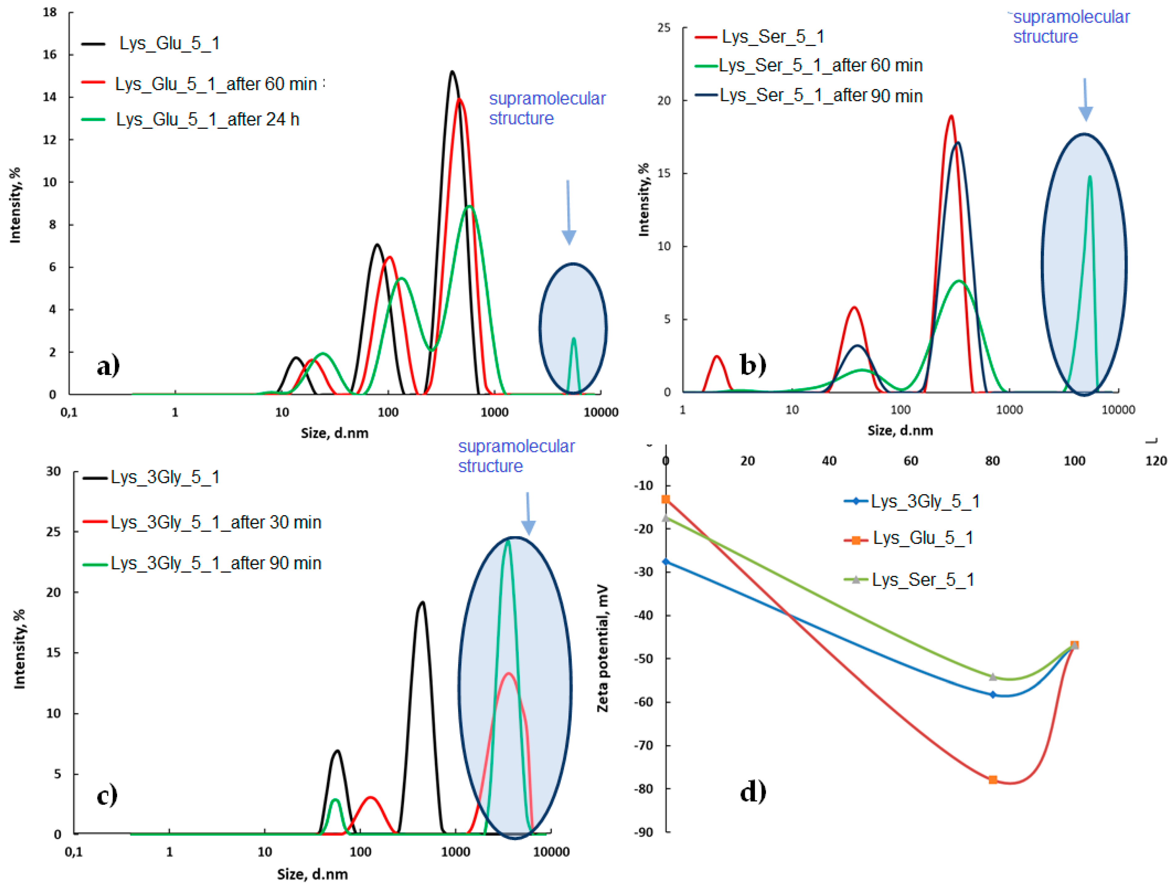
The Size Distribution Profile of Samples
Fluorescence Studies
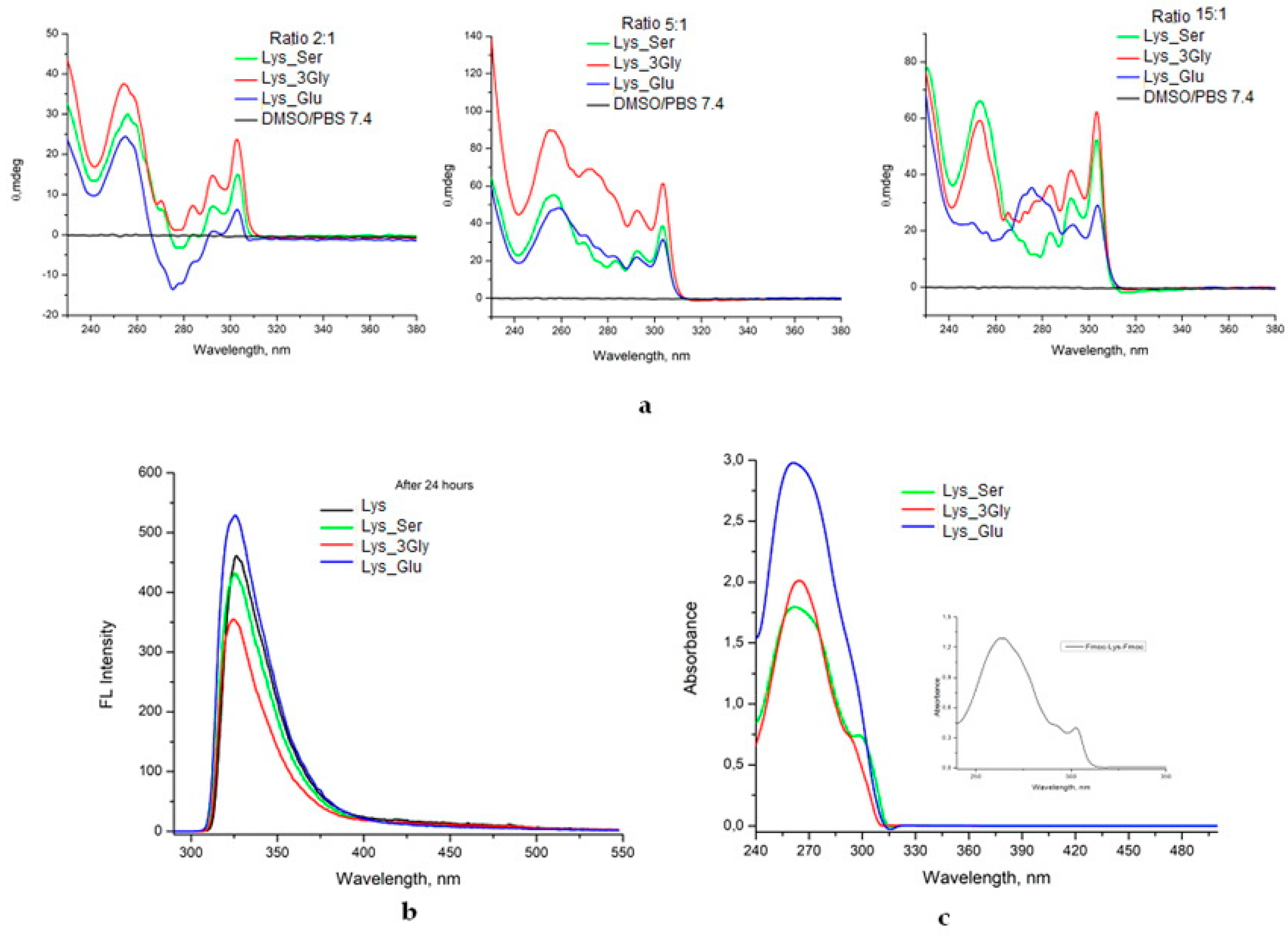
CD Spectra
UV-VIS Spectroscopy
2.3.2. Characterization of Gels in the Wet State
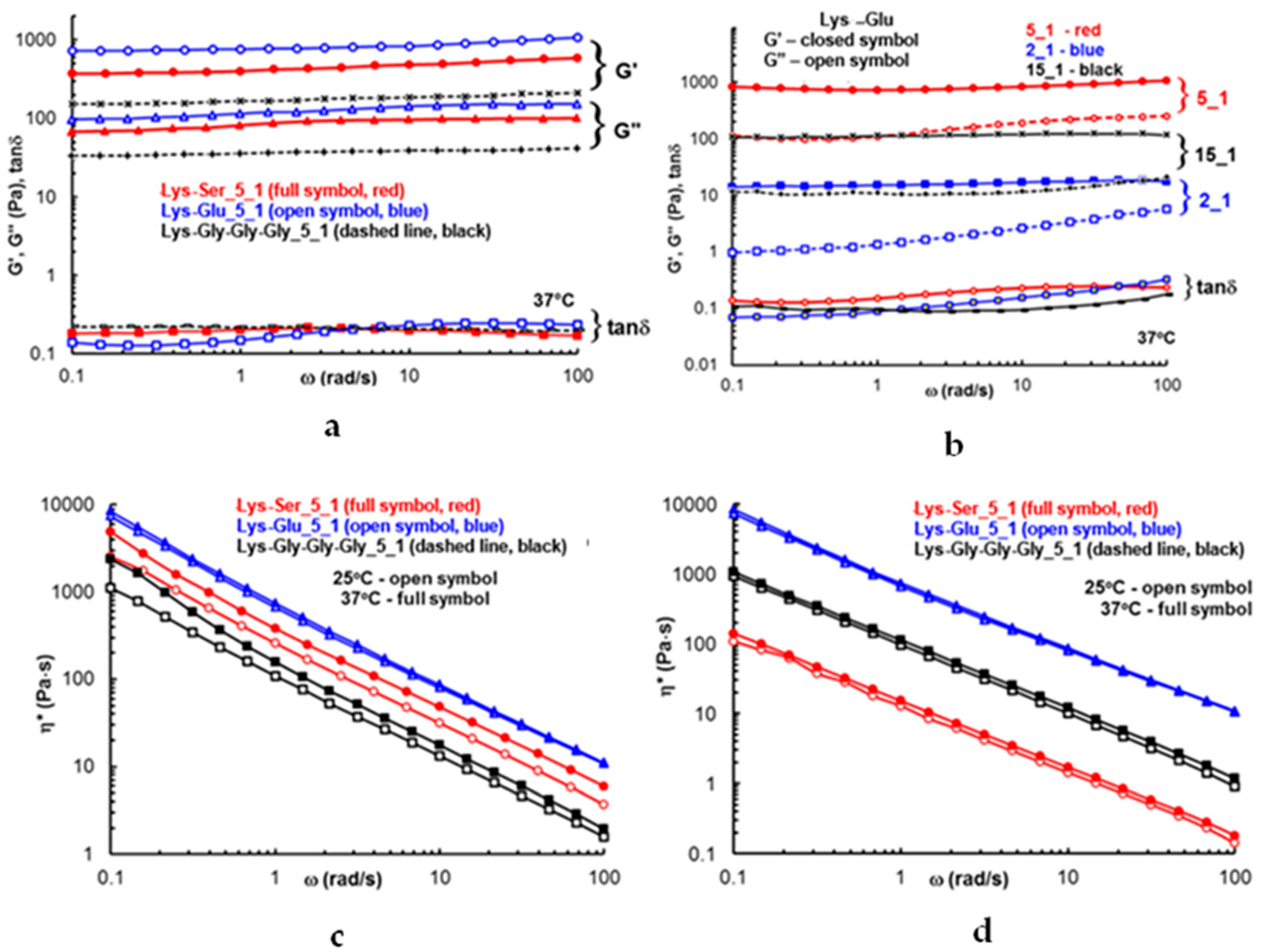
Rheological Studies
2.3.3. Characterization of Lyophilized Gels
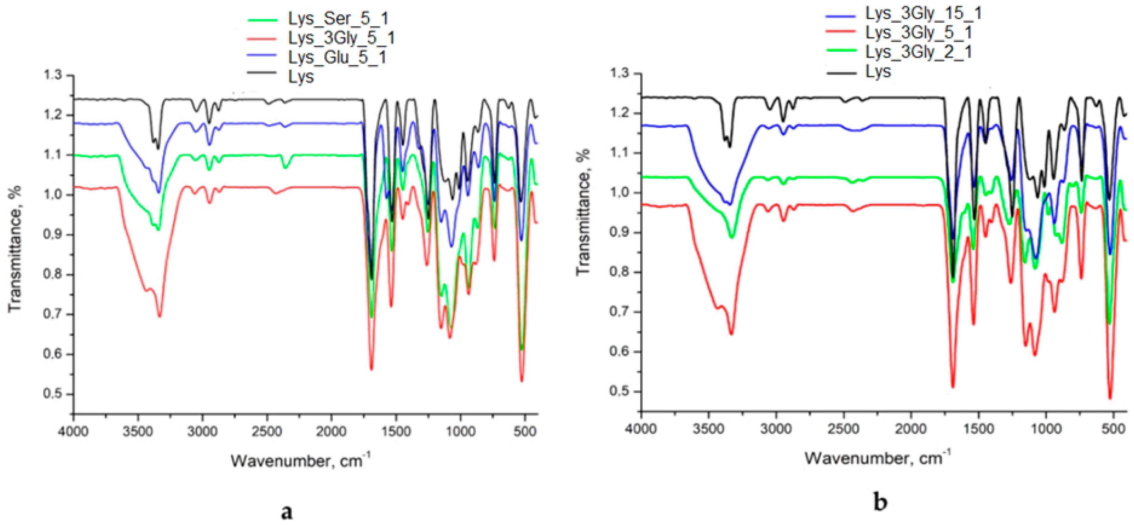
FTIR Spectroscopy
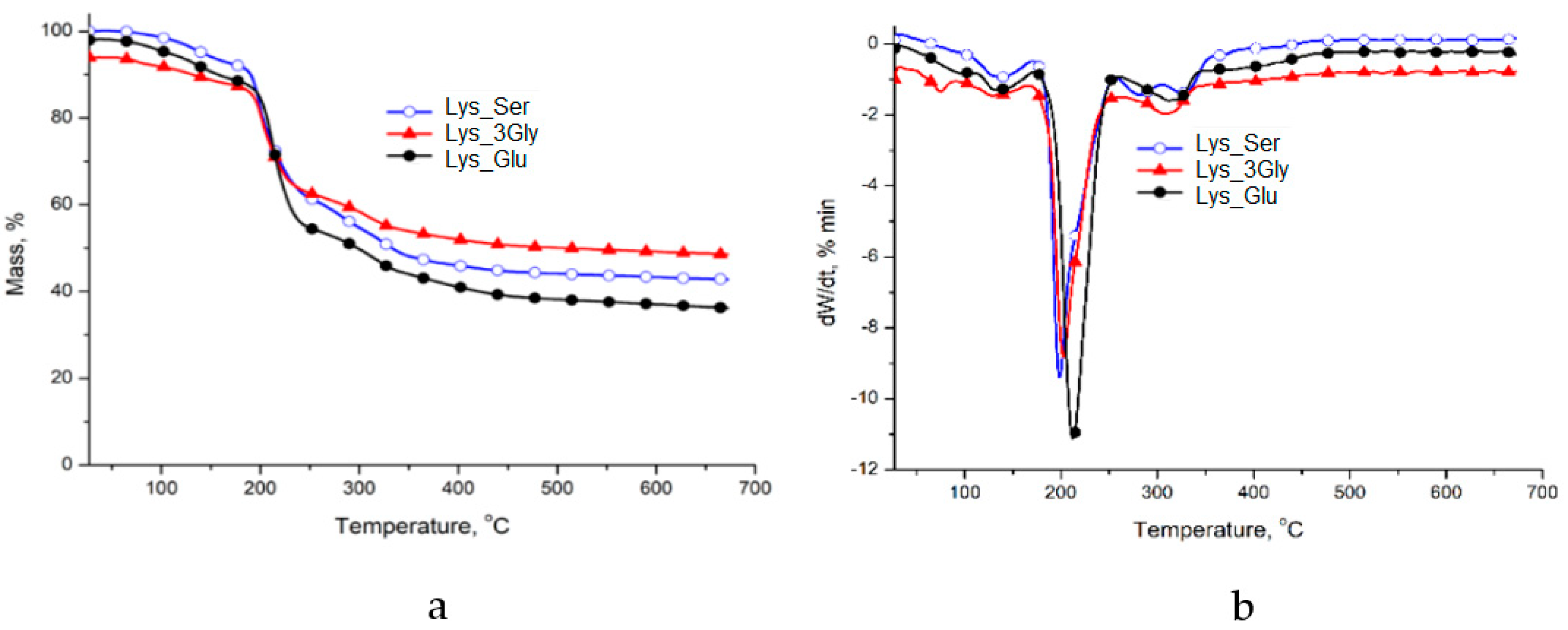
TG/DTG Analysis
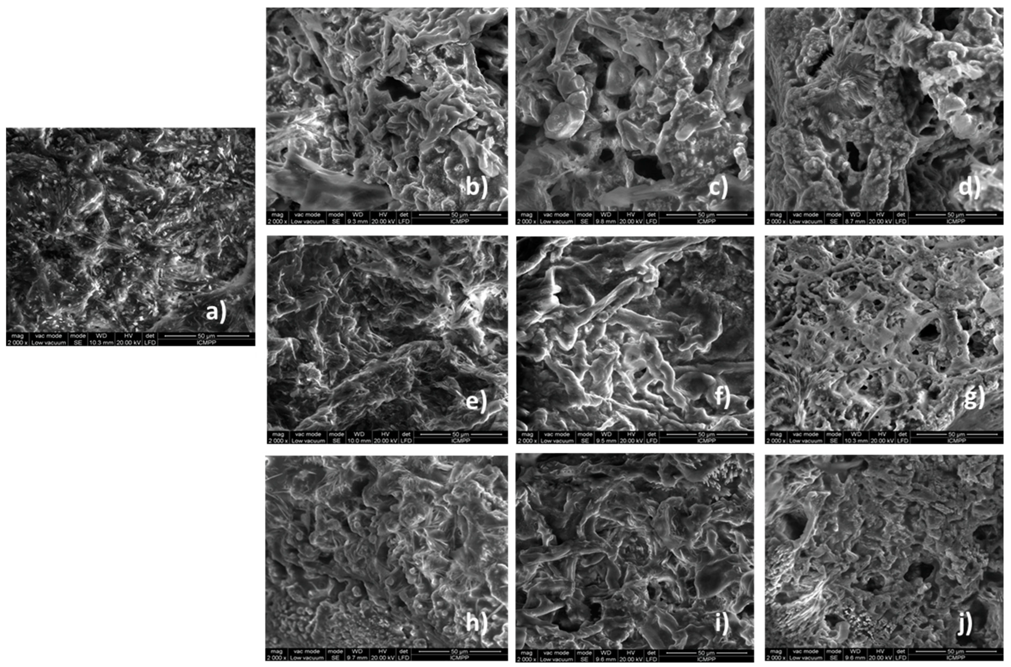
Morphology Analysis
3. Results
3.1. Hydrogelation Systems
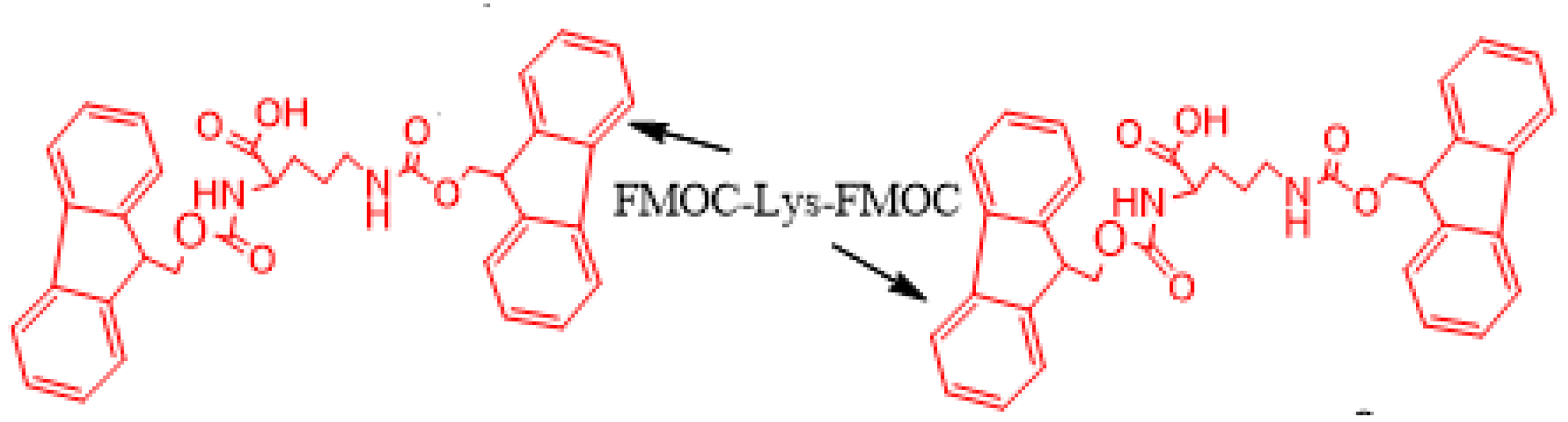
3.2. Co-Assembly Processes
3.2.1. FTIR Spectra
3.2.2. Thermal Analysis
3.2.3. DLS Studies
3.2.4. Molecular Arrangement
3.2.5. SEM Analysis
3.3. Rheological Studies
4. Conclusions
Author Contributions
Funding
Institutional Review Board Statement
Informed Consent Statement
Data Availability Statement
Conflicts of Interest
References
- Webber, M.J.; Dankers, P.W. Supramolecular Hydrogels for Biomedical Applications. Macromol. Biosci. 2019, 19, 1800452. [Google Scholar] [CrossRef]
- Debnath, S.; Shome, A.; Das, D.; Das, P.K. Hydrogelation through Self-Assembly of Fmoc-Peptide Functionalized Cationic Amphiphiles: Potent Antibacterial Agent. J. Phys. Chem. B 2010, 114, 4407–4415. [Google Scholar] [CrossRef] [PubMed]
- Bellotto, O.; Semeraro, S.; Bandiera, A.; Tramer, F.; Pavan, N.; Marchesan, S. Polymer Conjugates of Antimicrobial Peptides (AMPs) with d-Amino Acids (d-aa): State of the Art and Future Opportunities. Pharmaceutics 2022, 14, 446. [Google Scholar] [CrossRef] [PubMed]
- Nita, L.E.; Chiriac, A.P.; Rusu, A.G.; Bercea, M.; Ghilan, A.; Dumitriu, R.P.; Mititelu-Tartau, L. New self-healing hydrogels based on reversible physical interactions and their potential applications. Eur. Polym. J. 2019, 118, 176–185. [Google Scholar] [CrossRef]
- Chakraborty, P.; Tang, Y.; Yamamoto, T.; Yao, Y.; Guterman, T.; Zilberzwige-Tal, S.; Gazit, E. Unusual Two-Step Assembly of a Minimalistic Dipeptide-Based Functional Hypergelator. Adv. Mater. 2020, 32, 1906043. [Google Scholar] [CrossRef]
- Panja, S.; Dietrich, B.; Smith, A.J.; Seddon, A.; Adams, D.J. Controlling Self-Sorting versus Co-assembly in Supramolecular Gels. ChemSystemsChem 2022, 4, 202200008. [Google Scholar] [CrossRef]
- Colquhoun, C.; Draper, E.R.; Eden, G.B.E.; Cattoz, B.N.; Morris, K.L.; Chen, L.; McDonald, O.T.; Terry, A.E.; Griffiths, P.C.; Serpell, C.L.; et al. The Effect of Self-Sorting and Co-Assembly on the Mechanical Properties of Low Molecular Weight Hydrogels. Nanoscale 2012, 6, 13719–13725. [Google Scholar] [CrossRef] [PubMed]
- Abul-Haija, Y.M.; Roy, S.; Frederix, P.W.J.M.; Javid, N.; Jayawarna, V.; Ulijn, R.V. Biocatalytically Triggered Co-Assembly of Two-Component Core/Shell Nanofibers. Small 2013, 10, 973–979. [Google Scholar] [CrossRef] [PubMed]
- Çelik, E.; Bayram, C.; Akçapınar, R.; Türk, M.; Denkbaş, E.B. The effect of calcium chloride concentration on alginate/Fmoc-diphenylalanine hydrogel networks. Mater. Sci. Eng. C 2016, 66, 221–229. [Google Scholar] [CrossRef] [PubMed]
- Das, T.; Häring, M.; Haldar, D.; Díaz Díaz, D. Phenylalanine and derivatives as versatile low-molecular-weight gelators: Design, structure and tailored function. Biomater. Sci. 2018, 6, 38–59. [Google Scholar] [CrossRef]
- Croitoriu, A.; Nita, L.E.; Rusu, A.G.; Doroftei, F.; Verestiuc, L. Co-assembled peptides hierarchically oriented for supramolecular gel formation. Rev. Roum. Chim. 2021, 66, 449–458. [Google Scholar] [CrossRef]
- Reddy, S.M.M.; Shanmugam, G.; Duraipandy, N.; Kiran, M.S.; Mandal, A.B. An additional fluorenylmethoxycarbonyl (Fmoc) moiety in di-Fmoc-functionalizedL-lysine induces pH-controlled ambidextrous gelation with significant advantages. Soft Matter 2015, 11, 8126–8140. [Google Scholar] [CrossRef] [PubMed]
- Zheng, M.; Pan, M.; Zhang, W.; Lin, H.; Wu, S.; Lu, C.; Cai, J. Poly(α-L-lysine)-based nanomaterials for versatile biomedical applications: Current advances and perspectives. Bioact. Mater. 2021, 6, 1878–1909. [Google Scholar] [CrossRef] [PubMed]
- Idrees, M.; Mohammad, A.R.; Karodia, N.; Rahman, A. Multimodal Role of Amino Acids in Microbial Control and Drug Development. Antibiotics 2020, 9, 330. [Google Scholar] [CrossRef] [PubMed]
- Kapil, S.; Sharma, V. d-Amino acids in antimicrobial peptides: A potential approach to treat and combat antimicrobial resistance. Can. J. Microbiol. 2020, 67, 119–137. [Google Scholar] [CrossRef]
- Thompson, M.; Scholz, C. Highly Branched Polymers Based on Poly (amino acid)s for Biomedical Application. Nanomaterials 2021, 11, 1119. [Google Scholar] [CrossRef]
- Arokianathan, J.F.; Ramya, K.A.; Janeena, A.; Deshpande, A.P.; Ayyadurai, N.; Leemarose, A.; Shanmugam, G. Non-proteinogenic Amino Acid based Supramolecular Hydrogel Material for Enhanced Cell Proliferation. Colloids Surf. B 2019, 185, 110581. [Google Scholar] [CrossRef]
- Bustamante-Torres, M.; Romero-Fierro, D.; Arcentales-Vera, B.; Palomino, K.; Magaña, H.; Bucio, E. Hydrogels Classification According to the Physical or Chemical Interactions and as Stimuli-Sensitive Materials. Gels 2021, 7, 182. [Google Scholar] [CrossRef]
- Alonci, G.; Mocchi, R.; Sommatis, S.; Capillo, M.C.; Liga, E.; Janowska, A.; Zerbinati, N. Physico-Chemical Characterization and In Vitro Biological Evaluation of a Bionic Hydrogel Based on Hyaluronic Acid and L-lysine for Medical Applications. Pharmaceutics 2021, 13, 1194. [Google Scholar] [CrossRef]
- Persikov, A.V.; Ramshaw, J.A.M.; Kirkpatrick, A.A.; Brodsky, B. Electrostatic Interactions Involving Lysine Make Major Contributions to Collagen Triple-Helix Stability. Biochemistry 2005, 44, 1414–1422. [Google Scholar] [CrossRef]
- Johnson, E.K.; Adams, D.J.; Cameron, P.J. Peptide based low molecular weight gelators. J. Mater. Chem. 2011, 21, 2024–2027. [Google Scholar] [CrossRef]
- Okesola, B.O.; Redondo-Gómez, C.; Mata, A. Multicomponent self-assembly: Supramolecular design of complex hydrogels for biomedical applications. In Self-Assembling Biomaterials; Woodhead Publishing: Duxford, UK, 2018; pp. 371–397. [Google Scholar] [CrossRef]
- Zhang, Y.; Li, S.; Ma, M.; Yang, M.; Wang, Y.; Hao, A.; Xing, P. Tuning of gel morphology with supramolecular chirality amplification using a solvent strategy based on an Fmoc-amino acid building block. New J. Chem. 2016, 40, 5568–5576. [Google Scholar] [CrossRef]
- Yang, Z.; Zhong, Y.; Zheng, J.; Liu, Y.; Li, T.; Hu, E.; Wang, Y. Fmoc-amino acid-based hydrogel vehicle for delivery of amygdalin to perform neuroprotection. Smart Mater. Med. 2020, 2, 56–64. [Google Scholar] [CrossRef]
- Li, W.; Hu, X.; Chen, J.; Wei, Z.; Song, C.; Huang, R. N-(9-Fluorenylmethoxycarbonyl)-L-Phenylalanine/nano-hydroxyapatite hybrid supramolecular hydrogels as drug delivery vehicles with antibacterial property and cytocompatibility. J. Mater. Sci. Mater. Med. 2020, 31, 73. [Google Scholar] [CrossRef]
- Debnath, S.; Roy, S.; Abul-Haija, Y.M.; Frederix, P.; Ramalhete, S.; Hirst, A.; Ulijn, R. Tunable supramolecular gels by varying thermal history. Eur. J. Chem. 2019, 25, 7881–7887. [Google Scholar] [CrossRef]
- Schaberg, R.; Wroblowski, R.; Goertz, R. Comparative study of the thermal decomposition behaviour of different amino acids and peptides. J. Phys. Conf. Ser. 2018, 1107, 32013. [Google Scholar] [CrossRef]
- Ehlers, G.F.L.; Fisch, K.R.; Powell, W.R. Thermal degradation of polymers with phenylene units in the chain. IV. Aromatic polyamides and polyimides. J. Polym. Sci. Part A Polym. Chem. 1970, 8, 3511–3527. [Google Scholar] [CrossRef]
- Ruffoni, A.; Cavanna, M.V.; Argentiere, S.; Locarno, S.; Pellegrino, S.; Gelmi, M.L.; Clerici, F. Aqueous self-assembly of short hydrophobic peptides containing norbornene amino acid into supramolecular structures with spherical shape. RSC Adv. 2016, 6, 90754–90759. [Google Scholar] [CrossRef]
- Andersson, D.; Carlsson, U.; Freskgård, P.-O. Contribution of tryptophan residues to the CD spectrum of the extracellular domain of human tissue factor. Eur. J. Biochem. 2001, 268, 1118–1128. [Google Scholar] [CrossRef]
- Diaferia, C.; Rosa, E.; Gallo, E.; Smaldone, G.; Stornaiuolo, M.; Morelli, G.; Accardo, A. Self-Supporting Hydrogels Based on Fmoc-Derivatized Cationic Hexapeptides for Potential Biomedical Applications. Biomedicines 2021, 9, 678. [Google Scholar] [CrossRef]
- Sahoo, J.K.; Roy, S.; Javid, N.; Duncan, K.; Aitken, L.; Ulijn, R.V. Pathway-dependent gold nanoparticle formation by biocatalytic self-assembly. Nanoscale 2017, 9, 12330–12334. [Google Scholar] [CrossRef] [PubMed]
- Argudo, P.G.; Contreras-Montoya, R.; Alvarez de Cienfuegos, L.; Cuerva, J.M.; Cano, M.; Alba-Molina, D.; Giner-Casares, J.J. Unravelling the 2D Self-Assembly of Fmoc-Dipeptides at Fluid Interfaces. Soft Matter 2018, 14, 9343–9350. [Google Scholar] [CrossRef] [PubMed]
- Amdursky, N.; Stevens, M.M. Circular Dichroism of Amino Acids: Following the Structural Formation of Phenylalanine. Chem. Phys. Chem. 2015, 16, 2768–2774. [Google Scholar] [CrossRef] [PubMed]
- Rahman, M.S.; Islam, M.M.; Islam, M.S.; Zaman, A.; Ahmed, T.; Biswas, S.; Sharmeen, S.; Rashid, T.U.; Rahman, M.M. Morphological Characterization of Hydrogels. In Cellulose−Based Superabsorbent Hydrogels; Mondal, M., Ed.; Springer: Cham, Switzerland, 2018; pp. 1–46. [Google Scholar]
- Chavda, H.V.; Patel, R.D.; Modhia, I.P.; Patel, C.N. Preparation and characterization of superporous hydrogel based on different polymers. Int. J. Pharm. Investig. 2012, 2, 134–139. [Google Scholar] [CrossRef]
- Li, R.; Horgan, C.C.; Long, B.; Rodriguez, A.L.; Mather, L.; Barrow, C.J.; Williams, R.J. Tuning the mechanical and morphological properties of self-assembled peptide hydrogels via control over the gelation mechanism through regulation of ionic strength and the rate of pH change. RSC Adv. 2015, 5, 301–307. [Google Scholar] [CrossRef]


| Sample | Sample Code | Composition for 5 mL of Gel | |||
|---|---|---|---|---|---|
| Fmoc-Lys-Fmoc g | Second Compound g | DMSO µL | Water mL | ||
| Fmoc-Lys-Fmoc | Lys | 0.0250 | - | 120 | 4.88 |
| Fmoc-Lys-Fmoc _Fmoc-Ser_2_1 | Lys_Ser_2_1 | 0.00625 | 0.00375 | 60 | 4.94 |
| Fmoc-Lys-Fmoc _Fmoc-Ser_5_1 | Lys_Ser_5_1 | 0.0125 | 0.0025 | 80 | 4.92 |
| Fmoc-Lys-Fmoc _Fmoc-Ser_15_1 | Lys_Ser_15_1 | 0.0187 | 0.00125 | 110 | 4.89 |
| Fmoc-Lys-Fmoc _Fmoc-Glu_2_1 | Lys_Glu_2_1 | 0.00625 | 0.00375 | 60 | 4.94 |
| Fmoc-Lys-Fmoc _Fmoc-Glu_5_1 | Lys_Glu_5_1 | 0.0125 | 0.0025 | 80 | 4.92 |
| Fmoc-Lys-Fmoc _Fmoc-Glu_15_1 | Lys_Glu_15_1 | 0.0187 | 0.00125 | 110 | 4.89 |
| Fmoc-Lys-Fmoc_Fmoc-Gly-Gly-Gly_2_1 | Lys_3Gly_2_1 | 0.00625 | 0.00375 | 60 | 4.94 |
| Fmoc-Lys-Fmoc_Fmoc-Gly-Gly-Gly_5_1 | Lys_3Gly_5_1 | 0.0125 | 0.0025 | 80 | 4.92 |
| Fmoc-Lys-Fmoc_Fmoc-Gly-Gly-Gly_15_1 | Lys_3Gly_15_1 | 0.0187 | 0.00125 | 110 | 4.89 |
| Name and Structure of First Compound | Name and Structure of Second Compound | Images of Co-Assembled Gels | Conclusions |
|---|---|---|---|
Fmoc-Lys-Fmoc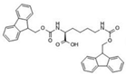 | Fmoc-Gly-Gly-Gly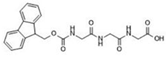 |  | Stable gels |
Fmoc-Lys-Fmoc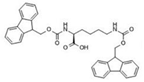 | Fmoc-Glutamic acid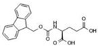 |  | Stable gels with translucent aspect |
Fmoc-Lys-Fmoc | Fmoc-serine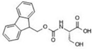 | 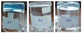 | Stable gels with opaque aspect |
| Sample | Degradation Stage | Tonset (°C) | Tpeak (°C) | Tendset (°C) | W (%) | Residue | T10 (°C) | T20 (°C) |
|---|---|---|---|---|---|---|---|---|
| Lys | I II III IV V | 62 182 262 316 361 | 144 189 280 322 365 | 172 196 307 350 398 | 10.44 33.00 8.77 6.85 6.76 | 34.18 | 178 | 193 |
| Lys_Ser | I II III IV | 27 187 213 305 | 138 197 280 323 | 183 204 303 357 | 9.82 15.10 21.03 11.28 | 42.77 | 190 | 206 |
| Lys_3Gly | I II III IV | 62 89 188 283 | 74 128 201 307 | 80 172 220 335 | 1.71 6.04 26.18 11.49 | 54.58 | 195 | 209 |
| Lys_Glu | I II III IV | 27 115 194 270 | 97 132 211 311 | 109 174 240 334 | 3.98 5.73 34.55 17.57 | 38.17 | 183 | 202 |
Publisher’s Note: MDPI stays neutral with regard to jurisdictional claims in published maps and institutional affiliations. |
© 2022 by the authors. Licensee MDPI, Basel, Switzerland. This article is an open access article distributed under the terms and conditions of the Creative Commons Attribution (CC BY) license (https://creativecommons.org/licenses/by/4.0/).
Share and Cite
Croitoriu, A.; Nita, L.E.; Rusu, A.G.; Ghilan, A.; Bercea, M.; Chiriac, A.P. New Fmoc-Amino Acids/Peptides-Based Supramolecular Gels Obtained through Co-Assembly Process: Preparation and Characterization. Polymers 2022, 14, 3354. https://doi.org/10.3390/polym14163354
Croitoriu A, Nita LE, Rusu AG, Ghilan A, Bercea M, Chiriac AP. New Fmoc-Amino Acids/Peptides-Based Supramolecular Gels Obtained through Co-Assembly Process: Preparation and Characterization. Polymers. 2022; 14(16):3354. https://doi.org/10.3390/polym14163354
Chicago/Turabian StyleCroitoriu, Alexandra, Loredana Elena Nita, Alina Gabriela Rusu, Alina Ghilan, Maria Bercea, and Aurica P. Chiriac. 2022. "New Fmoc-Amino Acids/Peptides-Based Supramolecular Gels Obtained through Co-Assembly Process: Preparation and Characterization" Polymers 14, no. 16: 3354. https://doi.org/10.3390/polym14163354
APA StyleCroitoriu, A., Nita, L. E., Rusu, A. G., Ghilan, A., Bercea, M., & Chiriac, A. P. (2022). New Fmoc-Amino Acids/Peptides-Based Supramolecular Gels Obtained through Co-Assembly Process: Preparation and Characterization. Polymers, 14(16), 3354. https://doi.org/10.3390/polym14163354