
Polymeric Nanoparticles for Antimicrobial Therapies: An up-to-date Overview


 ,
,  and
and
Abstract
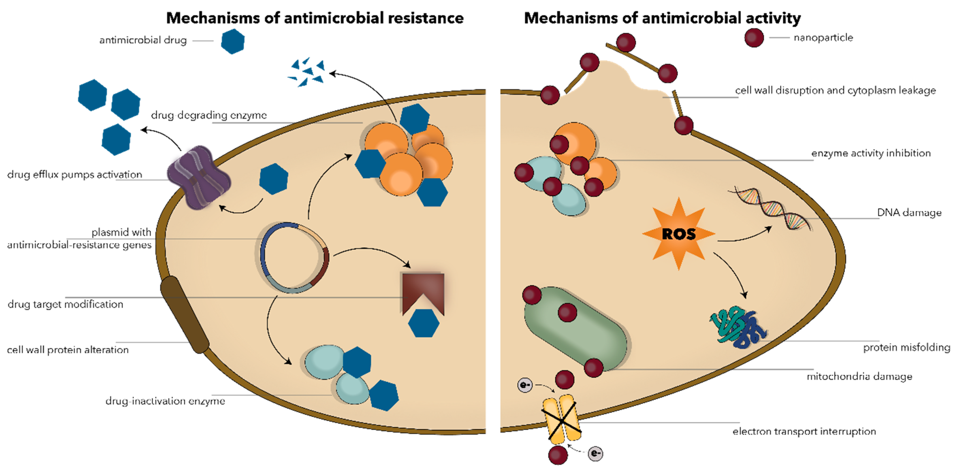
1. Introduction
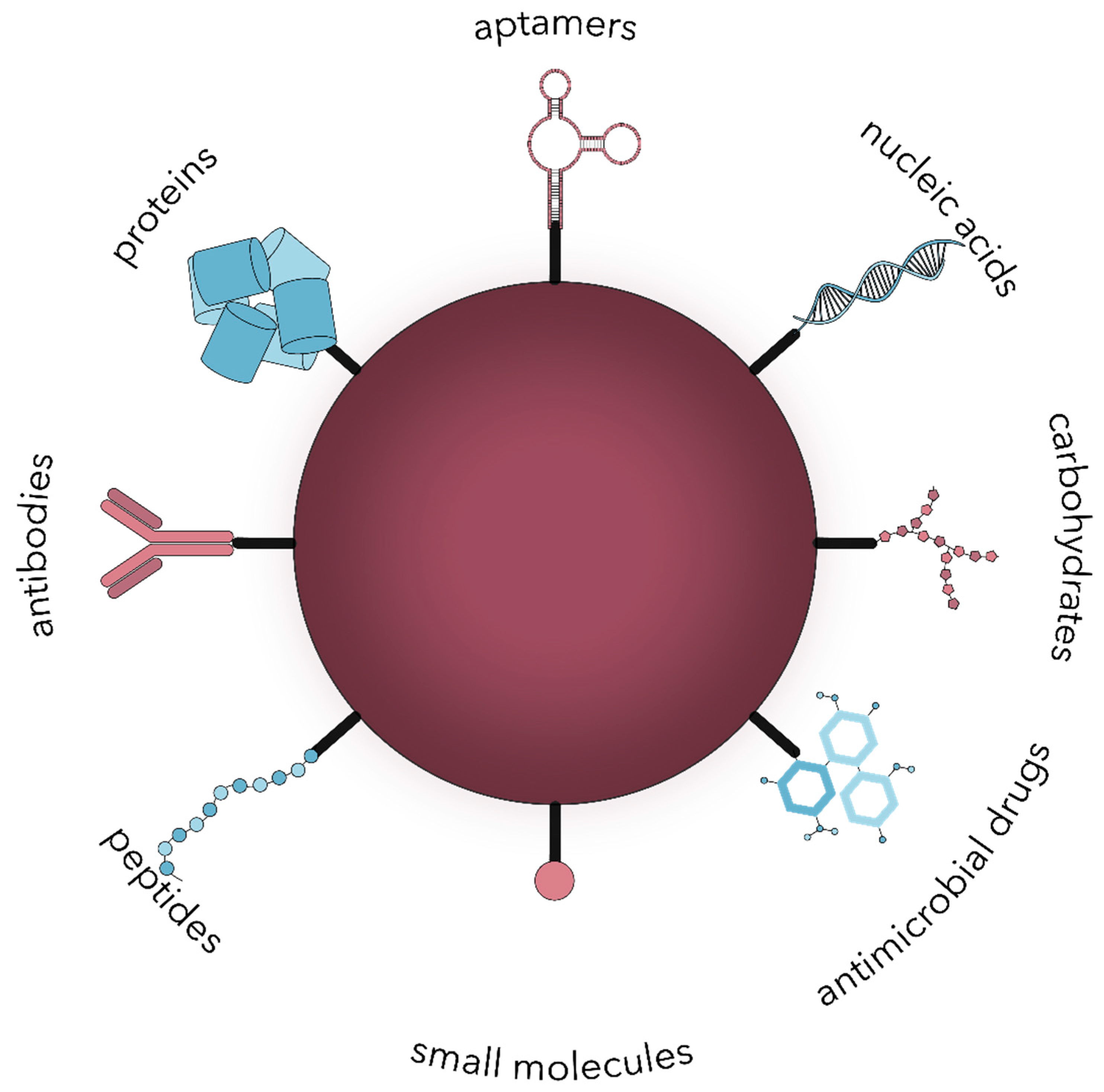
2. Microbial Targeting Strategies
2.1. Small Molecules
2.2. Peptides
2.3. Proteins
2.4. Nucleic Acids
2.5. Carbohydrates
2.6. Antimicrobial Drugs
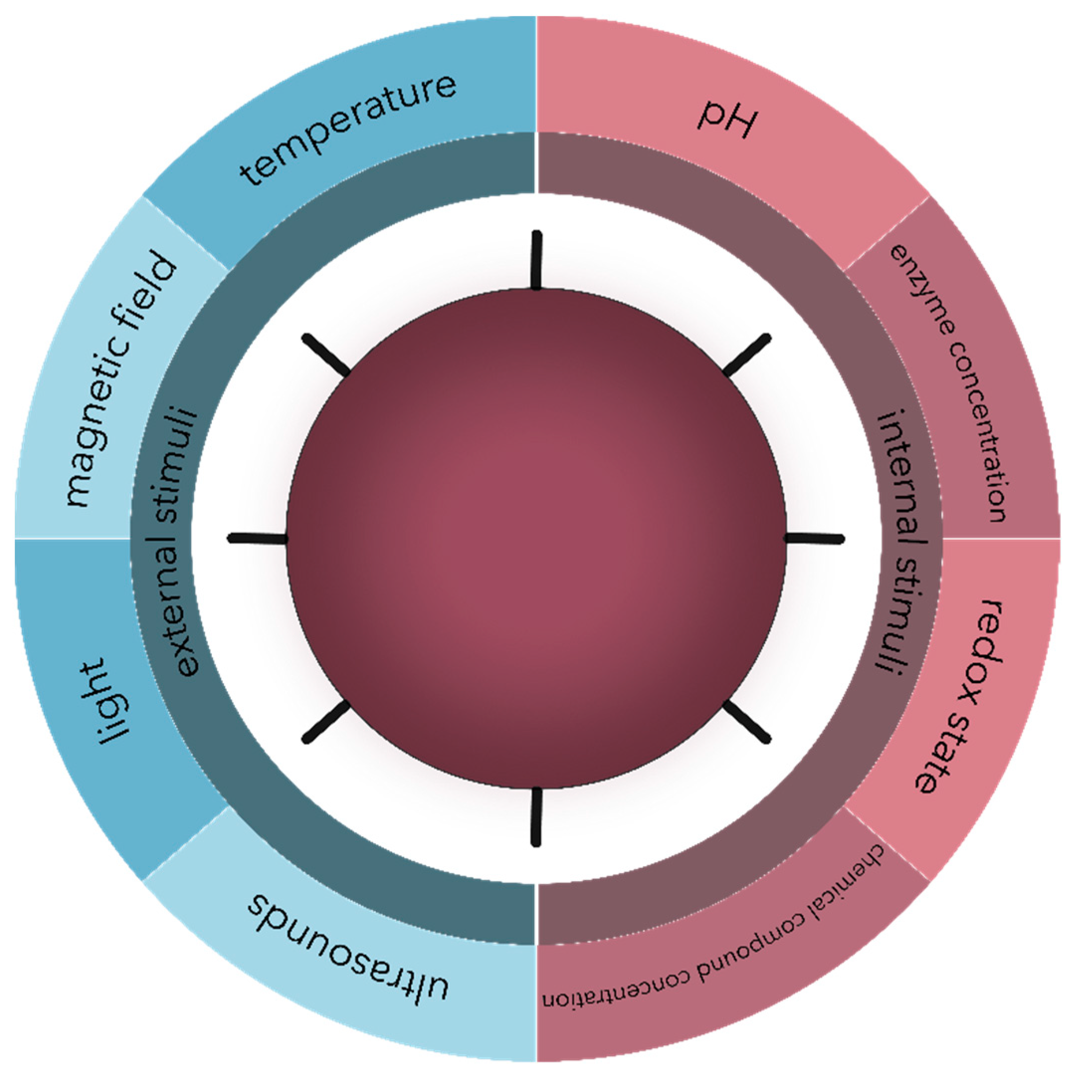
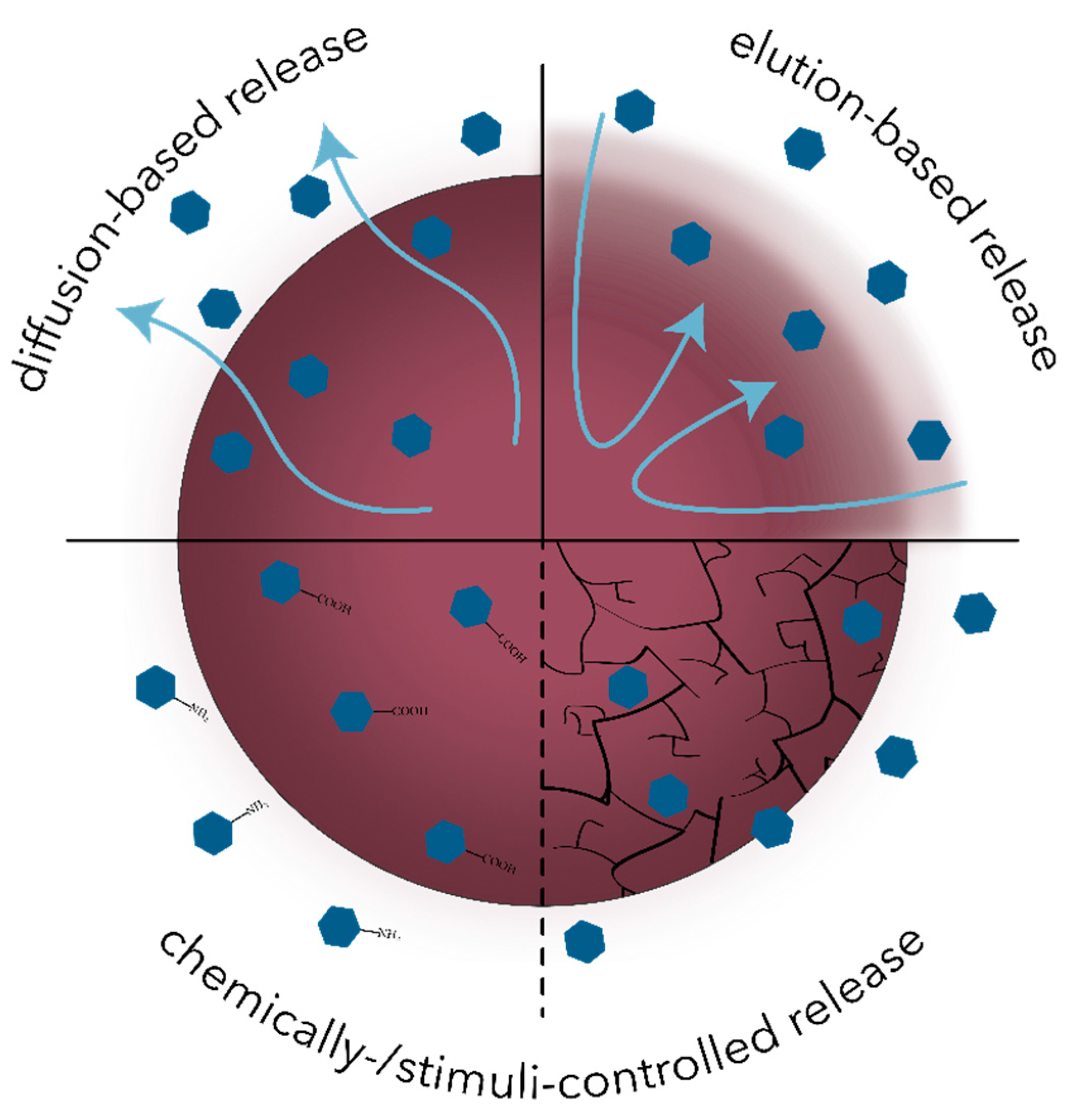
2.7. Stimuli-Responsive Nanosystems
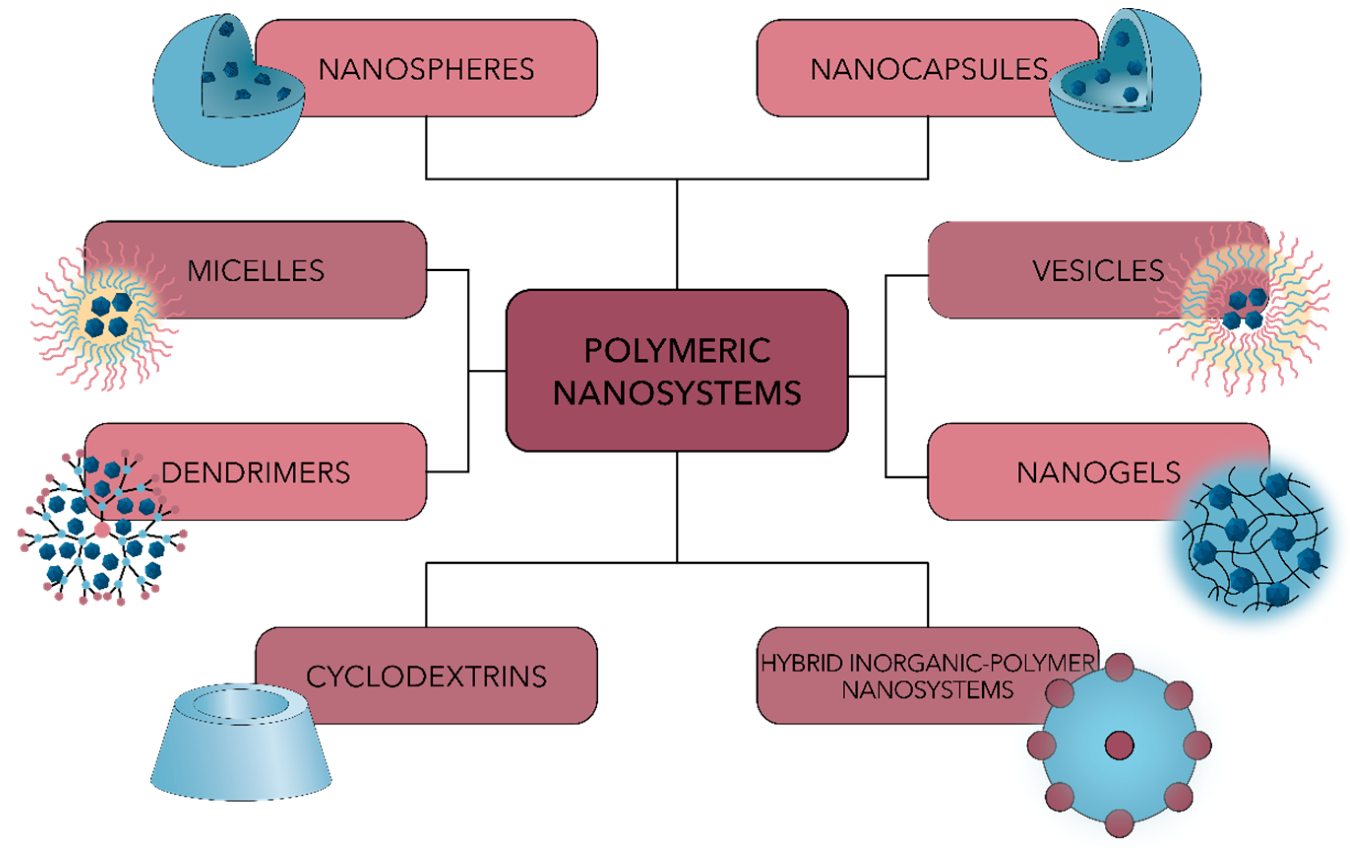
3. Antimicrobial Applications of Polymeric Nanoparticles
3.1. Antibacterial Nanoparticles
3.2. Antiviral Nanoparticles
3.3. Antifungal Nanoparticles
3.4. Antiparasitic Nanoparticles
4. Conclusions and Future Perspectives
Author Contributions
Funding
Institutional Review Board Statement
Informed Consent Statement
Data Availability Statement
Acknowledgments
Conflicts of Interest
References
- Chircov, C.; Grumezescu, A.M.; Holban, A.M. Magnetic Particles for Advanced Molecular Diagnosis. Materials 2019, 12, 2158. [Google Scholar] [CrossRef] [PubMed]
- Tripathi, L.P.; Chen, Y.-A.; Mizuguchi, K.; Morita, E. Network-Based Analysis of Host-Pathogen Interactions. Encycl. Bioinform. Comput. Biol. 2019, 3, 932–937. [Google Scholar]
- Sheikhzadeh, E.; Beni, V.; Zourob, M. Nanomaterial application in bio/sensors for the detection of infectious diseases. Talanta 2020, 122026. [Google Scholar] [CrossRef]
- Raza, A.; Sime, F.B.; Cabot, P.J.; Maqbool, F.; Roberts, J.A.; Falconer, J.R. Solid nanoparticles for oral antimicrobial drug delivery: A review. Drug Discov. Today 2019, 24, 858–866. [Google Scholar] [CrossRef]
- Roberts, C.A.; Buikstra, J.E. Bacterial infections. In Other’s Identification of Pathological Conditions in Human Skeletal Remains, 3rd ed.; Elsevier: Amsterdam, The Netherlands, 2019; pp. 321–349. [Google Scholar]
- Roberts, C.A. Infectious disease. In Other’s Identification of Pathological Conditions in Human Skeletal Remains, 3rd ed.; Elsevier: Amsterdam, The Netherlands, 2019; pp. 285–319. [Google Scholar]
- Ruiz, J.; Castro, I.; Calabuig, E.; Salavert, M. Non-antibiotic treatment for infectious diseases. Rev. Esp. Quim. Publ. Soc. Esp. Quim. 2017, 1, 66–71. [Google Scholar]
- Waller, D.G.; Sampson, A.P. Chemotherapy of infections. Med Pharmacol. Ther. 2018, 581–629. [Google Scholar] [CrossRef]
- Melander, R.J.; Zurawski, D.V.; Melander, C. Narrow-spectrum antibacterial agents. Med. Chem. Comm. 2018, 9, 12–21. [Google Scholar] [CrossRef]
- Devrim, B.; Bozkır, A. Nanocarriers and Their Potential Application as Antimicrobial Drug Delivery. In Nanostructures for Antimicrobial Therapy; Elsevier: Amsterdam, The Netherlands, 2017; pp. 169–202. [Google Scholar]
- Yuan, P.; Ding, X.; Yang, Y.Y.; Xu, Q.-H. Metal Nanoparticles for Diagnosis and Therapy of Bacterial Infection. Adv. Heal. Mater. 2018, 7, e1701392. [Google Scholar] [CrossRef]
- Antabe, R.; Ziegler, B.R. Diseases, Emerging and Infectious. Int. Encycl. Hum. Geogr. 2020, 389–391. [Google Scholar] [CrossRef]
- Eleraky, N.E.; Allam, A.; Hassan, S.B.; Omar, M.M. Nanomedicine Fight against Antibacterial Resistance: An Overview of the Recent Pharmaceutical Innovations. Pharmaceutics 2020, 12, 142. [Google Scholar] [CrossRef]
- Cheng, G.; Dai, M.; Ahmed, S.; Hao, H.; Wang, X.; Yuan, Z. Antimicrobial Drugs in Fighting against Antimicrobial Resistance. Front. Microbiol. 2016, 7, 470. [Google Scholar] [CrossRef]
- McMahon, D.P.; Wilfert, L.; Paxton, R.J.; Brown, M.J. Emerging Viruses in Bees: From Molecules to Ecology; Elsevier BV: Amsterdam, The Netherlands, 2018; Volume 101, pp. 251–291. [Google Scholar]
- Willyard, C. The drug-resistant bacteria that pose the greatest health threats. Nat. Cell Biol. 2017, 543, 15. [Google Scholar] [CrossRef]
- Lam, S.J.; Wong, E.H.; Boyer, C.; Qiao, G.G. Antimicrobial polymeric nanoparticles. Prog. Polym. Sci. 2018, 76, 40–64. [Google Scholar] [CrossRef]
- Mühlen, S.; Dersch, P. Anti-virulence Strategies to Target Bacterial Infections. In How to Overcome the Antibiotic Crisis; Springer: Berlin/Heidelberg, Germany, 2015; Volume 398, pp. 147–183. [Google Scholar] [CrossRef]
- Garland, M.; Loscher, S.; Bogyo, M. Chemical Strategies To Target Bacterial Virulence. Chem. Rev. 2017, 117, 4422–4461. [Google Scholar] [CrossRef] [PubMed]
- Niu, G.; Li, W. Next-Generation Drug Discovery to Combat Antimicrobial Resistance. Trends Biochem. Sci. 2019, 44, 961–972. [Google Scholar] [CrossRef] [PubMed]
- Principi, N.; Silvestri, E.; Esposito, S. Advantages and limitations of bacteriophages for the treatment of bacterial infections. Front. Pharm.. 2019, 10, 513. [Google Scholar] [CrossRef] [PubMed]
- Chen, C.H.; Lu, T.K. Development and Challenges of Antimicrobial Peptides for Therapeutic Applications. Antibiotics 2020, 9, 24. [Google Scholar] [CrossRef]
- Lai, J.Y.; Lim, T.S. Infectious disease antibodies for biomedical applications: A mini review of immune antibody phage library repertoire. Int. J. Biol. Macromol. 2020, 163, 640–648. [Google Scholar] [CrossRef]
- Wang, L.; Hu, C.; Shao, L. The antimicrobial activity of nanoparticles: Present situation and prospects for the future. Int. J. Nanomed. 2017, 12, 1227–1249. [Google Scholar] [CrossRef]
- Fernando, S.; Gunasekara, T.; Holton, J. Antimicrobial Nanoparticles: Applications and mechanisms of action. Sri Lankan J. Infect. Dis. 2018, 8, 2. [Google Scholar] [CrossRef]
- Luksiene, Z. Nanoparticles and their potential application as antimicrobials in the food industry. Food Preserv. 2017, 567–601. [Google Scholar] [CrossRef]
- Varier, K.M.; Gudeppu, M.; Chinnasamy, A.; Thangarajan, S.; Balasubramanian, J.; Li, Y.; Gajendran, B. Nanoparticles: An-timicrobial Applications and Its Prospects. In Advanced Nanostructured Materials for Environmental Remediation; Springer: Berlin/Heidelberg, Germany, 2019; pp. 321–355. [Google Scholar]
- Srividya, N.; Ghoora, M.D.; Padmanabh, P.R. Antimicrobial nanotechnology: Research implications and prospects in food safety. In Food Preservation; Elsevier: Amsterdam, The Netherlands, 2017; pp. 125–165. [Google Scholar]
- Lee, N.-Y.; Ko, W.-C.; Hsueh, P.-R. Nanoparticles in the Treatment of Infections Caused by Multidrug-Resistant Organisms. Front. Pharm. 2019, 10, 1153. [Google Scholar] [CrossRef] [PubMed]
- Abduraimova, A.; Molkenova, A.; Duisembekova, A.; Mulikova, T.; Kanayeva, D.; Atabaev, T. Cetyltrimethylammonium Bromide (CTAB)-Loaded SiO2–Ag Mesoporous Nanocomposite as an Efficient Antibacterial Agent. Nanomaterials 2021, 11, 477. [Google Scholar] [CrossRef]
- Liu, J.; Li, S.; Fang, Y.; Zhu, Z. Boosting antibacterial activity with mesoporous silica nanoparticles supported silver nanoclusters. J. Colloid Interface Sci. 2019, 555, 470–479. [Google Scholar] [CrossRef]
- Patil, J.S.; Dhadde, S.B.; Chandakavathe, B.N. Nanostructure Drug Delivery System Is an Option to Solve Anti-microbial Drug Resistance: Perspective Review. In Characterization and Biology of Nanomaterials for Drug Delivery; Mohapatra, S.S., Ranjan, S., Dasgupta, N., Mishra, R.K., Thomas, S., Eds.; Elsevier: Amsterdam, The Netherlands, 2019; pp. 165–197. [Google Scholar]
- Canaparo, R.; Foglietta, F.; Giuntini, F.; Della Pepa, C.; Dosio, F.; Serpe, L. Recent Developments in Antibacterial Therapy: Focus on Stimuli-Responsive Drug-Delivery Systems and Therapeutic Nanoparticles. Molecules 2019, 24, 1991. [Google Scholar] [CrossRef] [PubMed]
- Lin, Y.-S.; Lee, M.-Y.; Yang, C.-H.; Huang, K.-S. Active Targeted Drug Delivery for Microbes Using Nano-Carriers. Curr. Top. Med. Chem. 2015, 15, 1525–1531. [Google Scholar] [CrossRef]
- Bahrami, A.; Delshadi, R.; Jafari, S.M. Active delivery of antimicrobial nanoparticles into microbial cells through surface functionalization strategies. Trends Food Sci. Technol. 2020, 99, 217–228. [Google Scholar] [CrossRef]
- Samrot, A.V.; Sahithya, C.S.; Sruthi, D.P.; Selvarani, J.A.; Raji, P.; Prakash, P.; Ponnaiah, P.; Petchi, I.; Pattammadath, S.; Keeyari, S.; et al. Itraconazole Coated Super Paramagnetic Iron Oxide Nanoparticles for Antimicrobial Studies. Biointerface Res. Appl. Chem. 2020, 10, 6218–6225. [Google Scholar] [CrossRef]
- Thiruvengadam, V.; Bansod, A.V. Characterization of Silver Nanoparticles Synthesized using Chemical Method and its Antibacterial Property. Biointerface Res. Appl. Chem. 2020, 10, 7257–7264. [Google Scholar] [CrossRef]
- Tripathy, A.; Behera, M.; Rout, A.S.; Biswal, S.K.; Phule, A. Optical, Structural, and Antimicrobial Study of Gold nanoparticles Synthesized Using an Aqueous Extract of Mimusops elengi Raw Fruits. Biointerface Res. Appl. Chem. 2020, 10, 7085–7096. [Google Scholar] [CrossRef]
- Baptista, P.V.; McCusker, M.P.; Carvalho, A.; Ferreira, D.A.; Mohan, N.M.; Martins, M.; Fernandes, A.R. Nano-strategies to fight multidrug resistant bacteria—A Battle of the Titans. Front. Microbiol. 2018, 9, 1441. [Google Scholar] [CrossRef] [PubMed]
- Masri, A.; Anwar, A.; Khan, N.A.; Siddiqui, R. The Use of Nanomedicine for Targeted Therapy against Bacterial Infections. Antibiotics 2019, 8, 260. [Google Scholar] [CrossRef] [PubMed]
- Yetisgin, A.A.; Cetinel, S.; Zuvin, M.; Kosar, A.; Kutlu, O. Therapeutic Nanoparticles and Their Targeted Delivery Applications. Molecules 2020, 25, 2193. [Google Scholar] [CrossRef] [PubMed]
- Ulah, F.; Javed, F.; Khan, A.N.; Helmi, M.; Kudus, M.H.A.; Jamila, N.; Minhaz, A.; Akil, H. Synthesis and surface modification of chitosan built nanohydrogel with antiviral and antimicrobial agent for controlled drug delivery. Biointerface Res. Appl. Chem. 2019, 9, 4439–4445. [Google Scholar] [CrossRef]
- Smerkova, K.; Dolezelikova, K.; Bozdechova, L.; Heger, Z.; Zurek, L.; Adam, V. Nanomaterials with active targeting as advanced antimicrobials. Wiley Interdiscip. Rev. Nanomed. Nanobiotechnol. 2020, 12, e1636. [Google Scholar] [CrossRef]
- Yeh, Y.-C.; Huang, T.-H.; Yang, S.-C.; Chen, C.-C.; Fang, J.-Y. Nano-Based Drug Delivery or Targeting to Eradicate Bacteria for Infection Mitigation: A Review of Recent Advances. Front. Chem. 2020, 8, 286. [Google Scholar] [CrossRef]
- Lu, H.D.; Yang, S.S.; Wilson, B.K.; McManus, S.A.; Chen, C.V.H.-H.; Prud’Homme, R.K. Nanoparticle targeting of Gram-positive and Gram-negative bacteria for magnetic-based separations of bacterial pathogens. Appl. Nanosci. 2017, 7, 83–93. [Google Scholar] [CrossRef]
- Zhao, Z.; Ukidve, A.; Kim, J.; Mitragotri, S. Targeting Strategies for Tissue-Specific Drug Delivery. Cell 2020, 181, 151–167. [Google Scholar] [CrossRef]
- Gao, W.; Chen, Y.; Zhang, Y.; Zhang, Q.; Zhang, L. Nanoparticle-based local antimicrobial drug delivery. Adv. Drug Deliv. Rev. 2018, 127, 46–57. [Google Scholar] [CrossRef]
- Exploration of neem gum-chitosan and kheri gum-chitosan polyelectrolyte complex based film for transdermal delivery of protein/peptide. Biointerface Res. Appl. Chem. 2020, 10, 5860–5868. [CrossRef]
- Fernández-Villa, D.; Aguilar, M.R.; Rojo, L. Rojo Folic Acid Antagonists: Antimicrobial and Immunomodulating Mechanisms and Applications. Int. J. Mol. Sci. 2019, 20, 4996. [Google Scholar] [CrossRef] [PubMed]
- Maynard, C.; Cummins, I.; Green, J.; Weinkove, D. A bacterial route for folic acid supplementation. BMC Biol. 2018, 16, 1–10. [Google Scholar] [CrossRef] [PubMed]
- Kok, D.E.; Steegenga, W.T.; Smid, E.J.; Zoetendal, E.G.; Ulrich, C.M.; Kampman, E. Bacterial folate biosynthesis and colorectal cancer risk: More than just a gut feeling. Crit. Rev. Food Sci. Nutr. 2020, 60, 244–256. [Google Scholar] [CrossRef]
- Myllykallio, H.; Sournia, P.; Heliou, A.; Liebl, U. Unique Features and Anti-microbial Targeting of Folate- and Flavin-Dependent Methyltransferases Required for Accurate Maintenance of Genetic Information. Front. Microbiol. 2018, 9, 918. [Google Scholar] [CrossRef] [PubMed]
- Rai, M.; Ingle, A.P.; Paralikar, P.; Gupta, I.; Medici, S.; Santos, C.A. Recent advances in use of silver nanoparticles as antima-larial agents. Int. J. Pharm. 2017, 526, 254–270. [Google Scholar] [CrossRef]
- Varela-Aramburu, S.; Ghosh, C.; Goerdeler, F.; Priegue, P.; Moscovitz, O.; Seeberger, P.H. Targeting and inhibiting Plasmodium falciparum using ultra-small gold nanoparticles. ACS Appl. Mater. Interfaces 2020, 12, 43380–43387. [Google Scholar] [CrossRef]
- Gaafar, M.; El-Zawawy, L.; El-Temsahy, M.; Shalaby, T.; Hassan, A. Silver nanoparticles as a therapeutic agent in experimental cyclosporiasis. Exp. Parasitol. 2019, 207, 107772. [Google Scholar] [CrossRef]
- Field, L.D.; Delehanty, J.B.; Chen, Y.; Medintz, I.L. Peptides for Specifically Targeting Nanoparticles to Cellular Organelles: Quo Vadis? Acc. Chem. Res. 2015, 48, 1380–1390. [Google Scholar] [CrossRef]
- Malviya, R. Non-invasive drug delivery system for the delivery of protein/peptide using neem gum and its derivatives. Biointerface Res. Appl. Chem. 2020, 10, 5460–5465. [Google Scholar] [CrossRef]
- Pal, I.; Bhattacharyya, D.; Kar, R.K.; Zarena, D.; Bhunia, A.; Atreya, H.S. A Peptide-Nanoparticle System with Improved Efficacy against Multidrug Resistant Bacteria. Sci. Rep. 2019, 9, 1–11. [Google Scholar] [CrossRef]
- Singh, R.; Patil, S.; Singh, N.; Gupta, S. Dual functionality nanobioconjugates targeting intracellular bacteria in cancer cells with enhanced antimicrobial activity. Sci. Rep. 2017, 7, 1–10. [Google Scholar] [CrossRef]
- Biswaro, L.S.; da Costa Sousa, M.G.; Rezende, T.M.B.; Dias, S.C.; Franco, O.L. Antimicrobial Peptides and Nanotechnology, Recent Advances and Challenges. Front. Microbiol. 2018, 9, 855. [Google Scholar] [CrossRef] [PubMed]
- Podgoreanu, P.; Negrea, S.M.; Buia, R.; Delcaru, C.; Trusca, S.B.; Lazar, V.; Chifiriuc, M.C. Alternative strategies for fighting multidrug resistant bacterial infections. Biointerface Res. Appl. Chem. 2019, 9, 3834–3841. [Google Scholar] [CrossRef]
- Falanga, A.; Nigro, E.; De Biasi, M.G.; Daniele, A.; Morelli, G.; Galdiero, S.; Scudiero, O. Cyclic Peptides as Novel Therapeutic Microbicides: Engineering of Human Defensin Mimetics. Molecules 2017, 22, 1217. [Google Scholar] [CrossRef]
- Rajchakit, U.; Sarojini, V. Recent Developments in Antimicrobial-Peptide-Conjugated Gold Nanoparticles. Bioconjugate Chem. 2017, 28, 2673–2686. [Google Scholar] [CrossRef]
- Sharifi, S.; Samani, A.A.; Ahmadian, E.; Eftekhari, A.; Derakhshankhah, H.; Jafari, S.; Mokhtarpour, M.; Vahed, S.Z.; Salatin, S.; Dizaj, S.M. Oral delivery of proteins and peptides by mucoadhesive nanoparticles. Biointerface Res. Appl. Chem. 2019, 9, 3849–3852. [Google Scholar] [CrossRef]
- Vahdati, M.; Moghadam, T.T. Synthesis and Characterization of Selenium Nanoparticles-Lysozyme Nanohybrid System with Synergistic Antibacterial Properties. Sci. Rep. 2020, 10, 1–10. [Google Scholar] [CrossRef]
- Wang, J.; Liu, Y.; Tang, L.; Ren, X.; Zeng, G.; Zhu, J. Mesoporous Carbon-Based Enzyme Biocatalyst for Aquatic Recalcitrant Pollutant Treatment. Nanohybrid Nanoporous Mater. Aquat. Pollut. Control 2019, 103–124. [Google Scholar] [CrossRef]
- Wang, Y.; Li, S.; Jin, M.; Han, Q.; Liu, S.; Chen, X.; Han, Y. Enhancing the Thermo-Stability and Anti-Bacterium Activity of Lysozyme by Immobilization on Chitosan Nanoparticles. Int. J. Mol. Sci. 2020, 21, 1635. [Google Scholar] [CrossRef]
- Singh, R.S.; Singh, T.; Singh, A.K. Enzymes as Diagnostic Tools. Adv. Enzym. Technol. 2019, 225–271. [Google Scholar] [CrossRef]
- Maes, M.; Dyson, Z.A.; Smith, S.E.; Goulding, D.A.; Ludden, C.; Baker, S.; Kellam, P.; Reece, S.T.; Dougan, G.; Scott, J.B. A novel therapeutic antibody screening method using bacterial high-content imaging reveals functional antibody binding phenotypes of Escherichia coli ST131. Sci. Rep. 2020, 10, 1–13. [Google Scholar] [CrossRef]
- Mariathasan, S.; Tan, M.-W. Antibody–Antibiotic Conjugates: A Novel Therapeutic Platform against Bacterial Infections. Trends Mol. Med. 2017, 23, 135–149. [Google Scholar] [CrossRef]
- Zurawski, D.V.; McLendon, M.K. Monoclonal Antibodies as an Antibacterial Approach Against Bacterial Pathogens. Antibiotics 2020, 9, 155. [Google Scholar] [CrossRef]
- Soliman, C.; Walduck, A.K.; Yuriev, E.; Richards, J.S.; Cywes-Bentley, C.; Pier, G.B.; Ramsland, P.A. Structural basis for antibody targeting of the broadly expressed microbial polysaccharide poly-N-acetylglucosamine. J. Biol. Chem. 2018, 293, 5079–5089. [Google Scholar] [CrossRef]
- Glisic, S.; Veljkovic, V. Design of targeting peptides for nanodrugs for treatment of infectious diseases and cancer. Drug Target. Stimuli Sensitive Drug Deliv. Syst. 2018, 343–381. [Google Scholar] [CrossRef]
- Jovčevska, I.; Muyldermans, S. The Therapeutic Potential of Nanobodies. BioDrugs 2020, 34, 11–26. [Google Scholar] [CrossRef] [PubMed]
- Ganji, A.; Islami, M.; Ejtehadifar, M.; Zarei-Mehrvarz, E.; Darvish, M. Nanobody and aptamer as targeting moiety against bacterial toxins: Therapeutic and diagnostic applications. Rev. Med Microbiol. 2019, 30, 183–190. [Google Scholar] [CrossRef]
- Sroga, P.; Safronetz, D.; Stein, D.R. Nanobodies: A new approach for the diagnosis and treatment of viral infectious diseases. Future Virol. 2020, 15, 195–205. [Google Scholar] [CrossRef]
- Timmis, K.; Timmis, J.K.; Brüssow, H.; Fernández, L. Ángel Synthetic consortia of nanobody-coupled and formatted bacteria for prophylaxis and therapy interventions targeting microbiome dysbiosis-associated diseases and co-morbidities. Microb. Biotechnol. 2018, 12, 58–65. [Google Scholar] [CrossRef]
- Xiao, Y.; Shi, K.; Qu, Y.; Chu, B.; Qian, Z. Engineering Nanoparticles for Targeted Delivery of Nucleic Acid Therapeutics in Tumor. Mol. Methods Clin. Dev. 2019, 12, 1–18. [Google Scholar] [CrossRef]
- Fu, Z.; Xiang, J. Aptamer-Functionalized Nanoparticles in Targeted Delivery and Cancer Therapy. Int. J. Mol. Sci. 2020, 21, 9123. [Google Scholar] [CrossRef]
- Rabiee, N.; Ahmadi, S.; Arab, Z.; Bagherzadeh, M.; Safarkhani, M.; Nasseri, B.; Rabiee, M.; Tahriri, M.; Webster, T.J.; Tayebi, L. Aptamer Hybrid Nanocomplexes as Targeting Components for Antibiotic/Gene Delivery Systems and Diagnostics: A Review. Int. J. Nanomed. 2020, 15, 4237–4256. [Google Scholar] [CrossRef]
- Gutiérrez-Santana, J.C.; Toscano-Garibay, J.D.; López-López, M.; Coria-Jiménez, V.R. Aptamers coupled to nanoparticles in the diagnosis and treatment of microbial infections. Enferm. Infecc. Y Microbiol. Clin. 2020, 38, 331–337. [Google Scholar] [CrossRef]
- Debiais, M.; Lelievre, A.; Smietana, M.; Müller, S. Splitting aptamers and nucleic acid enzymes for the development of advanced biosensors. Nucleic Acids Res. 2020, 48, 3400–3422. [Google Scholar] [CrossRef]
- Davydova, A.; Vorobjeva, M.; Pyshnyi, D.; Altman, S.; Vlassov, V.; Venyaminova, A. Aptamers against pathogenic microorganisms. Crit. Rev. Microbiol. 2015, 42, 847–865. [Google Scholar] [CrossRef] [PubMed]
- Hu, J.; Xiao, Y.; Shao, S.-A.; Gu, R.; Shi, Q.-M.; Liu, Z.-H.; Yin, J. Construction and application of carbohydrate microarrays to detect foodborne bacteria. Chin. J. Nat. Med. 2020, 18, 219–225. [Google Scholar] [CrossRef]
- Nizet, V.; Varki, A.; Aebi, M. Microbial lectins: Hemagglutinins, Adhesins and Toxins. In Essentials of Glycobiology, 3rd ed.; Cold Spring Harbor Laboratory Press: New York, NY, USA, 2017. [Google Scholar]
- Sharma, S.; Shekhar, S.; Sharma, B.; Jain, P. Decoding glycans: Deciphering the sugary secrets to be coherent on the implication. RSC Adv. 2020, 10, 34099–34113. [Google Scholar] [CrossRef]
- Lin, B.; Qing, X.; Liao, J.; Zhuo, K. Role of Protein Glycosylation in Host-Pathogen Interaction. Cells 2020, 9, 1022. [Google Scholar] [CrossRef] [PubMed]
- Cross, B.W.; Ruhl, S. Glycan recognition at the saliva—oral microbiome interface. Cell. Immunol. 2018, 333, 19–33. [Google Scholar] [CrossRef] [PubMed]
- Watanabe, Y.; Bowden, T.A.; Wilson, I.A.; Crispin, M. Exploitation of glycosylation in enveloped virus pathobiology. Biochim. Biophys. Acta (BBA) Gen. Subj. 2019, 1863, 1480–1497. [Google Scholar] [CrossRef]
- Capeletti, L.B.; De Oliveira, J.F.A.; Loiola, L.M.D.; Galdino, F.E.; Santos, D.E.D.S.; Soares, T.A.; Freitas, R.D.O.; Cardoso, M.B. Gram-Negative Bacteria Targeting Mediated by Carbohydrate–Carbohydrate Interactions Induced by Surface-Modified Nanoparticles. Adv. Funct. Mater. 2019, 29, 1904216. [Google Scholar] [CrossRef]
- Zhang, C.; Wang, C.; Xiao, R.; Tang, L.; Huang, J.; Wu, D.; Liu, S.; Wang, Y.; Zhang, D.; Wang, S.; et al. Sensitive and specific detection of clinical bacteria via vancomycin-modified Fe3O4@Au nanoparticles and aptamer-functionalized SERS tags. J. Mater. Chem. B 2018, 6, 3751–3761. [Google Scholar] [CrossRef]
- Snodgrass, A.; Motaparthi, K. Systemic Antibacterial Agents. Compr. Dermatol. Drug Ther. 2021, 69–98.e13. [Google Scholar] [CrossRef]
- Pichavant, L.; Carrié, H.; Nguyen, M.N.; Plawinski, L.; Durrieu, M.-C.; Héroguez, V. Vancomycin Functionalized Nanoparticles for Bactericidal Biomaterial Surfaces. Biomacromolecules 2016, 17, 1339–1346. [Google Scholar] [CrossRef] [PubMed]
- Patidar, K.R.; Bajaj, J.S. Neurologic Consequences of Liver Disease. Zakim Boyer’s Hepatol. 2018, 203–219.e6. [Google Scholar] [CrossRef]
- Wanger, A.; Chavez, V.; Huang, R.S.; Wahed, A.; Actor, J.K.; Dasgupta, A. Antibiotics, Antimicrobial Resistance, Antibiotic Susceptibility Testing, and Therapeutic Drug Monitoring for Selected Drugs. Microbiol. Mol. Diagn. Pathol. 2017, 119–153. [Google Scholar] [CrossRef]
- Caires, R.A.; Silva, V.T.D.C.E.; Burdmann, E.A.; Coelho, F.O.; Costalonga, E.C. Drug-Induced Acute Kidney Injury. Crit. Care Nephrol. 2019, 214–221.e2. [Google Scholar] [CrossRef]
- Franz-Montan, M.; de Araújo, D.R.; de Morais Ribeiro, L.N.; de Melo, N.F.S.; de Paula, E. Nanostructured systems for trans-buccal drug delivery. In Nanostructures for Oral Medicine; Elsevier Inc.: Amsterdam, The Netherlands, 2017; pp. 87–121. [Google Scholar]
- Ma, X.; He, Y.; Cai, R.; Zeng, J.; Lu, Y.; Chen, C.; Huang, B. Polymyxins Resistance in Enterobacteriaceae. Ref. Modul. Biomed. Sci. 2018, 978–1016. [Google Scholar] [CrossRef]
- Yang, S.; Han, X.; Yang, Y.; Qiao, H.; Yu, Z.; Liu, Y.; Wang, J.; Tang, T. Bacteria-Targeting Nanoparticles with Microenvironment-Responsive Antibiotic Release To Eliminate Intracellular Staphylococcus aureus and Associated Infection. Acs Appl. Mater. Interfaces 2018, 10, 14299–14311. [Google Scholar] [CrossRef]
- Chircov, C.; Spoială, A.; Păun, C.; Crăciun, L.; Ficai, D.; Ficai, A.; Andronescu, E.; Turculeƫ, S.C. Mesoporous Silica Platforms with Potential Applications in Release and Adsorption of Active Agents. Molecules 2020, 25, 3814. [Google Scholar] [CrossRef]
- Pichavant, L.; Amador, G.; Jacqueline, C.; Brouillaud, B.; Héroguez, V.; Durrieu, M.-C. pH-controlled delivery of gentamicin sulfate from orthopedic devices preventing nosocomial infections. J. Control. Release 2012, 162, 373–381. [Google Scholar] [CrossRef]
- Ramos, M.A.D.S.; Dos Santos, K.C.; Da Silva, P.B.; De Toledo, L.G.; Marena, G.D.; Rodero, C.F.; De Camargo, B.A.F.; Fortunato, G.C.; Bauab, T.M.; Chorilli, M. Nanotechnological strategies for systemic microbial infections treatment: A review. Int. J. Pharm. 2020, 589, 119780. [Google Scholar] [CrossRef]
- Ding, X.; Wang, A.; Tong, W.; Xu, F.J. Biodegradable Antibacterial Polymeric Nanosystems: A New Hope to Cope with Mul-tidrug-Resistant Bacteria. Small 2019, 15, 1900999. [Google Scholar] [CrossRef]
- Abdelghany, A.M.; Meikhail, M.S.; El-Bana, A.A. Microbial activity and swelling behavior of chitosan/polyvinyl alcohol/sodium alginate semi-natural terpolymer interface containing amoxicillin for wound dressing applications. Biointerface Res. Appl. Chem. 2019, 9, 4368–4373. [Google Scholar] [CrossRef]
- Patra, J.K.; Das, G.; Fraceto, L.F.; Campos, E.V.R.; del Pilar Rodriguez-Torres, M.; Acosta-Torres, L.S.; Diaz-Torres, L.A.; Grillo, R.; Swamy, M.K.; Sharma, S.; et al. Nano based drug delivery systems: Recent developments and future prospects. J. Nanobiotechnol. 2018, 16, 71. [Google Scholar] [CrossRef]
- Li, Y.; Wen, H.; Dong, H.; Shen, A.; Ren, T.; Shi, D. Polymeric Nanosystems for Targeted Theranostics. Nanomater. Tumor Target. Theranostics 2016, 205–227. [Google Scholar] [CrossRef]
- Vodnar, D.C.; Mitrea, L.; Călinoiu, L.F.; Szabo, K.; Ştefănescu, B.E. Removal of bacteria, viruses, and other microbial entities by means of nanoparticles. Adv. Nanostructures Environ. Health 2020, 465–491. [Google Scholar] [CrossRef]
- Ali, I.; Alsehli, M.; Scotti, L.; Scotti, M.T.; Tsai, S.-T.; Yu, R.-S.; Hsieh, M.F.; Chen, J.-C. Progress in Polymeric Nano-Medicines for Theranostic Cancer Treatment. Polymers 2020, 12, 598. [Google Scholar] [CrossRef] [PubMed]
- Injorhor, P.; Ruksakulpiwat, Y.; Ruksakulpiwat, C. Effect of shrimp shell chitosan loading on antimicrobial, absorption and morphological properties of natural rubber composites reinforced with silica-chitosan hybrid filler. Biointerface Res. Appl. Chem. 2020, 10, 5656–5659. [Google Scholar] [CrossRef]
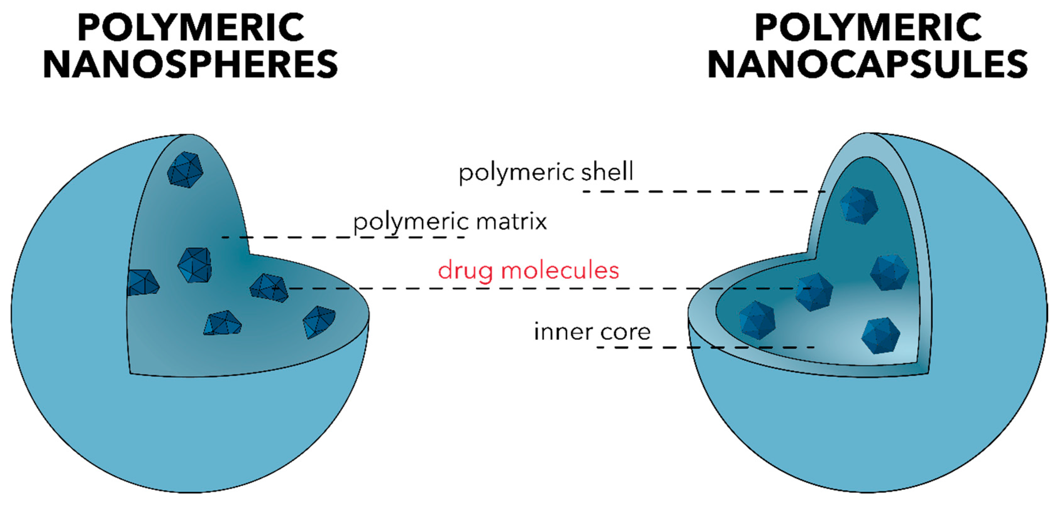
- Deng, S.; Gigliobianco, M.R.; Censi, R.; Di Martino, P. Polymeric Nanocapsules as Nanotechnological Alternative for Drug Delivery System: Current Status, Challenges and Opportunities. Nanomaterials 2020, 10, 847. [Google Scholar] [CrossRef] [PubMed]
- Zielińska, A.; Carreiró, F.; Oliveira, A.M.; Neves, A.; Pires, B.; Venkatesh, D.N.; Durazzo, A.; Lucarini, M.; Eder, P.; Silva, A.M.; et al. Polymeric Nanoparticles: Production, Characterization, Toxicology and Ecotoxicology. Molecules 2020, 25, 3731. [Google Scholar] [CrossRef]
- Allen, R.J.; Waclaw, B. Bacterial growth: A statistical physicist’s guide. Rep. Prog. Phys. 2019, 82, 16601. [Google Scholar] [CrossRef]
- Israni, N.; Shivakumar, S. Interface influence of materials and surface modifications. Fundam. Biomater. Met. 2018, 371–409. [Google Scholar] [CrossRef]
- Qiu, Y.; Xu, D.; Sui, G.; Wang, D.; Wu, M.; Han, L.; Mu, H.; Duan, J. Gentamicin decorated phosphatidylcholine-chitosan nanoparticles against biofilms and intracellular bacteria. Int. J. Biol. Macromol. 2020, 156, 640–647. [Google Scholar] [CrossRef] [PubMed]
- Alruwaili, N.K.; Zafar, A.; Imam, S.S.; Alharbi, K.S.; Alotaibi, N.H.; AlShehri, S.; Alhakamy, N.A.; AlZarea, A.I.; Afzal, M.; Elmowafy, M. Stimulus Responsive Ocular Gentamycin-Ferrying Chitosan Nanoparticles Hydrogel: Formulation Optimization, Ocular Safety and Antibacterial Assessment. Int. J. Nanomed. 2020, 15, 4717–4737. [Google Scholar] [CrossRef] [PubMed]
- Ciro, Y.; Rojas, J.; Oñate-Garzon, J.; Salamanca, C.H. Ciro Synthesis, Characterisation and Biological Evaluation of Ampicillin–Chitosan–Polyanion Nanoparticles Produced by Ionic Gelation and Polyelectrolyte Complexation Assisted by High-Intensity Sonication. Polymers 2019, 11, 1758. [Google Scholar] [CrossRef] [PubMed]
- Evangelista, T.F.; Andrade, G.R.; Nascimento, K.N.; Dos Santos, S.B.; Santos, M.D.F.C.; D’Oca, C.D.R.M.; Estevam, C.D.S.; Gimenez, I.F.; Almeida, L.E. Supramolecular polyelectrolyte complexes based on cyclodextrin-grafted chitosan and carrageenan for controlled drug release. Carbohydr. Polym. 2020, 245, 116592. [Google Scholar] [CrossRef] [PubMed]
- Walvekar, P.; Gannimani, R.; Salih, M.; Makhathini, S.; Mocktar, C.; Govender, T. Self-assembled oleylamine grafted hyaluronic acid polymersomes for delivery of vancomycin against methicillin resistant Staphylococcus aureus (MRSA). Colloids Surf. B Biointerfaces 2019, 182, 110388. [Google Scholar] [CrossRef]
- Oliveira, D.; Rezende, P.; Barbosa, T.; Nalone, L.; Bani, C.; Tavares, D.; Da Silva, C.; Chaud, M.; Padilha, F.; Cano, A.; et al. Double membrane based on lidocaine-coated polymyxin-alginate nanoparticles for wound healing: In vitro characterization and in vivo tissue repair. Int. J. Pharm. 2020, 591, 120001. [Google Scholar] [CrossRef] [PubMed]
- Ejaz, S.; Ihsan, A.; Noor, T.; Shabbir, S.; Imran, M. Mannose functionalized chitosan nanosystems for enhanced antimicrobial activity against multidrug resistant pathogens. Polym. Test. 2020, 91, 106814. [Google Scholar] [CrossRef]
- Kritchenkov, A.S.; Egorov, A.R.; Artemjev, A.A.; Kritchenkov, I.S.; Volkova, O.V.; Kurliuk, A.V.; Shakola, T.V.; Rubanik, V.V.; Tskhovrebov, A.G.; Yagafarov, N.Z.; et al. Ultrasound-assisted catalyst-free thiol-yne click reaction in chitosan chemistry: Antibacterial and transfection activity of novel cationic chitosan derivatives and their based nanoparticles. Int. J. Biol. Macromol. 2020, 143, 143–152. [Google Scholar] [CrossRef]
- Hadidi, M.; Pouramin, S.; Adinepour, F.; Haghani, S.; Jafari, S.M. Chitosan nanoparticles loaded with clove essential oil: Characterization, antioxidant and antibacterial activities. Carbohydr. Polym. 2020, 236, 116075. [Google Scholar] [CrossRef]
- Bagheri, R.; Ariaii, P.; Motamedzadegan, A. Characterization, antioxidant and antibacterial activities of chitosan nanoparticles loaded with nettle essential oil. J. Food Meas. Charact. 2020, 1–8. [Google Scholar] [CrossRef]
- Liakos, I.L.; Iordache, F.; Carzino, R.; Scarpellini, A.; Oneto, M.; Bianchini, P.; Grumezescu, A.M.; Holban, A.M. Cellulose acetate—essential oil nanocapsules with antimicrobial activity for biomedical applications. Colloids Surf. B Biointerfaces 2018, 172, 471–479. [Google Scholar] [CrossRef] [PubMed]
- Ivanova, K.; Ivanova, A.; Ramon, E.; Hoyo, J.; Sanchez-Gomez, S.; Tzanov, T. Antibody-Enabled Antimicrobial Nanocapsules for Selective Elimination of Staphylococcus aureus. Acs Appl. Mater. Interfaces 2020, 12, 35918–35927. [Google Scholar] [CrossRef]
- Bettencourt, A.F.; Tomé, C.; Oliveira, T.; Martin, V.; Santos, C.; Gonçalves, L.; Fernandes, M.H.; Gomes, P.S.; Ribeiro, I.A. Exploring the potential of chitosan-based particles as delivery-carriers for promising antimicrobial glycolipid biosurfactants. Carbohydr. Polym. 2021, 254, 117433. [Google Scholar] [CrossRef] [PubMed]
- Falciani, C.; Zevolini, F.; Brunetti, J.; Riolo, G.; Gracia, R.; Marradi, M.; Loinaz, I.; Ziemann, C.; Cossío, U.; Llop, J.; et al. Antimicrobial Peptide-Loaded Nanoparticles as Inhalation Therapy for Pseudomonas aeruginosa Infections. Int. J. Nanomed. 2020, 15, 1117–1128. [Google Scholar] [CrossRef]
- De Oliveira, M.S.; Oshiro-Junior, J.A.; Sato, M.R.; Conceição, M.M.; Medeiros, A.C.D. Polymeric Nanoparticle Associated with Ceftriaxone and Extract of Schinopsis Brasiliensis Engler against Multiresistant Enterobacteria. Pharmaceutics 2020, 12, 695. [Google Scholar] [CrossRef]
- Arzani, H.; Adabi, M.; Mosafer, J.; Dorkoosh, F.; Khosravani, M.; Maleki, H.; Nekounam, H.; Kamali, M. Preparation of curcumin-loaded PLGA nanoparticles and investigation of its cytotoxicity effects on human glioblastoma U87MG cells. Biointerface Res. Appl. Chem. 2018, 9, 4225–4231. [Google Scholar] [CrossRef]
- Ucak, S.; Sudagidan, M.; Borsa, B.A.; Mansuroglu, B.; Ozalp, V.C. Inhibitory effects of aptamer targeted teicoplanin encapsulated PLGA nanoparticles for Staphylococcus aureus strains. World J. Microbiol. Biotechnol. 2020, 36, 69. [Google Scholar] [CrossRef]
- Deepika, M.S.; Thangam, R.; Sundarraj, S.; Sheena, T.S.; Sivasubramanian, S.; Kulandaivel, J.; Thirumurugan, R. Co-delivery of Diverse Therapeutic Compounds Using PEG–PLGA Nanoparticle Cargo against Drug-Resistant Bacteria: An Improved Anti-biofilm Strategy. ACS Appl. Bio Mater. 2020, 3, 385–399. [Google Scholar] [CrossRef]
- Durak, S.; Arasoglu, T.; Ates, S.C.; Derman, S. Enhanced antibacterial and antiparasitic activity of multifunctional polymeric nanoparticles. Nanotechnology 2020, 31, 175705. [Google Scholar] [CrossRef]
- Parmar, A.; Kaur, G.; Kapil, S.; Sharma, V.; Sharma, S. Biogenic PLGA-Zinc oxide nanocomposite as versatile tool for enhanced photocatalytic and antibacterial activity. Appl. Nanosci. 2019, 9, 2001–2016. [Google Scholar] [CrossRef]
- Da Costa, D.; Exbrayat-Héritier, C.; Rambaud, B.; Megy, S.; Terreux, R.; Verrier, B.; Primard, C. Surface charge modulation of rifampicin-loaded PLA nanoparticles to improve antibiotic delivery in Staphylococcus aureus biofilms. J. Nanobiotechnology 2021, 19, 1–17. [Google Scholar] [CrossRef]
- Vrouvaki, I.; Koutra, E.; Kornaros, M.; Avgoustakis, K.; Lamari, F.N.; Hatziantoniou, S. Polymeric Nanoparticles of Pistacia lentiscus var. chia Essential Oil for Cutaneous Applications. Pharmaceutics 2020, 12, 353. [Google Scholar] [CrossRef] [PubMed]
- Srisang, S.; Nasongkla, N. Spray coating of foley urinary catheter by chlorhexidine-loadedpoly(ε-caprolactone) nanospheres: Effect of lyoprotectants, characteristics, and antibacterial activity evaluation. Pharm. Dev. Technol. 2018, 24, 402–409. [Google Scholar] [CrossRef]
- Li, X.; Wang, B.; Liang, T.; Wang, R.; Song, P.; He, Y. Synthesis of cationic acrylate copolyvidone-iodine nanoparticles with double active centers and their antibacterial application. Nanoscale 2020, 12, 21940–21950. [Google Scholar] [CrossRef] [PubMed]
- Montero, N.; Alhajj, M.J.; Sierra, M.; Oñate-Garzon, J.; Yarce, C.J.; Salamanca, C.H. Development of Polyelectrolyte Complex Nanoparticles-PECNs Loaded with Ampicillin by Means of Polyelectrolyte Complexation and Ultra-High Pressure Homogenization (UHPH). Polymers 2020, 12, 1168. [Google Scholar] [CrossRef]
- Gherasim, O.; Grumezescu, A.M.; Grumezescu, V.; Iordache, F.; Vasile, B.S.; Holban, A.M. Bioactive Surfaces of Polylactide and Silver Nanoparticles for the Prevention of Microbial Contamination. Materials 2020, 13, 768. [Google Scholar] [CrossRef]
- Grumezescu, A.M.; Stoica, A.E.; Dima-Bălcescu, M.-Ș.; Chircov, C.; Gharbia, S.; Baltă., C.; Roșu, M.; Herman, H.; Holban, A.M.; Ficai, A.; et al. Electrospun Polyethylene Terephthalate Nanofibers Loaded with Silver Nanoparticles: Novel Approach in Anti-Infective Therapy. J. Clin. Med. 2019, 8, 1039. [Google Scholar] [CrossRef]
- Grumezescu, V.; Negut, I.; Grumezescu, A.M.; Ficai, A.; Dorcioman, G.; Socol, G.; Iordache, F.; Truşcă, R.; Vasile, B.S.; Holban, A.M. MAPLE fabricated coatings based on magnetite nanoparticles embedded into biopolymeric spheres resistant to microbial colonization. Appl. Surf. Sci. 2018, 448, 230–236. [Google Scholar] [CrossRef]
- Grumezescu, V.; Negut, I.; Gherasim, O.; Birca, A.C.; Grumezescu, A.M.; Hudita, A.; Galateanu, B.; Costache, M.; Andronescu, E.; Holban, A.M. Antimicrobial applications of MAPLE processed coatings based on PLGA and lincomycin functionalized magnetite nanoparticles. Appl. Surf. Sci. 2019, 484, 587–599. [Google Scholar] [CrossRef]
- Ficai, D.; Grumezescu, V.; Fufă, O.M.; Popescu, R.C.; Holban, A.M.; Ficai, A.; Grumezescu, A.M.; Mogoanta, L.; Mogosanu, G.D.; Andronescu, E. Antibiofilm Coatings Based on PLGA and Nanostructured Cefepime-Functionalized Magnetite. Nanomaterials 2018, 8, 633. [Google Scholar] [CrossRef] [PubMed]
- Negut, I.; Grumezescu, V.; Ficai, A.; Grumezescu, A.M.; Holban, A.M.; Popescu, R.C.; Savu, D.; Vasile, B.S.; Socol, G. MAPLE deposition of Nigella sativa functionalized Fe3O4 nanoparticles for antimicrobial coatings. Appl. Surf. Sci. 2018, 455, 513–521. [Google Scholar] [CrossRef]
- Alamdaran, M.; Movahedi, B.; Mohabatkar, H.; Behbahani, M. In-vitro study of the novel nanocarrier of chitosan-based nanoparticles conjugated HIV-1 P24 protein-derived peptides. J. Mol. Liq. 2018, 265, 243–250. [Google Scholar] [CrossRef]
- Belgamwar, A.V.; Khan, S.A.; Yeole, P.G. Intranasal dolutegravir sodium loaded nanoparticles of hydroxypropyl-beta-cyclodextrin for brain delivery in Neuro-AIDS. J. Drug Deliv. Sci. Technol. 2019, 52, 1008–1020. [Google Scholar] [CrossRef]
- Costa, A.F.; Araujo, D.E.; Cabral, M.S.; Brito, I.T.; Leite, L.B.D.M.; Pereira, M.; Amaral, A.C. Development, characterization, andin vitro–in vivoevaluation of polymeric nanoparticles containing miconazole and farnesol for treatment of vulvovaginal candidiasis. Med Mycol. 2018, 57, 52–62. [Google Scholar] [CrossRef]
- Reddy, Y.C. Formulation and evaluation of chitosan nanoparticles for improved efficacy of itraconazole antifungal drug. Asian J. Pharm. Clin. Res. 2018, 11, 147–152. [Google Scholar] [CrossRef]
- Sombra, F.M.; Richter, A.R.; De Araújo, A.R.; Ribeiro, F.D.O.S.; Mendes, J.D.F.S.; Fontenelle, R.O.D.S.; Da Silva, D.A.; Paula, H.C.; Feitosa, J.P.; Goycoolea, F.M.; et al. Development of amphotericin B-loaded propionate Sterculia striata polysaccharide nanocarrier. Int. J. Biol. Macromol. 2020, 146, 1133–1141. [Google Scholar] [CrossRef] [PubMed]
- Real, D.; Hoffmann, S.; Leonardi, D.; Salomon, C.; Goycoolea, F.M. Chitosan-based nanodelivery systems applied to the development of novel triclabendazole formulations. PLoS ONE 2018, 13, e0207625. [Google Scholar] [CrossRef]


| Nanoparticle Type | Size Range [nm] | Zeta Potential [mV] | Targeted Bacteria | Targeting Strategy | Antibacterial Agent | Results | Ref. | ||
|---|---|---|---|---|---|---|---|---|---|
| Gram-Positive | Gram-Negative | Biofilm | |||||||
| phosphatidylcholine CS NPs | 137.2–231.8 | −27.6 to −31.8 | L. monocytogenes, S. aureus | P. aeruginosa, E. coli | L. monocytogenes, P. aeruginosa | passive | gentamycin | MIC results indicated similar antibacterial effects between the NPs and gentamycin alone; biofilm mass results showed a stronger inhibition capacity of the systems than gentamycin alone. | [114] |
| CS NPs and CS NPs dispersed into Carbopol sol–gel systems | 135.2 | +25.1 | S. aureus | E. coli | - | pH-responsive | gentamycin | ZOI was higher for NPs than for the marketed Gentacin eye drop, but lower than for sol–gel systems due to a sustained drug release in both bacterial types. | [115] |
| CS–polyanion NPs | 130.7–249.2 | +39.5 to +49.2 | S. aureus (ATCC25923, ATCC29213, and ATCC43300) | - | - | passive | ampicillin | MIC increased by 50% once the antibiotic was encapsulated into the NPs, independent of the ampicillin-resistance degree. | [116] |
| β-cyclodextrin-grafted CS and carrageenan SPECs | 10–60 | −40 to +42 | S. aureus (ATCC25923), E. durans/hirae (SS1225/ IAL 03/10) | K. pneumoniae (ATCC700603), E. coli (ATCC25922) | - | passive | silver sulfadiazine | ZOI for the drug-loaded SPECs was similar to the ZOI for the drug alone and gentamycin alone, especially in the case of Gram-positive bacteria; MIC values for the drug-loaded SPECs were equal to the values for the drug alone and half of the values for the gentamycin alone against both S. aureus and E. coli. | [117] |
| hyaluronic acid–oleylamine polymersomes | 201.4–360.9 | −20.4 to −17.6 | S. aureus and MRSA | - | - | passive | vancomycin | MIC values were considerably lower for the free gentamycin, but it lost its activity after 24 h; polymersomes were not as potent as the free vancomycin but were able to improve the antibacterial effects due to a slow and controlled release over a prolonged period of time. | [118] |
| Double-layer membrane comprising a sodium alginate NPs layer and a chitosan and hyaluronic acid layer | n.r. | n.r. | S. aureus (ATCC25923) | P. aeruginosa (ATCC27853) | - | passive | polymyxin B sulphate | MIC values for the NPs were lower than for the drug alone; MIC values for the biomembrane were lower than for the NPs due to the synergistic antibacterial effects of the components. | [119] |
| mannose-functionalized CS NPs | 180 | +25.4 | L. monocytogenes, S. aureus | E. coli, P. aeruginosa | L. monocytogenes, S. aureus, E. coli, P. aeruginosa | mannose-binding lectins | - | mannose functionalization increased inhibited bacterial growth more significantly due to the interaction with the bacterial membrane lectins; growth inhibition was higher for Gram-negative bacteria; NPs effectively reduced the adherence of bacteria in the polystyrene adherence assay; mannose-functionalized CS NPs exhibited the highest antibiofilm potential, as compared to the simple CS NPs, especially against E. coli and P. aeruginosa. | [120] |
| cationic betaine CS derivatives NPs | 108–807 | +33.1 to +69.1 | S. aureus | E. coli | - | passive | - | NPs possess higher antibacterial activity than pristine polymers; antibacterial activity is dependent upon the NPs size and the ξ-potential—smaller sizes and higher ξ-potentials leads to increased antibacterial activity. | [121] |
| CS NPs | 223.2–444.5 | +10.1 to +34.5 | L. monocytogenes, S. aureus | S. typhi, E. coli | - | passive | clove EOs | the highest inhibitory activity was achieved for EOs-encapsulated NPs, as compared to the pure EOs and unloaded NPs against all bacterial strains; IH values were higher for S. aureus and L. monocytogenes; MIV values were the lowest for the EOs-encapsulated NPs against all bacterial strains. | [122] |
| CS NPs | 208.3–369.4 | +14.4 to +30.1 | L. monocytogenes, S. aureus, B. cereus | S. typhi, E. coli | - | passive | nettle EOs | the highest inhibitory activity was achieved for EOs-encapsulated NPs, as compared to the pure EOs and unloaded NPs against all bacterial strains; IH values were higher for S. aureus; MIV values for the EOs-encapsulated NPs were similar to the values for the pure EOs and considerably lower than the unloaded NPs. | [123] |
| cellulose acetate NCs | 150–200 | −42 to −38 | S. aureus (ATCC25923) | P. aeruginosa (ATCC25324), E. coli (ATCC25922) | P. aeruginosa, E. coli, S. aureus | passive | peppermint, cinnamon, and lemongrass EOs | the most efficient were cinnamon EOs-encapsulated NCs, with significant growth inhibition of all bacterial strains, especially E. coli; peppermint EOs-encapsulated NCs demonstrated a low inhibitory activity against the growth of S. aureus and C. albicans; lemongrass EOs-encapsulated NCs slightly inhibited the development of E.coli; P. aeruginosa strain revealed the highest resistance to the tested NCs; lowest MIC values were obtained for the cinnamon EOs-encapsulated NCs; most significant antibiofilm formation was observed against S. aureus biofilms for cinnamon EOs-encapsulated NCs. | [124] |
| zein protein NCs | 134.9 | −28.6 | S. aureus (ATCC25923) | - | - | antibody-based targeting | oregano EOs | EOs encapsulation enhanced the antibacterial effects as compared to the pristine EOs; antibody attachment further enhanced the antibacterial activity; antibody attachment ensured a more specific activity against S. aureus co-cultured with the P. aeruginosa (ATCC10145) strain; antibody attachment inhibited S. aureus growth and protected human skin fibroblasts in co-culture. | [125] |
| CS NPs | 210.0/329.6 | +30.8/+37.4 | S. aureus (ATCC25923) | - | - | rhamnolipid-based targeting | sophorolipids and rhamnolipids | significantly higher MIC values for rhamnolipid-containing NPs and sophorolipid-containing NPs compared to the levofloxacin control; lower MIC values for both glycolipid-containing NPs compared to the unloaded NPs. | [126] |
| dextran NPs | 18 | −13 | - | P. aeruginosa (PAO1) | - | SET-M33 peptide | SET-M33 peptide | similar MIC values between the free peptide and the peptide-functionalized NPs; regrowth occurred after 24 h of exposure to the nanosystems. | [127] |
| CS and hydroxypropylmethylcellulose NPs | 440–1660 | +18.1 to +38.9 | - | E. coli (ATCC25922), E. coli producing extended-spectrum beta-lactamases, carbapenemase-producing K. pneumoniae | - | passive | ceftriaxone and S. brasiliensis extract | lowest MIC values for the nanosystems compared to the ceftriaxone-containing NPs and S. brasiliensis-containing NPs against all strains; lowest MBC values for the nanosystems compared to the ceftriaxone-containing NPs and S. brasiliensis-containing NPs against all strains. | [128] |
| Nanoparticle Type | Size Range [nm] | Zeta Potential [mV] | Targeted Bacteria | Targeting Strategy | Antibacterial Agent | Results | Ref. | ||
|---|---|---|---|---|---|---|---|---|---|
| Gram-Positive | Gram-Negative | Biofilm | |||||||
| PLGA NPs | 226 | -29 | S. aureus (ATCC29213, ATCC25923, ATCC43300), B. cereus (ATCC12228), MRSA (EGE-KK-13, EGE-KK-95) | - | - | aptamer-based targeting | teicoplanin | MIC values were considerably decreased upon the encapsulation of teicoplanin into the NPs for all bacterial strains; MIC values decreased even more after aptamer attachment for the S. aureus strains but considerably increased for the B. cereus. | [130] |
| PEG–PLGA NPs | 260–291 | −22.4 to −17.6 | S. aureus (MTCC96) | P. aeruginosa (MTCC2488) | S. aureus, P. aeruginosa | passive | rutin and benzamide | MIC values decreased with the encapsulation of the drugs into the NPs when compared to either drug alone; rutin and rutin-encapsulated NPs exhibited higher MIC values than benzamide and benzamide-encapsulated NPs, respectively; biofilm inhibition analysis followed a trend similar to the MIC assay. | [131] |
| PLGA NPs | 151.4–196.1 | −25.7 to −21.2 | S. aureus | E. coli | - | passive | caffeic acid and juglone | MIC values were similar or slightly lower for the drug-containing NPs; ZOI were similar or slightly lower for the drug-containing NPs. | [132] |
| PLGA–ZnO nanocomposites | 185.7 | −5.9 | S. aureus | E. coli | - | passive | - | ZOI were considerably higher for the nanocomposites than for the zinc oxide NPs or the standard antibiotic; ZOI were higher against S. aureus due to electrostatic interactions. | [133] |
| PLA NPs | 162 | +40 | S. aureus (SH1000) | - | S. aureus | poly-L-lysine attached on the surface | rifampicin | MIC values against planktonic S. aureus were similar for all tested systems, namely the free antibiotic, antibiotic-encapsulated NPs, and antibiotic-encapsulated NPs functionalized with poly-L-lysine; antibiofilm properties were similar for all tested systems, namely the free antibiotic, antibiotic-encapsulated NPs, and antibiotic-encapsulated NPs functionalized with poly-L-lysine; interactions between poly-L-lysine-functionalized nanoparticles are dose-dependent. | [134] |
| PLA NPs | 239.9/286.1 | −29.1/−34.5 | B. subtilis sub. spizizenii (DSM-347) | E. coli (DSM-1103) | - | passive | Pistacia lentiscus L. var. chia EOs | MIC values for the EOs-functionalized NPs were lower than for the EOs dissolved in organic solvents but higher than for gentamycin against E. coli and higher than all cases for B. subtilis. | [135] |
| PCL NSs | 152 | −10.2 | S. aureus (ATCC25423) | E. coli (ATCC25922) | - | passive | chlorhexidine | inhibition of 50% growth of the microorganisms up to 15 days. | [136] |
| cationic acrylate copolyvidone–iodine NPs | 200 | +11.7 | S. aureus | E. coli | - | passive | - | no bacterial growth in the presence of the NPs due to the synergistic effects of iodine and quaternary ammonium salts; NPs maintained antibacterial effects for 11 days; growth inhibition of S. aureus was lower than that of E. coli; NPs exhibited significant dose-dependent inhibitory effects. | [137] |
| PEC NPs | >200 | ≈0/>|40| | S. aureus (ATCC25923, ATCC29213, ATCC43300) | - | - | passive | ampicillin | different antibacterial behaviors depending on the family of the complex. | [138] |
Publisher’s Note: MDPI stays neutral with regard to jurisdictional claims in published maps and institutional affiliations. |
© 2021 by the authors. Licensee MDPI, Basel, Switzerland. This article is an open access article distributed under the terms and conditions of the Creative Commons Attribution (CC BY) license (http://creativecommons.org/licenses/by/4.0/).
Share and Cite
Spirescu, V.A.; Chircov, C.; Grumezescu, A.M.; Andronescu, E. Polymeric Nanoparticles for Antimicrobial Therapies: An up-to-date Overview. Polymers 2021, 13, 724. https://doi.org/10.3390/polym13050724
Spirescu VA, Chircov C, Grumezescu AM, Andronescu E. Polymeric Nanoparticles for Antimicrobial Therapies: An up-to-date Overview. Polymers. 2021; 13(5):724. https://doi.org/10.3390/polym13050724
Chicago/Turabian StyleSpirescu, Vera Alexandra, Cristina Chircov, Alexandru Mihai Grumezescu, and Ecaterina Andronescu. 2021. "Polymeric Nanoparticles for Antimicrobial Therapies: An up-to-date Overview" Polymers 13, no. 5: 724. https://doi.org/10.3390/polym13050724
APA StyleSpirescu, V. A., Chircov, C., Grumezescu, A. M., & Andronescu, E. (2021). Polymeric Nanoparticles for Antimicrobial Therapies: An up-to-date Overview. Polymers, 13(5), 724. https://doi.org/10.3390/polym13050724