Synthesis, Structural Characterization and Ligand-Enhanced Photo-Induced Color-Changing Behavior of Two Hydrogen-Bonded Ho(III)-Squarate Supramolecular Compounds

 ,
,  and
and
Abstract
1. Introduction
2. Materials and Methods
2.1. Materials and General Methods
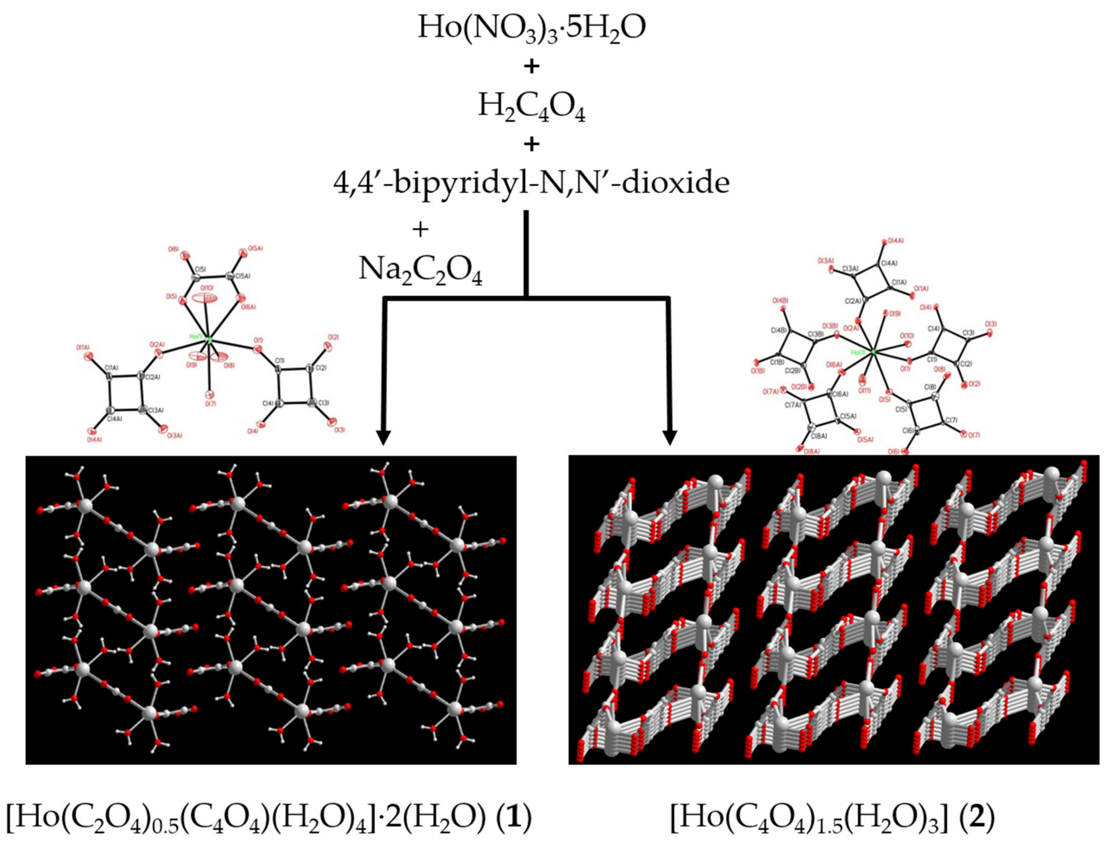
2.2. Synthesis of [Ho2(C2O4)(C4O4)2(H2O)8]·4H2O (1) and [Ho(C4O4)1.5(H2O)3]·H2O (2)
2.3. Crystallographic Data Collection and Refinements
2.4. In Situ Powder X-ray Diffraction
2.5. Spectral Measurement
3. Results and Discussion
3.1. Syntheses and Characterization of Compounds 1 and 2
3.2. Structure Description of {[Ho(C2O4)0.5(C4O4)(H2O)4]·2H2O}n (1)
3.3. Structure Description of [Ho(C4O4)1.5(H2O)3]n (2)
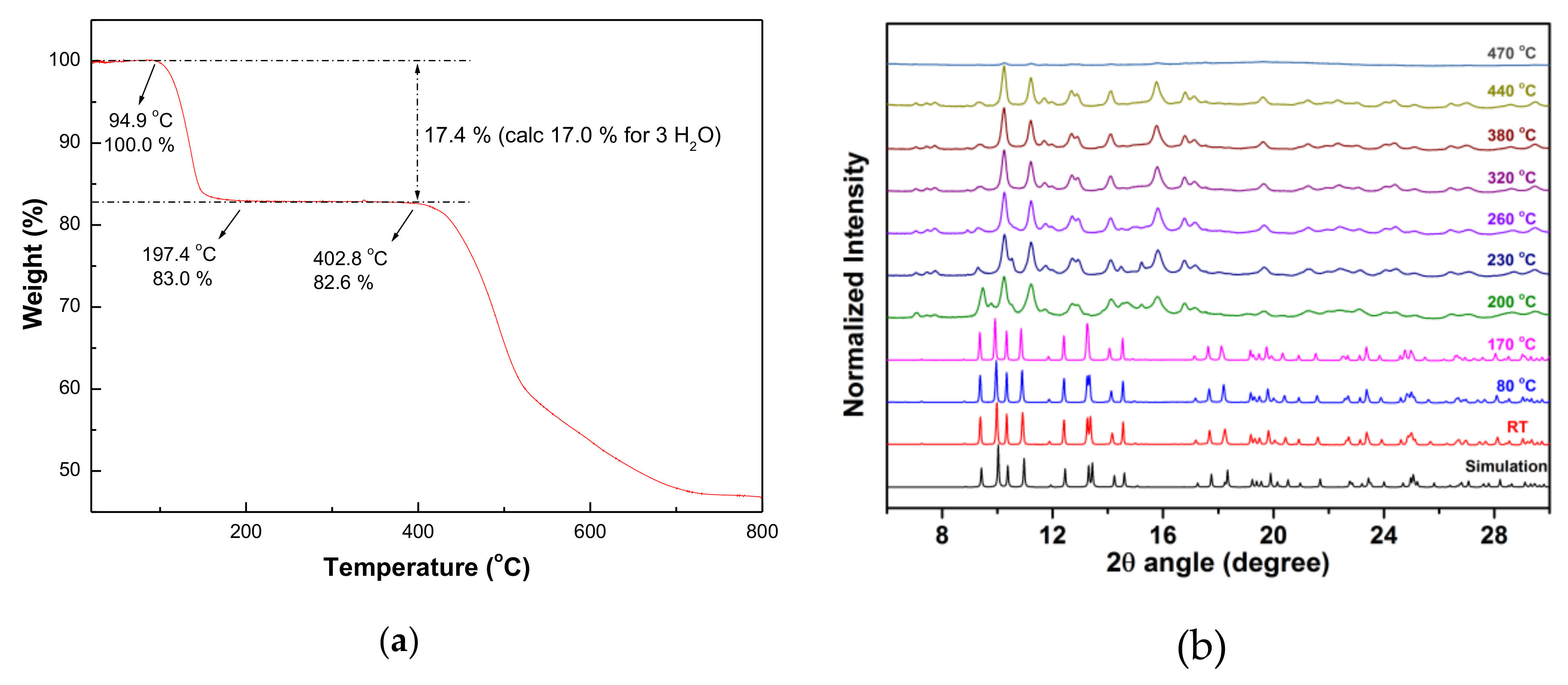
3.4. Thermal Stability of CPs 1 and 2
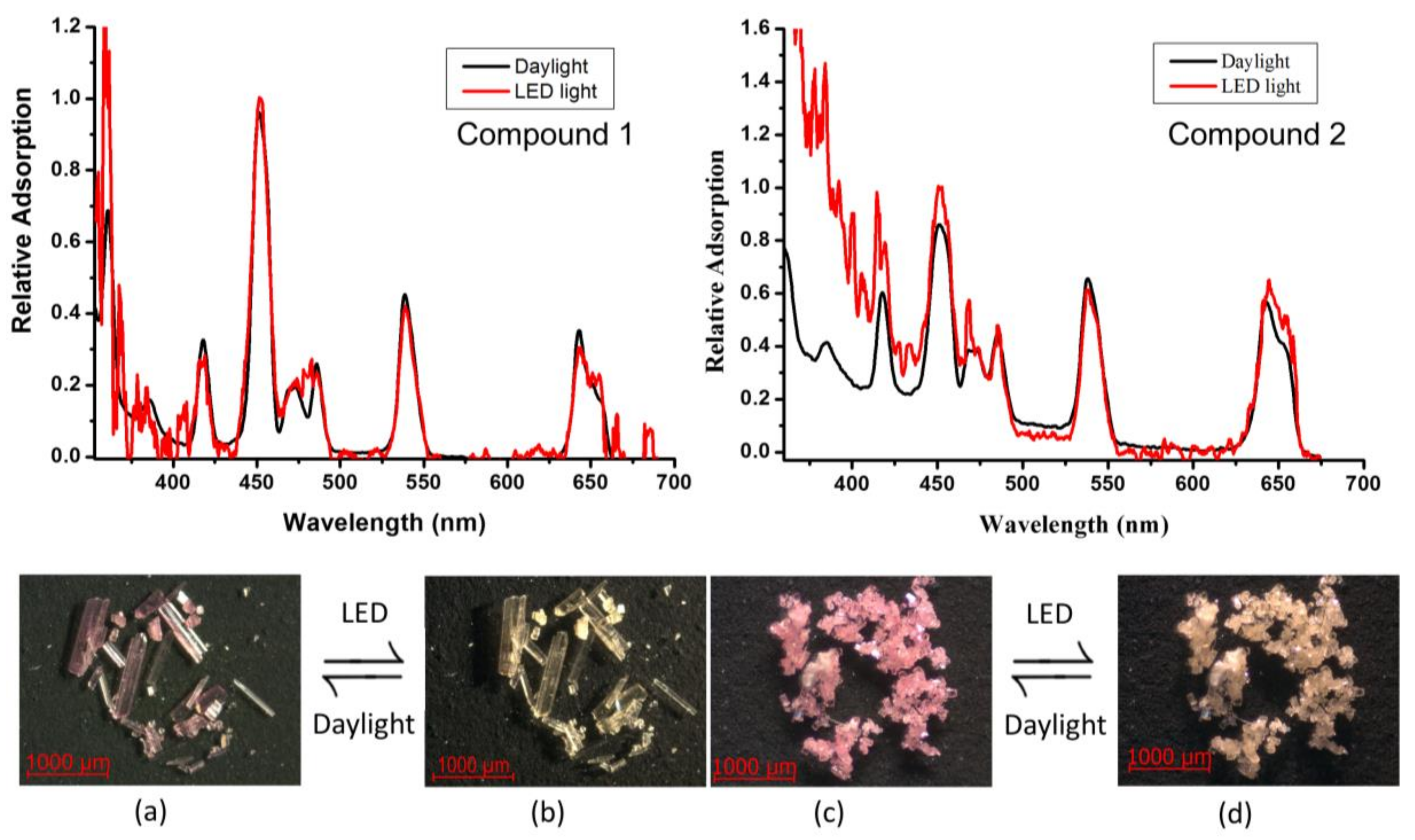
3.5. UV-Visible Spectroscopy of CPs 1 and 2
4. Conclusions
Author Contributions
Funding
Acknowledgments
Conflicts of Interest
References
- Bradshaw, D.; Claridge, J.B.; Cussen, E.J.; Prior, T.J.; Rosseinsky, M.J. Design, Chirality and Flexibility in Nanoporous Molecule-Based Materials. Acc. Chem. Res. 2015, 38, 273–282. [Google Scholar] [CrossRef] [PubMed]
- Kitagawa, S.; Kitaura, R.; Noro, S.I. Functional Porous Coordination Polymers. Angew. Chem. Int. Ed. 2004, 43, 2334–2375. [Google Scholar] [CrossRef] [PubMed]
- Uemura, T.; Horike, S.; Kitagawa, S. Polymerization in Coordination Nanospaces. Chem. Asian J. 2006, 1, 36–44. [Google Scholar] [CrossRef] [PubMed]
- Férey, G. Hybrid Porous Solids: Past, Present, Future. Chem. Soc. Rev. 2008, 37, 191–214. [Google Scholar] [CrossRef] [PubMed]
- Wang, B.; Côté, A.P.; Furukawa, H.; O’Keeffe, M.; Yaghi, O.M. Colossal Cages in Zeolitic Imidazolate Frameworks as Selective Carbon Dioxide reservoirs. Nature 2008, 453, 207–211. [Google Scholar] [CrossRef] [PubMed]
- Rosi, N.L.; Kim, J.; Eddaoudi, M.; Chen, B.L.; O’Keeffe, M.; Yaghi, O.M. Rod Packings and Metal-Organic Frameworks Constructed from Rod-Shaped Secondary Building Units. J. Am. Chem. Soc. 2005, 127, 1504–1518. [Google Scholar] [CrossRef] [PubMed]
- Hayashi, H.; Côté, A.P.; Furukawa, H.; O’keeffe, M.; Yaghi, O.M. Zeolite, A Imidazolate Frameworks. Nat. Mater. 2007, 6, 501–506. [Google Scholar] [CrossRef]
- Thallapally, P.K.; Mcgrail, B.P.; Dalgarno, S.J.; Schaef, H.T.; Tian, J.; Atwood, J.L. Gas-Induced Transformation and Expansion of a Non-Porous Organic Solid. Nat. Mater. 2008, 7, 146–150. [Google Scholar] [CrossRef]
- Banerjee, R.; Phan, A.; Wang, B.; Knobler, C.; Furukawa, H.; O’Keeffe, M.; Yaghi, O.M. High-Throughput Synthesis of Zeolitic Imidazolate Frameworks and Application to CO2 Capture. Science 2008, 319, 939–943. [Google Scholar] [CrossRef]
- Benelli, C.; Gatteschi, D. Magnetism of Lanthanides in Molecular Materials with Transition-Metal Ions and Organic Radicals. Chem. Rev. 2002, 102, 2369–2388. [Google Scholar] [CrossRef]
- Bünzli, J.C.G.; Piguet, C. Taking Advantage of Luminescent Lanthanide ions. Chem. Soc. Rev. 2005, 34, 1048–1077. [Google Scholar] [CrossRef]
- Chandler, B.D.; Cram, D.T.; Shimizu, G.K.H. Microporous Metal-Organic Frameworks Formed in a Stepwise Manner from Luminescent Building Blocks. J. Am. Chem. Soc. 2006, 128, 10403–10412. [Google Scholar] [CrossRef]
- Wang, P.; Ma, J.P.; Dong, Y.B.; Huang, R.Q. Tunable Luminescent Lanthanide Coordination Polymers Based on Reversible Solid-State Ion-Exchange Monitored by Ion-Dependent Photoinduced Emission Spectra. J. Am. Chem. Soc. 2007, 129, 10620–10621. [Google Scholar] [CrossRef]
- Guo, X.D.; Zhu, G.S.; Fang, Q.R.; Xue, M.; Tian, G.; Sun, J.Y.; Li, X.T.; Qiu, S.L. Synthesis, Structure and Luminescent Properties of Rare Earth Coordination Polymers Constructed from Paddle-Wheel Building Blocks. Inorg. Chem. 2005, 44, 3850–3855. [Google Scholar] [CrossRef]
- Guo, X.D.; Zhu, G.S.; Li, Z.Y.; Chen, Y.; Li, X.T.; Qiu, S.L. Rare Earth Coordination Polymers with Zeolite Topology Constructed from 4-Connected Building Units. Inorg. Chem. 2006, 45, 4065–4070. [Google Scholar] [CrossRef]
- Devic, T.; Serre, C.; Audebrand, N.; Marrot, J.; Férey, G. MIL-103, A 3-D Lanthanide-Based Metal Organic Framework with Large One-Dimensional Tunnels and A High Surface Area. J. Am. Chem. Soc. 2005, 127, 12788–12789. [Google Scholar] [CrossRef]
- Harbuzaru, B.V.; Corma, A.; Rey, F.; Atienzar, P.; Jordá, J.L.; García, H.; Ananias, D.; Carlos, L.D.; Rocha, J. Metal-Organic Nanoporous Structures With Anisotropic Photoluminescence and Magnetic Properties and Their Use as Sensors. Angew. Chem. Int. Ed. 2008, 47, 1080–1083. [Google Scholar] [CrossRef]
- Wang, J.G.; Huang, C.C.; Huang, X.H.; Liu, D.S. Three-Dimensional Lanthanide Thiophenedicarboxylate Framework with an Unprecedented (4,5)-Connected Topology. Cryst. Growth Des. 2008, 8, 795–798. [Google Scholar] [CrossRef]
- Liu, Y.L.; Kravtsov, V.C.; Eddaoudi, M. Template-Directed Assembly of Zeolite-Like Metal-Organic Frameworks (ZMOFs): A Usf-ZMOF with an Unprecedented Zeolite Topology. Angew. Chem. Int. Ed. 2008, 47, 8446–8449. [Google Scholar] [CrossRef]
- Pan, L.; Woodlock, E.B.; Wang, X. A New Porous Three-Dimensional Lanthanide Coordination Polymer. Inorg. Chem. 2000, 39, 4174–4178. [Google Scholar] [CrossRef]
- Lipstman, S.; Muniappan, S.; George, S.; Goldberg, I. Framework Coordination Polymers of Tetra(4-carboxyphenyl)porphyrin and Lanthanide Ions in Crystalline Solids. Dalton Trans. 2007, 30, 3273–3281. [Google Scholar] [CrossRef]
- Muniappan, S.; Lipstman, S.; George, S.; Goldberg, I. Porphyrin Framework Solids. Synthesis and Structure of Hybrid Coordination Polymers of Tetra(carboxyphenyl)porphyrins and Lanthanide-Bridging Ions. Inorg. Chem. 2007, 46, 5544–5554. [Google Scholar] [CrossRef]
- He, Y.; Furukawa, H.; Wu, C.; O’Keeffe, M.; Chen, B. A Mesoporous Lanthanide-Organic Framework Constructed from a Dendritic Hexacarboxylate with Cages of 2.4 nm†. CrystEngComm 2013, 15, 9328–9331. [Google Scholar] [CrossRef]
- He, Y.; Furukawa, H.; Wu, C.; O’Keeffe, M.; Krishna, R.; Chen, B. Low-energy regeneration and high productivity in a lanthanide–hexacarboxylate framework for high-pressure CO2–CH4–H2 separation. Chem. Commun. 2013, 49, 6773–6775. [Google Scholar] [CrossRef][Green Version]
- He, H.Y.; Ma, H.Q.; Sun, D.; Zhang, L.L.; Wang, R.M.; Sun, D.F. Porous Lanthanide-Organic Frameworks: Control over Interpenetration, Gas Adsorption and Catalyst Properties. Cryst. Growth Des. 2013, 13, 3154–3161. [Google Scholar] [CrossRef]
- Mihalcik, D.J.; Zhang, T.; Ma, L.; Lin, W. Highly Porous 4,8-Connected Metal-Organic Frameworks: Synthesis, Characterization and Hydrogen Uptake. Inorg. Chem. 2012, 51, 2503–2508. [Google Scholar] [CrossRef]
- Decadt, R.; Van Hecke, K.; Depla, D.; Leus, K.; Weinberger, D.; Van Driessche, I.; Van Der Voort, P.; Van Deun, R. Synthesis, Crystal Structures and Luminescence Properties of Carboxylate Based Rare-Earth Coordination Polymers. Inorg. Chem. 2012, 51, 11623–11634. [Google Scholar] [CrossRef]
- Yan, L.; Yue, Q.; Jia, Q.X.; Lemercier, G.; Gao, E.Q. Lanthanide Metal-Organic Frameworks Based on Octahedral Secondary Building Units: Rare Net Topology and Luminescence. Cryst. Growth Des. 2009, 9, 2984–2987. [Google Scholar] [CrossRef]
- Yang, J.; Yue, Q.; Li, G.D.; Cao, J.J.; Li, G.H.; Chen, J.S. Structures, Photoluminescence, Up-Conversion and Magnetism of 2D and 3D Rare-Earth Coordination Polymers with Multicarboxylate Linkages. Inorg. Chem. 2006, 45, 2857–2865. [Google Scholar] [CrossRef]
- Khan, M.I.; Chang, Y.D.; Chen, Q.; Salta, J.; Lee, Y.S.; O’Connor, C.J.; Zubieta, J. Synthesis and Characterization of Binuclear Oxo-Vanadium Complexes of Carbon Oxoanion Ligands. Crystal Structures of the Binuclear Vanadium(IV) Complex (NH4)[V2O2(OH)(C4O4)2(H2O)3].cntdot.H2O, of the Mixed-Valence Vanadium(V)/Vanadium(IV)-Squarate Species [(n-C4H9)4N] [V2O3(C4O4)2(H2O)3].cntdot.3H2O and [(C4H9)4N]4[V4O6(C4O4)5(H2O)4]. cntdot. 6H2O and of the Binuclear Vanadium(IV)-Oxalate Species [V2O2Cl2(C2O4)(CH3OH)4].cntdot.2Ph4PCl. Inorg. Chem. 1994, 33, 6340–6350. [Google Scholar]
- Lee, C.R.; Wang, C.C.; Wang, Y. Structural Relationship of Various Squarates. Acta. Cryst. 1996, 52, 966–975. [Google Scholar] [CrossRef]
- Trombe, J.C.; Petit, J.-F.; Gleizes, A. Lanthanide Oxalato-Squarates-Preparation and Crystal-Structures of (CE2 (H2O) 8 (C4O4) 2 (C2O4)). 3H2O and EU2 (H2O) 4 (C4O4) 2 (C2O4). Eur. J. Solid State Inorg. Chem. 1991, 28, 669–681. [Google Scholar]
- Trombe, J.C.; Sabadie, L.; Millet, P. Synthesis and Crystal Structure of La(H2O)(C2O4)2·(CN3H6) and of [Nd(H2O)]2(C2O4)4·(NH4)(CN3H6). Solid State Sci. 2002, 4, 1199–1208. [Google Scholar]
- Soules, R.; Dahan, F.J.; Laurent, P.; Castan, P. A Novel Co-Ordination Mode for the Squarate Ligand [Dihydroxycyclobutenedionate(2–)]: Synthesis, Crystal Structure and Magnetic Properties of Catena-Diaqua(2,2′-Bipyridyl)-µ-(Squarato- O1, O2)-Nickel( II) Dihydrate. J. Chem. Soc. Dalton Trans. 1988, 3, 587–590. [Google Scholar] [CrossRef]
- Hall, L.A.; Williams, D.J.; Menzer, S.; White, A.J.P. The Complexing Properties of 1-Aminosquarate Derivatives with Lead. Inorg. Chem. 1997, 36, 3096–3101. [Google Scholar] [CrossRef]
- Alleyne, B.D.; Hall, L.A.; Hosein, H.A.; Jaggernauth, H.; White, A.J.P.; Williams, D.J. Hydrogen-Bonding Interactions in the Deries of Complexes [M(C4O4)(OH2)2(dmf)2] and [M(C4O4)(OH2)4] (M = Mn, Co, Ni, Cu, Zn). J. Chem. Soc. Dalton Trans. 1998, 22, 3845–3850. [Google Scholar] [CrossRef]
- Lai, S.F.; Cheng, C.Y.; Lin, K. Hydrothermal Synthesis of a Thermally Stable Poroussupramolecular π–π Framework:[{Co2(C12H8N2)4(μ-C4O4)(OH2)2}C4O4]·8H2O. J. Chem. Commun. 2001, 1082–1083. [Google Scholar] [CrossRef]
- Grove, H.; Sletten, J.; Julve, M.; Lloret, F.; Cano, J. Syntheses, Crystal Structures and Magnetic Properties of One- and Two-Dimensional Pap-Containing Copper(II) Complexes (Pap = Pyrazino[2–f][4,7]Phenanthroline). J. Chem. Soc. Dalton Trans. 2001, 3, 259–265. [Google Scholar] [CrossRef]
- Nather, C.; Greve, J.; Jess, I. New Coordination Polymer Changing Its Color upon Reversible Deintercalation and Reintercalation of Water: Synthesis, Structure and Properties of Poly[Diaqua-(μ2-Squarato-O,O‘)-(μ2-4,4‘-Bipyridine-N,N‘)- Manganese(II)] Trihydrate. Chem. Mater. 2002, 14, 4536–4542. [Google Scholar] [CrossRef]
- Piggott, P.M.T.; Hall, L.A.; White, A.J.P.; Williams, D.J. Attempted Syntheses of Lanthanide(III) Complexes of the Anisole- and Anilinosquarate Ligands. Inorg. Chem. 2003, 42, 8344–8352. [Google Scholar] [CrossRef]
- Yang, B.P.; Mao, J.G. New Types of Metal Squarato-phosphonates: Condensation of Aminodiphosphonate with Squaric Acid under Hydrothermal Conditions. Inorg. Chem. 2005, 44, 566–571. [Google Scholar] [CrossRef]
- Konar, S.; Corbella, M.; Zangrando, E.; Ribas, J.; Chaudhuri, N.R. The First Unequivocally Ferromagnetically Coupled Squarato Complex: Origin of the Ferromagnetism in an Interlocked 3D Fe(II) System. Chem. Commun. 2003, 12, 1424–1425. [Google Scholar] [CrossRef]
- Ghosh, A.K.; Ghoshal, D.; Zangrando, E.; Ribas, J.; Chaudhuri, N.R. Structural Diversity in Manganese Squarate Frameworks Using N,N-Donor Chelating/Bridging Ligands: Syntheses, Crystal Structures and Magnetic Properties. Dalton Trans. 2006, 12, 1554–1563. [Google Scholar] [CrossRef]
- Chen, Q.; Liu, S.; Zubieta, J. Coordination Chemistry of Polyoxomolybdates: The Structure of a Dodecanuclear Molybdate Cage Incorporating Hydrogen Squarate Ligands, [(C4H9)4N]4 [Mo12O36(C4O4H)4] · 10Et2O. Angew Chem. Int. Ed. 1990, 29, 70–72. [Google Scholar] [CrossRef]
- Lin, K.J.; Lii, K.H. Binuclear Vanadium(III) Squarates with Layered and Framework Structures: Hydrothermal Synthesis and Structures of [{V(OH)(C4O4)(H2O)}2] and [{V(OH)(C4O4)}2]·4H2O. Angew. Chem. Int. Ed. Engl. 1997, 36, 2076–2077. [Google Scholar] [CrossRef]
- Hosein, H.A.; Hall, L.A.; Lough, A.J.; Desmarais, W.; Vela, M.J.; Foxman, B.M. Attempted Syntheses of Transition Metal and Lanthanide (Dialkylamino)squarates. The Hydrolysis Problem. Inorg. Chem. 1998, 37, 4184–4189. [Google Scholar] [CrossRef]
- Hosein, H.A.; Jaggernauth, H.; Alleyne, B.D.; Hall, L.A.; White, A.J.P.; Williams, D.J. First-Row Transition-Metal Complexes of the 1-Methoxycyclobutenedionate(1−) Ion. Inorg. Chem. 1999, 38, 3716–3720. [Google Scholar] [CrossRef]
- Spandl, J.; Brüdgam, I.; Hartl, H. Solvothermal Synthesis of a 24-Nuclear, Cube-Shaped Squarato-oxovanadium (IV) Framework: [N(nBu)4]8[V24O24(C4O4)12(OCH3)32]. Angew. Chem. Int. Ed. Engl. 2001, 40, 4018–4020. [Google Scholar] [CrossRef]
- Maji, T.K.; Mostafa, G.; Sain, S.; Prasad, J.S.; Chaudhuri, N.R. Construction of a 3D Array of Cadmium(II) Using Squarate as a Building Block. CrystEngComm 2001, 3, 155–158. [Google Scholar] [CrossRef]
- Mukherjee, P.S.; Konar, S.; Zangrando, E.; Diaz, C.; Chaudhuri, N.R. Synthesis, Crystal Structure and Magneto-Structural Correlation of Two Bi-Bridging 1D Copper(II) Chains. J. Chem. Soc. Dalton Trans. 2002, 18, 3471–3476. [Google Scholar] [CrossRef]
- Neeraj, S.; Noy, M.L.; Rao, C.N.R.; Cheetham, A.K. Sodalite Networks Formed by Metal Squarates. Solid State Sci. 2002, 4, 1231–1236. [Google Scholar] [CrossRef]
- Dan, M.; Rao, C.N.R. An Open-Framework Cobalt Oxalato-Squarate Containing a Ligated Amine. Solid State Sci. 2003, 5, 615–620. [Google Scholar] [CrossRef]
- Kurmoo, M.; Kumagai, H.; Chapman, K.W.; Kepert, C.J. Reversible Ferromagnetic-Antiferromagnetic Transformation Upon Dehydration-Hydration of the Nanoporous Coordination Framework, [Co3(OH)2(C4O4)2]·3H2O. Chem. Commun. 2005, 3012–3014. [Google Scholar] [CrossRef]
- Gandara, F.; Gomez-Lor, B.; Iglesias, M.; Snejko, N.; Gutierrez-Puebla, E. A New Scandium Metal Organic Framework Built up from Octadecasil Zeolitic Cages as Heterogeneous Catalyst. Chem. Commun. 2009, 2393–2395. [Google Scholar] [CrossRef]
- Gutschke, S.O.H.; Molinier, M.; Powell, A.K.; Wood, P.T. Hydrothermal Synthesis of Microporous Transition Metal Squarates: Preparation and Structure of [CO3(μ3-OH)2(C4O4)2]·3H2O. Angew. Chem. Int. Ed. Engl. 1997, 36, 991–992. [Google Scholar] [CrossRef]
- Yufit, D.S.; Price, D.J.; Howard, J.A.K.; Gutschke, S.O.H.; Powell, A.K.; Wood, P.T. New Type of Metal Squarates. Magnetic and Multi-Temperature X-ray Study of Di-Hydroxy(µ6-squarato)manganese†. Chem. Commun. 1999, 16, 1561–1562. [Google Scholar] [CrossRef]
- Trombe, J.C.; Sabadie, L.; Millet, P. Hydrothermal Synthesis and Structural Characterization of Fe(II)-squarate Fe2(OH)2(C4O4). Solid State Sci. 2002, 4, 1209–1212. [Google Scholar] [CrossRef]
- Yang, C.H.; Chou, C.M.; Lee, G.H.; Wang, C.C. Self-Assembly of Two Mixed-Ligands Metal-Organic Coordination Polymers,[MII2 (DPA)2(C4O4)(C2O4)](M= Cu, Zn). Inorg. Chem. Commun. 2003, 6, 135–140. [Google Scholar] [CrossRef]
- Wang, C.C.; Yang, C.H.; Tseng, S.M.; Lee, G.H.; Sheu, H.S.; Phyu, K.W. A New Moisture-Sensitive Metal-Coordination Solids {[Cd(C4O4)(bipy)(H2O)2] 3H2O}∞ (bipy = 4,4′-bipyridine). Inorg. Chim. Acta 2004, 357, 3759–3764. [Google Scholar] [CrossRef]
- Wang, C.C.; Yang, C.H.; Lee, G.H.; Tsai, H.L. Syntheses, Structures and Magnetic Properties of Two 1D, Mixed-Ligand,Metal Coordination Polymers, [M(C4O4)(dpa)(OH2)] (M = CoII, NiII and ZnII; dpa = 2,2’-dipyridylamine) and [Cu(C4O4)(dpa)(H2O)]2·(H2O). Eur. J. Inorg. Chem. 2005, 7, 1334–1342. [Google Scholar] [CrossRef]
- Wang, C.C.; Yang, C.H.; Lee, G.H. Hydrothermal Synthesis and Structural Characterization of Two pH-Controlled Cd–Squarate Coordination Frameworks, [Cd2(C4O4)2.5(H2O)4]·(dpaH)·1.5(H2O) and [Cd(C4O4)(dpa)(OH2)] (dpa = 2,2′-dipyridylamine). Eur. J. Inorg. Chem. 2006, 4, 820–826. [Google Scholar] [CrossRef]
- Wang, C.C.; Tseng, S.M.; Lin, S.Y.; Liu, F.C.; Dai, S.C.; Lee, G.H.; Shih, W.J.; Sheu, H.S. Assemblies of Two Mixed-Ligand Coordination Polymers with Two-Dimensional Metal-Organic Frameworks Constructed from M(II) Ions with Croconate and 1,2-Bis-(4-pyridyl)ethylene (M = Cd and Zn). Cryst. Growth Des. 2007, 7, 1783–1790. [Google Scholar] [CrossRef]
- Wang, C.C.; Ke, S.Y.; Chen, K.T.; Hsieh, Y.F.; Wang, T.H.; Lee, G.H.; Chuang, Y.C. Reversible Single-Crystal-to-Single-Crystal Structural Transformation in a Mixed-Ligand 2D Layered Metal-Organic Framework: Structural Characterization and Sorption Study. Crystals 2017, 7, 364. [Google Scholar] [CrossRef]
- Lin, X.Y.; Zhao, L.M.; Wang, D.H.; Wang, Y.K.; Li, M.; Li, H.H.; Chen, Z.R. Structural Diversities of Squarate-Based Complexes: Photocurrent Responses and Thermochromic Behaviours Enhanced by Viologens. Inorg. Chem. Front. 2018, 5, 189–199. [Google Scholar] [CrossRef]
- Liu, C.M.; Xiang, D.Z.; Hao, X.; Zhu, D.B. Field-Induced Relaxation of Magnetization in a Three-Dimensional LnMOF with the Second Bridging Ligand Squarate. ACS Omega 2016, 1, 286–292. [Google Scholar] [CrossRef]
- Wang, L.; Gu, W.; Deng, X.J.; Zeng, L.F.; Liao, S.Y.; Zhang, M.; Yang, L.Y.; Liu, X. A Series of 2D and 3D Novel Lanthanide Complexes Constructed from Squarate C4O42−: Syntheses, Structures, Magnetic Properties and Near-infrared Emission Properties. Aust. J. Chem. 2011, 64, 1373–1382. [Google Scholar] [CrossRef]
- Wang, C.M.; Lii, K.H. Synthesis of Novel Organic-Inorganic Hybrid Compounds: Lanthanide Phosphites Incorporating a Squarate Ligand. Inorg. Chem. 2009, 48, 6335–6337. [Google Scholar] [CrossRef]
- Akkari, H.; Bénard-Rocherullé, P.; Mérazig, H.; Roisnel, T.; Rocherullé, J. Hydrothermal Synthesis, Crystal Structure and Thermal Behavior of the First Lanthanide Sulfato-Squarate, La2(H2O)4(SO4)2(C4O4). Solid State Sci. 2006, 8, 704–715. [Google Scholar] [CrossRef]
- Mahé, N.; Bataille, T. Synthesis, Crystal Structure from Single-Crystal and Powder X-ray Diffraction Data and Thermal Behavior of Mixed Potassium Lanthanide Squarates: Thermal Transformations of layered [Ln(H2O)6]K(H2C4O4)(C4O4)2 into Pillared LnK(C4O4)2 (Ln = Y, La, Gd, Er). Inorg. Chem. 2004, 43, 8379–8386. [Google Scholar] [CrossRef]
- Heinl, U.; Hinse, P.; Mattes, R. Oxalato- and Quadratato-Komplexe Hochkoordinierter Metallionen: Die Raumnetzstrukturten von La2(C2O4)(C4O4)(H2O)8⋅2,5H2O und K[Bi(C2O4)2⋅5H2O. Z. Anorg. Allg. Chem. 2001, 627, 2173–2177. [Google Scholar] [CrossRef]
- SMART V 4 043 Software for CCD Detector System; Siemens Analytical Instruments Division: Madison, WI, USA, 1995.
- SAINT V 4 035 Software for CCD Detector System; Siemens Analytical Instruments Division: Madison, WI, USA, 1995.
- Sheldrick, G.M. Program for the Refinement of Crystal Structures. Shelxl97 1997. [Google Scholar]
- SHELXTL 5 03 (PC-Version), Program Liberary for Structure Solution and Molecular Graphics; Siemens Analytical Instruments Division: Madison, WI, USA, 1995.
- Toby, B.H.; Von Dreele, R.B. GSAS-II: The Genesis of a Modern Open-Source All Purpose Crystallography Software Package. J. Appl. Crystallogr. 2013, 46, 544–549. [Google Scholar] [CrossRef]
- Chen, Y.T.; Lin, C.Y.; Lee, G.H.; Ho, M.L. Four New Lead(II)–Iridium(III) Heterobimetallic Coordination Frameworks: Synthesis, Structures, Luminescence and Oxygen-Sensing Properties. CrystEngComm 2015, 17, 2129–2140. [Google Scholar] [CrossRef]
- Fabre, P.L.; Castan, P.; Deguenon, D.; Paillous, N. A Photo-Oxidation of Croconic Acid into Oxalic Acid. Can. J. Chem. 1995, 73, 1298–1304. [Google Scholar] [CrossRef]
- Reinoso, S.; Vitoria, P.; Felices, L.S.; Montero, A.; Lezama, L.; Gutirrez-Zorrilla, M.M. NEXTTetrahydroxy-p-benzoquinone as a Source of Polydentate O-Donor Ligands. Synthesis, Crystal Structure and Magnetic Properties of the [Cu(bpy)(dhmal)]2 Dimer and the Two-Dimensional [{SiW12O40}{Cu2(bpy)2(H2O)(ox)}2]·16H2O Inorganic-Metalorganic Hybrid. Inorg. Chem. 2007, 46, 1237–1249. [Google Scholar] [CrossRef]
- Ke, S.Y.; Yeh, C.T.; Wang, C.C.; Lee, G.H.; Hwo-Shuenn, S. A 3d-4f Complex Constructed by the Assembly of a Cationic Template, [Cu(en)2]2+ and a 3D Anionic Coordination Polymer, [Sm2(C2O4)3(C5O5)(H2O)2]2−. Z. Anorg. Allg. Chem. 2017, 643, 657–663. [Google Scholar] [CrossRef]
- Ito, M.; West, R. New Aromatic Anions. IV. Vibrational Spectra and Force Constants for C4O4−2 and C5O5−2. J. Am. Chem. Soc. 1963, 85, 2580–2584. [Google Scholar] [CrossRef]
- Binnemans, K.; Görller-Walrand, C. On the Color of the Trivalent Lanthanide Ions. Chem. Phys. Lett. 1995, 235, 163–174. [Google Scholar] [CrossRef]
- Marques, L.F.; Correa, C.C.; Ribeiro, S.J.L.; dos Santos, M.V.; Dutra, J.D.L.; Freire, R.O.; Machado, F.C. Synthesis, Structural Characterization, Luminescent Properties and Theoretical Study of Three Novel Lanthanide Metal-Organic Frameworks of Ho(III), Gd(III) and Eu(III) with 2,5-Thiophenedicarboxylate Anion. J. Solid State Chem. 2015, 27, 68–78. [Google Scholar] [CrossRef]


| Compound | 1 | 2 |
|---|---|---|
| empirical formula | C10H24Ho2O24 | C6H8Ho1O10 |
| formula mass (g mol−1) | 858.15 | 405.05 |
| crystal system | Triclinic | Monoclinic |
| space group | P-1 | P21/c |
| a (Å) | 7.2463(8) | 11.8844(11) |
| b (Å) | 7.4084(8) | 8.1321(7) |
| c (Å) | 12.6393(14) | 9.9939(9) |
| α (deg) | 96.599(2) | 90.00 |
| β (deg) | 93.691(2) | 96.097(2) |
| γ (deg) | 116.651(2) | 90.00 |
| V (Å3) | 597.26(11) | 960.40(15) |
| Z | 2 | 4 |
| T (K) | 150(2) | 250(2) |
| Dcalcd (g cm−3) | 2.386 | 2.801 |
| μ (mm−1) | 6.682 | 8.288 |
| θ range (deg) | 1.64−27.5 | 1.72−27.5 |
| total no. of data collected | 7686 | 6229 |
| no. of unique data | 2733 | 2215 |
| no. of obsd data (I > 2 σ (I)) | 2570 | 2037 |
| Rint | 0.0514 | 0.0424 |
| refine params | 163 | 181 |
| R1, wR21 (I > 2 σ (I)) | 0.0430, 0.1089 | 0.0391, 0.0926 |
| R1, wR21 (all data) | 0.0463, 0.1103 | 0.0424, 0.0940 |
| GOF 2 | 1.216 | 1.296 |
| Ho(1)−O(1) | 2.314(6) | Ho(1)−O(2)i | 2.317(6) |
|---|---|---|---|
| Ho(1)−O(10) | 2.319(7) | Ho(1)−O(8) | 2.300(7) |
| Ho(1)−O(7) | 2.326(6) | Ho(1)−O(9) | 2.343(7) |
| Ho(1)−O(5) | 2.431(6) | Ho(1)−O(6)ii | 2.470(6) |
| O(1)−Ho(1)−O(2)i | 153.3(2) | O(1)−Ho(1)−O(10) | 92.0(3) |
| O(2)i−Ho(1)−O(10) | 100.3(3) | O(1)−Ho(1)−O(8) | 90.3(3) |
| O(2)i−Ho(1)−O(8) | 91.9(3) | O(10)−Ho(1)−O(8) | 147.2(3) |
| O(1)−Ho(1)−O(7) | 78.8(2) | O(2)−Ho(1)−O(7) | 76.5(2) |
| O(10)−Ho(1)−O(7) | 140.2(3) | O(8)−Ho(1)−O(7) | 72.2(2) |
| O(1)−Ho(1)−O(9) | 83.6(3) | O(2)i−Ho(1)−O(9) | 79.4(3) |
| O(10)−Ho(1)−O(9) | 67.9(3) | O(8)−Ho(1)−O(9) | 144.8(2) |
| O(7)−Ho(1)−O(9) | 72.6(2) | O(1)−Ho(1)−O(5) | 136.0(2) |
| O(2)i−Ho(1)−O(5) | 70.3(2) | O(10)−Ho(1)−O(5) | 76.5(3) |
| O(8)−Ho(1)−O(5) | 79.3(2) | O(7)−Ho(1)−O(5) | 135.0(2) |
| O(9)−Ho(1)−O(5) | 127.5(3) | O(1)−Ho(1)−O(6)ii | 70.9(2) |
| O(2)i−Ho(1)−O(6)ii | 135.3(2) | O(10)−Ho(1)−O(6)ii | 73.2(3) |
| O(8)−Ho(1)−O(6)ii | 76.7(2) | O(7)−Ho(1)−O(6)ii | 136.0(2) |
| O(9)−Ho(1)−O(6)ii | 132.2(2) | O(5)−Ho(1)−O(6)ii | 65.1(2) |
| D−H⋅⋅⋅A | D−H (Å) | H⋅⋅⋅A (Å) | D⋅⋅⋅A (Å) | ∠ D−H⋅⋅⋅A (°) |
|---|---|---|---|---|
| O(7)−H(7A)⋅⋅⋅O(4) | 0.852(6) | 1.778(6) | 2.627(10) | 174.3(5) |
| O(7)−H(7B)⋅⋅⋅O(3)i | 0.851(6) | 1.812(6) | 2.654(10) | 169.8(6) |
| O(8)−H(8A)⋅⋅⋅O(3)ii | 0.853(6) | 1.918(6) | 2.761(10) | 169.6(6) |
| O(8)−H(8B)⋅⋅⋅O(11) | 0.850(6) | 1.915(6) | 2.816(10) | 170.2(6) |
| O(9)−H(9A)⋅⋅⋅O(4)iii | 0.851(6) | 1.832(6) | 2.675(10) | 170.2(6) |
| O(9)−H(9B)⋅⋅⋅O(12)iv | 0.849(6) | 1.887(6) | 2.706(10) | 161.7(6) |
| O(10)−H(10A)⋅⋅⋅O(12))iv | 0.849(6) | 1.831(6) | 2.678(10) | 174.5(6) |
| O(10)−H(10B)⋅⋅⋅O(11)v | 0.849(6) | 1.829(6) | 2.677(10) | 175.6(6) |
| O(11)−H(11A)⋅⋅⋅O(2)vi | 0.848(6) | 2.058(6) | 2.772(10) | 141.4(6) |
| O(11)−H(11B)⋅⋅⋅O(1)vi | 0.846(6) | 1.998(6) | 2.785(10) | 154.3(6) |
| O(12)−H(12A)⋅⋅⋅O(6)v | 0.846(6) | 1.946(6) | 2.782(10) | 169.2(6) |
| O(12)−H(12B)⋅⋅⋅O(5)vii | 0.852(6) | 1.997(6) | 2.825(10) | 163.6(6) |
| Ho(1)–O(9) | 2.305(5) | Ho(1)–O(6)i | 2.317(6) |
|---|---|---|---|
| Ho(1)–O(10) | 2.327(6) | Ho(1)–O(3)ii | 2.367(5) |
| Ho(1)–O(11) | 2.375(6) | Ho(1)–O(5) | 2.379(6) |
| Ho(1)–O(1) | 2.408(5) | Ho(1)–O(2)iii | 2.415(5) |
| O(9)–Ho(1)–O(6)i | 143.2(2) | O(9)–Ho(1)–O(10) | 81.6(2) |
| O(6)i–Ho(1)–O(10) | 109.8(2) | O(9)–Ho(1)–O(3)ii | 74.6(2) |
| O(6)i–Ho(1)–O(3)ii | 77.4(2) | O(10)–Ho(1)–O(3)ii | 145.0(2) |
| O(9)–Ho(1)–O(11) | 109.5(2) | O(6)i–Ho(1)–O(11) | 85.1(2) |
| O(10)–Ho(1)–O(11) | 139.2(3) | O(3)ii−Ho(1)−O(11) | 74.2(2) |
| O(9)–Ho(1)–O(5) | 142.9(2) | O(6)i–Ho(1)–O(5) | 72.1(2) |
| O(10)–Ho(1)–O(5) | 72.0(3) | O(3)ii–Ho(1)–O(5) | 139.7(2) |
| O(11)–Ho(1)–O(5) | 77.5(2) | O(9)–Ho(1)–O(1) | 74.4(2) |
| O(6)i–Ho(1)–O(1) | 141.6(2) | O(10)–Ho(1)–O(1) | 76.8(3) |
| O(3)ii–Ho(1)–O(1) | 119.6(2) | O(11)–Ho(1)–O(1) | 69.4(2) |
| O(5)–Ho(1)–O(1) | 74.6(2) | O(9)−Ho(1)–O(2)iii | 76.1(2) |
| O(6)i–Ho(1)–O(2)iii | 73.6(2) | O(10)−Ho(1)–O(2)iii | 75.2(2) |
| O(3)ii–Ho(1)–O(2)iii | 74.3(2) | O(11)–Ho(1)–O(2)iii | 145.0(2) |
| O(5)–Ho(1)–O(2)iii | 119.7(2) | O(1)–Ho(1)–O(2)iii | 141.6(2) |
| D–H⋅⋅⋅A | D–H (Å) | H⋅⋅⋅A (Å) | D⋅⋅⋅A (Å) | ∠ D–H⋅⋅⋅A (°) |
|---|---|---|---|---|
| O(9)–H(9A)⋅⋅⋅O(4) | 0.854(6) | 1.869(5) | 2.689(8) | 160.4(4) |
| O(9)–H(9B)⋅⋅⋅O(4)i | 0.848(6) | 1.848(5) | 2.696(8) | 177.7(4) |
| O(10)–H(10A)⋅⋅⋅O(1)ii | 0.856(6) | 1.886(5) | 2.668(8) | 151.1(4) |
| O(10)–H(10B)⋅⋅⋅O(7)iii | 0.856(6) | 2.011(5) | 2.774(5) | 147.9(4) |
| O(10)–H(10B)⋅⋅⋅O(8) | 0.856(6) | 2.026(5) | 2.678(5) | 132.2(4) |
| O(11)–H(11A)⋅⋅⋅O(2)iv | 0.857(6) | 1.918(5) | 2.746(5) | 162.1(4) |
| O(11)–H(11B)⋅⋅⋅O(4)v | 0.853(6) | 2.181(5) | 2.941(5) | 148.1(4) |
© 2019 by the authors. Licensee MDPI, Basel, Switzerland. This article is an open access article distributed under the terms and conditions of the Creative Commons Attribution (CC BY) license (http://creativecommons.org/licenses/by/4.0/).
Share and Cite
Wang, C.-C.; Ke, S.-Y.; Feng, Y.; Ho, M.-L.; Chang, C.-K.; Chuang, Y.-C.; Lee, G.-H. Synthesis, Structural Characterization and Ligand-Enhanced Photo-Induced Color-Changing Behavior of Two Hydrogen-Bonded Ho(III)-Squarate Supramolecular Compounds. Polymers 2019, 11, 1369. https://doi.org/10.3390/polym11081369
Wang C-C, Ke S-Y, Feng Y, Ho M-L, Chang C-K, Chuang Y-C, Lee G-H. Synthesis, Structural Characterization and Ligand-Enhanced Photo-Induced Color-Changing Behavior of Two Hydrogen-Bonded Ho(III)-Squarate Supramolecular Compounds. Polymers. 2019; 11(8):1369. https://doi.org/10.3390/polym11081369
Chicago/Turabian StyleWang, Chih-Chieh, Szu-Yu Ke, Yun Feng, Mei-Lin Ho, Chung-Kai Chang, Yu-Chun Chuang, and Gene-Hsiang Lee. 2019. "Synthesis, Structural Characterization and Ligand-Enhanced Photo-Induced Color-Changing Behavior of Two Hydrogen-Bonded Ho(III)-Squarate Supramolecular Compounds" Polymers 11, no. 8: 1369. https://doi.org/10.3390/polym11081369
APA StyleWang, C.-C., Ke, S.-Y., Feng, Y., Ho, M.-L., Chang, C.-K., Chuang, Y.-C., & Lee, G.-H. (2019). Synthesis, Structural Characterization and Ligand-Enhanced Photo-Induced Color-Changing Behavior of Two Hydrogen-Bonded Ho(III)-Squarate Supramolecular Compounds. Polymers, 11(8), 1369. https://doi.org/10.3390/polym11081369