Article

cfDNA Chimerism and Somatic Mutation Testing in Early Prediction of Relapse After Allogeneic Stem Cell Transplantation for Myeloid Malignancies

1
John Theurer Cancer Center, 92 Second St., Hackensack, NJ 07601, USA
2
Lombardi Comprehensive Cancer Center, Georgetown University School of Medicine, 3800 Reservoir Rd. NW, Washington, DC 20057, USA
3
Genomic Testing Cooperative, 25371 Commercentre Dr., Lake Forest, CA 92630, USA
*
Author to whom correspondence should be addressed.
Cancers 2025, 17(4), 625; https://doi.org/10.3390/cancers17040625
Submission received: 17 December 2024 / Revised: 3 February 2025 / Accepted: 6 February 2025 / Published: 13 February 2025
(This article belongs to the Section Transplant Oncology)

Simple Summary

Relapse of disease is a primary cause of treatment failure after allogeneic bone marrow transplantation. Patients may benefit from post-transplant consolidation therapy, but such therapy poses serious risks requiring careful balancing with the potential benefits. We performed a pilot study detecting disease-associated somatic mutations by measuring cell-free DNA (cfDNA) in the plasma after transplantation. We found that clearance or persistence of adverse-risk defining mutations tested as early as 28 days after transplantation identified patients at low or high risk of relapse. These data support the hypothesis that testing cfDNA early after transplantation will identify individual patients who may benefit from early modification of their treatment plans to reduce the risk of relapse. Testing for cfDNA may be also useful in the design and interpretation of clinical protocols testing various conditioning and GvHD regimens and post-transplant consolidation techniques in which disease relapse is a primary or secondary endpoint.

Abstract

Background: Disease relapse is a primary cause of treatment failure after hematopoietic stem cell transplantation in the treatment of malignancy. Consolidation therapy early after transplantation may reduce this risk, but it is difficult to administer in the setting of various post-transplant complications. We proposed that testing donor cell chimerism and for persistent minimal residual disease (MRD) with next-generation sequencing (NGS) of plasma cell-free DNA (cfDNA) early after transplantation would identify those patients at higher risk of relapse who would possibly benefit from consolidation therapy. Methods: We enrolled 20 subjects with known tumor-associated somatic mutations into this prospective pilot study, testing plasma samples before and at 28, 56, and 84 days after transplantation. Pre- and post-transplant bone marrow samples were also analyzed. All samples were subjected to an agnostic, commercially available panel covering 302 genes. Results: Significantly more mutations (p < 0.0001) were detected in the plasma cfDNA than in the bone marrow cells in pre-transplant testing (92 versus 61 mutations, respectively), most likely reflecting sampling variation when bone marrow was used. Two subjects were negative for MRD in staging studies immediately before transplants. Most (19/20) subjects had intermittent or sustained MRD detected in post-transplant plasma cfDNA testing, albeit with much lower average variant allele frequencies (VAFs). Six out of 20 subjects suffered relapses within 12 months after transplantation, and all 6 could be identified by adverse-risk driver mutations that persisted after transplantation. No patients who cleared the adverse-risk mutations relapsed. Donor chimerism using cfDNA fell for all relapsed patients and contributed to the identification of patients at early risk for relapse. Conclusions: These data demonstrate that testing plasma cfDNA for persistent leukemia-associated somatic mutations and donor chimerism as early as 28 days after transplantation will identify a subset of patients with high-risk mutations who are at high risk of relapse. This early assessment of relapse risk may facilitate modifications to the treatment plan, reducing the risk of treatment failure.

1. Introduction

Disease relapse is the primary cause of treatment failure after allogeneic hematopoietic stem cell transplantation (HSCT) in the treatment of malignancy. The risk of relapse is associated with various patient, disease, and treatment factors, including the presence of adverse-risk somatic mutations detected at the time of transplantation [1,2,3,4]. Consolidation therapy early after transplantation may reduce the risk of relapse, but most diseases lack a treatment target and require the use of non-specific approaches such as chemotherapy, early withdrawal of immunosuppression, or prophylactic or preemptive infusion of donor lymphocytes (DLI) [5,6,7]. Unfortunately, each of these approaches increases the risk of treatment-related toxicities and may not be suitable if the patient is experiencing a post-transplant course complicated by poor graft function, opportunistic infections, or graft-versus-host disease (GvHD).
The ability to detect and monitor residual disease (MRD) early after transplantation would be advantageous if such testing could accurately identify those patients who would benefit from modifications to each individual patient’s treatment plan, effectively reducing the risk of relapse and treatment failure. Recently, techniques for detecting tumor-derived circulating cell-free DNA (cfDNA) have been developed. The advantages of a “liquid biopsy” include the rapid clearance of plasma cfDNA within hours; allowing sampling early after treatment; possibly more accurate identification of residual disease burden compared with otherwise patchy bone marrow involvement; the ability to study the clonal evolution of disease; and avoidance of an invasive sampling technique, thereby facilitating serial measurements [8,9,10,11,12]. Furthermore, measuring plasma cfDNA allows us to evaluate various single-nucleotide polymorphisms (SNPs) to determine the donor chimerism level of hematopoietic cells [13].
We hypothesized that detection of plasma cfDNA for identifying MRD and its associated measurement of donor chimerism during the first 12 weeks after allogeneic HSC transplantation may be clinically useful in identifying patients likely to relapse, allowing intervention at an earlier stage of the patient’s transplant course when such treatment may be more effective. We performed a prospective pilot study to determine the ability to detect cfDNA, which may reflect sustained MRD early after transplantation in a cohort of subjects with known pre-transplant somatic mutations undergoing allogeneic transplantation.

2. Methods

This is a single-center, non-randomized, prospective pilot study of the feasibility and potential clinical utility of detecting leukemia-associated somatic mutations through NGS analysis of cfDNA early after transplantation. The study’s eligibility criteria included a diagnosis of a myeloid malignancy for which allogeneic HSCT was clinically indicated and with pre-transplant documentation of tumor-associated genetic mutation(s). Subjects were required to meet the transplant eligibility criteria set by the transplant program. The choice of conditioning and GvHD prophylaxis regimens; source of HSC; and recipient or donor age or sex, ABO match, HLA match, and CMV status were not defined by protocol and were per the discretion of the treating physician. Transplant conditioning and GvHD prophylaxis regimens and post-transplant care are described in the Supplementary Materials. Details regarding the enrolled subjects are described in Table 1 and Table 2. Per protocol, the subjects were followed for study purposes for 12 months after transplantation.
The institutional review board approved the study protocol, and all subjects provided informed consent for enrollment into the study. Twenty-one subjects were enrolled in the study. One subject (014) expired of disease-related complications before transplantation and was excluded from analysis.

2.1. Bone Marrow and Peripheral Blood Analysis

The day the HSC infusion was completed is defined as day 0 for this study’s purposes. Bone marrow (BM) samples for disease evaluation, including cytogenetic and NGS mutation analyses, were planned per transplant program standard practice within 28 days before the initiation of transplant conditioning and again on or after day 84 after transplantation (Supplementary Table S1). Plasma samples for cfDNA analyses were obtained before the initiation of transplant conditioning (baseline) and again at days 28, 56, and 84 after transplantation. Study chimerism analyses using cfDNA analysis were also performed on the three samples obtained after transplantation. Testing was performed by Genomic Testing Cooperative (Lake Forest, CA, USA) using commercially available panels (Liquid Trace® (cfDNA) and Hematology Profile Plus (cell samples)) of 302 genes (Supplementary Table S7).
CD3+ (lymphoid) and CD15+ (myeloid) donor cell chimerism through PCR analyses of the short tandem repeat (STR) markers in peripheral blood (PB) samples enriched for CD3+ or CD15+ cells were performed per transplant program standard practice on PB samples obtained at 4 week intervals beginning at day 28 and up to day 84. Routine post-transplant BM samples scheduled at day 84 were evaluated for evidence of relapse using standard techniques, including NGS analysis of the cell sample, and for donor chimerism using CD34+ cell-enriched samples. The timing of the routine PB cellular chimerism analysis and collection of BM samples could be modified by the physician caring for the patient.

2.2. Post-Transplant Consolidation Therapy

Consolidation therapy (Table 2) after transplantation was not defined under the study protocol but was allowed per the decision of the treating physician. Several subjects were candidates for interventional studies of post-transplant consolidation, which required BM samples be obtained earlier than the standard day 84 time point.

2.3. Diagnosis of Relapse

Relapse was defined as the persistence or recurrence of disease meeting standard definitions of disease relapse, as documented by BM biopsy or a peripheral blood (PB) sample, and requiring reinitiation of therapy or infusion of donor lymphocytes (DLI). Persistence or recurrence of minimal residual disease (MRD) based on NGS testing of PB or BM samples was not defined as a relapse. The date of relapse was the date the BM biopsy or diagnostic PB sample confirming relapse was obtained.

2.4. DNA and RNA Extraction and Sequencing

The details regarding sample collection and analysis are described in the Supplementary Materials.
cfDNA Analysis: Mutation identification and variant allele frequency (VAF) values were based on plasma cfDNA data. (Mutations identified by cfRNA analysis also performed on these samples (Supplementary Tables S2–S4 and S8) were not analyzed or otherwise reported in this manuscript.) Multiple mutations in a single gene were identified for several subjects (Supplementary Tables S1 and S2). Each of these mutations was included in calculations of the geometric mean (GM) for VAFs found at each time point of sampling. We excluded from this analysis presumed germline mutations of a host origin and any attributed to donor transmission.
DNA Chimerism Analysis: Study samples were tested for residual host cfDNA and used for the calculation of donor chimerism (Supplementary Table S5). Sixteen different SNPs were selected from the genomic DNA panel. The median difference in the VAF using the informative SNPs for each patient was used to determine the level of chimerism.

2.5. Statistical Analysis

Patients’ characteristics were summarized (Table 1 and Table 2) using standard statistical analysis. Continuous variables were summarized with medians and ranges, and categorical variables were summarized with counts and percentages. The Pearson correlation coefficient was used to determine the linear relationship between the chimerism methods. A Kruskal–Wallis test was used to compare the numbers of mutations detected in BM and plasma analyses. A Wilcoxon matched-pair test was used to compare the allele frequency of each mutation detected between the BM and plasma, assigning a value of 0 for mutations which were detected in one specimen but not the other.
Overall survival was defined as the time from day 0 to the day of death. Relapse-free survival was defined as the time from day 0 to the day of either relapse or death.

3. Results

Twenty subjects underwent transplantation, with 19 subjects followed through the planned day 84 cfDNA sampling (Supplementary Tables S1 and S2). One subject expired without evidence of relapse of transplant-related complications at day 59 after transplantation, and two others succumbed to infections at days 268 and 357. Three subjects expired due to a relapse of disease at 125, 250, and 285 days after transplantation. The median survival time for all subjects was >365 days at the time of data analysis (study follow-up was 12 months).
Consolidation Therapy: Nine subjects received post-transplant consolidation therapy commencing at a median of 77 days after transplantation (range: 35–121; Table 2) using either targeted (n = 4) or non-targeted (n = 5) techniques. Only one subject (009) was treated in a research study exploring modified DLI.
Relapse of Disease: Six subjects relapsed at a median of 153 days (range: 52–170; Table 2). Three of these subjects expired, but three subjects remained alive at >365 days at the time of data analysis.
Chimerism: Routine day 28 donor CD3+ cell chimerism analyses were obtained for all subjects, as well as at day 56 for 18 subjects and day 84 for 15 subjects (Supplementary Table S5). The median day 28, 56, and 84 donor CD3+ cell chimerisms were 86% (range: 15–100), 82% (range: 18–100), and 88% (range: 15–100), respectively. Post-transplant BM samples were obtained at a median of 79 days (range: 51–110), with samples not obtained for the one subject who expired at day 59. The median donor CD34+ cell chimerism was 95% (range: 3–100; Supplementary Table S5).
The median donor chimerisms for the cfDNA analysis were 95.5% (range: 60–99), 90% (range: 40–99), and 84% (range: 12–97) for testing at days 28, 56, and 84, respectively (Supplementary Table S5). We found no correlation between the donor chimerism determined by cfDNA analysis and the CD3+ cell (or CD15+ cell) chimerism determined by STR analysis at each time point nor for day 84 NGS and BM donor CD34+ cell chimerism in the day 84 post-transplant sample (Supplementary Table S6 and Supplementary Figure S1A–D).
However, the pattern of cfDNA chimerism appeared to more accurately reflect relapses than the donor CD3+ cell chimerism. Two subjects (002 and 010, Supplementary Table S5) showed a drop of >50% in donor DNA detected in the NGS analysis, which was consistent with relapses. All relapsed patients showed a constant downward trend in cfDNA chimerism (Figure 1B,D). In contrast, CD3+ cell chimerism did not show the same trend (Figure 1A,C).

3.1. Concordance of cfDNA and Bone Marrow Samples

Table 3 shows mutations detected in BM samples obtained pre-transplant and post-transplant compared with mutations detected in plasma cfDNA at the same time points. (Complete lists of all mutations detected (including germline mutations) are shown on Supplementary Tables S1 and S2.) Significantly more mutations (Table 3, p < 0.0001, Kruskal–Wallis test) were detected in the cfDNA than in the bone marrow cells in pre-transplant testing (92 versus 61 mutations), most likely reflecting sampling variation when bone marrow was used. Subjects with MDS or MPN were more likely to have greater numbers of detectable mutations in cfDNA testing. The median VAF of mutations in the pre-transplant bone marrow samples was 7.53% versus 3.02% in cfDNA (p = 0.02) (Wilcoxon matched-pair test). After transplantation, both fewer mutations and lower VAF values were detected, consistent with the clinical response to the initial transplant procedures (Table 2). Overall, 36 mutations were detected in the BM samples, and 36 mutations were detected in the cfDNA analysis. The median VAF was 0.70% in the post-transplant BM cells versus 0.39% in the cfDNA (p = 0.13). We note that BM testing was not consistently performed per physician discretion on day 84 when plasma cfDNA testing was performed, which may have degraded the correlation between these two samples.
Table 2 shows the VAF geometric means of mutations obtained in BM or plasma samples at days 28, 56, and 84. All subjects showed a drop in the number of mutations and VAF values compared with the pre-transplant cfDNA samples. However, most subjects with mutations at day 28 showed persistent mutations, with sustained and stable VAF values in the subsequent testing at days 56 and 84. Our data appear to show mutation stability, although the limited number of samples and time course of the study limit this interpretation [14].

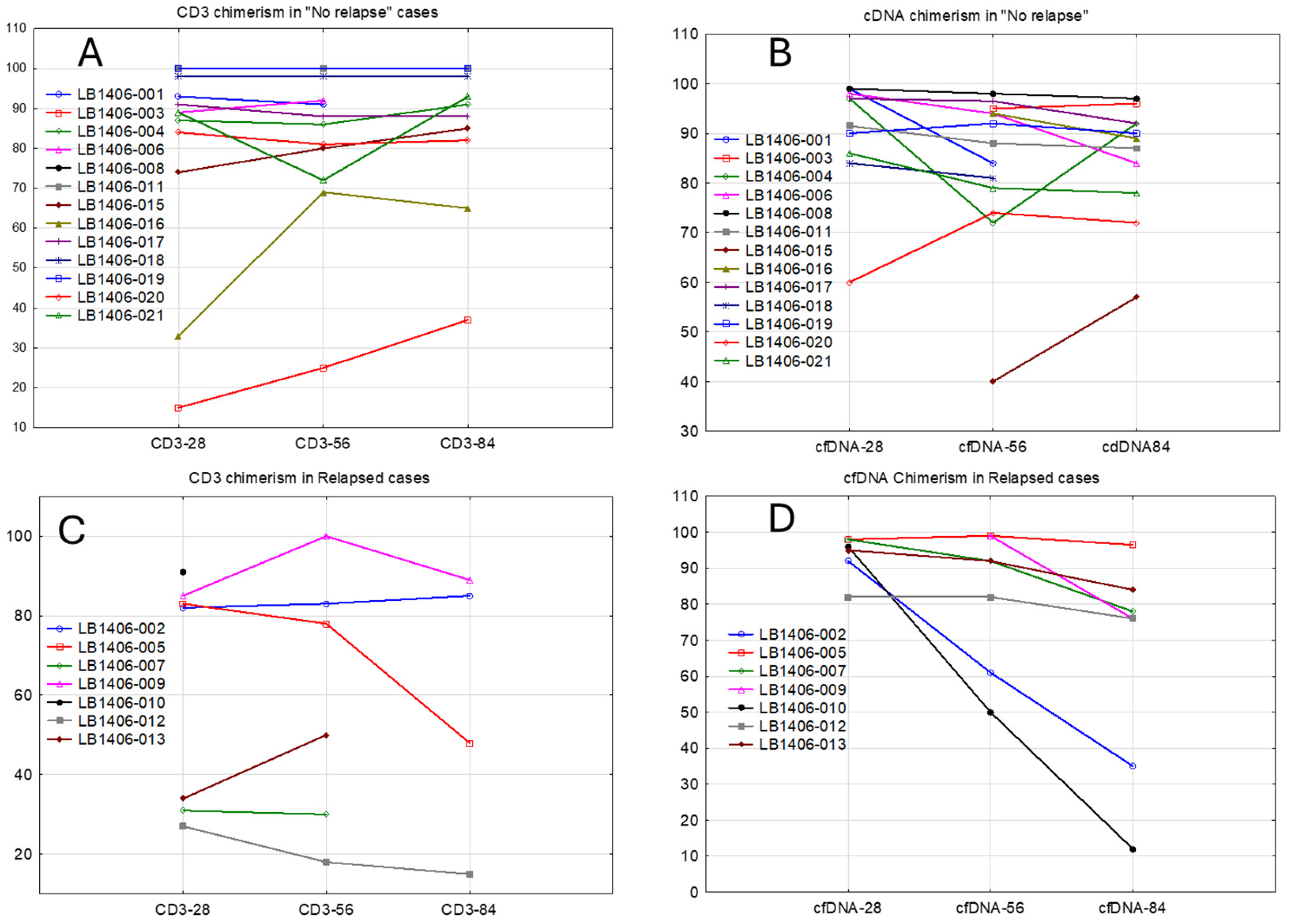
3.2. Correlation of cfDNA Detection with Relapse

All subjects came to transplantation with somatic mutations identified either at the time of diagnosis or in subsequent pre-transplant NGS testing, per the study’s eligibility criteria (Supplementary Table S1). Adverse risk-defining somatic mutations (ASXL1, BCOR, EZH2, FLT3-ITD, RUNX1, SETBP1,SF3B1, SRSF2, STAG2, TP53, U2AF1, ZRSR2, and WT1), as currently defined by the European Leukemia Network (ELN), Pethema Registry, and National Cancer Research Institution (NCRI), were detected before transplantation in all but one (004) of these subjects [14,15,16]. Pre-transplant staging studies for two subjects (008 and 015) were without mutations (MRD-negative) detected in either the pre-transplant BM or plasma samples, and one subject (003) had mutations detected only in the pre-transplant BM sample (Table 2 and Table 3). A fourth subject (021) cleared adverse-risk mutations but was still MRD+ for non-adverse-risk mutations in plasma at day 0.
The testing of cfDNA on days 28, 56, and 84 was without mutations in 6/20, 4/20, and 6/19 evaluable subjects, respectively. Only 1 subject (008) remained without any mutations in all three post-transplant plasma samples tested, while 19 subjects had mutations detected in the plasma at any of the three study time points. The lack of testing at later time points precludes analysis of the durability of clearance of the mutations.
Two subjects (001 and 016) died in remission of regimen-related complications before completing the 12-month follow-up (Table 2). Nine of the 19 subjects with disease characterized by adverse risk-defining mutations at diagnosis or before transplantation had a similar mutation profile detected in plasma samples tested after transplantation, and six of these nine (excluding from this analysis the subject who expired at day 59) experienced early relapses (Table 2 and Table 3, Supplementary Table S2, and Figure 2). Four of the subjects (including the two who were MRD-negative in plasma testing) cleared adverse-risk mutations before transplantation, and 5 of the 19 cleared the adverse risk-defining mutations after transplantation. None of these nine subjects suffered relapses.
Several subjects cleared somatic mutations in longer-term, off-study follow-up cfDNA testing (Supplementary Table S3), including resolution of TET2 (subject 006), SF3B1 (016), CALR and U2AF1 (019), DMNT3A (020), and CBL (021) mutations. Two of these subjects (016 and 019) cleared adverse-risk mutations and remained in remission. These subjects remained in remission through the end of the study period. The intermittent non-study testing obtained from this limited number of subjects did not allow comprehensive analysis or interpretation of these follow-up data, however.
Both subjects who relapsed before day 100 (subjects 007 and 010) showed a dramatic rise in the VAF GM or number of mutations detected prior to the date of relapse, in association with a fall in donor chimerism (Table 2 and Figure 1D).

4. Discussion

Serial measurement of plasma cfDNA via NGS analysis of leukemia-associated somatic mutations during the first 84 days after transplantation demonstrated intermittent or sustained disease markers for most (19 out of 20) subjects undergoing allogeneic PBSC transplantation. These low levels of measurable markers indicative of persistent MRD do not necessarily predict a relapse of disease, although a lack of clearance of mutations of high-risk driver genes appears to be associated with a much higher probability of relapse early after transplantation, possibly justifying modifications in the post-transplant treatment regimen for such patients (Figure 2). In contrast, mutations in epigenetic modifiers and other low-risk mutations did not appear to be as clearly associated with relapse. Our data confirmed the finding of others that failure of clearance of MRD after transplantation is associated with disease relapse (Figure 2) [17,18]. Our data are also consistent with those of others who reported that the genetic profile of the disease at diagnosis is more important in predicting the control of disease than mere MRD positivity at pre-transplant staging (Figure 2) [19,20], possibly explained by distinct genomic aberrations influencing the immunogenicity of malignant cells [21,22,23,24,25].
The detection of adverse-risk somatic mutations in plasma samples obtained after transplantation identified a specific population with a high risk of relapse. If the two subjects (Supplementary Table S3) who cleared adverse-risk mutations after the day 84 sampling were included in this analysis, then relapses occurred in six out of seven subjects with sustained adverse-risk mutations. This observation agrees with the NCRI results, demonstrating that classification of patients by adverse-risk mutations in pre-transplant sampling identifies patients predicted to be at a higher risk of relapse after transplantation [16]. The difference in relapses observed in our study, based on the presence or absence of adverse-risk somatic mutations characterizing post-transplant MRD, may be a reflection of particular disease subclones that either persist or are eliminated after transplantation. It is also possible that for the patients with somatic mutations demonstrated by cfDNA analysis but without persisting adverse-risk mutations, we were actually detecting clonal hematopoiesis of indeterminant prognosis (CHIP) present in the individual patients before the development of a myeloid malignancy and not MRD. CHIP may not be associated with the relapse of a myeloid malignancy after transplantation (and conceivably may be eliminated over time through a GvH effect on host hematopoiesis). Larger studies with longer-term testing are required to determine the clinical significance and natural history of persistent detection of non-adverse-risk somatic mutations after transplantation, which would also include the significance of donor-transmitted somatic mutations [22,26,27,28].
The results of this pilot study support our hypothesis that detection of cfDNA identifies persistently stable levels of MRD as early as day 28 after transplantation and furthermore supports our hypothesis that detection of MRD early after transplantation using this technique identifies patients for whom modifications of the transplant treatment plan are required to reduce the risk of relapse. In contrast, we propose that patients who are MRD-negative in pre-transplant evaluation using cfDNA testing or those patients who clear MRD through the transplant process may be managed without post-transplant consolidation therapy, avoiding the recognized toxicity of such treatment.
We were able to closely correlate the VAF values for the BM and plasma samples, as reported by other laboratories [18,29]. These results support the use of serial cfDNA analyses of plasma samples for disease monitoring without resorting to invasive bone marrow sampling. Such testing as that conducted in this study would appear to be quite valuable in the design and interpretation of transplant trials, including studies involving targeted or non-targeted post-transplant consolidation approaches in which relapse is a designated primary or secondary endpoint. Prospective studies are required to determine if a choice of conditioning or GvHD regimens as well as a donor or cell source may be more effective in clearing adverse-risk somatic mutations through the transplant process. Prospective studies using cfDNA analysis will be required to test our hypothesis that modifications to the post-transplant treatment regimen, such as with consolidation therapy, will reduce the risk of relapse for patients with persistent adverse-risk somatic mutations.
Although our data beyond day 84 are limited, we did observe at least transient clearing of somatic mutations for five subjects and the specific clearing of adverse-risk somatic mutations detected at day 84 for the two patients still in remission, signifying that relapse may not be inevitable for these patients. The clinical relevance of a sustained persistence of low-level epigenetic gene mutations such as DNMT3A and TET2 or other low-risk mutations is uncertain and will require larger studies that can focus on the clinical importance of any individual gene mutation or classes of genes not considered adverse-risk somatic mutations. Longer-term testing of this study population may show that these low-risk mutations may be detected months after transplantation and are evidence of persistence of disease (or CHIP), albeit of uncertain clinical relevance. We suspect that these subjects could suffer relapses in the more distant future if the immunologic graft-versus-leukemia effect of transplantation weakens. The ease of this noninvasive testing, however, facilitates monitoring of such patients with serial testing to address this risk.
The addition of chimerism testing using the same cfDNA technique appeared to provide a warning of an impending relapse, as shown by the decrease in donor chimerism before documentation of a relapse. We note that the BM samples obtained early after transplantation were insensitive to the pending relapse, in contrast to the data developed via plasma cfDNA analysis. We did not find a correlation between the chimerism results determined by NGS of cfDNA and STR analysis of specific cell subsets. Chimerism analysis using cfDNA from plasma samples is expected to reflect the entire body’s DNA, but it has been shown to be significantly more enriched by hematologic cells that are immersed in blood [18]. Accordingly, the results of cfDNA testing may differ from the CD3+ or CD15+ cell chimerism measurements. The pattern of cfDNA chimerism appears to reflect relapsing disease more accurately than that of CD3 chimerism. All patients who relapsed early after transplantation showed a constant downward trend in cfDNA chimerism, a trend that could be seen even on day 56. Although low or falling donor T-cell chimerism has been associated with a higher probability of relapse [30,31], chimerism testing is not a measure of MRD and does not appear to be useful in identifying individual patients who may benefit from consolidation therapy.
Although not analyzed in this study, mutations can be detected using RNA sequencing. RNA mutation data may add another level of sensitivity for some mutations or chromosomal translocations. We detected multiple variants in the samples obtained before or after transplantation using RNA sequencing (Supplementary Table S4). Some of the variants detected may be related to disease, but the expression of multiple unique variants was detected, especially after transplantation, that likely reflect events other than relapse of malignancy. We previously reported that RNA transcriptome analysis of post-transplant bone marrow samples could identify upregulation of genes associated with the occurrence of acute GvHD and with overall survival [32], and such analysis may be helpful in monitoring post-transplant events.
This study is limited by the small number of cases; by selecting subjects with known mutations identified at the time of diagnosis or in subsequent pre-transplant testing; and by the heterogeneity of donor types, conditioning regimens, GvHD prophylaxis regimens, and variable use of post-transplant consolidation, any of which may have affected the clearance of MRD. We used a single commercially available source for the pre- and post-transplant testing for this study, and other NGS platforms may have different sensitivities and specificities or use more limited gene profiles. The clinical importance of most of the genes included in the agnostic panel used in this study is currently not defined and may require larger study populations and long-term follow-ups to determine, including distinguishing between MRD and CHIP.

5. Conclusions

Our study explored the use of cfDNA to demonstrate the persistence of disease after transplantation as a possible technique for identifying patients who may benefit from post-transplant consolidation therapy and did not focus on any particular mutation. Based on the non-invasiveness of NGS testing of plasma samples, we propose that serial testing for leukemia-associated cfDNA, especially if combined with chimerism testing using NGS techniques, is an appropriate test for monitoring patients after transplantation for clinically relevant MRD predictive of post-transplant relapses. Such testing allows discussion with the transplant recipient about modification of the individual patient’s treatment plan, with possible improvement in treatment success.

Supplementary Materials

The following supporting information can be downloaded at https://www.mdpi.com/article/10.3390/cancers17040625/s1, Included are an additional description of experimental technique, eight tables listing additional clinical details and various lists of mutations, including RNA mutations, and one figure showing correlation between the chimerism tests and supplemental references. Table S1: Initial diagnosis details and pre- and post-transplant cytogenetics; Table S2: Detected mutations and VAF in study samples; Table S3: NGS analysis of samples obtained in long-term follow-up of subjects; Table S4: Study samples with RNA mutations identified; Table S5: Chimerism values; Table S6: Correlation of chimerism results using NGS and STR analyses; Table S7: List of 302 genes validated and included in the DNA analysis; Table S8: List of 1408 genes included in the RNA analysis; Figure S1: Correlation between chimerism tests by technique. References [33,34] are cited in the Supplementary Materials.

Author Contributions

Conceptualization, S.D.R. and M.A.; formal analysis, S.D.R., M.A. and M.F.B.; investigation, S.D.R. and M.F.B.; methodology, S.D.R. and M.A.; writing—original draft, S.D.R.; writing—review and editing, S.D.R., M.A., M.F.B., A.A., S.K., H.C.S., A.G. and M.L.D. All authors have read and agreed to the published version of the manuscript.

Funding

This research received no external funding. Internal funding was provided by the John Theurer Cancer Center.

Institutional Review Board Statement

This study was conducted according to the guidelines of the Declaration of Helsinki and approved by the Institutional Review Board of Georgetown University School of Medicine (Study00003857) on 21 September 2021.

Informed Consent Statement

Informed consent was obtained from all subjects involved in this study.

Data Availability Statement

The human sequence data generated in this study are included within the article and its online Supplementary Materials data files. Further inquiries can be directed toward the corresponding author.

Conflicts of Interest

M.F.B., A.A., H.C.S. and S.K. declare no conflicts of interest. M.A. works for and own stocks in a diagnostic company which offers DNA and RNA testing. S.D.R. is a consultant for ReAlta Life Sciences, an advisory board member for SIRPant Immunotherapeutics and stock owner for COTA, Genomic Testing Cooperative. M.L.D. owns stocks in COTA, Genomic Testing Cooperative. A.G. owns stocks in COTA, Genetic Testing Cooperative, Alloplex, and Resilience, is part of the consulting faculty for Michael J Hennessey Associated, Physician Education Resource, the Society of Hematology Oncology, and OncLive, is on the scientific advisory board for Alloplex and Vincerx, and is on the steering committee for Astrazeneca.

References

  1. Dillon, L.W.; Gui, G.; Page, K.M.; Ravindra, N.; Wong, Z.C.; Andrew, G.; Mukherjee, D.; Zeger, S.L.; El Chaer, F.; Spellman, S.; et al. DNA Sequencing to detect residual disease in adults with acute myeloid leukemia prior to hematopoietic cell transplant. JAMA 2023, 329, 745–755. [Google Scholar] [CrossRef]
  2. Buckley, S.A.; Wood, B.L.; Othus, M.; Hourigan, C.S.; Ustun, C.; Linden, M.A.; DeFor, T.E.; Malagola, M.; Anthias, C.; Valkova, V.; et al. Minimal residual disease prior to allogeneic hematopoietic cell transplantation in acute myeloid leukemia: A meta-analysis. Haematologica 2017, 102, 865–873. [Google Scholar] [CrossRef]
  3. Moukalled, N.; Labopin, M.; Versluis, J.; Socié, G.; Blaise, D.; Salmenniemi, U.; Rambaldi, A.; Gedde-Dahl, T.; Tholouli, E.; Kröger, N.; et al. Complex karyotype but not other cytogenetic abnormalities is associated with worse posttransplant survival of patients with nucleophosmin 1-mutated acute myeloid leukemia: A study from the European Society for Blood and Marrow Transplantation Acute Leukemia Working Party. Am. J. Hematol. 2024, 99, 360–369. [Google Scholar]
  4. Dillon, L.W.; Gui, G.; Ravindra, N.; Andrew, G.; Mukherjee, D.; Wong, Z.C.; Huang, Y.; Gerhold, J.; Holman, M.; D’Angelo, J.; et al. Measurable residual flt3 internal tandem duplication before allogeneic transplant for acute myeloid leukemia. JAMA Oncol. 2024, 10, 1104–1110. [Google Scholar] [CrossRef] [PubMed] [PubMed Central]
  5. Tsirigotis, P.; Byrne, M.; Schmid, C.; Baron, F.; Ciceri, F.; Esteve, J.; Gorin, N.C.; Giebel, S.; Mohty, M.; Savani, B.N.; et al. Relapse of AML after hematopoietic stem cell transplantation: Methods of monitoring and preventive strategies. A review from the ALWP of the EBMT. Bone Marrow Transplant. 2016, 51, 1431–1438. [Google Scholar] [CrossRef]
  6. DeFilipp, Z.; Chen, Y.B. How I treat with maintenance therapy after allogeneic HCT. Blood 2023, 141, 39–48. [Google Scholar] [CrossRef]
  7. Biederstädt, A.; Rezvani, K. How I treat high-risk acute myeloid leukemia using preemptive adoptive cellular immunotherapy. Blood 2023, 141, 22–38. [Google Scholar] [CrossRef]
  8. Chen, K.; Zhao, H.; Shi, Y.; Yang, F.; Wang, L.T.; Kang, G.; Nie, Y.; Wang, J. Perioperative Dynamic Changes in Circulating Tumor DNA in Patients with Lung Cancer (DYNAMIC). Clin. Cancer Res. 2019, 25, 7058–7067. [Google Scholar] [CrossRef]
  9. Cescon, D.W.; Bratman, S.V.; Chan, S.M.; Siu, L.L. Circulating tumor DNA and liquid biopsy in oncology. Nat. Cancer 2020, 1, 276–290. [Google Scholar] [CrossRef]
  10. Crowley, E.; Di Nicolantonio, F.; Loupakis, F.; Bardelli, A. Liquid biopsy: Monitoring cancer-genetics in the blood. Nat. Rev. Clin. Oncol. 2013, 10, 472–484. [Google Scholar] [CrossRef]
  11. Waldeck, S.; Mitschke, J.; Wiesemann, S.; Rassner, M.; Andrieux, G.; Deuter, M.; Mutter, J.; Lüchtenborg, A.M.; Kottmann, D.; Titze, L.; et al. Early assessment of circulating tumor DNA after curative-intent resection predicts tumor recurrence in early-stage and locally advanced non-small-cell lung cancer. Mol. Oncol. 2022, 16, 527–537. [Google Scholar] [CrossRef] [PubMed]
  12. Pellini, B.; Chaudhuri, A.A. Circulating tumor DNA minimal residual disease detection of non-small-cell lung cancer treated with curative intent. J. Clin. Oncol. 2022, 40, 567–575. [Google Scholar] [CrossRef] [PubMed]
  13. Picard, C.; Frassati, C.; Cherouat, N.; Maioli, S.; Moskovtchenko, P.; Cherel, M.; Chiaroni, J.; Pedini, P. New methods for the quantification of mixed chimerism in transplantation. Front. Immunol. 2023, 14, 1023116. [Google Scholar] [CrossRef] [PubMed]
  14. Sargas, C.; Ayala, R.; Larráyoz, M.J.; Chillón, M.C.; Carrillo-Cruz, E.; Bilbao-Sieyro, C.; Prados de la Torre, E.; Martínez-Cuadrón, D.; Rodríguez-Veiga, R.; Boluda, B.; et al. Molecular Landscape and Validation of New Genomic Classification in 2668 Adult AML Patients: Real Life Data from the PETHEMA Registry. Cancers 2023, 15, 438. [Google Scholar] [CrossRef]
  15. Döhner, H.; Wei, A.H.; Appelbaum, F.R.; Craddock, C.; DiNardo, C.D.; Dombret, H.; Bert, B.L.; Fenaux, P.; Godley, L.A.; Hasserjian, R.P.; et al. Diagnosis and management of AML in adults: 2022 recommendations from an international expert panel on behalf of the ELN. Blood 2022, 140, 1345–1377. [Google Scholar] [CrossRef] [PubMed]
  16. Tazi, Y.; Arango-Ossa, J.E.; Zhou, Y.; Bernard, E.; Thomas, I.; Gilkes, A.; Freeman, S.; Pradat, Y.; Johnson, S.J.; Hills, R.; et al. Unified classification and risk-stratification in Acute Myeloid Leukemia. Nat. Commun. 2022, 13, 4622. [Google Scholar] [CrossRef]
  17. Tobiasson, M.; Pandzic, T.; Illman, J.; Nilsson, L.; Weström, S.; Ejerblad, E.; Olesen, G.; Björklund, A.; Olsnes Kittang, A.; Werlenius, O.; et al. Patient-specific measurable residual disease markers predict outcome in patients with myelodysplastic syndrome and related diseases after hematopoietic stem-cell transplantation. J. Clin. Oncol. 2024, 42, 1378–1390. [Google Scholar] [CrossRef] [PubMed]
  18. Pasca, S.; Guo, M.Z.; Wang, S.; Stokvis, K.; Shedeck, A.; Pallavajjala, A.; Shams, C.; Pallavajjala, R.; DeZern, A.E.; Varadhan, R.; et al. Cell-free DNA measurable residual disease as a predictor of postallogeneic hematopoietic cell transplant outcomes. Blood Adv. 2023, 7, 4660–4670. [Google Scholar] [CrossRef]
  19. Murdock, H.M.; Kim, H.T.; Denlinger, N.; Vachhani, P.; Hambley, B.; Manning, B.S.; Gier, S.; Cho, C.; Tsai, H.K.; McCurdy, S.; et al. Impact of diagnostic genetics on remission MRD and transplantation outcomes in older patients with AML. Blood 2022, 139, 3546–3557. [Google Scholar] [CrossRef]
  20. Bernard, E.; Tuechler, H.; Greenberg, P.L.; Hasserjian, R.P.; Arango Ossa, J.E.; Nannya, Y.; Devlin, S.M.; Creignou, M.; Pinel, P.; Monnier, L.; et al. Molecular International Prognostic Scoring System for myelodysplastic syndromes. NEJM Evid. 2022, 1, EVIDoa2200008. [Google Scholar] [CrossRef] [PubMed]
  21. Austin, R.J.; Straube, J.; Halder, R.; Janardhanan, Y.; Bruedigam, C.; Witkowski, M.; Cooper, L.; Porter, A.; Braun, M.; Souza-Fonseca-Guimaraes, F.; et al. Oncogenic drivers dictate immune control of acute myeloid leukemia. Nat. Commun. 2023, 14, 2155. [Google Scholar] [CrossRef] [PubMed]
  22. Gibson, C.J.; Kim, H.T.; Zhao, L.; Murdock, H.M.; Hambley, B.; Ogata, A.; Madero-Marroquin, R.; Wang, S.; Green, L.; Fleharty, M.; et al. Donor clonal hematopoiesis and recipient outcomes after transplantation. J. Clin. Oncol. 2022, 40, 189–201. [Google Scholar] [CrossRef] [PubMed]
  23. Vago, L.; Gojo, I. Immune escape and immunotherapy of acute myeloid leukemia. J. Clin. Investig. 2020, 130, 1552–1564. [Google Scholar] [CrossRef] [PubMed]
  24. Tettamanti, S.; Pievani, A.; Biondi, A.; Dotti, G.; Serafini, M. Catch me if you can: How AML and its niche escape immunotherapy. Leukemia 2022, 36, 13–22. [Google Scholar] [CrossRef]
  25. Limongello, R.; Marra, A.; Mancusi, A.; Bonato, S.; Hoxha, E.; Ruggeri, L.; Hui, S.; Velardi, A.; Pierini, A. Novel immune cell-based therapies to eradicate high-risk acute myeloid leukemia. Front. Immunol. 2021, 12, 695051. [Google Scholar] [CrossRef] [PubMed]
  26. Frick, M.; Chan, W.; Arends, C.M.; Hablesreiter, R.; Halik, A.; Heuser, M.; Michonneau, D.; Blau, O.; Hoyer, K.; Christen, F.; et al. Role of donor clonal hematopoiesis in allogeneic hematopoietic stem-cell transplantation. J. Clin. Oncol. 2019, 37, 375–385. [Google Scholar] [CrossRef]
  27. Newell, L.F.; Williams, T.; Liu, J.; Yu, Y.; Chen, Y.; Booth, G.C.; Knight, R.J.; Goslee, K.R.; Cook, R.J.; Leonard, J.; et al. Engrafted donor-derived clonal hematopoiesis after allogenic hematopoietic cell transplantation is associated with chronic graft-versus-host disease requiring immunosuppressive therapy, but no adverse impact on overall survival or relapse. Transplant. Cell Ther. 2021, 27, 662.e1–662.e9. [Google Scholar] [CrossRef]
  28. Boettcher, S.; Wilk, C.M.; Singer, J.; Beier, F.; Burcklen, E.; Beisel, C.; Ventura Ferreira, M.S.; Gourri, E.; Gassner, C.; Frey, B.M.; et al. Clonal hematopoiesis in donors and long-term survivors of related allogeneic hematopoietic stem cell transplantation. Blood 2020, 135, 1548–1559. [Google Scholar] [CrossRef]
  29. Jansko-Gadermeir, B.; Leisch, M.; Gassner, F.J.; Zaborsky, N.; Dillinger, T.; Hutter, S.; Risch, A.; Melchardt, T.; Egle, A.; Drost, M.; et al. Myeloid NGS analyses of paired samples from bone marrow and peripheral blood yield concordant results: A prospective cohort analysis of the AGMT Study Group. Cancers 2023, 15, 2305. [Google Scholar] [CrossRef] [PubMed] [PubMed Central]
  30. Nahas, G.; Gandhi, A.P.; Donato, M.L.; Nyirenda, T.; Vesole, D.H.; Pecora, A.L.; Goldberg, S.; Goy, A.; Feldman, T.; Mato, A.R.; et al. Donor CD3 chimerism after allogeneic transplantation predicts probability of relapse for patients treated for acute myelogenous leukemia (AML), acute lymphoblastic leukemia (ALL), and non-Hodgkin’s lymphoma (NHL). Blood 2013, 122, 4545. [Google Scholar]
  31. Huisman, C.; de Weger, R.A.; de Vries, L.; Tilanus, M.G.; Verdonck, L.F. Chimerism analysis within 6 months of allogeneic stem cell transplantation predicts relapse in acute myeloid leukemia. Bone Marrow Transplant. 2007, 39, 285–291. [Google Scholar] [CrossRef] [PubMed]
  32. Rowley, S.D.; Gunning, T.S.; Pelliccia, M.; Della Pia, A.; Lee, A.; Behrmann, J.; Bangolo, A.; Jandir, P.; Zhang, H.; Kaur, S.; et al. Using targeted transcriptome and machine learning of pre- and post-transplant bone marrow samples to predict acute graft-versus-host disease and overall survival after allogeneic stem cell transplantation. Cancers 2024, 16, 1357. [Google Scholar] [CrossRef] [PubMed] [PubMed Central]
  33. Bacigalupo, A.; Ballen, K.; Rizzo, D.; Giralt, S.; Lazarus, H.; Ho, V.; Apperley, J.; Slavin, S.; Pasquini, M.; Sandmaier, B.M.; et al. Defining the intensity of conditioning regimens: Working definitions. Biol. Blood Marrow Transplant. 2009, 15, 1628–1633. [Google Scholar] [CrossRef] [PubMed]
  34. Khoury, J.D.; Solary, E.; Abla, O.; Akkari, Y.; Alaggio, R.; Apperley, J.F.; Bejar, R.; Berti, E.; Busque, L.; Chan, J.K.; et al. The 5th edition of the World Health Organization Classification of Haematolymphoid Tumours: Myeloid and Histiocytic/Dendritic Neoplasms. Leukemia 2022, 36, 1703–1719. [Google Scholar] [CrossRef] [PubMed]
Figure 1. The chimerism values using cfDNA analysis (B,D) and for CD3+ cells (A,C) for individual subjects in post-transplant relapse (C,D) or remission (A,B).
Cancers 17 00625 g001
Figure 2. The number of subjects with relapses within 12 months after transplantation, as defined by the presence of adverse-risk or any mutations at various time points before and after transplantation (Table 3). The group listing any mutations includes subjects with adverse-risk or non-adverse-risk mutations.
Cancers 17 00625 g002
Table 1. Transplant Subject and donor demographics.
Subject Age, Median (Range)59 (27–77)Donor Age, Median (Range)25 (16–56)
Subject Sex (N) Donor Sex (N)
Male13Male11
Female7Female9
Transplant Diagnosis (N) Donor (N)
Primary AML6HLA-Matched Sibling2
Secondary AML6Haploidentical3
MDS7URD Matched12
CML, Blast Crisis1URD Mismatched3
ABO (N) CMV (Recipient or Donor, N)
Match11+/+3
Minor Mismatch3+/−4
Major or Bidirectional Mismatch6−/+4
−/−9
HSC Source (N) Conditioning Regimen (N)
PBSC20MA6
BM0RIC10
Non-MA4
GvHD Regimen (N)
Tac + MTX3Post-Transplant Consolidation (N)
Tac, MTX + abatacept11Yes9
PTCy2No11
PTCy or abatacept4
Subject and donor characteristics are shown. AML = acute myelogenous leukemia; MDS = myelodysplastic syndrome; CML = chronic myelogenous leukemia; GvHD = graft-versus-host disease; CMV = cytomegalovirus status; HLA = histocompatibility locus antigen; URD = unrelated donor; MA = myeloablative; RIC = reduced intensity; Tac = tacrolimus; MTX = methotrexate; PTCy = post-transplant cyclophosphamide.
Table 2. Subject and donor demographics and mutations detected through day 84.
Sub No.DXDonor/HLA MatchCondRegGvHD
Reg
Post-Transplant ConsolidationRelapseCurrent StatusNumber of Mutations/Geometric Mean of VAF
RegimenDay StartedY/NDayPre-Transplant BMPre cfDNADay 28Day 56Day 84Post-Transplant BM
001AMLURD 10/10 MA + ATGMTX NA N Expired, RRT, day 593/4.582/20.925/0.13/0.51NDND
002t-MDSURD 10/10MA + ATGMTX + AbatNA Y170Alive, >day 3652/34.422/42.723/2.43/4.023/2.772/1.13
003MDSURD 8/10NMAPTCy + AbatNA N Alive, >day 3652/11.040/NE0/NE1/2.310/NE0/NE
004AMLURD 10/10MA + ATGMTX + AbatSorafenib +HMA51N Expired, infection, day 3571/44.481/44.871/0.51/0.260/NE1/0.28
005t-MDSURD 10/10RIC + ATGMTX + AbatHMA +DLI121N Alive, >day 3655/3.5416/0.972/0.542/0.302/0.345/0.37
0062nd-AML (CMML)URD 10/10RIC + ATGMTX + AbatNA N Alive, >day 3654/37.624/35.910/NE0/NE2/0.111/2.55
0072nd-AML (MDS)URD 10/10NMAPTCy + AbatNA Y62Expired, relapse, day 2508/22.979/18.721/0.666/1.077/1.5210/4.29
008AMLHaploRIC PTCyCrenolinib +sorafenib91N Alive, >day 3650/NE0/NE0/NE0/NE0/NE0/NE
009AMLRD 10/10RIC + ATGMTXHMA +MT401 DLI Group 276Y168Expired, relapse, day 2852/0.666/0.840/NE0/NE7/0.94/0.24
0102nd-AMLHaploNMAPTCy + AbatNA Y52Expired, relapse, day 1253/8.143/23.983/0.642/19.282/33.484/5.92
0112nd-AML (MDS)URD 9/10RICPTCy + AbatHMA88N Alive, >day 3653/19.833/9.661/1.591/3.401/6.320/NE
012MDSURD 10/10MA + ATGMTX + AbatNA Y167Alive, relapse, day 3474/7.6711/5.685/0.623/0.047/0.364/0.46
013MDSURD 10/10RIC + ATGMTX + AbatNA Y139Alive, >day 3651/48.785/2.501/0.262/1.632/1.602/4.23
015AMLRD 10/10MAMTX Midostaurin + gilteritinib35N Alive, >day 3650/NE0/NE1/36.071/30.31/31.22/0.94
016MDSURD 10/10RIC + ATGMTX + AbatHMA89N Expired, sepsis, day 2681/33.185/1.461/0.981/0.031/0.060/NE
017CMLURD 10/10RICMTX + AbatImatinib52N Alive, >day 3651/6.663/3.780/NE1/0.210/NE0/NE
0182nd-AML (BrCa)URD 10/10RICMTX + AbatNA N Alive, >day 3656/1.428/0.632/0.570/NE0/NEND
0192nd AML (MF)HaploNMAPTCyNA N Alive, >day 3659/5.796/9.433/0.185/0.542/0.231/0.06
020AMLURD 10/10MA + ATGMTX + AbatNA N Alive, >day 3655/4.117/1.711/0.971/0.370/NE1/0.37
021MDSURD 10/10RIC + ATGMTX + AbatSorafenib77N Alive, >day 3652/0.694/1.200/NE1/0.111/0.110/NE
Individual subject and transplant characteristics, post-transplant relapse and survival, and number of mutations and VAF GM at defined time points after transplantation. Host germline mutations (subjects 001, 006, 019, 020, and 021) and donor germline mutations (subjects 002, 004, 017, and 021) were excluded from mutation counts and calculations of VAF GM. Mutations identified via analysis of cfRNA were not included in this analysis. Sub No = subject number; GM = geometric mean; URD = unrelated donor; Haplo = haploidentical related donor without the number of matched alleles shown; HLA = histocompatibility locus antigen; HMA = hypomethylating agent; NA = not applicable; ND = not done; NE = VAF frequency not evaluable; CondReg = conditioning regimens are described by intensity (myeloablative (MA); non-myeloablative (NMA); and reduced intensity (RIC)) with or without rabbit antithymocyte globulin (ATG); GvHD Reg = GvHD prophylaxis was either methotrexate (MTX)-or cyclophosphamide (PTCy)-based, with the addition of abatacept (abat) per physician discretion. Complete details regarding conditioning and GvHD prophylaxis regimens are provided in the Supplementary Materials.
Table 3. Comparison of pre-and post-transplant mutations detected in bone marrow and plasma samples.
Sub NoRelapse (Y/N)Pre-Transplant BMVAFPre-Transplant cfDNAVAFDay 84 BMVAFDay 84 cfDNAVAF
001NDNMT3a
SRSF2
IDH1
15.85
4.66
1.3
DNMT3A
SRSF2
28.57
15.38
ND ND
002YTP53
HNF1A
48.34
24.54
TP53
HNF1A
63.57
28.71
TP53
HNF1A
2.5
0.51
TP53
HNF1A
3.19
0.84
003NBCOR
KMT2D
13.85
8.8
Neg Neg Neg
004NDNMT3A44.48DNMT3A44.87DNMT3A0.28Neg
005NTP53
PPM1D
CHEK2
NOTCH3
PPM1D
5.2
2.18
7.53
3.95
1.66
TP53
PPM1D
CHEK2
NOTCH3
PPM1D
BRAF
PPM1D
CARD11
KMT2A
PMS1
CDK12
PBRM1
TP53
KEAP1
CARD11
KMT2C
5.53
5.13
4.76
2.79
0.67
5.97
3.23
0.76
0.7
0.58
0.56
0.52
0.35
0.21
0.18
0.11
KMT2A
GATA3
MAP3Ki4
TET2
KRCC2
0.19
0.95
0.47
0.37
0.21
TP53
PPM1D
0.37
0.32
006NTET2
EZH2
ASXL1
TET2
52.71
47.1
33.71
23.92
TET2
EZH2
ASXL1
TET2
49.13
36.36
34
27.4
ASXL12.55TET2
TET2
0.25
0.05
007YSRSF2
ASXL1
NRAS
MTOR
MTOR
MTOR
KDM6A
ARAF
55.44
50.62
41.9
31.37
30.0
27.27
6.15
4.17
SRSF2
ASXL1
NRAS
MTOR
MTOR
MTOR
FH
GNAQ
IRF4
45.55
48.27
48.59
40.26
38.3
36.09
2.71
4.17
4.11
SRSF2
ASXL1
NRAS
MTOR
MTOR
MTOR
GNAS
GNAS
GNAS
BCL6
9.4
10.24
7.63
0/73
0.48
0.8
10.41
8.46
8.67
13.43
SRSF2
ASXL1
NRAS
MTOR
MTOR
FH
DNMT3A
5.62
8.29
4.46
0.55
0.31
1.85
0.27
008NNeg Neg Neg Neg
009YTET2
NRAS
0.28
1.54
TET2
TET2
WT1
NFKBIA
NRAS
FLT3-ITD
5.78
1.38
0.83
0.6
0.39
0.23
TET2
WT1
NFKBIA
FLT3-ITD
0.15
0.69
0.25
0.14
TET2
WT1
NFKBIA
NRAS
FLT3-ITD
WT1
WT1
2.73
3.02
3.77
0.13
2.4
0.12
0.41
010YSRSF2
MPL
IDH2
18.93
4.31
6.61
SRSF2
MPL
IDH2
37.89
23.93
15.2
SRSF2
MPL
IDH2
KMT2C
36.08
0.54
16.24
3.87
SRSF2
IDH2
31.04
36.11
011NTP53
TET2
PDGFRB
31.54
16.72
14.79
TP53
TET2
PDGFRB
5.26
8.62
19.86
Neg PDGFRB6.32
012YAXIN1
SF3B1
ASXL1
ASXL1
AMER1
16.83
17.7
6.4
4
8.05
AXIN1
SF3B1
ASXL1
ASXL1
AMER1
H3F3A
EGFR
RUNX1
KMT2C
ASXL1
34.8
33.02
3.03
3.01
2.14
17.7
3.46
2.61
1.79
1.4
AXIN1
SF3B1
RUNX1
KMT2B
0.25
0.49
0.28
1.68
AXIN1
SF3B1
AMER1
EGFR
RUNX1
KMT2C
KMT2C
0.5
0.35
0.05
0.36
0.28
0.48
1.86
013YNeg TP53
TET2
NOTCH1
TET2
43.38
0.78
0.47
0.19
NF20.37TP530/37
015NNeg Neg KMT2B
DNMT3A
1.25
0.7
Neg
016NSF3B133.18SF3B1
TNFRSF14
KMT2D
DNMT3A
MAP3K1
43.33
1.48
0.83
0.33
0.26
Neg SF3B10.06
017NASXL16.66ASXL1
CEBPA
DNMT3A
12.81
5.2
0.81
Neg Neg
018NALK
SRSF2
TET2
SRSF2
DDX41
0.97
0.8
0.51
0.29
1.82
ALK
SRSF2
TET2
SF3B1
SF3B1
ALK
FGFR4
1.46
0.17
0.2
0.21
0.34
0.44
0.35
ND Neg
019NCALR
U2AF1
ASXL1
GNAS
KRAS
RUNX1
GALNT12
TP31
GRIN2A
39.23
38.39
34.88
20.25
20.11
2.04
0.87
0.6
0.32
CALR
U2AF1
ASXL1
GNAS
KRAS
RUNX1
17.76
15.04
19.57
12.98
12.92
0.8
ASXL10.06CALR
U2AF1
0.29
0.18
020NDMNT3A.
NF1
ASXL1
TET2
SMC3
32.74
4.4
2.52
3.72
0.87
DMNT3A.
NF1
ASXL1
TET2
SMC3
PDGFRB
KMT2C
33.87
4.27
3.64
2.2
0.4
0.4
0.23
DNMT3A0.37DNMT3A0.36
021NDNMT3A
DNMT3A
1.02
0.47
DNMT3A
DNMT3A
CBL
NFI I
1.38
1.01
1.03
1.46
Neg CBL0.11
The gene mutations and VAF values identified in BM or plasma testing before transplant conditioning and at day 84 after transplantation. See Supplementary Table S1 for complete gene descriptions. All subjects are listed, and relapses within 12 months of transplantation are indicated. Mutations shown in bold type are considered high-risk driver mutations [14,15,16]. Sub No = subject number; ND = not done; Neg = no mutations were identified; VAF = variant allele frequency.
Disclaimer/Publisher’s Note: The statements, opinions and data contained in all publications are solely those of the individual author(s) and contributor(s) and not of MDPI and/or the editor(s). MDPI and/or the editor(s) disclaim responsibility for any injury to people or property resulting from any ideas, methods, instructions or products referred to in the content.

