Simple Summary
Cutaneous T-cell lymphomas (CTCLs) represent a heterogeneous group of rare extranodal non-Hodgkin lymphomas with variable clinical presentation. In the Middle East and North Africa (MENA region), where darker skin colors are more common than in the West, CTCL generally presents at a younger age and with distinct clinical features that necessitate special expertise and management across disciplines: rare forms of CTCL are more common (hypo- and hyperpigmented mycosis fungoides (MF)) and a higher prevalence of pediatric MF is noticed.
Abstract
The high cancer burden in the Middle East and North Africa (MENA region) is coupled with an increasing cancer incidence. While the MENA region constitutes 6% of the world’s population, it remains underrepresented in clinical trials. Cutaneous T-cell lymphomas (CTCLs) represent a heterogeneous group of rare extranodal non-Hodgkin lymphomas with variable clinical presentation. In the MENA region, where darker skin colors are more common than in the West, CTCL generally presents at a younger age and with distinct clinical features that necessitate special expertise and management across disciplines: rare forms of CTCL are more common (hypo- and hyperpigmented MF) and a higher prevalence of pediatric MF is noticed. The multidisciplinary approach to cancer management is growing worldwide and is necessary for the comprehensive management of CTCL. The MENA CTCL group was established with the aim of creating a collaborative environment for the diagnosis and treatment of CTCL in the region. Its first meeting was held in May 2023. The group plans to increase the global representation of the MENA region and establish CTCL registries and patient advocacy groups in the region.
1. Introduction
The Middle East and North Africa (MENA) is the geographic region extending between Iran in the east and Morocco in the west, including 20 countries. The MENA population is estimated to be around 500 million people, constituting around 6% of the world’s population (Figure 1) [1].
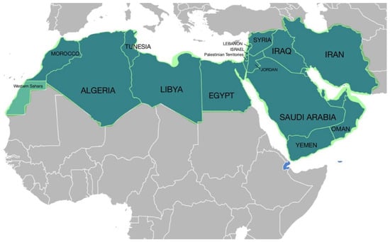
Figure 1.
The Middle East and North Africa (MENA) is a geographic region, which comprises the Middle East and North Africa together.
While the cancer burden is a global concern, it remains much higher in low- and middle-income countries than in other parts of the world [2]. Nevertheless, by 2030 [3], there is projected to be a more than 60% increase in the cancer burden in these countries. A study by Hofmarcher et al. included data from eight MENA countries between the years 2000 and 2019 and found the increase in the cancer incidence to be 8% in Lebanon, 14% in Jordan, 65% in the UAE, and 104% in Algeria. This increase was mainly attributed to three factors: the aging population (demographic changes), lifestyle changes, and increased rates of cancer screening [4].
Despite having a wide pool of patients, the MENA region still falls behind in terms of participation in global clinical trials—including oncology trials. While the MENA region includes around 6% of the global population, it only participates in around 3% of clinical trials worldwide [5].
Cutaneous T-cell lymphomas (CTCLs) represent a heterogeneous group of rare extranodal non-Hodgkin lymphomas, with the most common subtypes being mycosis fungoides (MF) and Sézary syndrome. CTCL is characterized by the localization of neoplastic T-lymphocytes to the skin without evidence of extracutaneous disease at the time of diagnosis [6]. While the pathogenesis of CTCL is not fully understood, some studies suggest a potential role of environmental exposure [7]. The CTCL incidence in the USA is estimated to be 0.64 to 0.87 per 100,000 person-years [7].
The clinical presentation of CTCL is highly variable, often including visible skin changes such as patches, plaques, tumors and/or erythroderma [8]. In addition to the medical history and physical exam, the initial workup for patients with suspected CTCL includes multiple tests that assist in the differential diagnosis of CTCL, identifying its subtype, as well as disease staging, including biopsy of the affected skin, molecular analysis to detect clonal T-cell receptor (TCR) gene rearrangements, assessment of peripheral blood involvement, among others [9]. The International Society for Cutaneous Lymphomas (ISCL) and EORTC developed the TNMB classification and clinical staging of CTCL, which considers T (skin), N (node), M (visceral), and B (blood involvement) [9]. Early-stage MF is limited to the skin; however, it may progress to skin nodules and/or involve extracutaneous sites such as the lymph nodes, blood or visceral organs [10]. As the disease advances, survival is greatly reduced. Agar et al. reported a median overall survival of 4.7–1.4 years for MF patients with stage IIB–IVB disease [11].
The updated 2023 EORTC consensus for the treatment of mycosis fungoides/Sézary syndrome includes recommendations for the first- and second-line treatment by disease stage, while acknowledging that the choice of treatment would depend on the clinical presentation and treatment availability in each individual case [12]. Table 1 summarizes the recommendations for MF treatment by line of therapy and disease stage.

Table 1.
Treatment recommendation for MF, modified according to [12].
Although the treatment options have increased in recent years, e.g., with the approval of chlormethine gel or, for the advanced stage, the highly effective antibodies mogamulizumab and brentuximab vedotin, the availability of several drugs is limited in many countries in the MENA region. There is a huge difference regarding the availability of first-line treatments, e.g., interferon and retinoids, as well as second-line treatments, e.g., the antibody-based treatments, from country to country. As an example, mogamulizumab or brentuximab vedotin are currently unavailable or not covered by medical insurance in Egypt. On the other hand, all the drugs listed in the guidelines, including the antibody-based treatments, are available in Kuwait and Saudi Arabia, indicating the difference in resources in the countries in the MENA region [13].
The purpose of this paper is to clarify the local difficulties in the diagnosis and treatment of cutaneous T-cell lymphomas (CTCLs) in the Middle East and North Africa (MENA) area. It also aims to highlight the objectives and projects of the MENA CTCL working group in tackling these issues and encouraging teamwork for better patient care.
2. CTCL in the MENA Region
CTCL in the MENA region exhibits distinct characteristics compared to Western populations in the EU and USA. The MENA region is characterized by a young average population age, and the diverse Middle Eastern populations generally exhibit darker skin tones compared to Europeans or Americans. Accordingly, there are distinct clinical features to consider in MF presenting in Middle Eastern patients. In Middle Eastern MF patients, it is noted that hyper- and hypopigmented MF (Figure 2A,B, Figure 3A) is more common, patients present at a younger age, and pediatric MF is more common, as compared to Western populations.
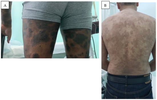
Figure 2.
Hyperpigmented MF: (A) widespread hyperpigmented infiltrated scaly plaques (MF IB), and (B) generalized slightly scaly hyperpigmented patches of mycosis fungoides on the trunk (MF IB).
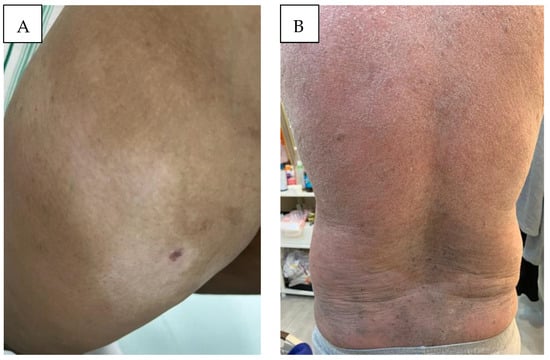
Figure 3.
(A) Hypopigmented MF: localized hypopigmented patches of mycosis fungoides, restricted to the sun-protected area of the thigh. (B) Erythrodermic mycosis fungoides: scaly infiltrated melanoerythroderma covering >90% of the body surface area (MF III).
Erythema, for instance, appears differently on darker skin tones (melanoerythroderma, Figure 3B), which is one possible explanation why MF is diagnosed at more advanced stages and has worse outcomes in skin of color [14,15]. In addition, rare forms of MF are more common in skin of color [16].
In the MENA region, MF management is shared across different disciplines, where it tends to be managed by dermatologists in the early stages, while hemato-oncologists generally manage the stages that are more advanced, e.g., tumor stage MF (Figure 4A,B). These differences in the CTCL presentation and management highlight the need for special expertise when dealing with MF in Middle Eastern patients.
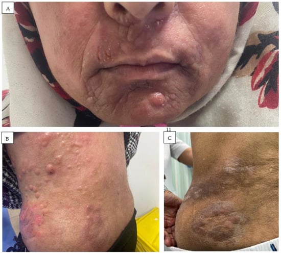
Figure 4.
Tumor stage MF (IIB): multiple nodules and plaques, partly with erosions on the face (A) and trunk (B,C), histologically with large cell transformation.
2.1. Studies by Country
Studies by country show differences in the clinical presentation and age of the patients (for an overview, see Table 2).
Table 2.
Epidemiological and clinical insights into CTCL: a multinational retrospective analysis.
2.1.1. Iran
Naeini et al. described the epidemiological characteristics of 95 primary CTCL patients diagnosed between 2003 and 2013 in Isfahan, Iran. The study highlighted the absence of a male predominance and lower age at diagnosis in this group, with a male-to-female ratio of 1:1.2 and a mean age at diagnosis of 41.78 years [17].
Another study during the same period provided clinical information on patients with MF specifically and reported the same male-to-female ratio and mean age at diagnosis. It included 86 patients, 6% of whom were children, and highlighted that 21% of cases had unusual variants of MF, including hypopigmented and poikilodermatous MF [18].
Nasimi et al. studied 30 cases of pediatric MF and concluded that persistent hypopigmented, purpuric and papulosquamous lesions in children is suggestive of MF as a differential diagnosis. The study reported excellent results with phototherapy in this patient group [19].
2.1.2. UAE
Hamodat et al. conducted a retrospective review and identified 40 patients with mycosis fungoides between 2013 and 2019. The male-to-female ratio was 1.22 and the median age at diagnosis was 47 years. In addition, 20% of patients presented with hypopigmented patches and 12.5% with hyperpigmented patches [20].
2.1.3. KSA
One study included 34 cases of MF diagnosed between 2011 and 2016 and reported that the hypopigmented type was the most common type of MF in the study, affecting a younger age group. The study reported a good response to phototherapy NB-UVB combined with topical corticosteroids in the study group [21].
Another study from Riyadh included 125 CTCL patients (2010–2016) and reported that the hypopigmented type occurred in 15.2% of cases. The male-to-female ratio was 1.36, the median age at diagnosis was 41 years, and 13% of patients were younger than 20 years of age [22].
Alghubaywi et al. enrolled 73 patients diagnosed with MF between 2016 and 2022 and reported an early onset of disease, with a mean age of 44 years, a slightly higher prevalence in females, with a male-to-female ratio of 1.3:1, and a higher prevalence of hypopigmented MF (20.5%) [23].
Another study included 66 skin biopsies generated from 58 patients with suspected/early patch stage MF between 2002 and 2006 and concluded that observing the hyperconvoluted dermal and epidermal lymphocytes, among other parameters, plays an important role in the histopathological diagnosis of early MF lesions and their discrimination from inflammatory simulators [24].
2.1.4. Morocco
One study conducted on 114 MF cases diagnosed from 1993 to 2022 reported a male-to-female ratio of 2.56, a median age at diagnosis of 56 years, and a high proportion of classic MF with a favorable prognosis [25].
2.1.5. Kuwait
One study included 193 MF cases diagnosed between 1991 and 2006 and reported a male-to-female ratio of 2:1, a mean age at diagnosis of 35 years, and a proportion of patients diagnosed by the age 20 years of 16%. In addition, 22% of patients had a pure hypopigmented variant, and they were observed to have a younger mean age at diagnosis (27.6 years) compared to other MF cases (38.1 years). The calculated annual incidence rate of MF in Kuwait was 0.43 cases per 100,000 persons [26].
Another study included 738 subjects registered with non-Hodgkin’s lymphoma between 1998 and 2006 in the population-based cancer registry at the Kuwait Cancer Control Center. The study reported that the prevalence of MF was 9.3% in this group [27].
Nanda et al. studied the clinico-epidemiologic features of juvenile onset MF in Kuwait and included 36 patients diagnosed between 1991 and 2009. Juvenile onset MF constituted 16.6% of the total number of MF patients and the age-adjusted incidence rate of MF in children and adolescents among the total population was 0.29/100,000 persons/year. The study reported a male-to-female ratio of 1.25:1 and a mean age at onset of disease of 9 years and at diagnosis of 13 years. Patients had a predominantly hypopigmented presentation (56% of the cases, Figure 5) [28].
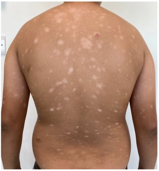
Figure 5.
Pediatric MF, hypopigmented variant: generalized hypopigmented macules and patches in a 13-year-old male (MF IB).
2.1.6. Egypt
Abdel-Halim et al. studied the frequency of hypopigmented MF in a cohort of 100 Egyptian patients presenting with hypopigmented lesions involving the trunk (with or without other sites’ involvement). The frequency of hypopigmented MF was found to be 16%. Hypopigmented MF was significantly associated with a progressive disease course, affection of the distal upper limbs, proximal lower limbs, large-sized lesions (>5 cm), a well-defined margin, scaliness, erythema, atrophy, and mottled pigmentation, as compared to other hypopigmented disorders [29].
2.1.7. Jordan
A retrospective study analyzing the clinical and pathological features of MF included 63 patients diagnosed between 2000 and 2015 and reported a male-to-female ratio of 2.15:1, a mean age at diagnosis of 45 years, and the following clinical variants: 39.6% classical MF, 11% hypopigmented MF, 9.5% poikilodermatous MF, and 3.1% hyperpigmented MF [30].
2.2. Multidisciplinary Approach in CTCL Management
The multidisciplinary team approach to disease management is growing in popularity across different therapeutic areas, especially oncology, and CTCL is no exception [30]. Vitiello et al. suggest that a multidisciplinary team, including dermatologists, hematologists, oncologists, and support staff, should be involved in the management of CTCL [31]. The authors suggest that while dermatologists usually handle treatment in the early phases, they should still be involved in CTCL management even in advanced stages where systemic therapies are needed. Dermatologists’ involvement in CTCL management is necessary considering their knowledge of the topical and systemic therapies for CTCL and the use of the TNMB classification and staging system, their ability to perform frequent skin examinations to monitor the response to treatments and toxicities, and their role in the evaluation and treatment of pruritus, skin infections, and wound care [8]. A review by Dai et al. suggested that the multidisciplinary approach to diagnosing and treating MF and SS should integrate anticancer therapies with skincare and bacterial decolonization to ensure comprehensive management, which should typically be performed by a multidisciplinary team that includes dermatologists, oncologists, radiation oncologists, and bone marrow transplant specialists [32].
2.3. Establishment of the MENA CTCL Group
The inaugural meeting of the MENA CTCL group took place in Dubai on 6 May 2023. This meeting marked the beginning of a collaborative interdisciplinary effort to address the challenges faced in the diagnosis and treatment of CTCL in the MENA region. The CTCL MENA group’s aims and tasks are as follows. (1) To establish regular communication between CTCL experts in the MENA region in the form of regular interdisciplinary tumor boards. The board meetings will provide a platform for participants to present and discuss challenging CTCL cases. Collaborative discussions among board members will contribute to the accuracy and effectiveness of diagnoses and treatment plans, ultimately improving the quality of care for CTCL patients in the MENA region. (2) To involve different multidisciplinary actors (dermatologists, hematologists, oncologists, pathologists, as well as radiotherapists) in the care of patients. (3) To increase representation of the MENA region, e.g., in guidelines, publications, clinical studies, etc., considering the special clinical ethnic features and therapeutic limitations in the management of CTCL patients. The board plans to document and publish regional-specific data, such as patient characteristics (age, skin color), clinical cases, and guidelines. These publications will contribute to the body of knowledge regarding CTCL and improve future treatment approaches in this region, including establishing CTCL registries and establishing CTCL patient advocacy groups in the MENA region.
3. Conclusions
In conclusion, the heterogenous nature of CTCL, in addition to the unique ethnic features of the population in the MENA region, such as hypo- and hyperpigmented MF and increased pediatric cases, contributes to the distinct clinical features of CTCL specific to this population. These differences from Western patients highlight the need for special expertise and multidisciplinary management of CTCL. The MENA CTCL group was established with the objective of improving the diagnosis and treatment of CTCL in the region through creating a collaborative environment and increasing the global representation of the MENA region. Further research is needed to elucidate epidemiological trends, establish CTCL registries, and develop tailored diagnostic and therapeutic strategies.
Author Contributions
Conceptualization, methodology, validation, formal analysis, writing—original draft preparation, writing—review and editing: all authors. All authors have read and agreed to the published version of the manuscript.
Funding
This publication was fully sponsored by Recordati Rare Diseases.
Informed Consent Statement
Informed consent was obtained from all subjects involved in the study.
Conflicts of Interest
S.A.: advisory board member of Hikma, Novartis, Janssen, Abbvie, Leo, Pfizer. C.A. has served as a consultant for 4SC, Takeda, Helsinn, Innate Pharma, Recordati Rare Diseases, Kyowa Kirin. The remaining authors declare that the research was conducted in the absence of any commercial or financial relationships that could be construed as a potential conflict of interest.
References
- United Nations, Department of Economic and Social Affairs, Population Division. World Population Prospects: The 2017 Revi-sion, Key Findings and Advance Tables; Working Paper No. ESA/P/WP/248; New York, NY, USA, 2017; pp. 2–8. Available online: https://population.un.org/wpp/Publications/Files/WPP2017_KeyFindings.pdf (accessed on 27 December 2020).
- Stefan, D.C.; Tang, S. Addressing cancer care in low- to middle-income countries: A call for sustainable innovations and impactful research. BMC Cancer 2023, 23, 756. [Google Scholar] [CrossRef] [PubMed]
- Znaor, A.; Eser, S.; Anton-Culver, H.; Fadhil, I.; Ryzhov, A.; Silverman, B.G.; Bendahou, K.; Demetriou, A.; Nimri, O.; Yakut, C.; et al. Cancer surveillance in northern Africa, and central and western Asia: Challenges and strategies in support of developing cancer registries. Lancet Oncol. 2018, 19, E85–E92. [Google Scholar] [CrossRef] [PubMed]
- Hofmarcher, T.; García, A.M.; Wilking, N.; Lindgren, P. The Disease Burden and Economic Burden of Cancer in 9 Countries in the Middle East and Africa. Value Heal. Reg. Issues 2023, 37, 81–87. [Google Scholar] [CrossRef] [PubMed]
- Sameh, K.; Khalife, N. Oncology clinical research landscape in Middle East and North Africa (MENA) region: Challenges and proposed solutions. J. Clin. Oncol. 2021, 39, e13567. [Google Scholar] [CrossRef]
- Willemze, R.; Cerroni, L.; Kempf, W.; Berti, E.; Facchetti, F.; Swerdlow, S.H.; Jaffe, E.S. The 2018 update of the WHO-EORTC classification for primary cutaneous lymphomas. Blood 2019, 133, 1703–1714. [Google Scholar] [CrossRef]
- Dobos, G.; Pohrt, A.; Ram-Wolff, C.; Lebbé, C.; Bouaziz, J.-D.; Battistella, M.; Bagot, M.; de Masson, A. Epidemiology of Cutaneous T-Cell Lymphomas: A Systematic Review and Meta-Analysis of 16,953 Patients. Cancers 2020, 12, 2921. [Google Scholar] [CrossRef]
- Poligone, B.; Querfeld, C. Management of advanced cutaneous T-cell lymphoma: Role of the dermatologist in the multidisciplinary team. Br. J. Dermatol. 2015, 173, 1081–1083. [Google Scholar] [CrossRef]
- Mehta-Shah, N.; Horwitz, S.M.; Ansell, S.; Ai, W.Z.; Barnes, J.; Barta, S.K.; Clemens, M.W.; Dogan, A.; Fisher, K.; Goodman, A.M.; et al. NCCN Guidelines Insights: Primary Cutaneous Lymphomas, Version 2.2020. J. Natl. Compr. Cancer Netw. 2020, 18, 522–536. [Google Scholar] [CrossRef]
- Olsen, E.A.; Whittaker, S.; Willemze, R.; Pinter-Brown, L.; Foss, F.; Geskin, L.; Schwartz, L.; Horwitz, S.; Guitart, J.; Zic, J.; et al. Primary cutaneous lymphoma: Recommendations for clinical trial design and staging update from the ISCL, USCLC, and EORTC. Blood 2021, 140, 419–437. [Google Scholar] [CrossRef]
- Agar, N.S.; Wedgeworth, E.; Crichton, S.; Mitchell, T.J.; Cox, M.; Ferreira, S.; Robson, A.; Calonje, E.; Stefanato, C.M.; Wain, E.M.; et al. Survival Outcomes and Prognostic Factors in Mycosis Fungoides/Sézary Syndrome: Validation of the Revised International Society for Cutaneous Lymphomas/European Organisation for Research and Treatment of Cancer Staging Proposal. J. Clin. Oncol. 2010, 28, 4730–4739. [Google Scholar] [CrossRef]
- Latzka, J.; Assaf, C.; Bagot, M.; Cozzio, A.; Dummer, R.; Guenova, E.; Gniadecki, R.; Hodak, E.; Jonak, C.; Klemke, C.-D.; et al. EORTC consensus recommendations for the treatment of mycosis fungoides/Sézary syndrome—Update 2023. Eur. J. Cancer 2023, 195, 113343. [Google Scholar] [CrossRef] [PubMed]
- Ibrahim, M.A.-H.; Eltayeb, N.; Ibrahim, M.M.; Nassar, A.; Daruish, M.; El-Zimaity, M.; El-Lithy, M.; Mostafa, A.; El-Afifi, A.; Abdelbary, H.; et al. Suggested Guidelines for the Treatment of Mycosis Fungoides in Countries with Limited Resources. Dermatol. Res. Pr. 2023, 2023, 1360740. [Google Scholar] [CrossRef] [PubMed]
- Allen, P.B.; Goyal, S.; O′eary, C.; Ayers, A.; Niyogusaba, T.; Khan, M.K.; Lechowicz, M.J. Racial differences in clinical presentation and outcomes in mycosis fungoides and Sézary syndrome in the United States: A large singe center retrospective analysis. Eur. J. Cancer 2021, 156, S34. [Google Scholar] [CrossRef] [PubMed]
- Huang, A.H.; Kwatra, S.G.; Khanna, R.; Semenov, Y.R.; Okoye, G.A.; Sweren, R.J. Racial Disparities in the Clinical Presentation and Prognosis of Patients with Mycosis Fungoides. J. Natl. Med Assoc. 2019, 111, 633–639. [Google Scholar] [CrossRef] [PubMed]
- Rager, T.; Lake, E. Mycosis Fungoides in Skin of Color. J. Dermatol. Nurses Assoc. 2022, 14, 261–264. [Google Scholar] [CrossRef]
- Naeini, F.F.; Sadeghiyan, H.; Pourazizi, M.; Najafian, J.; Abtahi-Naeini, B. Characteristics of Primary Cutaneous T-Cell Lymphoma in Iran: A 10-Year Retrospective Study. Int. Sch. Res. Not. 2014, 2014, 820921. [Google Scholar] [CrossRef]
- Naeini, F.F.; Abtahi-Naeini, B.; Sadeghiyan, H.; Nilforoushzadeh, M.A.; Najafian, J.; Pourazizi, M. Mycosis Fungoides in Iranian Population: An Epidemiological and Clinicopathological Study. J. Ski. Cancer 2015, 2015, 306543. [Google Scholar] [CrossRef]
- Nasimi, M.; Kamyab, K.; Aghahi, T.; Fahim, S.; Ghandi, N. Childhood mycosis fungoides: A clinicopathologic study of 30 cases from Iran. Australas. J. Dermatol. 2020, 61, E259–E261. [Google Scholar] [CrossRef]
- Hamodat, M.M.; Al Maashari, R.S.; Al Zaabi, E. Mycosis Fungoides in UAE. Saudi J. Pathol. Microbiol. 2021, 6, 258–260. [Google Scholar]
- Alojail, H.Y.; Alshehri, H.; Kaliyadan, F. Clinical Patterns and Treatment Response of Patients with Mycosis Fungoides a Retrospective Study. Cureus 2022, 14, e21231. [Google Scholar] [CrossRef]
- Binamer, Y. Cutaneous T-cell lymphoma in Saudi Arabia: Retrospective single-center review. Ann. Saudi Med. 2017, 37, 212–215. [Google Scholar] [CrossRef] [PubMed][Green Version]
- Alghubaywi, F.A.; Alharthi, S.A.; Aldharman, S.S.; Najjar, R.H.; Aleissa, M.Y.; Aljarbou, O.Z.; AlJasser, M.I.; Almohideb, M.A. Clinicopathologic characteristics and outcomes of patients with mycosis fungoides. SciVee 2023, 44, 394–400. [Google Scholar] [CrossRef] [PubMed]
- Arafah, M.; Zaidi, S.N.; Kfoury, H.K.; Al Rikabi, A.; Al Ghamdi, K. The Histological Spectrum of Early Mycosis Fungoides: A Study of 58 Saudi Arab patients. Oman Med. J. 2012, 27, 134–139. [Google Scholar] [CrossRef] [PubMed]
- Titou, H.; Bouhamidi, A. Epidemiology and prognostic factors of 114 patients with mycosis fungoides in a Moroccan cohort: A 29-year review. Clin. Exp. Med. 2023, 23, 3751–3758. [Google Scholar] [CrossRef]
- Alsaleh, Q.A.; Nanda, A.; Al-Ajmi, H.; Al-Sabah, H.; Elkashlan, M.; Al-Shemmari, S.; Demierre, M. Clinicoepidemiological features of mycosis fungoides in Kuwait, 1991–2006. Int. J. Dermatol. 2010, 49, 1393–1398. [Google Scholar] [CrossRef]
- Ameen, R.; Sajnani, K.P.; Albassami, A.; Refaat, S. Frequencies of non-Hodgkin’s lymphoma subtypes in Kuwait: Comparisons between different ethnic groups. Ann. Hematol. 2009, 89, 179–184. [Google Scholar] [CrossRef]
- Nanda, A.; AlSaleh, Q.A.; Al-Ajmi, H.; Al-Sabah, H.; Elkashlan, M.; Al-Shemmari, S.; Demierre, M.-F. Mycosis Fungoides in Arab Children and Adolescents: A Report of 36 Patients from Kuwait. Pediatr. Dermatol. 2010, 27, 607–613. [Google Scholar] [CrossRef]
- Abdel-Halim, M.; El-Nabarawy, E.; El Nemr, R.; Hassan, A.M. Frequency of Hypopigmented Mycosis Fungoides in Egyptian Patients Presenting with Hypopigmented Lesions of the Trunk. Am. J. Dermatopathol. 2015, 37, 834–840. [Google Scholar] [CrossRef]
- Al-Tarawneh, A.H. Clinical and Histopathological Spectrum of Mycosis Fungoides. Bahrain Med. Bull. 2018, 40, 103–107. [Google Scholar] [CrossRef]
- Vitiello, P.; Sagnelli, C.; Ronchi, A.; Franco, R.; Caccavale, S.; Mottola, M.; Pastore, F.; Argenziano, G.; Creta, M.; Calogero, A.; et al. Multidisciplinary Approach to the Diagnosis and Therapy of Mycosis Fungoides. Healthcare 2023, 11, 614. [Google Scholar] [CrossRef]
- Dai, J.; Duvic, M. Cutaneous T-Cell Lymphoma: Current and Emerging Therapies. Oncology 2023, 37, 55–62. [Google Scholar] [CrossRef] [PubMed]
Disclaimer/Publisher’s Note: The statements, opinions and data contained in all publications are solely those of the individual author(s) and contributor(s) and not of MDPI and/or the editor(s). MDPI and/or the editor(s) disclaim responsibility for any injury to people or property resulting from any ideas, methods, instructions or products referred to in the content. |
© 2024 by the authors. Licensee MDPI, Basel, Switzerland. This article is an open access article distributed under the terms and conditions of the Creative Commons Attribution (CC BY) license (https://creativecommons.org/licenses/by/4.0/).