

Robotic Stereotactic Radiotherapy for Intracranial Meningiomas—An Opportunity for Radiation Dose De-Escalation

 , and
, and
Abstract
:Simple Summary
Abstract
1. Introduction
2. Materials and Methods
2.1. Patient Characteristics
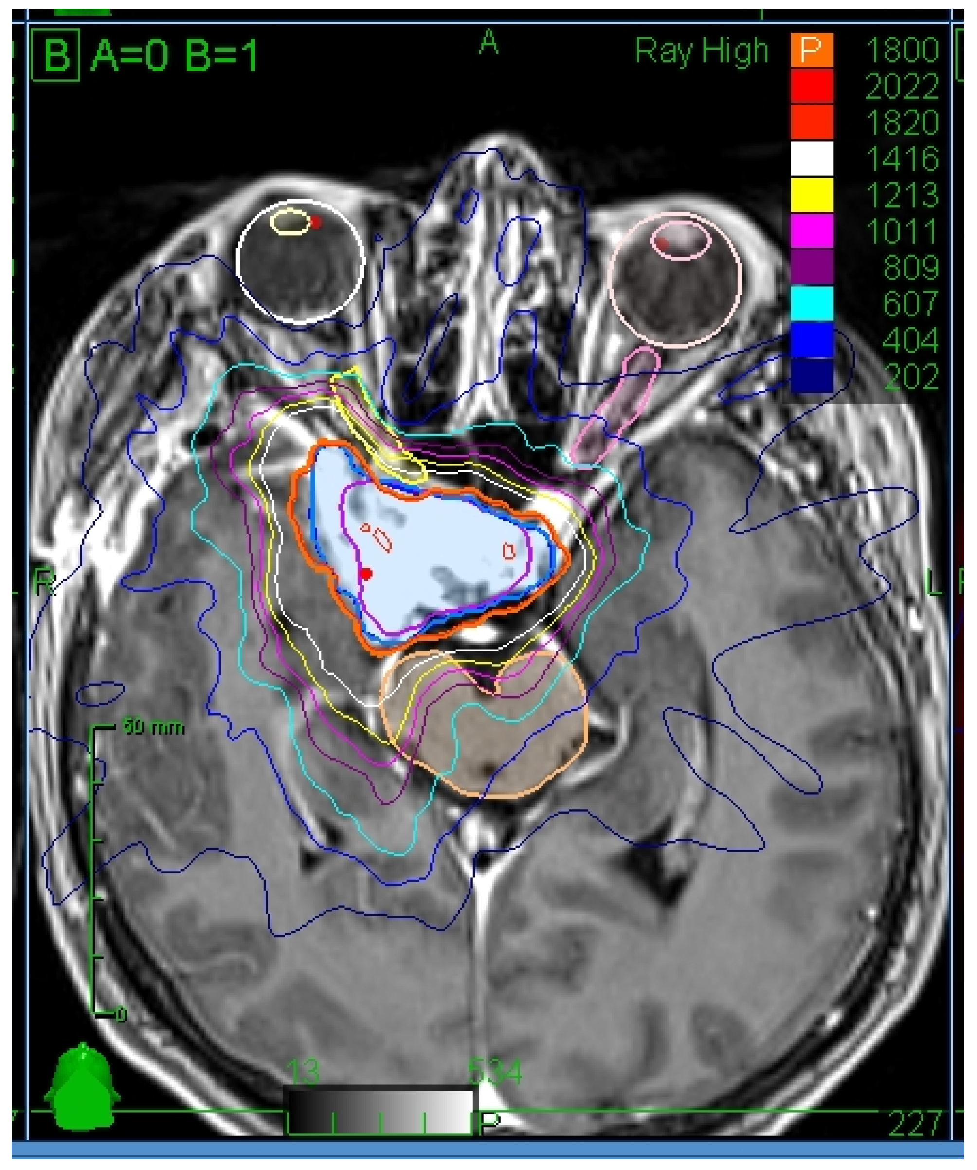
2.2. Radiotherapy Treatment Planning and Delivery
2.3. Follow-Up
2.4. Statistical Analysis
3. Results
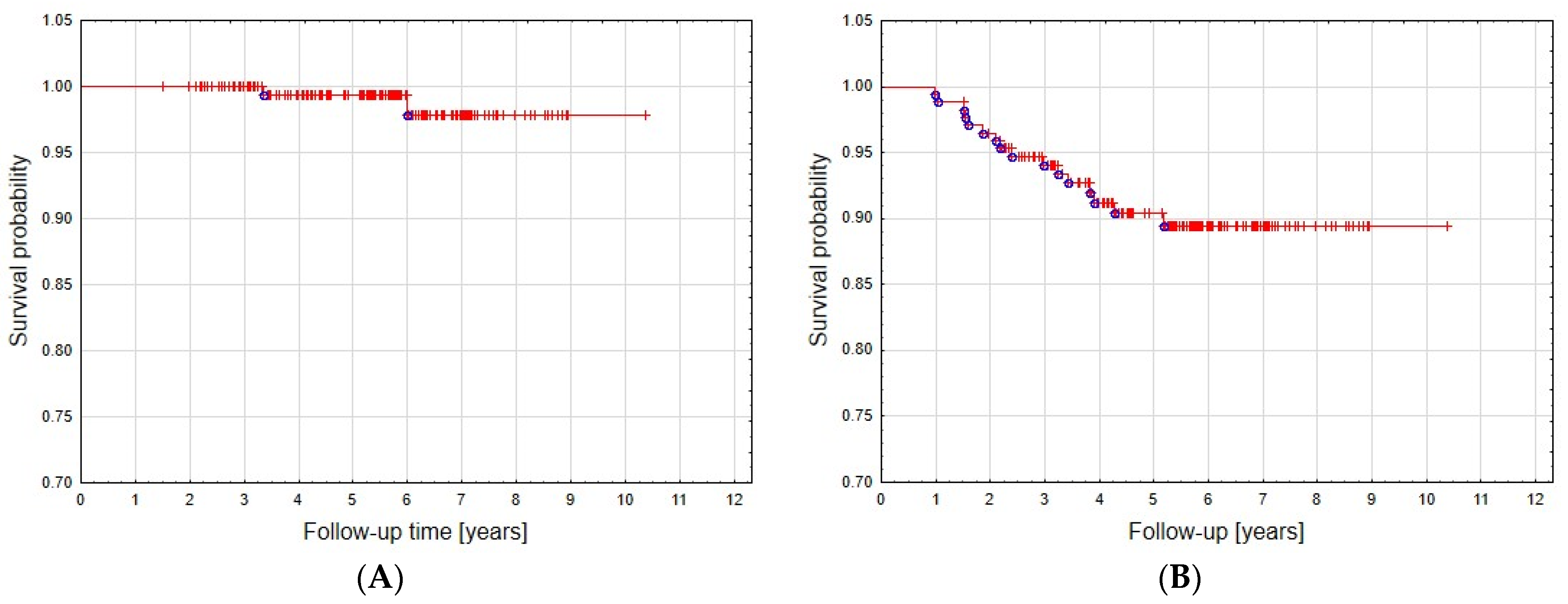
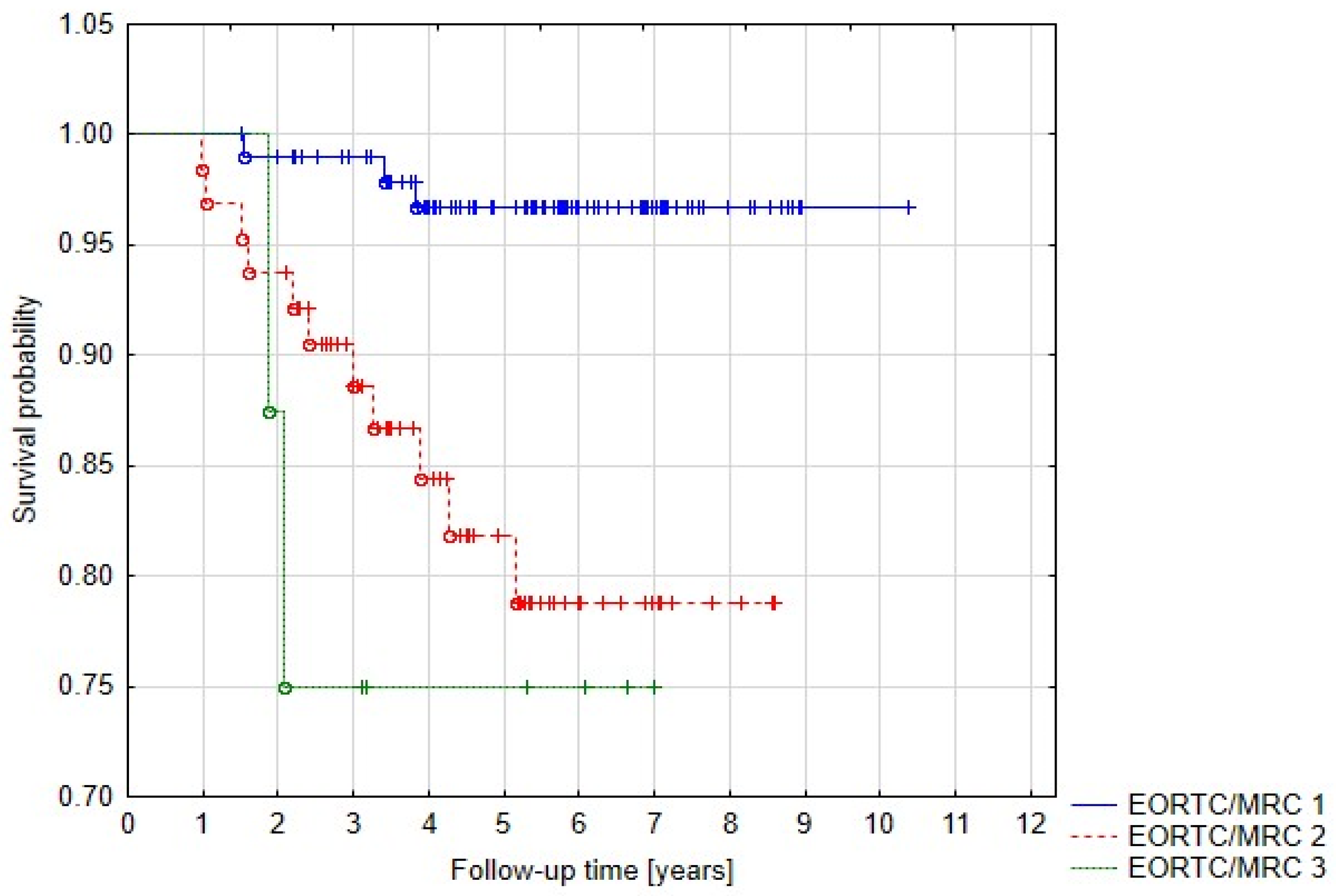
3.1. Local Control
3.2. Patients with Tumor Progression
3.3. Tolerance of Treatment
3.4. Clinical Effects of Radiotherapy
3.5. Other Neoplasms Diagnosed during Follow-Up Period
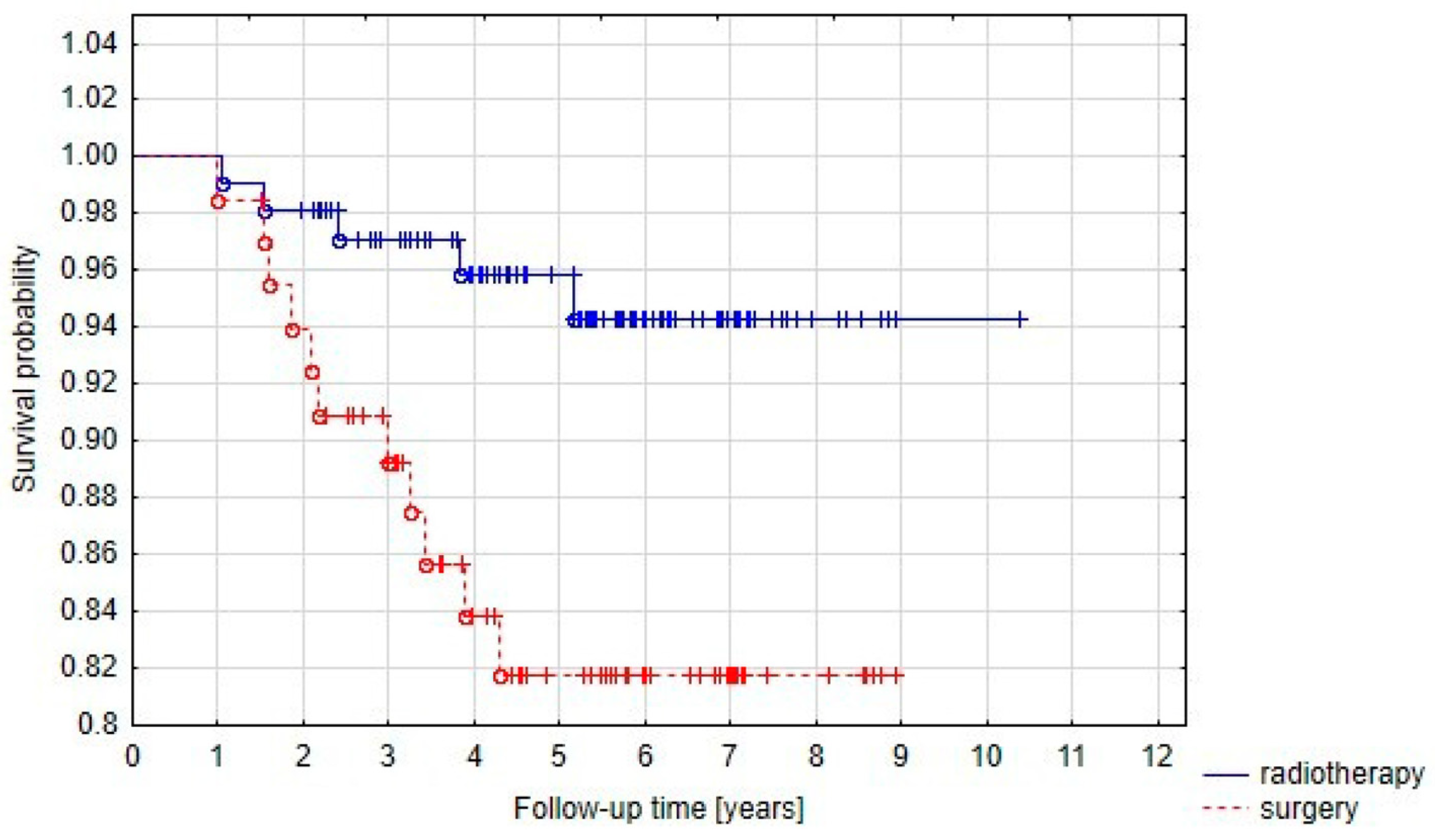
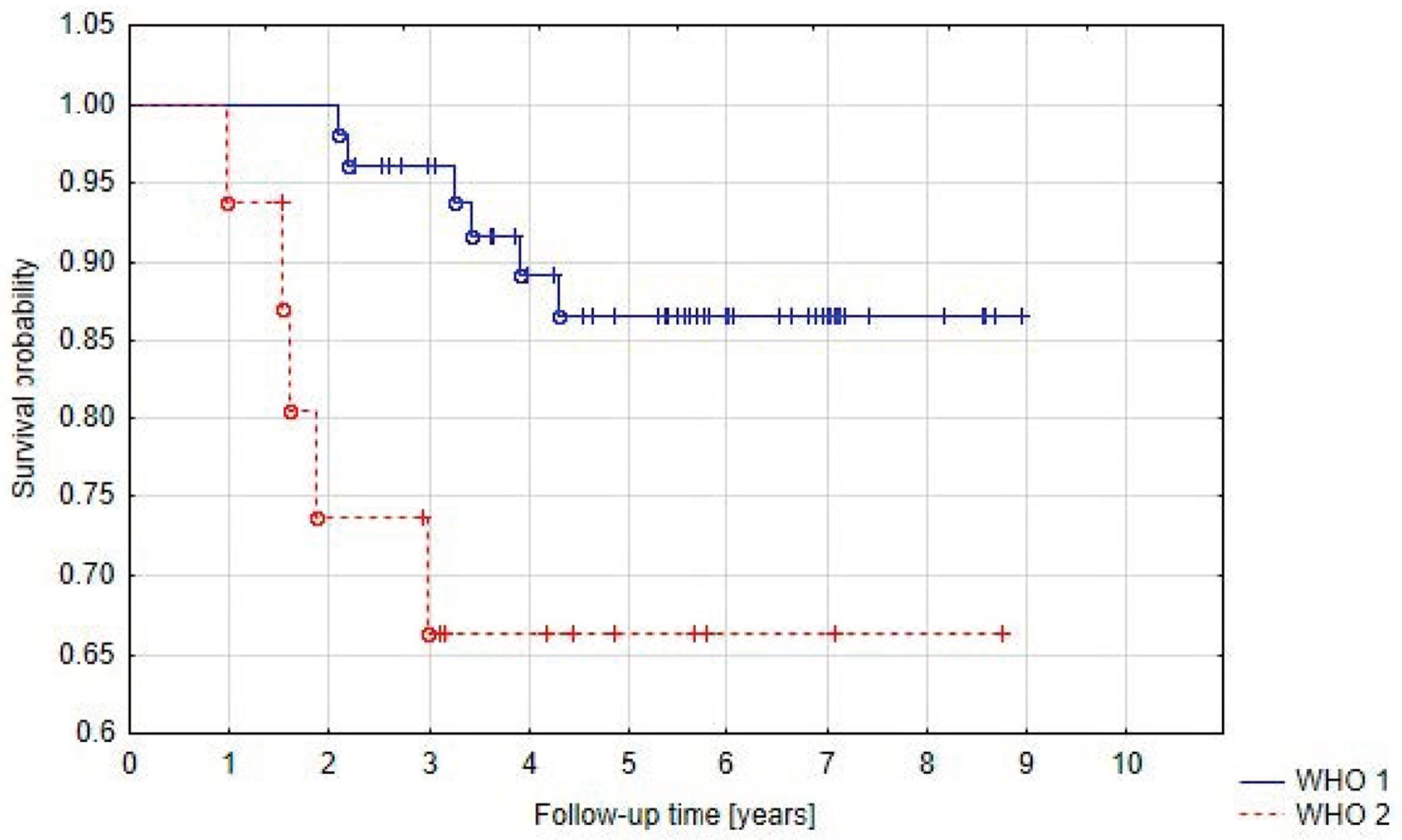
3.6. Univariate and Multivariate Analysis
4. Discussion
4.1. Study Population
4.2. Treatment Results
4.3. Treatment Tolerance
4.4. Prognostic Factors
5. Conclusions
Author Contributions
Funding
Institutional Review Board Statement
Informed Consent Statement
Data Availability Statement
Conflicts of Interest
References
- Marosi, C.; Hassler, M.; Roessler, K.; Reni, M.; Sant, M.; Mazza, E.; Vecht, C. Meningioma. Crit. Rev. Oncol./Hematol. 2008, 67, 153–171. [Google Scholar] [PubMed]
- Riemenschneider, M.; Perry, A.; Reifenberger, G. Histological classification and molecular genetics of meningiomas. Lancet Neurol. 2006, 5, 1045–1054. [Google Scholar] [PubMed]
- Buerki, R.A.; Horbinski, C.M.; Kruser, T.; Horowitz, P.M.; James, C.D.; Lukas, R.V. An overview of meningiomas. Future Oncol. 2018, 14, 2161–2177. [Google Scholar]
- Horbinski, C.; Nabors, L.B.; Portnow, J.; Baehring, J.; Bhatia, A.; Bloch, O.; Brem, S.; Butowski, N.; Cannon, D.M.; Chao, S. NCCN Guidelines Insights: Central Nervous System Cancers; Version 2.2022. J. Natl. Compr. Canc. Netw. 2023, 21, 12–20. [Google Scholar] [PubMed]
- Simpson, D. The recurrence of intracranial meningiomas after surgical treatment. J. Neurol. Neurosurg. Psychiatry 1957, 20, 22. [Google Scholar] [CrossRef]
- Naumann, M.; Meixensberger, J. Factors influencing meningioma recurrence rate. Acta Neurochir. 1990, 107, 108–111. [Google Scholar]
- Timmerman, R.D. An overview of hypofractionation and introduction to this issue of seminars in radiation oncology. Semin. Radiat. Oncol. 2008, 18, 215–222. [Google Scholar]
- Orski, M.; Tarnawski, R.; Wylegala, E.; Tarnawska, D. The Impact of Robotic Fractionated Radiotherapy for Benign Tumors of Parasellar Region on the Eye Structure and Function. J. Clin. Med. 2023, 12, 404. [Google Scholar] [CrossRef]
- Nakasu, S.; Hirano, A.; Shimura, T.; Llena, J.F. Incidental meningiomas in autopsy study. Surg. Neurol. 1987, 27, 319–322. [Google Scholar]
- Klaeboe, L.; Lonn, S.; Scheie, D.; Auvinen, A.; Christensen, H.C.; Feychting, M.; Johansen, C.; Salminen, T.; Tynes, T. Incidence of intracranial meningiomas in Denmark, Finland, Norway, and Sweden, 1968–1997. Int. J. Cancer 2005, 117, 996–1001. [Google Scholar] [CrossRef]
- Radhakrishnan, K.; Mokri, B.; Parisi, J.E.; O’Fallon, W.M.; Sunku, J.; Kurland, L.T. The trends in incidence of primary brain tumors in the population of Rochester, Minnesota. Ann. Neurol. 1995, 37, 67–73. [Google Scholar] [CrossRef] [PubMed]
- Chakravarthy, V.; Kaplan, B.; Gospodarev, V.; Myers, H.; De Los Reyes, K.; Achiriloaie, A. Houdini Tumor: Case Report and Literature Review of Pregnancy-Associated Meningioma. World Neurosurg. 2018, 114, 1261–1265. [Google Scholar] [CrossRef] [PubMed]
- Central Brain Tumor Registry of the United States. CBTRUS (2009–2010) CDTRUS Statistical Report: Primary Brain and Central nervous System Tumors Diagnosed in Eighteeen States in 2002–2006; Central Brain Tumor Registry of the United States: Hisdale, IL, USA, 2010. [Google Scholar]
- Park, B.J.; Kim, H.K.; Sade, B.; Lee, J.H. Meningiomas: Diagnosis, Treatment, and Outcome; Springer: Berlin/Heidelberg, Germany, 2009. [Google Scholar]
- Bhat, A.R.; Wani, M.A.; Kirmani, A.R.; Ramzan, A.U. Histological subtypes and anatomical location correlated in meningeal brain tumors (meningiomas). J. Neurosci. Rural. Pract. 2014, 5, 244–249. [Google Scholar] [CrossRef] [PubMed]
- Wellenreuther, R.; Kraus, J.A.; Lenartz, D.; Menon, A.G.; Schramm, J.; Louis, D.N.; Ramesh, V.; Gusella, J.F.; Wiestler, O.D.; von Deimling, A. Analysis of the neurofibromatosis 2 gene reveals molecular variants of meningioma. Am. J. Pathol. 1995, 146, 827–832. [Google Scholar] [PubMed]
- Sahm, F.; Schrimpf, D.; Stichel, D. DNA methylation-based classification and grading system for meningioma: A multi-centre, retrospective analysis. Lancet Oncol. 2017, 17, 155–159. [Google Scholar] [CrossRef]
- Claus, E.B.; Calvocoressi, L.; Bondy, M.L.; Schildkraut, J.M.; Wiemels, J.L.; Wrensch, M. Family and personal medical history and risk of meningioma. J. Neurosurg. 2011, 115, 1072–1077. [Google Scholar] [CrossRef]
- Hill, D.A.; Linet, M.S.; Black, P.M.; Fine, H.A.; Selker, R.G.; Shapiro, W.R.; Inskip, P.D. Meningioma and schwannoma risk in adults in relation to family history of cancer. Neuro-Oncology 2004, 6, 274–280. [Google Scholar] [CrossRef]
- Kirsch, M.; Zhu, J.J.; Black, P.M. Analysis of the BRCA1 and BRCA2 genes in sporadic meningioma. Genes Chromosomes Cancer 1997, 20, 53–59. [Google Scholar] [CrossRef]
- Custer, B.S.; Koepsell, T.D.; Mueller, B.A. The association between breast carcinoma and meningioma in women. Cancer 2002, 94, 1626–1635. [Google Scholar] [CrossRef]
- Markwalder, T.M.; Seiler, R.W.; Zava, D.T. Antiestrogenic therapy of meningiomas—A pilot study. Surg. Neurol. 1985, 24, 245–249. [Google Scholar] [CrossRef]
- Goodwin, J.W.; Crowley, J.; Eyre, H.J.; Stafford, B.; Jaeckle, K.A.; Townsend, J.J. A phase II evaluation of tamoxifen in unresectable or refractory meningiomas: A Southwest Oncology Group study. J. Neurooncol. 1993, 15, 75–77. [Google Scholar] [CrossRef] [PubMed]
- Ji, J.; Sundquist, J.; Sundquist, K. Association of tamoxifen with meningioma: A population-based study in Sweden. Eur. J. Cancer Prev. 2016, 25, 29–33. [Google Scholar] [CrossRef] [PubMed]
- Sun, L.M.; Lin, C.L.; Sun, S.; Hsu, C.Y.; Shae, Z.; Kao, C.H. Long-term use of tamoxifen is associated with a decreased subsequent meningioma risk in patients with breast cancer: A nationwide population-based cohort study. Front. Pharmacol. 2019, 10, 674. [Google Scholar] [CrossRef] [PubMed]
- Champeaux-Depond, C.; Weller, J. Tamoxifen. A treatment for meningioma? Cancer Treat. Res. Commun. 2021, 27, 100343. [Google Scholar] [CrossRef]
- Rogers, L.; Barani, I.; Chamberlain, M.; Kaley, T.; McDermott, M.; Raizer, J.; Schiff, D.; Weber, D.C.; Wen, P.Y.; Vogelbaum, M.A. Meningiomas: Knowledge base, treatment outcomes, and uncertainties: A RANO review. J. Neurosurg. 2015, 122, 4–23. [Google Scholar] [CrossRef]
- Pollock, B.E.; Stafford, S.L.; Utter, A.; Giannini, C.; Schreiner, S.A. Stereotactic radiosurgery provides equivalent tumor control to Simpson Grade 1 resection for patients with small- to medium-size meningiomas. Int. J. Radiat. Oncol. Biol. Phys. 2003, 55, 1000–1005. [Google Scholar] [CrossRef]
- Cohen-Inbar, O.; Lee, C.; Sheehan, J.P. The Contemporary Role of Stereotactic Radiosurgery in the Treatment of Meningiomas. Neurosurg. Clin. N. Am. 2016, 27, 215–228. [Google Scholar] [CrossRef]
- Unger, K.R.; Lominska, C.E.; Chanyasulkit, J.; Randolf-Jackson, P.; White, R.L.; Aulisi, E.; Jacobson, J.; Jean, W.; Gagnon, G.J. Risk factors for posttreatment edema in patients treated with stereotactic radiosurgery for meningiomas. Neurosurgery 2012, 70, 639–645. [Google Scholar] [CrossRef]
- Biswas, T.; Sandhu, A.P.; Singh, D.P.; Schell, M.C.; Maciunas, R.J.; Bakos, R.S.; Muhs, A.G.; Okunieff, P. Low-Dose Radiosurgery for Benign Intracranial Lesions. Am. J. Clin. Oncol. 2003, 26, 325–331. [Google Scholar] [CrossRef]
- Oh, H.J.; Cho, Y.H.; Kim, J.H.; Kim, C.J.; Kwon, D.H.; Lee, D.; Yoon, K.J. Hypofractionated stereotactic radiosurgery for large-sized skull base meningiomas. J. Neurooncol. 2020, 149, 87–93. [Google Scholar] [CrossRef]
- Tuniz, F.; Soltys, S.G.; Choi, C.Y.; Chang, S.D.; Gibbs, I.C.; Fischbein, N.J.; Adler, J.R., Jr. Multisession cyberknife stereotactic radiosurgery of large, benign cranial base tumors: Preliminary study. Neurosurgery 2009, 65, 898–907. [Google Scholar] [CrossRef] [PubMed]
- Conti, A.; Pontoriero, A.; Midili, F.; Iati, G.; Siragusa, C.; Tomasello, C.; La Torre, D.; Cardalli, S.M.; Pergolizzi, S.; De Renzis, C. CyberKnife multisession stereotactic radiosurgery and hypofractionated stereotactic radiotherapy for perioptic meningiomas: Intermediate-term results and radiobiological considerations. Springerplus 2015, 4, 37. [Google Scholar] [CrossRef] [PubMed]
- Meniai-Merzouki, F.; Bernier-Chastagner, V.; Geffrelot, J.; Tresch, E.; Lacornerie, T.; Coche-Dequeant, B.; Lartigau, E.; Pasquier, D. Hypofractionated Stereotactic Radiotherapy for Patients with Intracranial Meningiomas: Impact of radiotherapy regimen on local control. Sci. Rep. 2018, 8, 13666. [Google Scholar] [CrossRef] [PubMed]
- Shrieve, D.C.; Hazard, L.; Boucher, K.; Jensen, R.L. Dose fractionation in stereotactic radiotherapy for parasellar meningiomas: Radiobiological considerations of efficacy and optic nerve tolerance. J. Neurosurg. 2004, 101, 390–395. [Google Scholar] [CrossRef]
- Vernimmen, F.J.; Slabbert, J.P. Assessment of the alpha/beta ratios for arteriovenous malformations, meningiomas, acoustic neuromas, and the optic chiasma. Int. J. Radiat. Biol. 2010, 86, 486–498. [Google Scholar] [CrossRef]
- Santacroce, A.; Walier, M.; Régis, J.; Liščak, R.; Motti, E.; Lindquist, C.; Kemeny, A.; Kitz, K.; Lippitz, B.; Álvarez, R.M.; et al. Long-term tumor control of benign intracranial meningiomas after radiosurgery in a series of 4565 patients. Neurosurgery 2012, 70, 32–39. [Google Scholar] [CrossRef]
- Przybylowski, C.J.; Raper, D.M.; Starke, R.M.; Xu, Z.; Liu, K.C.; Sheehan, J.P. Stereotactic radiosurgery of meningiomas following resection: Predictors of progression. J. Clin. Neurosci. 2015, 22, 161–165. [Google Scholar] [CrossRef]
- Harat, M.; Lebioda, A.; Lasota, J.; Makarewicz, R. Evaluation of brain edema formation defined by MRI after LINAC-based stereotactic radiosurgery. Radiol. Oncol. 2017, 51, 137–141. [Google Scholar] [CrossRef]
- Gallagher, M.J.; Jenkinson, M.D.; Brodbelt, A.R.; Mills, S.J.; Chavredakis, E. WHO grade I meningioma recurrence: Are location and Simpson grade still relevant? Clin. Neurol. Neurosurg. 2016, 141, 117–121. [Google Scholar] [CrossRef]
- Narayan, V.; Bir, S.C.; Mohammed, N.; Savardekar, A.R.; Patra, D.P.; Nanda, A. Surgical Management of Giant Intracranial Meningioma: Operative Nuances, Challenges and Outcome. World Neurosurg. 2018, 110, 32–41. [Google Scholar] [CrossRef]
- Champeaux, C.; Dunn, L. World Health Organization Grade II Meningioma: A 10-Year Retrospective Study for Recurrence and Prognostic Factor Assessment. World Neurosurg. 2016, 89, 180–186. [Google Scholar] [CrossRef] [PubMed]
- Zhang, G.; Zhang, Y.; Zhang, G.; Yan, X.; Li, C.; Zhang, L.; Li, D.; Wu, Z.; Zhang, J. Prognostic Factors, Survival, and Treatment for Intracranial World Health Organization Grade II Chordoid Meningiomas and Clear-Cell Meningiomas. World Neurosurg. 2018, 117, 57–66. [Google Scholar] [CrossRef] [PubMed]
- Fokas, E.; Henzel, M.; Surber, G.; Hamm, K.; Engenhart-Cabillic, R. Stereotactic Radiation Therapy for Benign Meningioma: Long-Term Outcome in 318 Patients. Int. J. Radiat. Oncol. Biol. Phys. 2014, 89, 569–575. [Google Scholar] [CrossRef] [PubMed]
- Kołodziej, I.; Ślosarek, Z.; Blamek, S. Radiochirurgia stereotaktyczna jako leczenie pierwotne lub pooperacyjne u chorych na oponiaki. Onkol. W Prakt. Klin. 2017, 3 (Suppl. C), 23. [Google Scholar]
- Champeaux, C.; Houston, D.; Dunn, L. Atypical meningioma. A study on recurrence and disease-specific survival. Neurochirurgie 2017, 63, 273–281. [Google Scholar] [CrossRef]
- Zaher, A.; Mattar, M.A.; Zayed, D.H.; Ellatif, R.A.; Ashamallah, S.A. Atypical meningioma: A Study of Prognostic Factors. World Neurosurg. 2013, 80, 549–553. [Google Scholar] [CrossRef]
- Solda, F.; Wharram, B.; DeIeso, P.B.; Bonner, J.; Ashley, S.; Brada, M. Long-term efficacy of fractionated radiotherapy for benign meningiomas. Radiother. Oncol. 2013, 109, 330–334. [Google Scholar] [CrossRef]
- Dos Santos, M.A.; De Salcedo, J.B.; Gutierrez Diaz, J.A.; Calvo, F.A.; Samblas, J.; Marsiglia, H.; Sallabanda, K. Long-term outcomes of stereotactic radiosurgery for treatment of cavernous sinus meningiomas. Int. J. Radiat. Oncol. Biol. Phys. 2011, 81, 1436–1441. [Google Scholar] [CrossRef]
- Wang, W.H.; Lee, C.C.; Yang, H.C.; Liu, K.D.; Wu, H.M.; Shiau, C.Y.; Guo, W.Y.; Pan, D.H.C.; Chung, W.Y.; Chen, M.T. GammaKnife radiosurgery for atypical and anaplastic meningiomas. World Neurosurg. 2016, 87, 557–564. [Google Scholar] [CrossRef]
- Pollock, B.E.; Stafford, S.L.; Link, M.J.; Garces, Y.I.; Foote, R.L. Single-fraction Radiosugery for Presumed Intracranial Meningiomas: Efficacy and Complications From a 22-Year Experience. Int. J. Radiat. Oncol. Biol. Phys. 2012, 83, 1414–1418. [Google Scholar] [CrossRef]
- Kondziolka, D.; Flickinger, J.C.; Perez, B. Judicious resection and/or radiosurgery for parasagittal meningioma: Outcomes from a multicenter review. GammaKnife Meningioma Study Group. Neurosurgery 1998, 43, 405–414. [Google Scholar] [CrossRef] [PubMed]
- Kępka, L.; Żółciak, A.; Leszczyk, C.; Fijuth, J. Rola radioterapii w leczeniu chorych na oponiaka mózgu. Nowotwory 2000, 50, 134–140. [Google Scholar]
- Soyuera, S.; Changa, E.L.; Seleka, U.; Shib, W.; Maora, M.H.; DeMonteb, F. Radiotherapy after surgery for benign cerebral meningioma. Radiother. Oncol. 2004, 71, 85–90. [Google Scholar] [CrossRef] [PubMed]

| Topography | Number of Patients | % |
|---|---|---|
| Skull base | 95 | 55.2 |
| (incl. cavernous sinus) | (42) | (24.4) |
| Falx or parasagittal | 30 | 17.4 |
| Convexity | 28 | 16.3 |
| Cerebellopontine angle | 8 | 4.7 |
| Optic nerve sheath | 6 | 3.5 |
| Lateral fissure | 5 | 2.9 |
| Subtype | Number of Patients | % | |
|---|---|---|---|
| WHO 1 | Meningothelial | 32 | 47.8 |
| Fibrous | 9 | 13.4 | |
| Angiomatous | 2 | 3 | |
| Psammomatous | 2 | 3 | |
| Transitional | 6 | 8.9 | |
| WHO 2 | Atypical | 15 | 22.4 |
| Clear Cell | 1 | 1.5 |
| Symptom | Number of Patients | Complete Regression | Improvement | Worsening |
|---|---|---|---|---|
| Hemiparesis | 18 | 6 | 1 | 2 |
| Visual impairment | 36 | 9 | - | - |
| Hearing impairment | 3 | - | - | - |
| Facial nerve palsy | 12 | 7 | 1 | - |
| Epilepsy | 2 | - | 2 | - |
| Severe headaches | 1 | 1 | - | - |
| Prognostic Factor | 5-Year PFS | p | HR | CI (95%) |
|---|---|---|---|---|
| Prior surgery: | 0.016 | |||
| -yes | 82% | 3.67 | 1.27–10.55 | |
| -no | 96% | |||
| Age at diagnosis: | ||||
| <50 years | 86% | 0.335 | 0.61 | 0.22–1.67 |
| >50 years | 91% | |||
| Sex: | ||||
| -female | 92% | 0.19 | 0.51 | 0.18–1.4 |
| -male | 87% | |||
| GTV: | ||||
| <5 cm3 | 92% | 0.298 | 1.82 | 0.59–5.66 |
| >5 cm3 | 89% | |||
| Presence of neurological symptoms: | ||||
| -no | 96% | 0.00226 | 7.09 | 2.02–24.95 |
| -yes | 81% | |||
| Group A | ||||
| Simpson Grade: | ||||
| I-III | 86% | 0.34 | 1.82 | 0.53–6.22 |
| IV-V | 77% | |||
| Histology: | ||||
| WHO 1 | 87% | 0.027 | 3.86 | 1.17–12.72 |
| WHO 2 | 66% | |||
| Group B | ||||
| Progression prior to radiotherapy: | ||||
| -yes | 89% | 0.089 | 6.66 | 0.74–59.63 |
| -no | 100% | |||
| Prognostic Factor | p | HR | CI (95%) |
|---|---|---|---|
| Surgery and radiotherapy vs. radiotherapy alone | 0.13 | 2.32 | 0.78–6.88 |
| No symptoms vs. neurological deficits | 0.0095 | 5.54 | 1.52–20.23 |
| Study | Median Total Dose [Gy] | Number of Fractions | Median Follow-Up [Months] | Local Control [%] | BED Alpha/Beta = 3.28 | BED Alpha-Beta = 3.76 |
|---|---|---|---|---|---|---|
| Grzbiela et al. | 18 | 3 | 67.5 | 90.7 | 50.9 | 46.7 |
| Oh et al. [32] | 27.8 | 5 | 57 | 90.3 | 74.9 | 68.9 |
| Tuniz et al. [33] | 24 | 3 | 31 | 100 | 82.5 | 75.1 |
| Conti et al. [34] | 23 | 5 | 60 | 100 | 55.3 | 51.1 |
| Meniai-Merzouki et al. [35] | 25 | 5 | 20.3 | 74 | 63.1 | 58.2 |
Disclaimer/Publisher’s Note: The statements, opinions and data contained in all publications are solely those of the individual author(s) and contributor(s) and not of MDPI and/or the editor(s). MDPI and/or the editor(s) disclaim responsibility for any injury to people or property resulting from any ideas, methods, instructions or products referred to in the content. |
© 2023 by the authors. Licensee MDPI, Basel, Switzerland. This article is an open access article distributed under the terms and conditions of the Creative Commons Attribution (CC BY) license (https://creativecommons.org/licenses/by/4.0/).
Share and Cite
Grzbiela, H.; Nowicka, E.; Gawkowska, M.; Tarnawska, D.; Tarnawski, R. Robotic Stereotactic Radiotherapy for Intracranial Meningiomas—An Opportunity for Radiation Dose De-Escalation. Cancers 2023, 15, 5436. https://doi.org/10.3390/cancers15225436
Grzbiela H, Nowicka E, Gawkowska M, Tarnawska D, Tarnawski R. Robotic Stereotactic Radiotherapy for Intracranial Meningiomas—An Opportunity for Radiation Dose De-Escalation. Cancers. 2023; 15(22):5436. https://doi.org/10.3390/cancers15225436
Chicago/Turabian StyleGrzbiela, Hanna, Elzbieta Nowicka, Marzena Gawkowska, Dorota Tarnawska, and Rafal Tarnawski. 2023. "Robotic Stereotactic Radiotherapy for Intracranial Meningiomas—An Opportunity for Radiation Dose De-Escalation" Cancers 15, no. 22: 5436. https://doi.org/10.3390/cancers15225436
APA StyleGrzbiela, H., Nowicka, E., Gawkowska, M., Tarnawska, D., & Tarnawski, R. (2023). Robotic Stereotactic Radiotherapy for Intracranial Meningiomas—An Opportunity for Radiation Dose De-Escalation. Cancers, 15(22), 5436. https://doi.org/10.3390/cancers15225436