
Microtechnologies for Cell Microenvironment Control and Monitoring


Abstract
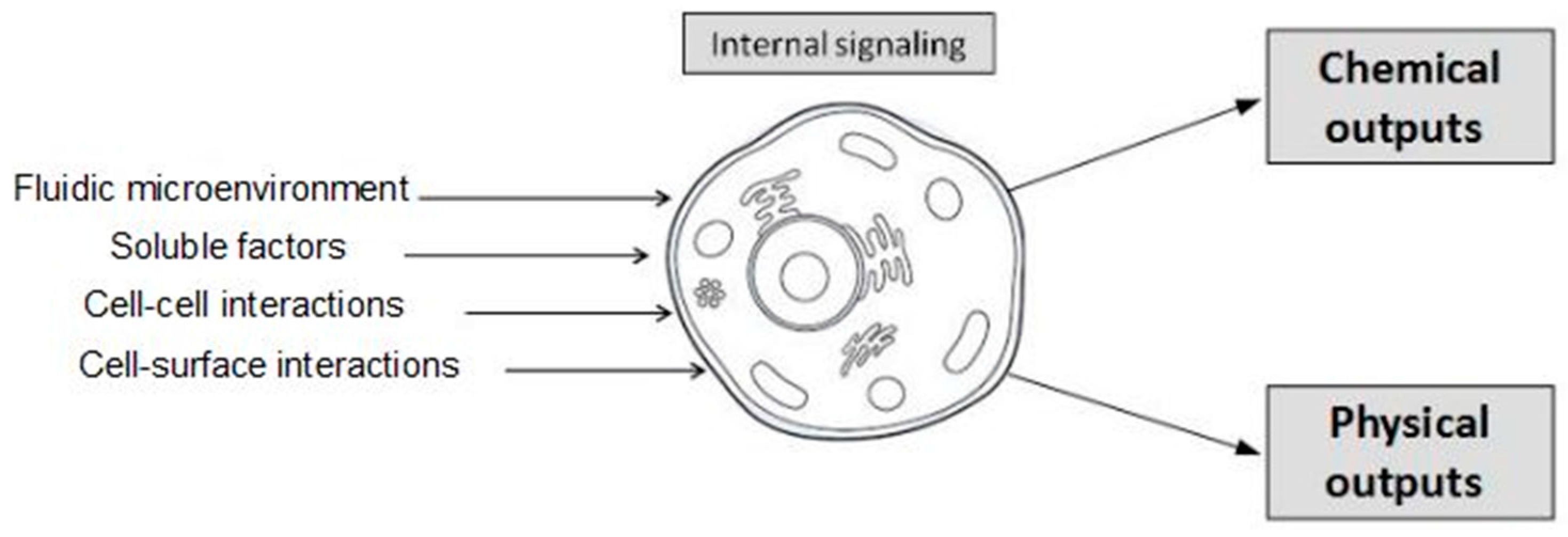
:1. Introduction
2. Microtechnologies for Cell Microenvironment Control
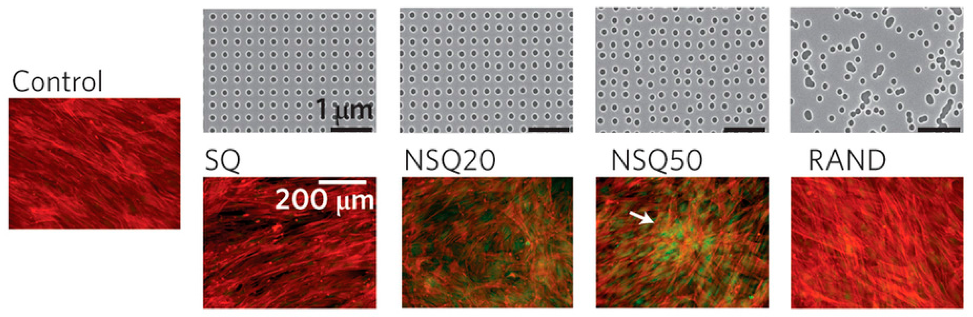
2.1. Topography
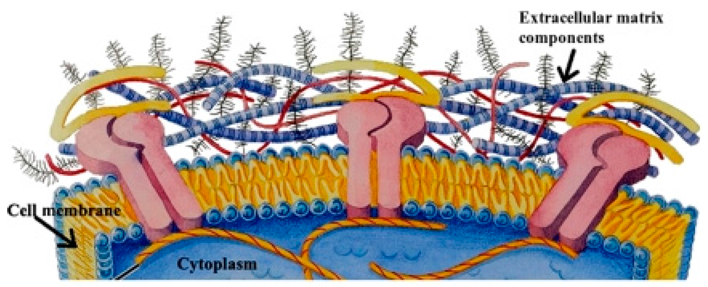
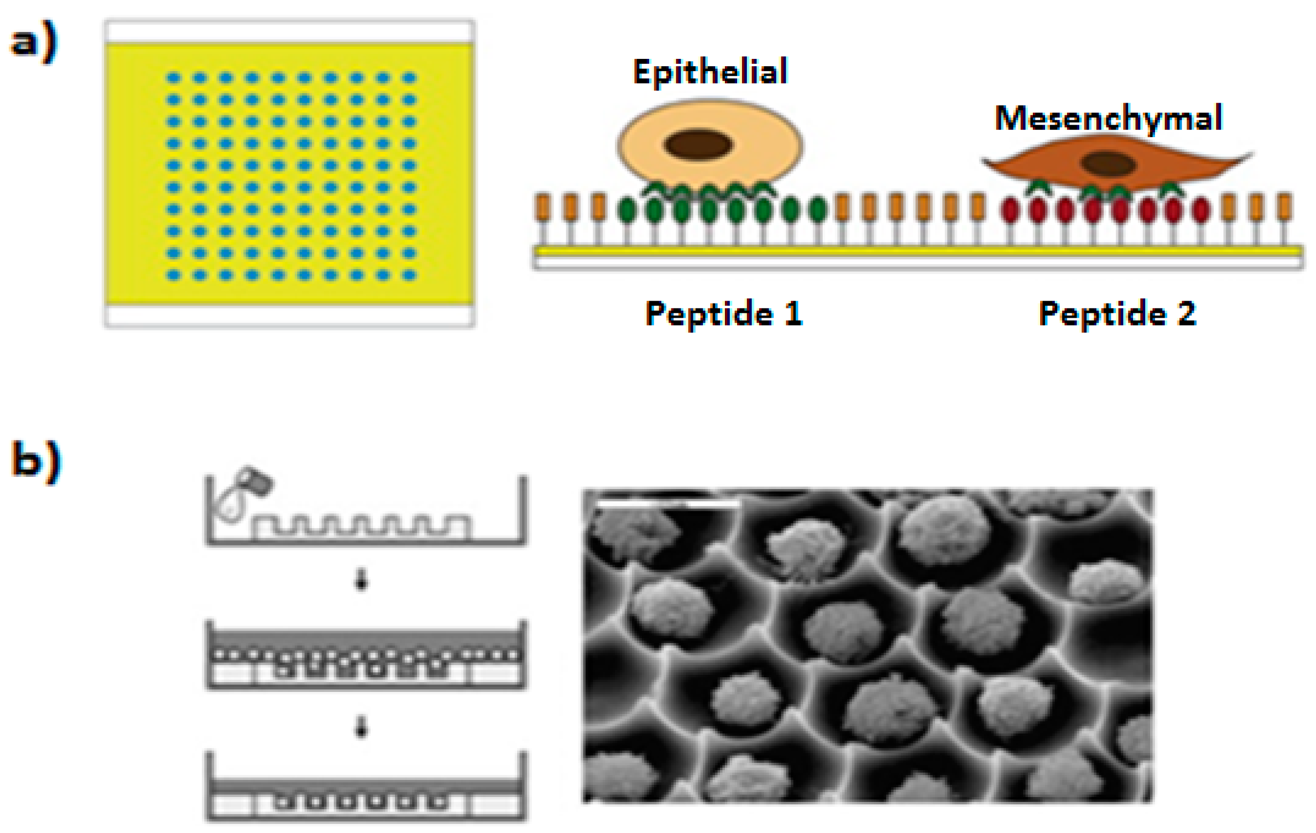
2.2. Biochemical Factors: Surface Chemistry and Soluble Factors
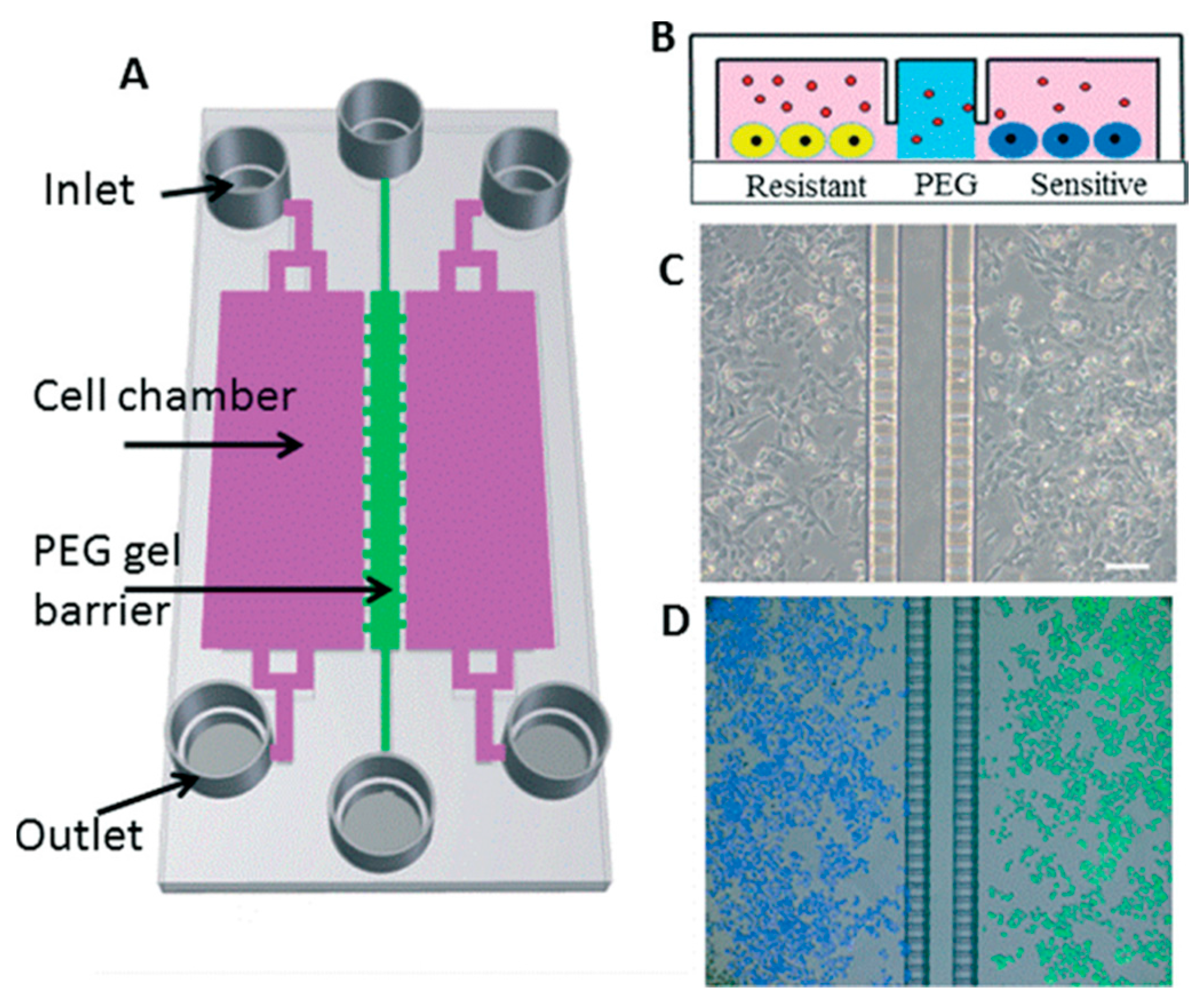
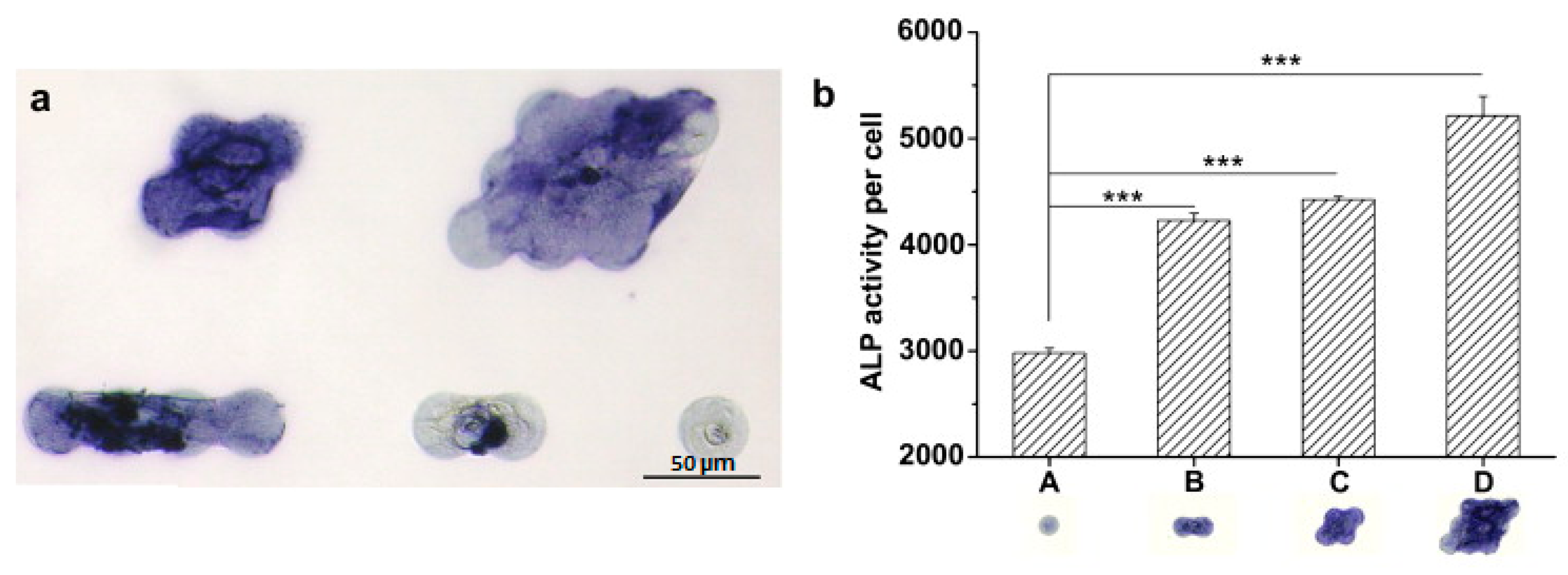
2.3. Cell–Cell Contact
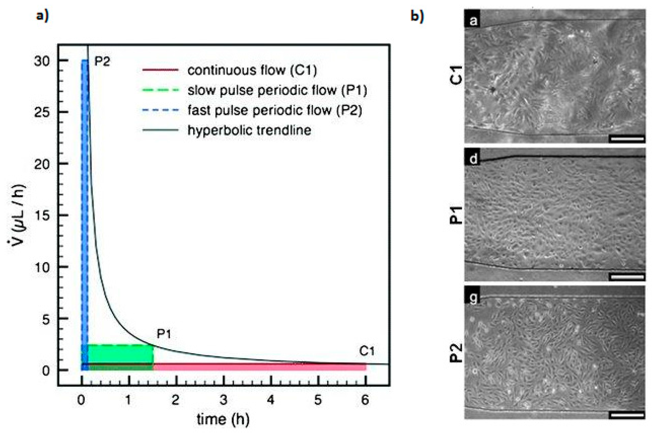
2.4. Fluidic Microenvironment
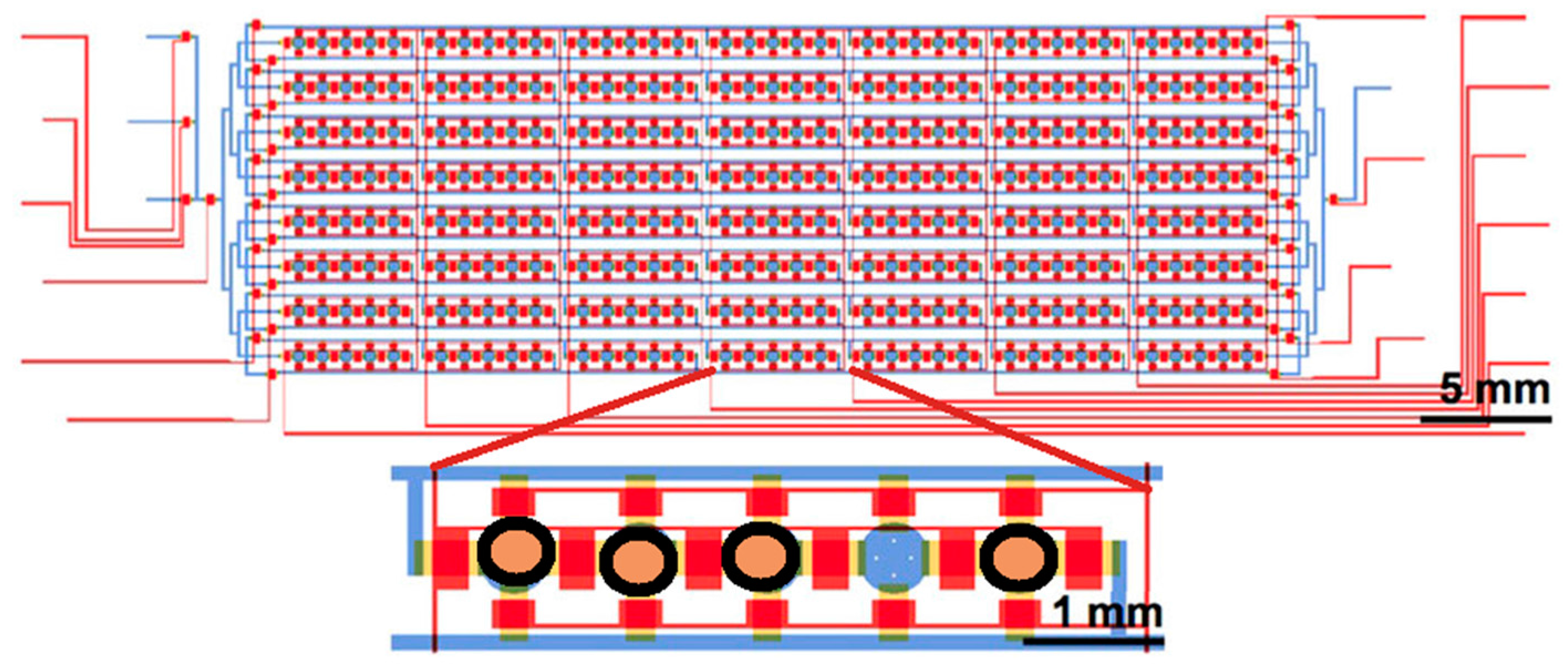
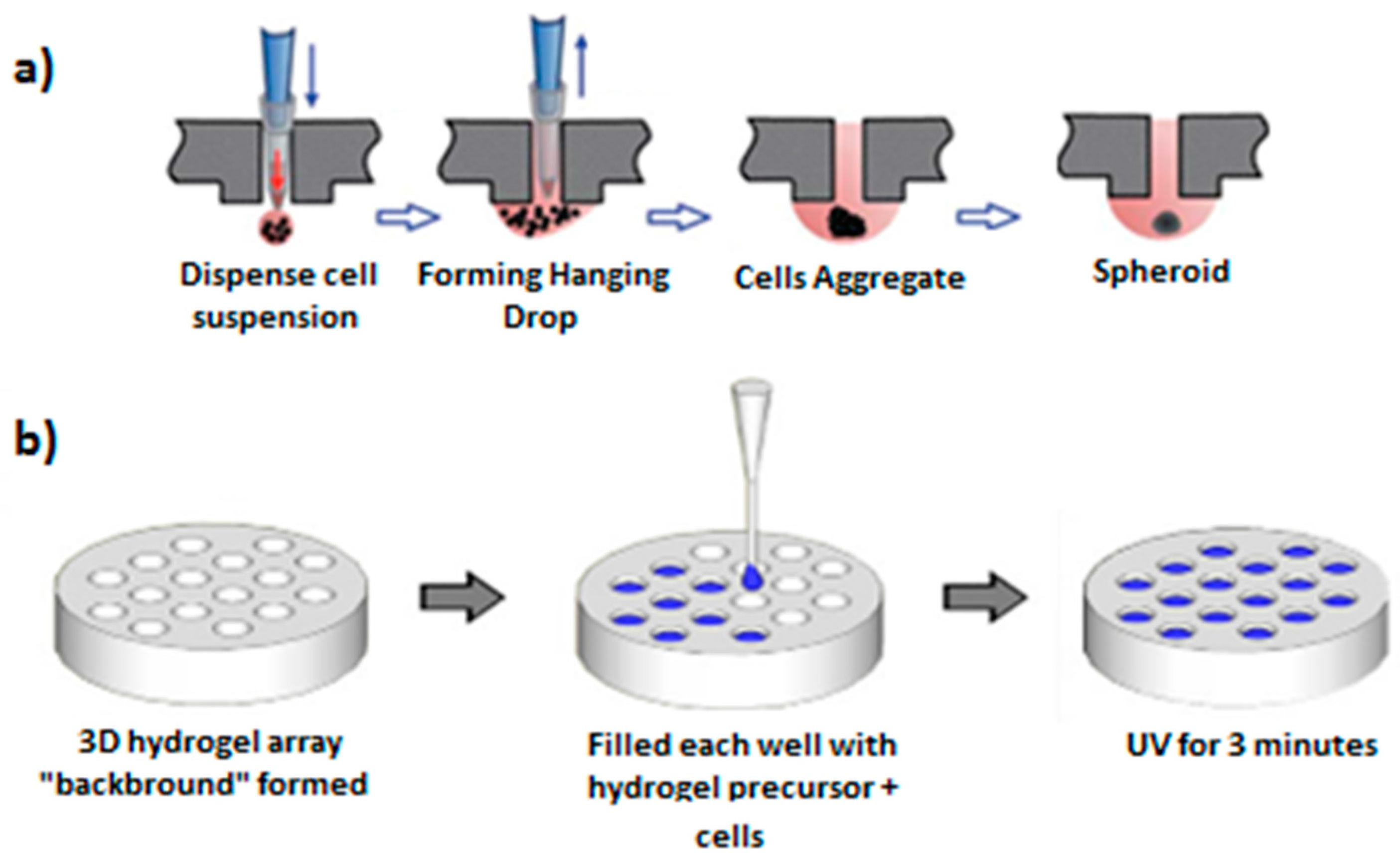
3. Large Arrays of Individually Addressable Cellular Systems under Controlled Microenvironment
4. Monitoring
5. Conclusions
Acknowledgments
Author Contributions
Conflicts of Interest
References
- Halldorsson, S.; Lucumi, E.; Gómez-Sjöberg, R.; Fleming, R.M.T. Advantages and challenges of microfluidic cell culture in polydimethylsiloxane devices. Biosens. Bioelectron. 2015, 63, 218–231. [Google Scholar] [CrossRef] [PubMed]
- Lautenschläger, F.; Piel, M. Microfabricated devices for cell biology: All for one and one for all. Curr. Opin. Cell Biol. 2013, 25, 116–124. [Google Scholar] [CrossRef] [PubMed]
- Weaver, W.M.; Tseng, P.; Kunze, A.; Masaeli, M.; Chung, A.J.; Dudani, J.S.; Kittur, H.; Kulkarni, R.P.; Di Carlo, D. Advances in high-throughput single-cell microtechnologies. Curr. Opin. Biotechnol. 2014, 25, 114–123. [Google Scholar] [CrossRef] [PubMed]
- Bloemen, V.; Schoenmaker, T.; de Vries, T.J.; Everts, V. Direct cell-cell contact between periodontal ligament fibroblasts and osteoclast precursors synergistically increases the expression of genes related to osteoclastogenesis. J. Cell. Physiol. 2010, 222, 565–573. [Google Scholar] [CrossRef] [PubMed]
- Karabacak, N.M.; Spuhler, P.S.; Fachin, F.; Lim, E.J.; Pai, V.; Ozkumur, E.; Martel, J.M.; Kojic, N.; Smith, K.; Chen, P.; et al. Microfluidic, marker-free isolation of circulating tumor cells from blood samples. Nat. Protoc. 2014, 9, 694–710. [Google Scholar] [CrossRef] [PubMed]
- Kim, J.; Taylor, D.; Agrawal, N.; Wang, H.; Kim, H.; Han, A.; Rege, K.; Jayaraman, A. A programmable microfluidic cell array for combinatorial drug screening. Lab Chip 2012, 12, 1813–1822. [Google Scholar] [CrossRef] [PubMed]
- Song, H.; Chen, T.; Zhang, B.; Ma, Y.; Wang, Z. An integrated microfluidic cell array for apoptosis and proliferation analysis induction of breast cancer cells. Biomicrofluidics 2010, 4, 044104. [Google Scholar] [CrossRef] [PubMed]
- Patel, D.; Gao, Y.; Son, K.; Siltanen, C.; Neve, R.M.; Ferrara, K.; Revzin, A. Microfluidic co-cultures with hydrogel-based ligand trap to study paracrine signals giving rise to cancer drug resistance. Lab Chip 2015, 15, 4614–4624. [Google Scholar] [CrossRef] [PubMed]
- Wolf, K.; Müller, R.; Borgmann, S.; Bröcker, E.; Friedl, P. Amoeboid shape change and contact guidance: T-lymphocyte crawling through fibrillar collagen is independent of matrix remodeling by MMPs and other proteases. Blood 2003, 102, 3262–3269. [Google Scholar] [CrossRef] [PubMed]
- Dalby, M.J.; Gadegaard, N.; Oreffo, R.O.C. Harnessing nanotopography and integrin-matrix interactions to influence stem cell fate. Nat. Mater. 2014, 13, 558–569. [Google Scholar] [CrossRef] [PubMed]
- Jaggy, M.; Zhang, P.; Greiner, A.M.; Autenrieth, T.J.; Nedashkivska, V.; Efremov, A.N.; Blattner, C.; Bastmeyer, M.; Levkin, P.A. Hierarchical Micro-Nano Surface Topography Promotes Long-Term Maintenance of Undifferentiated Mouse Embryonic Stem Cells. Nano Lett. 2015, 15, 7146–7154. [Google Scholar] [CrossRef] [PubMed]
- Van Midwoud, P.M.; Janse, A.; Merema, M.T.; Groothuis, G.M.M.; Verpoorte, E. Comparison of Biocompatibility and Adsorption Properties of Different Plastics for Advanced Microfluidic Cell and Tissue Culture Models. Anal. Chem. 2012, 84, 3938–3944. [Google Scholar] [CrossRef] [PubMed]
- Ren, K.; Chen, Y.; Wu, H. New materials for microfluidics in biology. Curr. Opin. Biotechnol. 2014, 25, 78–85. [Google Scholar] [CrossRef] [PubMed]
- Zhu, W.; O’Brien, J.R.; O’Brien, C.; Zhang, L.G. 3D nano/microfabrication techniques and nanobiomaterials for neural tissue regeneration. Nanomedicine 2014, 9, 859–875. [Google Scholar] [CrossRef] [PubMed]
- Suri, S.; Singh, A.; Nguyen, A.H.; Bratt-Leal, A.M.; McDevitt, T.C.; Lu, H. Microfluidic-based patterning of embryonic stem cells for in vitro development studies. Lab Chip 2013, 13, 4617–4624. [Google Scholar] [CrossRef] [PubMed]
- Jeon, H.; Koo, S.; Reese, W.M.; Loskill, P.; Grigoropoulos, C.P.; Healy, K.E. Directing cell migration and organization via nanocrater-patterned cell-repellent interfaces. Nat. Mater. 2015, 14, 918–923. [Google Scholar] [CrossRef] [PubMed]
- Kwon, K.W.; Choi, J.-C.; Suh, K.Y.; Doh, J. Multiscale fabrication of multiple proteins and topographical structures by combining capillary force lithography and microscope projection photolithography. Langmuir 2011, 27, 3238–3243. [Google Scholar] [CrossRef] [PubMed]
- Cheng, Z.A.; Zouani, O.F.; Glinel, K.; Jonas, A.M.; Durrieu, M.-C. Bioactive chemical nanopatterns impact human mesenchymal stem cell fate. Nano Lett. 2013, 13, 3923–3929. [Google Scholar] [CrossRef] [PubMed]
- Hamon, C.; Novikov, S.; Scarabelli, L.; Basabe-Desmonts, L.; Liz-Marzán, L.M. Hierarchical self-assembly of gold nanoparticles into patterned plasmonic nanostructures. ACS Nano 2014, 8, 10694–10703. [Google Scholar] [CrossRef] [PubMed]
- Kweon, S.; Song, K.H.; Park, H.; Choi, J.; Doh, J. Dynamic Micropatterning of Cells on Nanostructured Surfaces Using a Cell-friendly Photoresist. ACS Appl. Mater. Interfaces 2016, 8, 4266–4274. [Google Scholar] [CrossRef] [PubMed]
- Bershadsky, A.D.; Spatz, J.P.; Geiger, B. Environmental sensing through focal adhesions. Nat. Rev. Mol. Cell Biol. 2009, 10, 21–33. [Google Scholar] [CrossRef]
- Clause, K.C.; Barker, T.H. Extracellular matrix signaling in morphogenesis and repair. Curr. Opin. Biotechnol. 2013, 24, 830–833. [Google Scholar] [CrossRef] [PubMed]
- Frantz, C.; Stewart, K.M.; Weaver, V.M. The extracellular matrix at a glance. J. Cell Sci. 2010, 123, 4195–4200. [Google Scholar] [CrossRef] [PubMed]
- Gevertz, J.L.; Torquato, S. A novel three-phase model of brain tissue microstructure. PLoS Comput. Biol. 2008, 4, e1000152. [Google Scholar] [CrossRef] [PubMed]
- Petit, V.; Thiery, J. Focal adhesions: Structure and dynamics. Biol. Cell 2000, 92, 477–494. [Google Scholar] [CrossRef]
- Roca-Cusachs, P.; del Rio, A.; Puklin-Faucher, E.; Gauthier, N.C.; Biais, N.; Sheetz, M.P. Integrin-dependent force transmission to the extracellular matrix by α-actinin triggers adhesion maturation. Proc. Natl. Acad. Sci. USA 2013, 110, E1370. [Google Scholar] [CrossRef] [PubMed]
- Malmström, J.; Christensen, B.; Jakobsen, H.P.; Lovmand, J.; Foldbjerg, R.; Sørensen, E.S.; Sutherland, D.S. Large area protein patterning reveals nanoscale control of focal adhesion development. Nano Lett. 2010, 10, 686. [Google Scholar] [CrossRef] [PubMed]
- Peerani, R.; Zandstra, P.W. Enabling stem cell therapies through synthetic stem cell-niche engineering. J. Clin. Investig. 2010, 120, 60–70. [Google Scholar] [CrossRef] [PubMed]
- Ngangan, A.V.; Waring, J.C.; Cooke, M.T.; Mandrycky, C.J.; McDevitt, T.C. Soluble factors secreted by differentiating embryonic stem cells stimulate exogenous cell proliferation and migration. Stem Cell Res. Ther. 2014, 5, 26. [Google Scholar] [CrossRef] [PubMed]
- Kumacheva, E.; Nie, Z. Patterning surfaces with functional polymers. Nat. Mater. 2008, 7, 277–290. [Google Scholar] [CrossRef]
- Wen, J.H.; Vincent, L.G.; Fuhrmann, A.; Choi, Y.S.; Hribar, K.C.; Taylor-Weiner, H.; Chen, S.; Engler, A.J. Interplay of matrix stiffness and protein tethering in stem cell differentiation. Nat. Mater. 2014, 13, 979–987. [Google Scholar] [CrossRef] [PubMed]
- Ginger, D.S.; Zhang, H.; Mirkin, C.A. The evolution of dip-pen nanolithography. Angew. Chem. 2004, 43, 30–45. [Google Scholar] [CrossRef] [PubMed]
- Mai, Y.; Eisenberg, A. Self-assembly of block copolymers. Chem. Soc. Rev. 2012, 41, 5969–5985. [Google Scholar] [CrossRef] [PubMed]
- Deeg, J.A.; Louban, I.; Aydin, D.; Selhuber-Unkel, C.; Kessler, H.; Spatz, J.P. Impact of local versus global ligand density on cellular adhesion. Nano Lett. 2011, 11, 1469–1476. [Google Scholar] [CrossRef] [PubMed]
- Tan, K.Y.; Lin, H.; Ramstedt, M.; Watt, F.M.; Huck, W.T.S.; Gautrot, J.E. Decoupling geometrical and chemical cues directing epidermal stem cell fate on polymer brush-based cell micro-patterns. Integr. Biol. 2013, 5, 899–910. [Google Scholar] [CrossRef] [PubMed]
- Nelson, C.M.; Chen, C.S. Cell-cell signaling by direct contact increases cell proliferation via a PI3K-dependent signal. FEBS Lett. 2002, 514, 238–242. [Google Scholar] [CrossRef]
- Tang, J.; Peng, R.; Ding, J. The regulation of stem cell differentiation by cell-cell contact on micropatterned material surfaces. Biomaterials 2010, 31, 2470–2476. [Google Scholar] [CrossRef] [PubMed]
- Guo, F.; Li, P.; French, J.B.; Mao, Z.; Zhao, H.; Li, S.; Nama, N.; Fick, J.R.; Benkovic, S.J.; Huang, T.J. Controlling cell–cell interactions using surface acoustic waves. Proc. Natl. Acad. Sci. USA 2015, 112, 43–48. [Google Scholar] [CrossRef] [PubMed]
- Christakou, A.E.; Ohlin, M.; Vanherberghen, B.; Khorshidi, M.A.; Kadri, N.; Frisk, T.; Wiklund, M.; Oenfelt, B. Live cell imaging in a micro-array of acoustic traps facilitates quantification of natural killer cell heterogeneity. Integr. Biol. 2013, 5, 712–719. [Google Scholar] [CrossRef] [PubMed]
- Gauvin, R.; Parenteau-Bareil, R.; Dokmeci, M.R.; Merryman, W.D.; Khademhosseini, A. Hydrogels and microtechnologies for engineering the cellular microenvironment. Wiley Interdiscip. Rev. Nanomed. Nanobiotechnol. 2012, 4, 235–246. [Google Scholar] [CrossRef] [PubMed]
- Gruene, M.; Pflaum, M.; Hess, C.; Diamantouros, S.; Schlie, S.; Deiwick, A.; Koch, L.; Wilhelmi, M.; Jockenhoevel, S.; Haverich, A.; et al. Laser Printing of Three-Dimensional Multicellular Arrays for Studies of Cell–Cell and Cell–Environment Interactions. Tissue Eng. Part C Methods 2011, 17, 973–982. [Google Scholar] [CrossRef] [PubMed]
- Maruthamuthu, V.; Sabass, B.; Schwarz, U.S.; Gardel, M.L. Cell-ECM traction force modulates endogenous tension at cell-cell contacts. Proc. Natl. Acad. Sci. USA 2011, 108, 4708–4713. [Google Scholar] [CrossRef] [PubMed]
- Moe, A.A.K.; Suryana, M.; Marcy, G.; Lim, S.K.; Ankam, S.; Goh, J.Z.W.; Jin, J.; Teo, B.K.K.; Law, J.B.K.; Low, H.Y.; et al. Microarray with micro- and nano-topographies enables identification of the optimal topography for directing the differentiation of primary murine neural progenitor cells. Small 2012, 8, 3050–3061. [Google Scholar] [CrossRef] [PubMed]
- Ricotti, L.; Fujie, T.; Vazão, H.; Ciofani, G.; Marotta, R.; Brescia, R.; Filippeschi, C.; Corradini, I.; Matteoli, M.; Mattoli, V.; et al. Boron nitride nanotube-mediated stimulation of cell co-culture on micro-engineered hydrogels. PLoS ONE 2013, 8, e71707. [Google Scholar] [CrossRef] [PubMed]
- Tsai, M.; Kita, A.; Leach, J.; Rounsevell, R.; Huang, J.N.; Moake, J.; Ware, R.E.; Fletcher, D.A.; Lam, W.A. In vitro modeling of the microvascular occlusion and thrombosis that occur in hematologic diseases using microfluidic technology. J. Clin. Investig. 2012, 122, 408–418. [Google Scholar] [CrossRef] [PubMed]
- Schober, A.; Augspurger, C.; Fernekorn, U.; Weibezahn, K.; Schlingloff, G.; Gebinoga, M.; Worgull, M.; Schneider, M.; Hildmann, C.; Weise, F.; et al. Microfluidics and biosensors as tools for NanoBioSystems research with applications in the “Life Science”. Mater. Sci. Eng. B 2010, 169, 174–181. [Google Scholar] [CrossRef]
- Etxebarria, J.; Berganzo, J.; Elizalde, J.; Fernández, L.J.; Ezkerra, A. Highly integrated COP monolithic membrane microvalves by robust hot embossing. Sens. Actuators B Chem. 2014, 190, 451–458. [Google Scholar] [CrossRef]
- Huang, M.; Fan, S.; Xing, W.; Liu, C. Microfluidic cell culture system studies and computational fluid dynamics. Math. Comput. Model. 2010, 52, 2036–2042. [Google Scholar] [CrossRef]
- Giulitti, S.; Magrofuoco, E.; Prevedello, L.; Elvassore, N. Optimal periodic perfusion strategy for robust long-term microfluidic cell culture. Lab Chip 2013, 13, 4430–4441. [Google Scholar] [CrossRef] [PubMed]
- Chung, B.G.; Flanagan, L.A.; Rhee, S.W.; Schwartz, P.H.; Lee, A.P.; Monuki, E.S.; Jeon, N.L. Human neural stem cell growth and differentiation in a gradient-generating microfluidic device. Lab Chip 2005, 5, 401–406. [Google Scholar] [CrossRef] [PubMed]
- Kawada, J.; Kimura, H.; Akutsu, H.; Sakai, Y.; Fujii, T. Spatiotemporally controlled delivery of soluble factors for stem cell differentiation. Lab Chip 2012, 12, 4508–4515. [Google Scholar] [CrossRef] [PubMed]
- Paguirigan, A.L.; Beebe, D.J. Microfluidics meet cell biology: Bridging the gap by validation and application of microscale techniques for cell biological assays. Bioessays News Rev. Mol. Cell. Dev. Biol. 2008, 30, 811–821. [Google Scholar] [CrossRef] [PubMed]
- Ekerdt, B.L.; Segalman, R.A.; Schaffer, D.V. Spatial organization of cell-adhesive ligands for advanced cell culture. Biotechnol. J. 2013, 8, 1411–1423. [Google Scholar] [CrossRef] [PubMed]
- Chen, K.; Mallon, B.; McKay, R.; Robey, P. Human Pluripotent Stem Cell Culture: Considerations for Maintenance, Expansion, and Therapeutics. Cell Stem Cell 2014, 14, 13–26. [Google Scholar] [CrossRef] [PubMed]
- Williamson, A.; Singh, S.; Fernekorn, U.; Schober, A. The future of the patient-specific Body-on-a-chip. Lab Chip 2013, 13, 3471–3480. [Google Scholar] [CrossRef] [PubMed]
- Araci, I.E.; Quake, S.R. Microfluidic very large scale integration (mVLSI) with integrated micromechanical valves. Lab Chip 2012, 12, 2803. [Google Scholar] [CrossRef] [PubMed]
- Unger, M.A.; Chou, H.; Thorsen, T.; Scherer, A.; Quake, S.R. Monolithic Microfabricated Valves and Pumps by Multilayer Soft Lithography. Science 2000, 288, 113–116. [Google Scholar] [CrossRef] [PubMed]
- Park, E.S.; Brown, A.C.; DiFeo, M.A.; Barker, T.H.; Lu, H. Continuously perfused, non-cross-contaminating microfluidic chamber array for studying cellular responses to orthogonal combinations of matrix and soluble signals. Lab Chip 2010, 10, 571–580. [Google Scholar] [CrossRef] [PubMed]
- Gawad, C.; Koh, W.; Quake, S.R. Single-cell genome sequencing: Current state of the science. Nat. Rev. Genet. 2016, 17, 175–188. [Google Scholar] [CrossRef] [PubMed]
- Petriv, O.I.; Kuchenbauer, F.; Delaney, A.D.; Lecault, V.; White, A.; Kent, D.; Marmolejo, L.; Heuser, M.; Berg, T.; Copley, M.; et al. Comprehensive microRNA expression profiling of the hematopoietic hierarchy. Proc. Natl. Acad. Sci. USA 2010, 107, 15443–15448. [Google Scholar] [CrossRef] [PubMed]
- White, A.K.; VanInsberghe, M.; Petriv, O.I.; Hamidi, M.; Sikorski, D.; Marra, M.A.; Piret, J.; Aparicio, S.; Hansen, C.L. High-throughput microfluidic single-cell RT-qPCR. Proc. Natl. Acad. Sci. USA 2011, 108, 13999–14004. [Google Scholar] [CrossRef] [PubMed]
- Woodruff, K.; Maerkl, S.J. A High-Throughput Microfluidic Platform for Mammalian Cell Transfection and Culturing. Sci. Rep. 2016, 6. [Google Scholar] [CrossRef] [PubMed]
- Teh, S.; Lin, R.; Hung, L.; Lee, A.P. Droplet microfluidics. Lab Chip 2008, 8, 198–220. [Google Scholar] [CrossRef] [PubMed]
- Shembekar, N.; Chaipan, C.; Utharala, R.; Merten, C.A. Droplet-based microfluidics in drug discovery, transcriptomics and high-throughput molecular genetics. Lab Chip 2016, 16, 1314–1331. [Google Scholar] [CrossRef] [PubMed]
- Yu, L.; Chen, M.C.W.; Cheung, K.C. Droplet-based microfluidic system for multicellular tumor spheroid formation and anticancer drug testing. Lab Chip 2010, 10, 2424–2432. [Google Scholar] [CrossRef] [PubMed]
- Mazutis, L.; Gilbert, J.; Ung, W.L.; Weitz, D.A.; Griffiths, A.D.; Heyman, J.A. Single-cell analysis and sorting using droplet-based microfluidics. Nat. Protoc. 2013, 8, 870–891. [Google Scholar] [CrossRef] [PubMed]
- Jakiela, S.; Kaminski, T.S.; Cybulski, O.; Weibel, D.B.; Garstecki, P. Bacterial Growth and Adaptation in Microdroplet Chemostats. Angew. Chem. Int. Ed. 2013, 52, 8908–8911. [Google Scholar] [CrossRef] [PubMed]
- Macosko, E.; Basu, A.; Satija, R.; Nemesh, J.; Shekhar, K.; Goldman, M.; Tirosh, I.; Bialas, A.; Kamitaki, N.; Martersteck, E.; et al. Highly Parallel Genome-wide Expression Profiling of Individual Cells Using Nanoliter Droplets. Cell 2015, 161, 1202–1214. [Google Scholar] [CrossRef] [PubMed]
- Leung, K.; Zahn, H.; Leaver, T.; Konwar, K.M.; Hanson, N.W.; Pagé, A.P.; Lo, C.-C.; Chain, P.S.; Hallam, S.J.; Hansen, C.L. A programmable droplet-based microfluidic device applied to multiparameter analysis of single microbes and microbial communities. Proc. Natl. Acad. Sci. USA 2012, 109, 7665–7670. [Google Scholar] [CrossRef] [PubMed]
- Stroock, A.D. Microfluidics; McGraw-Hill Education: New York, NY, USA, 2014. [Google Scholar]
- Choi, J.; Kang, D.; Park, H.; deMello, A.J.; Chang, S. High-throughput analysis of protein-protein interactions in picoliter-volume droplets using fluorescence polarization. Anal. Chem. 2012, 84, 3849–3854. [Google Scholar] [CrossRef] [PubMed]
- Tumarkin, E.; Tzadu, L.; Csaszar, E.; Seo, M.; Zhang, H.; Lee, A.; Peerani, R.; Purpura, K.; Zandstra, P.W.; Kumacheva, E. High-throughput combinatorial cell co-culture using microfluidics. Integr. Biol. Quant. Biosci. Nano Macro 2011, 3, 653–662. [Google Scholar] [CrossRef] [PubMed]
- Fernandes, T.G.; Kwon, S.; Lee, M.; Clark, D.S.; Cabral, J.M.S.; Dordick, J.S. On-chip, cell-based microarray immunofluorescence assay for high-throughput analysis of target proteins. Anal. Chem. 2008, 80, 6633–6639. [Google Scholar] [CrossRef] [PubMed]
- Charwat, V.; Purtscher, M.; Tedde, S.F.; Hayden, O.; Ertl, P. Standardization of microfluidic cell cultures using integrated organic photodiodes and electrode arrays. Lab Chip 2013, 13, 785–797. [Google Scholar] [CrossRef] [PubMed]
- Coyle, R.; Jia, J.; Mei, Y. Polymer microarray technology for stem cell engineering. Acta Biomater. 2016, 34, 60–72. [Google Scholar] [CrossRef] [PubMed]
- Jose, B.; McCluskey, P.; Gilmartin, N.; Somers, M.; Kenny, D.; Ricco, A.J.; Kent, N.J.; Basabe-Desmonts, L. Self-Powered Microfluidic Device for Rapid Assay of Antiplatelet Drugs. Langmuir 2016, 32, 2820–2828. [Google Scholar] [CrossRef] [PubMed]
- Lovchik, R.; von Arx, C.; Viviani, A.; Delamarche, E. Cellular microarrays for use with capillary-driven microfluidics. Anal. Bioanal. Chem. 2008, 390, 801–808. [Google Scholar] [CrossRef] [PubMed]
- Lee, M.-Y.; Kumar, R.A.; Sukumaran, S.M.; Hogg, M.G.; Clark, D.S.; Dordick, J.S. Three-Dimensional Cellular Microarray for High-Throughput Toxicology Assays. Proc. Natl. Acad. Sci. USA 2008, 105, 59–63. [Google Scholar] [CrossRef] [PubMed]
- Tung, Y.; Hsiao, A.Y.; Allen, S.G.; Torisawa, Y.; Ho, M.; Takayama, S. High-throughput 3D spheroid culture and drug testing using a 384 hanging drop array. Analyst 2011, 136, 473–478. [Google Scholar] [CrossRef] [PubMed]
- Jongpaiboonkit, L.; King, W.J.; Lyons, G.E.; Paguirigan, A.L.; Warrick, J.W.; Beebe, D.J.; Murphy, W.L. An adaptable hydrogel array format for 3-dimensional cell culture and analysis. Biomaterials 2008, 29, 3346–3356. [Google Scholar] [CrossRef] [PubMed]
- Arshak, K.; Korostynska, O.; Cunniffe, C. Nanopatterning Using the Bioforce Nanoenabler. In Functionalized Nanoscale Materials, Devices and Systems; Springer: Dordrecht, The Netherlands, 2008; pp. 299–304. [Google Scholar]
- Basabe-Desmonts, L.; Ramstrom, S.; Meade, G.; O’Neill, S.; Riaz, A.; Lee, L.P.; Ricco, A.J.; Kenny, D. Single-step separation of platelets from whole blood coupled with digital quantification by interfacial platelet cytometry (iPC). Langmuir 2010, 26, 14700–14706. [Google Scholar] [CrossRef] [PubMed]
- Lin, E.; Sikand, A.; Wickware, J.; Hao, Y.; Derda, R. Peptide microarray patterning for controlling and monitoring cell growth. Acta Biomater. 2016, 34, 53–59. [Google Scholar] [CrossRef] [PubMed]
- Helma, J.; Cardoso, M.C.; Muyldermans, S.; Leonhardt, H. Nanobodies and recombinant binders in cell biology. J. Cell Biol. 2015, 209, 633–644. [Google Scholar] [CrossRef] [PubMed]
- Jen, C.-P.; Hsiao, J.-H.; Maslov, N.A. Single-Cell Chemical Lysis on Microfluidic Chips with Arrays of Microwells. Sensors 2012, 12, 347–358. [Google Scholar] [CrossRef] [PubMed]
- Chen, S.; Bremer, A.W.; Scheideler, O.J.; Na, Y.S.; Todhunter, M.E.; Hsiao, S.; Bomdica, P.R.; Maharbiz, M.M.; Gartner, Z.J.; Schaffer, D.V. Interrogating cellular fate decisions with high-throughput arrays of multiplexed cellular communities. Nat. Commun. 2016, 7. [Google Scholar] [CrossRef] [PubMed]
- Vasdekis, A.E.; Stephanopoulos, G. Review of methods to probe single cell metabolism and bioenergetics. Metab. Eng. 2015, 27, 115–135. [Google Scholar] [CrossRef] [PubMed]
- Sun, Y.-S. Label-Free Sensing on Microarrays. In Small Molecule Microarrays, 8th International ed.; Springer: New York, NY, USA, 2013; pp. 515–517. [Google Scholar]
- Sun, Y. Use of Microarrays as a High-Throughput Platform for Label-Free Biosensing. J. Lab. Autom. 2015, 20, 334–353. [Google Scholar] [CrossRef] [PubMed]
- Chen, J.; Zhou, G.; Chang, C.; Lee, A.; Chang, F. Label-free DNA detection using two-dimensional periodic relief grating as a visualized platform for diagnosis of breast cancer recurrence after surgery. Biosens. Bioelectron. 2014, 54, 35–41. [Google Scholar] [CrossRef] [PubMed]
- Rothbauer, M.; Wartmann, D.; Charwat, V.; Ertl, P. Recent advances and future applications of microfluidic live-cell microarrays. Biotechnol. Adv. 2015, 33, 948–961. [Google Scholar] [CrossRef] [PubMed]
- Zhang, Q.; Li, Z.; Zhao, S.; Wen, W.; Chang, L.; Yu, H.; Jiang, T. Analysis of red blood cells’ dynamic status in a simulated blood circulation system using an ultrahigh-speed simultaneous framing optical electronic camera. Cytometry A 2016, 91, 126–132. [Google Scholar] [CrossRef] [PubMed]
- Shi, C.; Luu, D.K.; Yang, Q.; Liu, J.; Chen, J.; Ru, C.; Xie, S.; Luo, J.; Ge, J.; Sun, Y. Recent advances in nanorobotic manipulation inside scanning electron microscopes. Microsyst. Nanoeng. 2016, 2. [Google Scholar] [CrossRef]
- Booth, R.; Kim, H. Characterization of a microfluidic in vitro model of the blood-brain barrier (μBBB). Lab Chip 2012, 12, 1784–1792. [Google Scholar] [CrossRef] [PubMed]
- Yea, C.-H.; An, J.H.; Kim, J.; Choi, J.-W. In situ electrochemical detection of embryonic stem cell differentiation. J. Biotechnol. 2013, 166, 1–5. [Google Scholar] [CrossRef] [PubMed]
- Gijs, M.A.M.; Lacharme, F.; Lehmann, U. Microfluidic applications of magnetic particles for biological analysis and catalysis. Chem. Rev. 2010, 110, 1518–1563. [Google Scholar] [CrossRef] [PubMed]
- Shoshi, A.; Schotter, J.; Schroeder, P.; Milnera, M.; Ertl, P.; Charwat, V.; Purtscher, M.; Heer, R.; Eggeling, M.; Reiss, G.; et al. Magnetoresistive-based real-time cell phagocytosis monitoring. Biosens. Bioelectron. 2012, 36, 116–122. [Google Scholar] [CrossRef] [PubMed]
- Chen, Q.; Wu, J.; Zhang, Y.; Lin, J. Qualitative and quantitative analysis of tumor cell metabolism via stable isotope labeling assisted microfluidic chip electrospray ionization mass spectrometry. Anal. Chem. 2012, 84, 1695–1701. [Google Scholar] [CrossRef] [PubMed]


| Technique | Definition | Cell Isolation Capacity | 2D/3D Culture | Other Characteristics |
|---|---|---|---|---|
| Microfluidic large-scale integration systems (mLSI) | Interconnected addressable microchambers for isolation and recovering of cells | Multiple and single cell | Two-dimensional cultures | Thousands of addressable events |
| Droplet microfluidic platforms | Droplet-based systems for cell entrapping in discrete volumes of immiscible phases | Multiple and single cell | Two- and three-dimensional cultures | Thousands of addressable events Low volume requirement |
| 3D microfluidic platforms | Spotting of biological suspension on addressable spots | Multiple cells | Three-dimensional cultures | Low volume requirement |
| Hanging drops platforms | Entrapping of spheroid cultures inside a hanging drop | Multiple cells | Three-dimensional cultures | Hundreds of addressable events |
| Patterned substrates | Patterning of chemical adhesive areas for cell immobilization | Multiple and single cell | Two-dimensional cultures | Thousands of addressable events Low volume requirement Fast processes |
| Microwell structures | Molding of microsieves or microwells for cellular trapping | Multiple and single cell | Two-dimensional cultures | Thousands of addressable events |
© 2017 by the authors. Licensee MDPI, Basel, Switzerland. This article is an open access article distributed under the terms and conditions of the Creative Commons Attribution (CC BY) license (http://creativecommons.org/licenses/by/4.0/).
Share and Cite
Azuaje-Hualde, E.; García-Hernando, M.; Etxebarria-Elezgarai, J.; De Pancorbo, M.M.; Benito-Lopez, F.; Basabe-Desmonts, L. Microtechnologies for Cell Microenvironment Control and Monitoring. Micromachines 2017, 8, 166. https://doi.org/10.3390/mi8060166
Azuaje-Hualde E, García-Hernando M, Etxebarria-Elezgarai J, De Pancorbo MM, Benito-Lopez F, Basabe-Desmonts L. Microtechnologies for Cell Microenvironment Control and Monitoring. Micromachines. 2017; 8(6):166. https://doi.org/10.3390/mi8060166
Chicago/Turabian StyleAzuaje-Hualde, Enrique, Maite García-Hernando, Jaione Etxebarria-Elezgarai, Marian M. De Pancorbo, Fernando Benito-Lopez, and Lourdes Basabe-Desmonts. 2017. "Microtechnologies for Cell Microenvironment Control and Monitoring" Micromachines 8, no. 6: 166. https://doi.org/10.3390/mi8060166
APA StyleAzuaje-Hualde, E., García-Hernando, M., Etxebarria-Elezgarai, J., De Pancorbo, M. M., Benito-Lopez, F., & Basabe-Desmonts, L. (2017). Microtechnologies for Cell Microenvironment Control and Monitoring. Micromachines, 8(6), 166. https://doi.org/10.3390/mi8060166