
Chromobacterium violaceum Periareolar Infection, First Non-Lethal Case in Colombia: Case Report and Literature Review

 , , ,
, , ,
Abstract
1. Introduction
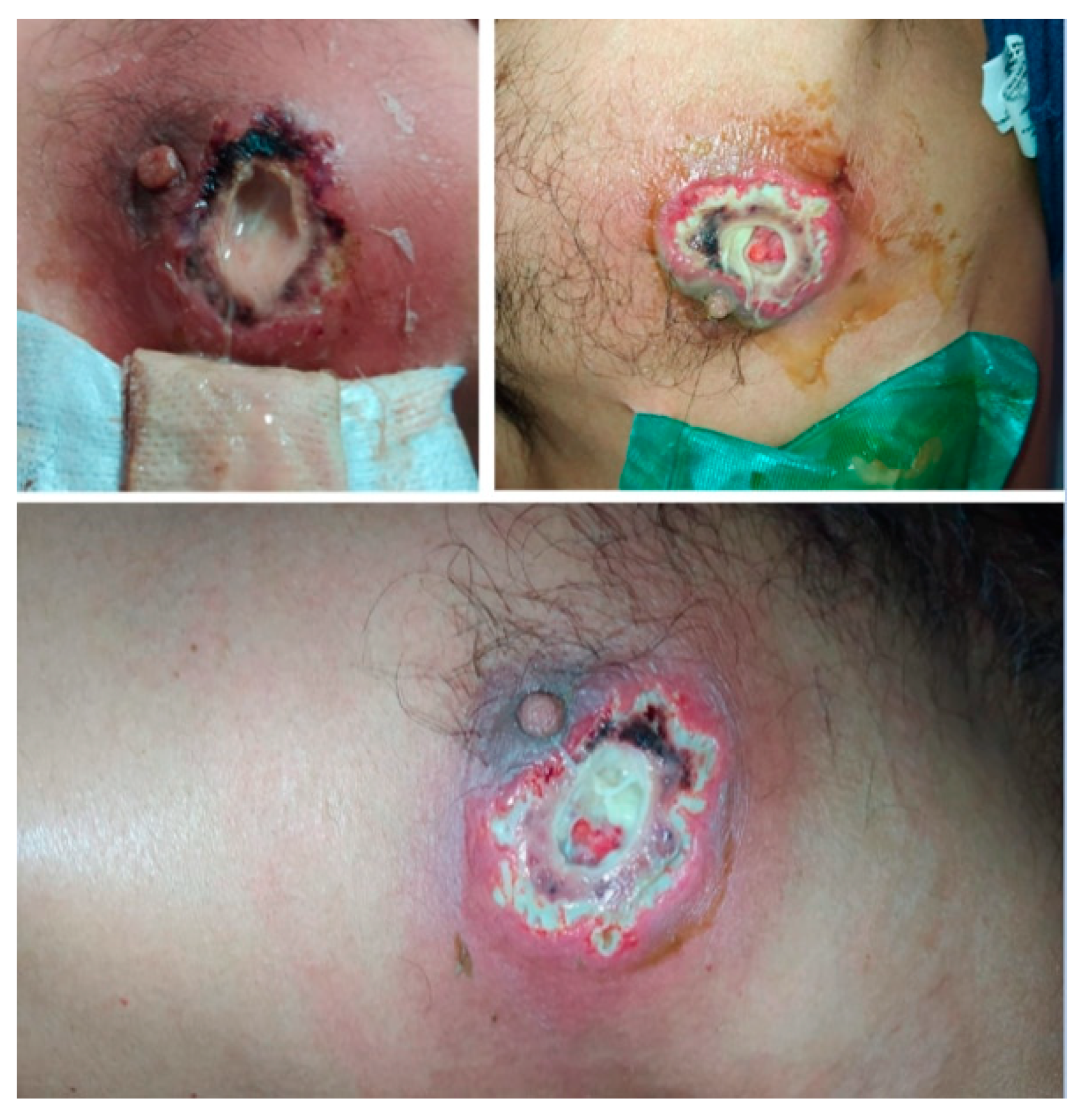
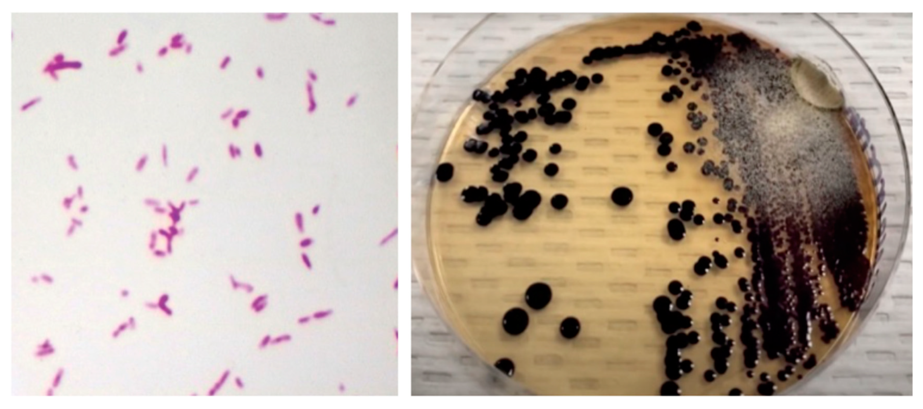
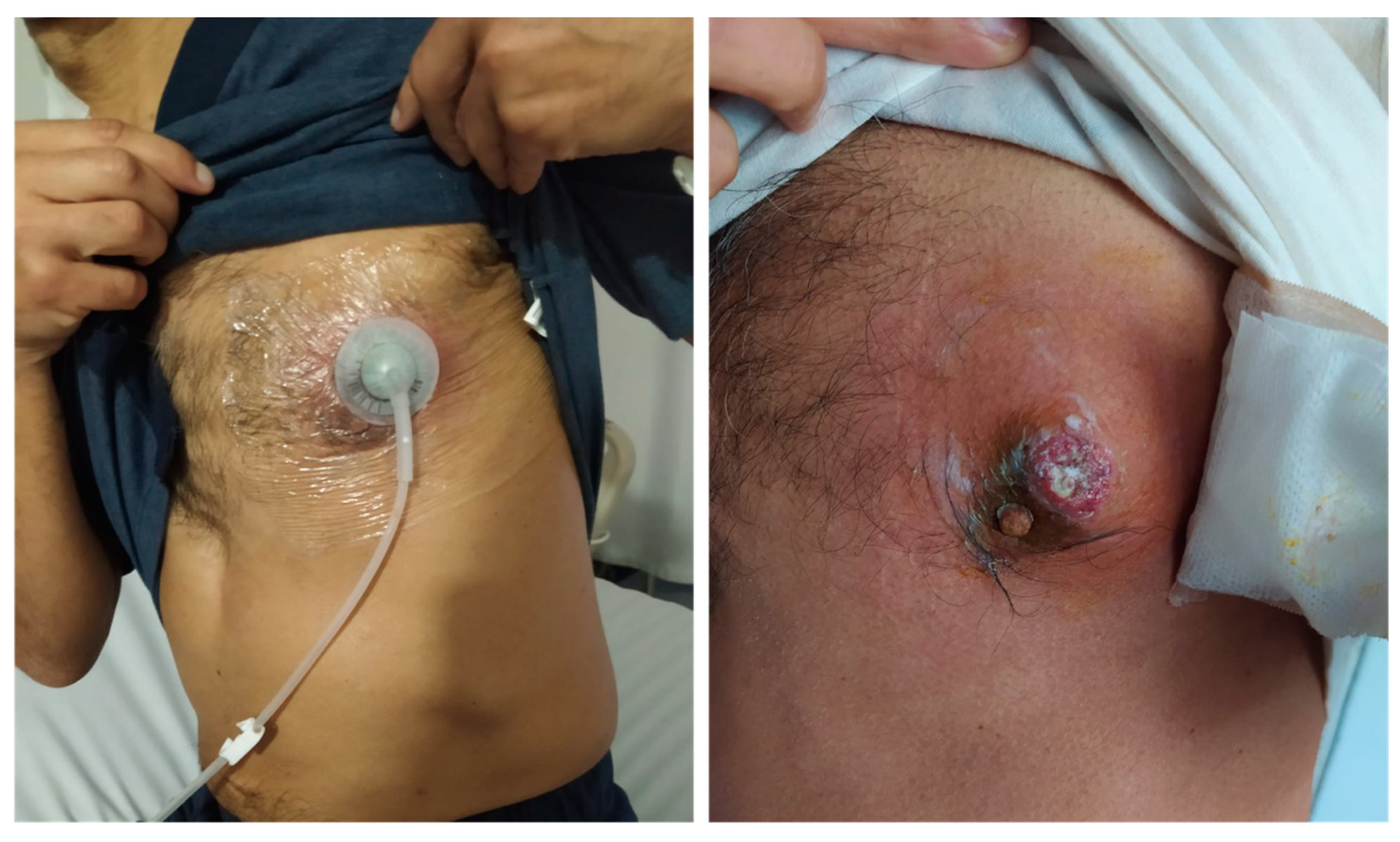
2. Case Report
3. Discussion
4. Conclusions
Author Contributions
Funding
Institutional Review Board Statement
Informed Consent Statement
Data Availability Statement
Conflicts of Interest
References
- Kim, M.H.; Lee, H.J.; Suh, J.T.; Chang, B.S.; Cho, K.S. A case of chromobacterium infection after car accident in korea. Yonsei Med. J. 2005, 46, 700–702. [Google Scholar] [CrossRef] [PubMed]
- Gómez, M.; Santos, A.; Guevara, A.; Rodríguez, C. Reporte de dos casos de inusual infección no letal por Chromobacterium violaceum. Revisión literaria. Infectio 2017, 21, 129–131. [Google Scholar] [CrossRef][Green Version]
- Mattar, S.; Martinez, P. Case report fatal septicemia caused by Chromobacterium violaceum in a child from colombia. Rev. Inst. Med. Trop. São Paulo 2007, 49, 391–393. [Google Scholar]
- Alexander Díaz Pérez, J.; García, J.; Andrea Rodriguez Villamizar, L.; Alexander Díaz, J. Sepsis by Chromobacterium violaceum: First Case Report from Colombia [Internet]. Braz. J. Infect. Dis. 2007, 11, 441–442. [Google Scholar] [CrossRef] [PubMed]
- Justo, G.Z.; Durán, N. Action and function of Chromobacterium violaceum in health and disease: Violacein as a promising metabolite to counteract gastroenterological diseases. Best Pract. Res. Clin. Gastroenterol. 2017, 31, 649–656. [Google Scholar] [CrossRef]
- Yang, C.H.; Li, Y.H. Chromobacterium violaceum infection: A clinical review of an important but neglected infection. J. Chin. Med. Assoc. 2011, 74, 435–441. [Google Scholar] [CrossRef]
- Segal, B.H.; Ding, L.; Holland, S.M. Phagocyte NADPH oxidase, but not inducible nitric oxide synthase, is essential for early control of Burkholderia cepacia and Chromobacterium violaceum infection in mice. Infect Immun. 2003, 71, 205–210. [Google Scholar] [CrossRef]
- Abais, J.M.; Zhang, C.; Xia, M.; Liu, Q.; Gehr, T.W.B.; Boini, K.M.; Li, P.L. NADPH oxidase-mediated triggering of inflammasome activation in mouse podocytes and glomeruli during hyperhomocysteinemia. Antioxidants Redox Signal. 2013, 18, 1537–1548. [Google Scholar] [CrossRef] [PubMed]
- Batista, J.H.; da Silva Neto, J.F. Chromobacterium violaceum pathogenicity: Updates and insights from genome sequencing of novel Chromobacterium species. Front. Microbiol. 2017, 8, 2213. [Google Scholar] [CrossRef]
- Banerjee, D.; Raghunathan, A. Constraints-based analysis identifies NAD + recycling through metabolic reprogramming in antibiotic resistant Chromobacterium violaceum. PLoS ONE 2019, 14, e0210008. [Google Scholar] [CrossRef]
- Sneath, P.H.A.; Bhagwan, S.R.; Whelan, J.P.F.; Edwards, D. Fatal infection by Chromobacterium violaceum. Lancet 1953, 262, 276–277. [Google Scholar] [CrossRef]
- Kaufman, S.C.; Ceraso, D.; Schugurensky, A. First Case Report from Argentina of Fatal Septicemia Caused by Chromobacterium violaceum. J. Clin. Microbiol. 1986, 23, 956–958. [Google Scholar] [CrossRef] [PubMed]
- Georghiou, P.R.; O’Kane, G.M.; Siu, S.; Kemp, R.J. Near-fatal septicaemia with Chromobacterium violaceum. Med. J. Aust. 1989, 150, 720–721. [Google Scholar] [CrossRef]
- Huffam, S.E.; Nowotny, M.J.; Currie, B.J. Notable Cases Chromobacterium violaceum in tropical northern Australia. Med. J. Aust. 1998, 168, 335–337. [Google Scholar] [PubMed]
- Dan, M.; Poch, F.; Shpitz, D.; Sheinberg, B. An Unusual Bacterium Causing a Brain Abscess. Report on emerging issues. Emerg. Infect. Dis. 2001, 7, 159. [Google Scholar]
- Teoh, A.Y.B.; Hui, M.; Ngo, K.Y.; Wong, J.; Lee, K.F.; Lai, P.B.S. Case Report Fatal septicaemia from Chromobacterium violaceum: Case reports and review of the literature. Hong Kong Med. J. 2006, 12, 228–231. [Google Scholar]
- Lim, I.W.M.; Stride, P.J.; Horvath, R.L.; Hamilton-Craig, C.R.; Chau, P.P. Chromobacterium violaceum endocarditis and hepatic abscesses treated successfully with meropenem and ciprofloxacin. Med. J. Aust. 2009, 190, 386–387. [Google Scholar] [CrossRef]
- Yang, C.H. Nonpigmented Chromobacterium violaceum bacteremic cellulitis after fish bite. J. Microbiol. Immunol. Infect. 2011, 44, 401–405. [Google Scholar] [CrossRef]
- Kumar, M. Chromobacterium violaceum: A rare bacterium isolated from a wound over the scalp. Int. J. Appl. Basic Med. Res. 2012, 2, 70. [Google Scholar] [CrossRef]
- Ansari, S.; Paudel, P.; Gautam, K.; Shrestha, S.; Thapa, S.; Gautam, R. Chromobacterium violaceum Isolated from a Wound Sepsis: A Case Study from Nepal. Case Rep. Infect. Dis. 2015, 2015, 1–4. [Google Scholar]
- Madi, D.R.; Vidyalakshmi, K.; Ramapuram, J.; Shetty, A.K. Case report: Successful treatment of Chromobacterium violaceum sepsis in a south indian adult. Am. J. Trop. Med. Hyg. 2015, 93, 1066–1067. [Google Scholar] [CrossRef] [PubMed]
- Lin, Y.D.; Majumdar, S.S.; Hennessy, J.; Baird, R.W. The spectrum of Chromobacterium violaceum infections from a single geographic location. Am. J. Trop. Med. Hyg. 2016, 94, 710–716. [Google Scholar] [CrossRef] [PubMed]
- Matsuura, N.; Miyoshi, M.; Doi, N.; Yagi, S.; Aradono, E.; Imamura, T.; Koga, R. Multiple liver abscesses with a skin pustule due to Chromobacterium violaceum. Intern Med. 2017, 56, 2519–2522. [Google Scholar] [CrossRef] [PubMed][Green Version]
- Sachu, A.; Antony, S.; Mathew, P.; Sunny, S.; Koshy, J.; Kumar, V.; Mathew, R. Chromobacterium violaceum causing deadly sepsis [Internet]. Iran. J. Microbiol. 2020, 12, 364. [Google Scholar]
- Khadanga, S.; Karuna, T.; Dugar, D.; Satapathy, S.P. Chromobacterium violaceum-induced sepsis and multiorgan dysfunction, resembling melioidosis in an elderly diabetic patient: A case report with review of literature. J. Lab. Physicians 2017, 9, 325–328. [Google Scholar]
- Mazumder, R.; Sadique, T.; Sen, D.; Mozumder, P.; Rahman, T.; Chowdhury, A.; Halim, F.; Akter, N.; Ahmed, D. Agricultural Injury–Associated Chromobacterium violaceum Infection in a Bangladeshi Farmer. Am. J. Trop. Med. Hyg. 2020, 103, 1039–1042. [Google Scholar] [CrossRef]
- Zhang, P.; Li, J.; Zhang, Y.Z.; Li, X.N. Chromobacterium violaceum infection on lower limb skin: A case report. Medicine 2021, 100, e24696. [Google Scholar] [CrossRef]
- Meher-Homji, Z.; Mangalore, R.P.; Johnson, P.D.R.; Chua, K.Y.L. Chromobacterium violaceum infection in chronic granulomatous disease: A case report and review of the literature. JMM Case Rep. 2017, 4, e005084. [Google Scholar] [CrossRef] [PubMed]
- Thwe, P.M.; Ortiz, D.A.; Wankewicz, A.L.; Hornak, J.P.; Williams-Bouyer, N.; Ren, P. The Brief Case: Recurrent Chromobacterium violaceum. J. Clin. Microbiol. 2020, 58, 6–11. [Google Scholar]
- Mamlok, R.J.; Mamiok, V.; Mills, G.C.; Daeschner, C.W.; Schmalstieg, F.C.; Anderson, D.C. Glucose-6-phosphate dehydrogenase deficiency, neutrophil dysfunction and Chromobacterium violaceum sepsis. J. Pediatr. 1987, 111, 852–854. [Google Scholar] [CrossRef]


| Laboratory | Admission | Discharge |
|---|---|---|
| White blood cell count | 22.03 × 10³/µL | 11.92 × 10³/µL |
| Neutrophils | 18.9 × 10³/µL (85.9%) | 6.6 × 10³/µL (55.2%) |
| Lymphocytes | 1.33 × 10³/µL (6%) | 3.56 × 10³/µL (29.9%) |
| Monocytes | 1.61 × 10³/µL (7.3%) | 1.18 × 10³/µL (9.90%) |
| Red blood cells | 3.89 × 10⁶/µL | 3.35 × 10⁶/µL |
| Hematocrit | 33.3% | 29% |
| Hemoglobin | 11.2 g/dL | 9.70 g/dL |
| MCV | 85.6 fl | 86.6 fl |
| MCH | 28.8 pg | 29 pg |
| MCHC | 33.6 g/dL | 33.4 g/dL |
| RDW | 12.7% | 12.5% |
| Platelets | 264 × 103/µL | 407 × 103/µL |
| C-reactive protein | 176.4 mg/L | 12.29 mg/L |
| Blood urea nitrogen | 89.7 mg/dL | 74.3 mg/dL |
| Creatinine | 9.14 mg/dL | 5.16 mg/dL |
| Author/Year | Age and Gender | Clinical Manifestations | Related Condition | Time to Positive Culture | Bacteremia | Sepsis | Resistance Profile | Antibiotic Treatment | Outcome |
|---|---|---|---|---|---|---|---|---|---|
| Sneath et al. [11] (1953) | Not reported (M) | Left thigh ulcer with inguinal lymphadenopathy fever and hepatomegaly. | Not reported. | 24 h | Yes | Yes | Not reported | TCY | Septic shock and multi-organ dysfunction. Death 26 days after admission. |
| Kaufman et al. [12] (1986) | 44 yo (F) | Ulcerated nodules and purpura in the abdomen, limbs, and back. Fever, abdominal pain, hepatomegaly, and jaundice. | Exposure to contaminated water. Wasp sting. | 24 h | Yes | Yes | Sensitive: MZL, GEN, CLO, TCY, CMX CBN, NMC, and NTM Resistant: PEN, AMP, AMK, TOB, PMX B, and CFS | MEZ and GEN | Septic shock. Death at 6 weeks after admission. |
| Georghiou et al. [13] (1989) | 35 yo (M) | Posterior neck wound. Twelve days later: extension to the right shoulder with abscess, abdominal pain, fever, and diarrhoea. | Abrasion while carrying old damp floor-covering on that shoulder | 24 h | Yes | Yes | Sensitive: GEN, CIP, and CLO, IPM, TOB, and AMC Resistant: ATM, AMX, SXT, and CEP | GEN, IPM, and CIP | Alive, fully recovered. |
| Huffman et al. [14] (1998) | 46 yo (M) | Three weeks of malaise, hyporexia, nausea, abdominal pain, vomiting, 7 kg loss, left thigh wound, and liver abscess. | Heavy smoking. | 24 h | Yes | Yes | Sensitive: GEN, CIP, CLO, TCY, IPM, and SXT Resistant: CRO, AMC, and CAZ | CLO, GEN, and TCY | Resolved. Neuropathy as a sequel of the disease. |
| Huffman et al. [14] (1998) | 53 yo (M) | 7 mm wound on the sole of the left foot; six days later: fever, pain, and purulent discharge. | Diabetes mellitus. Wound with rusty metal. | Not reported | No | No | Sensitive: GEN, CIP, IPM, PIP, TOB, and AMC Resistant: CAZ | AMC, GEN, DOX, and CIP | Alive, fully recovered. |
| Dan et al. [15] (2001) | 24 yo (M) | Pimple in right cheek with purulent discharge for three weeks. Fever, headache, and dizziness for 2 weeks. Brain abscess was reported. | Farm laboring. | 24 h | No | No | Sensitive: IPM and CIP Resistant: CTX and CRO | CIP | Alive, fully recovered. |
| Hee kim et al. [1] (2005) | 38 yo (M) | Polytrauma with multiple wounds, rib fractures, empyema, hemothorax, tibial fracture, kidney, and liver hematoma. | Hit by a car while fishing. Contaminated water exposure. | Not reported | No | No | Sensitive: CIP, AMK, GEN, TZP, LVX, and SXT, Resistant: CRO, AMP, TOB, SAM, and FEP | CFA and AZM | Alive, fully recovered. |
| Teoh et al. [16] (2006) | 40 yo (M) | 1 cm forearm abscess. One day later: purulent discharge epigastric pain and fever. Peritonitis was reported. | Forearm wound during camping near a lagoon. | Not reported | Yes | Yes | Not reported | CIP, MDZ, and CTX | Septic shock. Death at 20 h after admission. |
| Lim et al. [17] (2009) | 40 yo (F) | Anterior chest wall wound. Malaise, fever, chills, lumbar pain, and headache. Apical pansystolic murmur, liver abscess, and vegetations on the coronary cusps. | Wound with a tree branch during lake swimming. | 28–39 h | Yes | Yes | Sensitive: CIP, TZP, and MEM, IPM, SXT, and CFX Resistant: AMP, CXM, and CAZ | MEM and CIP | Liver abscess and infective endocarditis resolved after 6–11 weeks, respectively. Alive. |
| Yang et al. [18] (2011) | 64 yo (M) | Right foot erythematous lesion with pain and edema. Headache and fever. | Diabetes mellitus and hepatitis B infection. Fish bite in a river. | 10 days | Yes | Yes | Sensitive: CIP, TZP, and MEM, LVX, ATM, CAS, and FEP Intermediate: GEN and AMK Resistant: SAM | CRO, CIP, and DOX | Alive, fully recovered. |
| Kumar et al. [19] (2012) | 42 yo (M) | Head and leg lesions. Seven days later: pain, swelling, and purulent discharge. | Exposure to contaminated water. | 24 h | No | No | Sensitive: GEN, CIP, and AMK, CLO, TCY, CAZ, and IPM Intermediate: CTX Resistant: PEN and CFX | GEN | Alive, fully recovered. |
| Ansari et al. [20] (2015) | 45 yo (M) | Wound in the middle finger of the left hand. Seven days later: fever, epigastric pain, swelling, and purulent discharge. | Stinging wound with unknown object. | 12 h | Yes | Yes | Sensitive: AMK, GEN, CIP, TZP, MEM, CRO, SXT, AMC, MDZ, and FLX. | TZP, FLX, and MDZ. | Septic shock and death 21 h after admission. |
| Madi et al. [21] (2015) | 53 yo (F) | Left leg lesion. Two weeks later: abdominal pain and vomiting; the lesion then ulcerated. Hepatomegaly and jaundice. | Skin trauma during farm laboring. | Not reported | Yes | Yes | Sensitive: CIP, TZP, MEM IPM, SXT, and CFX Resistant: AMP, CXM, and CAZ | IPM, CIP, and TZP | Alive, fully recovered. |
| Lin et al. [22] (2016) | 23 yo (F) | Limb injury. Eight months later: granulation tissue in the wound. | Wound contamination in muddy field. | Not reported | No | No | All isolates were sensitive to CIP, MEM FEP, and GEN | TZP | Alive, fully recovered. |
| Lin et al. [22] (2016) | 42 yo (F) | Chest wall wound. | Not reported | Not reported | Yes | Yes | TZP, MEM, and GEN | Sepsis, mesenteric ischemia, abdominal abscess and fat necrosis. Deceased. | |
| Lin et al. [22] (2016) | 53 yo (F) | Limb wound, diabetic foot with infection, and abscess. | Diabetes mellitus | Not reported | No | No | GEN and SXT | Alive, fully recovered. | |
| Lin et al. [22] (2016) | 52 yo (M) | Post-surgical infected sternotomy wound. | Myocardial revascularization | Not reported | Not reported | Yes | TZP | Alive, fully recovered. | |
| Lin et al. [22] (2016) | 32 yo (M) | Lacerating injury on the toes with purulent discharge. | Wound during swamp hike. | Not reported | No | No | SXT | Alive, fully recovered. | |
| Lin et al. [22] (2016) | 57 yo (M) | Anterior chest wall wound. | Found unconscious in a swamp. | Not reported | Not reported | Not reported | MEM | Alive, fully recovered. | |
| Lin et al. [22] (2016) | 45 yo (M) | Wound on the palm of the hand. | Stab wound. Possible contaminated metal. | Not reported | No | No | DOX | Alive, fully recovered. | |
| Lin et al. [22] (2016) | 50 yo (M) | Wound on toe | Not reported | Not reported | No | No | CZO and DOX | Alive, fully recovered. | |
| Lin et al. [22] (2016) | 21 yo (M) | Stinging wound with abscess in upper limb. | Spider bite. | Not reported | No | No | FLX and AMC | Alive, fully recovered. | |
| Matsuura et al. [23] (2017) | 69 yo (F) | Internal malleolus wound in right leg, fever, and liver abscess. | Rice cultivation and contaminated water. | 72 h | Yes | Yes | Sensitive: LVX, GEN, TCY, and SXT Resistant: All betalactams. | CIP | Alive, fully recovered. |
| Sachu et al. [24] (2020) | 76 yo (F) | Several pustulous skin lesions predominantly in the right forearm; intermittent fever. | Myelodysplastic syndrome, autoimmune hemolytic anemia, diabetes mellitus type 2, typhoid fever, and recurrent urinary tract infection. Exposure to contaminated water. | 15 h | Yes | Yes | Sensitive: AMK, GEN, CIP, MEM, and LVX Resistant: AMP, AMC, and CFS | MEM | Septic shock, acute kidney failure and death 72 h later. |
| Khadanga et al. [25] (2020) | 62 yo (M) | Left foot ulcer with necrosis associated with five days of abdominal pain and hepatomegaly. | Diabetes mellitus/ | 8 h | Yes | Yes | Sensitive: CIP, GEN, AMK, TZP, TCY, CAZ, FEP, IPM, NTM, and GFX. Resistant: AMP, AMC, and CTX | CRO, VAN, and CIP | Alive, fully recovered. |
| Mazumder et al. [26] (2020) | 40 yo (M) | Left ankle contusion. Fifteen days later: fever, chills, abdominal pain, sweating, and purulent discharge from the wound. | Contusion in rice crop fields and contaminated water exposure. | 12 h | No | No | Sensitive: AMK, AZM, CIP, NIT, TZP, MEM, IPM, LVX, ETP, SXT, GEN, TOB, and NTM Resistant: CRO, AMP, CAZ, CTX, AMC, COL, PMX B, and CFM | MEM and CIP | Alive, fully recovered. |
| Zhang et al. [27] (2021) | 50 yo (M) | Left leg wound. Seven days later: ulceration, bleeding, purulent discharge, and necrosis. | Wound while laboring in the field. | 24 h | No | No | Sensitive: TZP, CIP, AMK, MEM, LVX, IPM, TGC, and CFP Resistant: AMP, PEN, and CFS | TZP and LVX | Alive, fully recovered. |
| Author | Year | Age | Gender | Bacteremia | Resistance Profile | Antibiotic Treatment | Outcome |
|---|---|---|---|---|---|---|---|
| Díaz J, et al. [22] | 2007 | 38 | M | Yes | Sensitive: IPM, MEM, and TZP Resistant: GEN, AZM, AMK, CRO, FEP, CTX, CAZ, CIP, and PIP | CIP, CRO, and OXA | Death |
| Mattar S, et al. [23] | 2007 | 4 | M | Yes | Sensitive: CLO, TCC, SXT, and CIP Resistant: AMP, CFS, CTN, CRO, CTX, CAZ, ATM, IPM, AMK, GEN, and TOB | CRO, AMK, and CIP | Death |
Publisher’s Note: MDPI stays neutral with regard to jurisdictional claims in published maps and institutional affiliations. |
© 2021 by the authors. Licensee MDPI, Basel, Switzerland. This article is an open access article distributed under the terms and conditions of the Creative Commons Attribution (CC BY) license (https://creativecommons.org/licenses/by/4.0/).
Share and Cite
Cubides Diaz, D.A.; Arsanios Martin, D.; Bernal Ortiz, N.; Ovalle Monroy, A.L.; Hernandez Angarita, V.; Mantilla Florez, Y.F. Chromobacterium violaceum Periareolar Infection, First Non-Lethal Case in Colombia: Case Report and Literature Review. Infect. Dis. Rep. 2021, 13, 571-581. https://doi.org/10.3390/idr13020053
Cubides Diaz DA, Arsanios Martin D, Bernal Ortiz N, Ovalle Monroy AL, Hernandez Angarita V, Mantilla Florez YF. Chromobacterium violaceum Periareolar Infection, First Non-Lethal Case in Colombia: Case Report and Literature Review. Infectious Disease Reports. 2021; 13(2):571-581. https://doi.org/10.3390/idr13020053
Chicago/Turabian StyleCubides Diaz, Diego Alejandro, Daniel Arsanios Martin, Nicolas Bernal Ortiz, Ana Lucia Ovalle Monroy, Valentina Hernandez Angarita, and Yesid Fabian Mantilla Florez. 2021. "Chromobacterium violaceum Periareolar Infection, First Non-Lethal Case in Colombia: Case Report and Literature Review" Infectious Disease Reports 13, no. 2: 571-581. https://doi.org/10.3390/idr13020053
APA StyleCubides Diaz, D. A., Arsanios Martin, D., Bernal Ortiz, N., Ovalle Monroy, A. L., Hernandez Angarita, V., & Mantilla Florez, Y. F. (2021). Chromobacterium violaceum Periareolar Infection, First Non-Lethal Case in Colombia: Case Report and Literature Review. Infectious Disease Reports, 13(2), 571-581. https://doi.org/10.3390/idr13020053