Abstract
Granular cell tumors (GCTs), also known as Abrikossoff tumors, are rare tumors that originate from Schwann cells that primarily localize in the tongue, skin and submucosal tissues and involve the gastrointestinal tract in 11% of cases. We present a case of a young woman who first presented to our center in 2018 for an EGDS to assess a thickening of the esophageal wall, seen on a CT. On that occasion, a diagnosis of Abrikossoff tumor was made. She underwent endoscopic resection with subsequent yearly follow-up without evidence of recurrence. Five years later, during a routine colonoscopy, we found numerous white submucosal formations in all of the explored tracts, with a histological examination compatible with GCTs. Her daughter presented with a white nodule on her tongue, also diagnosed as a GCT. Her daughter was also diagnosed with a GCT of the tongue a few months later. Our research represents a significant contribution to the field given that it presents the first documented case of a patient with multifocal gastrointestinal GCTs and suggests a potential hereditary component.
1. Introduction
The granular cell tumor (GCT), also known as Abrikossoff tumor, is a rare tumor that originates from Schwann cells [1]. It was first described in 1854 by Weber and Virchow [2] but in 1926 Russian pathologist Alexei Ivanovich Abrikosoff described it in a patient presenting with a tongue lesion [3] and it was believed to stem from muscular cells and was, therefore, named “granular cell myoblastoma”.
It localizes, primarily, in the tongue, skin and submucosal tissues; roughly 11% of cases involve the gastrointestinal tract, especially the esophagus.
Gastrointestinal GCTs appear in endoscopy as isolated submucosal nodules covered by white-yellow mucosa. In immunohistochemistry, these lesions are, typically, positive for the S-100 protein [4,5]. Most are benign, but cases of malignant GCTs have been reported [6]. The Fanburg-Smith histopathological criteria can help to predict this evolution. The preferred treatment for gastrointestinal GCTs is endoscopic removal [7,8], while surgical removal or instrumental follow-up may be considered in some cases.
2. Materials and Methods
A 42-year-old patient started a workup for chronic long-lasting constipation, presenting without alarm symptoms and as responsive to laxatives. She came to our center for a colonoscopy.
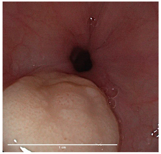
We gathered her past medical history which included a prolactin-secreting pituitary microadenoma for which she was treated with Cabergoline and Drospirenone, migraine with aura treated with Triptans and autoimmune chronic euthyroid-thyroiditis. She also had a pregnancy with spontaneous birth in 2011. The patient had never undergone surgery and she did not declare food or drug allergies. Her familial medical history was unremarkable for gastrointestinal tumors. Moreover, in 2018, the patient underwent upper gastrointestinal endoscopy (EGDS) following the detection of esophageal wall thickening on a chest CT for a suspected pulmonary disease due to the presence of chronic cough. She denied having any upper GI symptoms. During the endoscopic examination, a white nodular formation, measuring approximately 10 mm, was found in the middle esophagus (Figure 1), along with subcentimetric submucosal lesions in the stomach and duodenal bulb; 4 oriented biopsies were taken from the esophageal nodule. Histological examination resulted in the diagnosis of granular cell tumor. The patient was then referred to a tertiary center for endoscopic removal of the esophageal lesion, confirmed as a granular cell tumor, and underwent annual follow-up with endoscopic ultrasound (EUS) without evidence of recurrence. The gastric lesions remained stable in number and size during the subsequent follow-up.

Figure 1.
EGDS 2018: white nodule, granular cell tumor on histology. White bar: 1 cm length.
On the day of colonoscopy, written consent for the endoscopic examination and for conscious sedation was obtained, together with consent for the eventual anonymous publication of clinical and instrumental data, according to the policies of our Institution and to our national laws.
The examination was performed after 12 h fasting and after bowel cleansing with a 2 L “split dose” polyethylene glycol solution. Conscious sedation with intravenous midazolam and meperidine was controlled at the beginning, followed by continuous monitoring of vital parameters. The examination was performed with a high-definition (HD) standard colonoscope (Pentax Medical®, Tokyo, Japan). Oriented biopsies with standard biopsy forceps were collected and sent to the Pathological Anatomy Institute of our hospital within the same day for standard and immunohistochemical evaluation.
3. Results
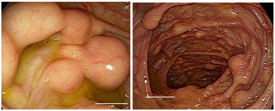
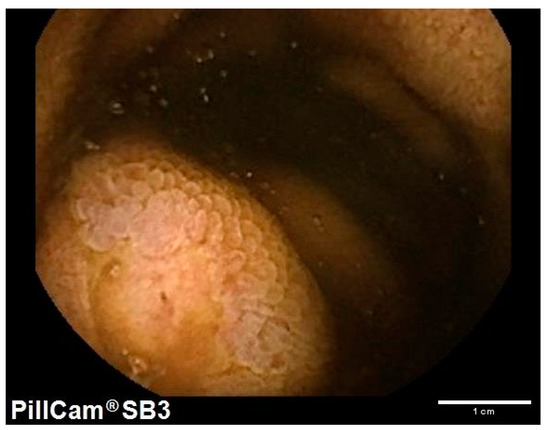
Colonoscopy was performed in April 2023 and showed numerous whitish and slightly elevated submucosal lesions throughout the entire colon and terminal ileum. The size of the lesions varied from 5 to 20 mm (Figure 2), with a rigid consistency on biopsy and a negative “pillow sign”. Two biopsies from all the lesions > 1 cm length were taken and collected in separated tubes. Histological examination described “submucosal lesion, composed of uniform epithelioid cells with abundant granular eosinophilic cytoplasm and small nuclei covered by hyperplastic esophageal epithelium”. Moreover, immunohistochemistry was positive for S100, Calretinin and PAS and negative for CD1a, CD25 and CD163. The Ki67 index was <1–2%, devoid of mitoses (evaluated with PHH3). These features confirmed the presence of multifocal granular cell tumors (Abrikossoff tumors) and were identical to those found in the biopsies of the gastric lesions in 2018, which were revised by the pathologist. To complete the GI tract study, Video Capsule Endoscopy (VCE) was thus performed, revealing the presence of more than 20 lesions throughout the whole small intestine, ranging in size from 2 to 10 mm and macroscopically compatible with granular cell tumors (Figure 3).

Figure 2.
Colonoscopy 2023: numerous submucosal formations present in the whole colon. White bar: 1 cm length.

Figure 3.
VCE 2023: multiple white submucosal nodules seen in the small intestine.
The patient’s 12 year-old daughter, during a routine dental checkup, was found to have a white and painless nodule on the tongue, at high clinical suspicion for a granular cell tumor. She was asymptomatic and denied any symptoms of dysphagia, food impaction or weight loss, her past medical history was unremarkable and her physical and psychological development were within the norm; she regularly attended school.
The nodule was subsequently biopsied, resulting in an histological confirmation of GCT. Immunohistochemical evaluation was totally comparable. The girl has not yet undergone endoscopy due to the parents’ wishes.
Both the patient and her daughter are asymptomatic and have normal full blood count at blood exams.
4. Discussion
The granular cell tumor (GCT), known as Abrikossoff tumor, is a rare tumor that originates from Schwann cells [1].
This type of tumor can potentially affect soft tissues of any organ, but it most frequently involves the head and neck region, the oral cavity, the skin or subcutaneous tissues. The tongue is the most frequently affected site [9].
Individuals of any age can present with this tumor, most commonly during the fourth, fifth and sixth decades of life, with an incidence peaking between the fourth and the sixth decade [9]. Females are more frequently affected than males, with an M/F ratio = ½.
Pediatric cases are rare, but some reports are available, including a case series of neonatal or congenital oral GCTs [10].
Histologically, they are composed of nests or sheets of plump epithelioid or spindle cells (or both) with a small, round nucleus and abundant granular eosinophilic cytoplasm. These histological characteristics are highly suggestive of a GCT, making it possible to diagnose with scarce samples of just a few cells. From an immunohistochemistry point of view, gastrointestinal GCTs are characterized by positivity for S-100 protein (100%), CD56 (95%), CD68 (95%), SOX-10 (93%) and inhibin alpha (52%) [7]. Cases of S-100-negative GCTs have also been reported in the literature [11]. While most of these tumors are benign, cases of malignant GCTs have been reported.
Malignancy is defined using the Fanburg-Smith histopathological criteria [12]: necrosis, spindling, vesicular nuclei with large nucleoli, mitotic activity (>2/10 HPF), increased nucleus/cytoplasm ratio and pleomorphism. Lesions exhibiting none of the aforementioned features or with isolated focal pleomorphism are classified as benign, those presenting with one or two are classified as atypical and those with three or more characteristics are categorized as malignant [12].
Cases of metastatic GCTs have been reported, both in cases of metastatic benign GCTs and in cases of metastatic malignant GCTs. The most common sites for metastasis are regional lymph nodes, lungs, liver and bones [13].
The gastrointestinal tract is involved in up to 11% of cases, with the esophagus, especially the distal esophagus, being the most frequently affected site, followed by the colon and the stomach [14]. There have also been reports of GCTs detected in the pancreas [15], biliary tree [16] or the appendix [17], but these locations are less common.
Typically, these lesions present as solitary nodules covered by white or yellow mucosa. GCTs of the colon and stomach tend to be larger in size than those found in the esophagus, with an average dimension of 0.75 cm, 0.6 cm and 0.27 cm, respectively [14]. Gastrointestinal GCTs are histologically similar to GCTs found in other locations, even though those located in the colon tend to exhibit more nuclear atypia, a feature that has not been correlated with malignancy [4]. Most gastrointestinal granular cell tumors are incidentally detected during endoscopic examinations performed for other reasons, with the majority of patients being asymptomatic. Rarely, esophageal GCTs present in patients reporting voice changes such as hoarseness or dysphagia.
Endoscopic removal with EMR or ESD is the safest and most effective treatment when feasible [18], especially for single lesions. The benefits of endoscopic treatment versus surgical removal are a less invasive procedure that can prevent skin scars caused by incisions, a reduction in the perceived pain associated with surgical trauma and a lower risk of postoperative infections. No specific guidelines are available to aid in the decision of the best removal technique, but some of the factors involved, such as the size and dimension or location of the nodule, could be applied to the guidelines used for the endoscopic removal of different subepithelial lesions [19]. EMR is the easier and less time-consuming technique; however, it can only be applied with smaller lesions, usually up to 10 mm in diameter, in order to maximize the probability of “en-bloc” curative resection. Seldomly, this technique can lead to serious complications, such as perforation [20,21,22]. Cap-assisted EMR has also been employed for small esophageal GCTs [7]. An attempt to remove a 13 mm esophageal GCT by EMR was unsuccessful in a report from the Mayo Clinic [23].
It must be noted that the majority of esophageal GCTs are not confined to the mucosa but rather involve the submucosa, which results in frequent involvement of the resection margins. ESD removal consists of the dissection of submucosal tissues beneath the lesion; its use is described for GCTs only in the right colon [24]. Despite a higher probability of obtaining an “en-bloc” curative resection, this technique is not yet widespread. New strategies to facilitate the procedure, especially applying counter-traction strategies (i.e., the clip and rubber band approaches) and using new knives, have been proposed [25], but the procedure remains quite challenging and has limited experience being executed in the context of GCTs and should be applied only in tertiary referral centers. However, when conducted by expert endoscopists, this technique lasts between 25 and 60 min and guarantees complete resection rates of more than 90%. Further benefits include lower complications and recurrence-free periods of up to 18 months after resection [26]. Surgical removal can be considered for lesions larger than 3 cm, those invading the muscularis propria or those with suspicious malignancy features. Cases of endoscopic surveillance for small, stable-sized lesions with follow-up endoscopies have been reported [24].
The etiology and pathophysiology of GCTs are poorly understood, as is their specific association with genetic syndromes. A possible aspect of GCTs’ pathogenesis could be trauma; there have been several reports of GCTs occurring at trauma sites, such as surgical scars, vaccination sites or within a tattoo.
An interesting association between Eosinophilic Esophagitis (EoE) and esophageal granular cell tumors has been reported by different authors, both in children and in adults [27,28,29]. A possible cause of this association is believed to be chronic esophageal inflammation found in patients affected by EOE, even though further data and research are needed to understand the immunological pathway linking EoE and esophageal GCTs.
No specific genetic mutation has been linked to the development of GCTs.
Somatic mutations of ATP6AP1 and ATP6AP2 genes in Schwann cells, which result in an accumulation of intracytoplasmic granules typical of GCTs, have been proposed as drivers of GCTs and appear to be pathognomonic for these tumors [30].
The presence of multiple granular cell tumors has been associated with various syndromes, such as Noonan syndrome [31], neurofibromatosis type I [32] and LEOPARD syndrome [33]. A possible feature present in all three syndromes is an abnormal RAS/MAPK pathway but mutations of the PTPN11 [33] gene link LEOPARD and Noonan syndrome.
To our knowledge, there are no reported cases in the literature of multifocal GCTs involving the entire gastrointestinal tract.
Currently, there are no guidelines for the treatment and follow-up of gastrointestinal GCTs.
The role of conventional chemotherapy and/or radiation is still debated and should be considered in patients with recurrent malignant or metastatic GCTs. For these reasons, given the impossibility of an operative approach through endoscopic and/or surgical removal due to the presence of too many lesions, we propose that the best management in our case could include annual endoscopic monitoring with EGDS, VCE and colonoscopy, annual mammography and Pap tests, and monitoring for any newly appearing formations on the skin and oral cavity with regular dermatological and odontostomatological check-ups. Furthermore, considering the possible familial component to this tumor in our case, we believe it could be beneficial to conduct genetic counseling as part of research protocols to understand the potential presence of a predisposing genetic mutation.
5. Conclusions
Granular cell tumors are rare tumors that may affect the gastrointestinal tract with benign or potentially malignant behavior in both pediatric and adult life. Their appearance is typically described as submucosal isolated lesions of the esophagus or the stomach, making them potentially amenable for endoscopic or surgical resection. Our research presents one of the first documented cases of a patient with panenteric gastrointestinal GCTs, while also suggesting a potential hereditary component due to the concomitant diagnosis of a tongue GCT affecting the patient’s daughter. We believe that reporting such cases is of utmost importance for such conditions as no clinical or endoscopic guidelines exist to manage these rare tumors. Genetic counseling is essential to identify possible genetic mutations, such as those involving ATP6 or the RAS/MAPK pathway.
Author Contributions
Conceptualization, M.C.; writing—original draft preparation, R.S. and L.F.; writing—reviewing and editing, F.M.; supervision, M.C. and F.M. All authors have read and agreed to the published version of the manuscript.
Funding
This research received no external funding.
Institutional Review Board Statement
Not applicable.
Informed Consent Statement
Written informed consent has been obtained from the patient to publish this paper.
Data Availability Statement
Clinical and endoscopic data of the patient are available on our hospital server.
Conflicts of Interest
The authors declare no conflicts of interest.
References
- Stefansson, K.; Wollmann, R.L. S-100 protein in granular cell tumors (granular cell myoblastomas). Cancer 1982, 49, 1834–1838. [Google Scholar] [CrossRef] [PubMed]
- Weber, C.O.; Virchow, R. Anatomische Untersuchung einer hypertrophischen Zunge nebst Bemerkungen über die Neubildung quergestreifter Muskelfasern. Arch. Pathol. Anat. Physiol. Klin. Med. 1854, 7, 115–125. [Google Scholar] [CrossRef]
- Abrikossoff, A. Über myome: Ausgehend von der quergestreiften willkürlichen Muskulatur. Virchows Arch. Pathol. Anat. Physiol. Klin. Med. 1926, 260, 215–233. [Google Scholar] [CrossRef]
- Na, J.; Kim, H.; Jung, J.; Kim, Y.; Kim, S.; Lee, J.; Lee, K.; Park, J. Granular cell tumours of the colorectum: Histopathological and immunohistochemical evaluation of 30 cases. Histopathology 2014, 65, 764–774. [Google Scholar] [CrossRef] [PubMed]
- Fahim, S.; Aryanian, Z.; Ebrahimi, Z.; Kamyab-Hesari, K.; Mahmoudi, H.; Alizadeh, N.; Heidari, N.; Livani, F.; Ghanadan, A.; Goodarzi, A. Cutaneous granular cell tumor: A case series, review, and update. J. Family Med. Prim. Care 2022, 11, 6955–6958. [Google Scholar] [PubMed]
- Salaouatchi, M.T.; De Breucker, S.; Rouvière, H.; Lesage, V.; Rocq, L.J.A.; Vandergheynst, F.; Beernaert, L. A Rare Case of a Metastatic Malignant Abrikossoff Tumor. Case Rep. Oncol. 2021, 14, 1868–1875. [Google Scholar] [CrossRef] [PubMed]
- Canavesi, A.; Berrueta, J.; Pillajo, S.; Gaggero, P.; Olano, C. Endoscopic resection of a granular cell tumor (Abrikossoff’s tumor) in the esophagus using cap-assisted band ligation. Endoscopy 2023, 55, E796–E797. [Google Scholar] [CrossRef]
- De Vincentis, F.; Manzi, I.; Di Giorgio, V.; Mussetto, A. Endoscopic full-thickness resection of a residual scar in ascending colon to assess post-EMR complete removal of an Abrikossoff tumor. Dig. Liver Dis. 2023, 55, 985–986. [Google Scholar] [CrossRef]
- van de Loo, S.; Thunnissen, E.; Postmus, P.; van der Waal, I. Granular cell tumor of the oral cavity; a case series including a case of metachronous occurrence in the tongue and the lung. Med. Oral. Patol. Oral. Cir. Bucal. 2015, 20, e30–e33. [Google Scholar] [CrossRef]
- Zheng, C.; Su, J.; Liang, X.; Wu, J.; Gu, W.; Zhao, X. Clinical and pathological analysis of congenital granular cell tumor. Hua Xi Kou Qiang Yi Xue Za Zhi. 2022, 40, 710–715. [Google Scholar]
- Parfitt, J.R.; McLean, C.A.; Joseph, M.G.; Streutker, C.J.; Al-Haddad, S.; Driman, D.K. Granular cell tumours of the gastrointestinal tract: Expression of nestin and clinicopathological evaluation of 11 patients. Histopathology 2006, 48, 424–430. [Google Scholar] [CrossRef]
- Fanburg-Smith, J.C.; Meis-Kindblom, J.M.; Fante, R.; Kindblom, L.G. Malignant granular cell tumor of soft tissue: Diagnostic criteria and clinicopathologic correlation. Am. J. Surg. Pathol. 1998, 22, 779–794. [Google Scholar] [CrossRef]
- Menaker, G.M.; Sanger, J.R. Granular Cell Tumor of Uncertain Malignant Potential. Ann. Plast. Surg. 1997, 38, 658–660. [Google Scholar] [CrossRef]
- An, S.; Jang, J.; Min, K.; Kim, M.-S.; Park, H.; Park, Y.S.; Kim, J.; Lee, J.H.; Song, H.J.; Kim, K.-J.; et al. Granular cell tumor of the gastrointestinal tract: Histologic and immunohistochemical analysis of 98 cases. Hum. Pathol. 2015, 46, 813–819. [Google Scholar] [CrossRef] [PubMed]
- Kanno, A.; Satoh, K.; Hirota, M.; Hamada, S.; Umino, J.; Itoh, H.; Masamune, A.; Egawa, S.; Motoi, F.; Unno, M.; et al. Granular cell tumor of the pancreas: A case report and review of literature. World J. Gastrointest. Oncol. 2010, 2, 121–124. [Google Scholar] [CrossRef] [PubMed]
- Mackenzie, D.J.; Klapper, E.; Gordon, L.A.; Silberman, A.W. Granular cell tumor of the biliary system. Med. Pediatr. Oncol. 1994, 23, 50–56. [Google Scholar] [CrossRef] [PubMed]
- Lv, X.; Sun, X.; Zhou, J.; Zhang, Y.; Lv, G. Granular cell tumor of the appendix: A case report and literature review. J. Int. Med. Res. 2022, 50, 030006052211093. [Google Scholar] [CrossRef]
- Chen, W.S.; Zheng, X.L.; Jin, L.; Pan, X.J.; Ye, M.F. Novel diagnosis and treatment of esophageal granular cell tumor: Report of 14 cases and review of the literature. Ann. Thorac. Surg. 2014, 97, 296–302. [Google Scholar] [CrossRef] [PubMed]
- Deprez, P.H.; Moons, L.M.; Oʼtoole, D.; Gincul, R.; Seicean, A.; Pimentel-Nunes, P.; Fernández-Esparrach, G.; Polkowski, M.; Vieth, M.; Borbath, I.; et al. Endoscopic management of subepithelial lesions including neuroendocrine neoplasms: European Society of Gastrointestinal Endoscopy (ESGE) Guideline. Endoscopy 2022, 54, 412–429. [Google Scholar] [CrossRef]
- Battaglia, G.; Rampado, S.; Bocus, P.; Guido, E.; Portale, G.; Ancona, E. Single-band mucosectomy for granular cell tumor of the esophagus: Safe and easy technique. Surg. Endosc. 2006, 20, 1296–1298. [Google Scholar] [CrossRef]
- Wehrmann, T.; Martchenko, K.; Nakamura, M.; Riphaus, A.; Stergiou, N. Endoscopic Resection of Submucosal Esophageal Tumors: A Prospective Case Series. Endoscopy 2004, 36, 802–807. [Google Scholar] [CrossRef] [PubMed]
- Sakamoto, H.; Suga, M.; Ozeki, I.; Kobayashi, T.; Sugaya, T.; Sasaki, Y.; Azuma, N.; Itoh, F.; Sakamoto, S.-I.; Yachi, A.; et al. Subcapsular hematoma of the liver and pylethrombosis in the setting of cholestatic liver injury. J. Gastroenterol. 1996, 31, 880–884. Available online: https://pubmed.ncbi.nlm.nih.gov/9027656/ (accessed on 12 November 2022). [CrossRef] [PubMed]
- Zhong, N.; Katzka, D.A.; Smyrk, T.C.; Wang, K.K.; Topazian, M. Endoscopic diagnosis and resection of esophageal granular cell tumors. Dis. Esophagus 2011, 24, 538–543. [Google Scholar] [CrossRef] [PubMed]
- Biscay, M.; Chabrun, E.; Menguy, S.; Cesbron-Métivier, E.; Barthet, M.; Marty, M.; Pioche, M. Colonic Abrikossoff tumor: Fortuitous discovery at colonoscopy for serrated adenomas polyposis, and resection by endoscopic submucosal dissection. Endoscopy 2019, 51, E176–E178. [Google Scholar] [CrossRef]
- Utzeri, E.; Jacques, J.; Charissoux, A.; Rivory, J.; Legros, R.; Ponchon, T.; Pioche, M. Traction strategy with clips and rubber band allows complete en bloc endoscopic submucosal dissection of laterally spreading tumors invading the appendix. Endoscopy 2017, 49, 820–822. [Google Scholar] [CrossRef] [PubMed]
- Lu, W.; Xu, M.D.; Zhou, P.H.; Zhang, Y.Q.; Chen, W.F.; Zhong, Y.S.; Yao, L.Q. Endoscopic submucosal dissection of esophageal granular cell tumor. World J. Surg. Oncol. 2014, 12, 221. [Google Scholar] [CrossRef] [PubMed]
- Malik, F.; Bernieh, A.; Saad, A.G. Esophageal Granular Cell Tumor in Children: A Clinicopathologic Study of 11 Cases and Review of the Literature. Am. J. Clin. Pathol. 2023, 160, 106–112. [Google Scholar] [CrossRef]
- Riffle, M.E.; Polydorides, A.D.; Niakan, J.; Chehade, M. Eosinophilic Esophagitis and Esophageal Granular Cell Tumor. Am. J. Surg. Pathol. 2017, 41, 616–621. [Google Scholar] [CrossRef]
- Reddi, D.; Chandler, C.; Cardona, D.; Schild, M.; Westerhoff, M.; McMullen, E.; Tomizawa, Y.; Clinton, L.; Swanson, P.E. Esophageal granular cell tumor and eosinophils: A multicenter experience. Diagn. Pathol. 2021, 16, 49. [Google Scholar] [CrossRef]
- Pareja, F.; Brandes, A.H.; Basili, T.; Selenica, P.; Geyer, F.C.; Fan, D.; Da Cruz Paula, A.; Kumar, R.; Brown, D.N.; Gularte-Mérida, R.; et al. Loss-of-function mutations in ATP6AP1 and ATP6AP2 in granular cell tumors. Nat. Commun. 2018, 9, 3533. [Google Scholar] [CrossRef]
- Castagna, J.; Clerc, J.; Dupond, A.S.; Laresche, C. Tumeurs à cellules granuleuses multiples chez un enfant atteint d’un syndrome de Noonan compliqué de leucémie myélomonocytaire juvénile. Ann. Dermatol. Venereol. 2017, 144, 705–711. [Google Scholar] [CrossRef] [PubMed]
- Weinreb, I.; Bray, P.; Ghazarian, D. Plexiform intraneural granular cell tumour of a digital cutaneous sensory nerve. J. Clin. Pathol. 2007, 60, 725–726. [Google Scholar] [CrossRef] [PubMed]
- Schrader, K.; Nelson, T.; De Luca, A.; Huntsman, D.; McGillivray, B. Multiple granular cell tumors are an associated feature of LEOPARD syndrome caused by mutation in PTPN11. Clin. Genet. 2009, 75, 185–189. [Google Scholar] [CrossRef] [PubMed]
Disclaimer/Publisher’s Note: The statements, opinions and data contained in all publications are solely those of the individual author(s) and contributor(s) and not of MDPI and/or the editor(s). MDPI and/or the editor(s) disclaim responsibility for any injury to people or property resulting from any ideas, methods, instructions or products referred to in the content. |
© 2024 by the authors. Licensee MDPI, Basel, Switzerland. This article is an open access article distributed under the terms and conditions of the Creative Commons Attribution (CC BY) license (https://creativecommons.org/licenses/by/4.0/).