Next Issue
Volume 15, June
Previous Issue
Volume 14, December


Cardiogenetics, Volume 15, Issue 1 (March 2025) – 9 articles

Cover Story (view full-size image): 
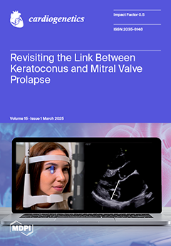
The proposed association between keratoconus and mitral valve prolapse (MVP) suggests a shared pathophysiological mechanism involving collagen defects. However, previous studies have reported conflicting results. This case–control study investigated the prevalence of MVP and mitral valve billowing in 101 patients with keratoconus treated with corneal collagen cross-linking, compared to matched controls. Using advanced echocardiographic imaging, we found no significant association between keratoconus and MVP prevalence. Our findings challenge earlier reports and suggest that routine cardiac screening in patients with keratoconus may not be warranted, though further research in diverse populations is needed. View this paper
  • Issues are regarded as officially published after their release is announced to the table of contents alert mailing list.
  • You may sign up for e-mail alerts to receive table of contents of newly released issues.
  • PDF is the official format for papers published in both, html and pdf forms. To view the papers in pdf format, click on the "PDF Full-text" link, and use the free Adobe Reader to open them.
Order results
Result details
Section
Select all
Export citation of selected articles as:
8 pages, 563 KiB  
Review
Brugada Syndrome and GPD1L: Definite Genotype-Phenotype Association?
by Andrea Greco, Estefanía Martínez-Barrios, José Cruzalegui, Sergi Cesar, Fredy Chipa, Nuria Díez-Escuté, Patricia Cerralbo, Irene Zschaeck, Paula Loredo, Georgia Sarquella-Brugada and Oscar Campuzano
Cardiogenetics 2025, 15(1), 9; https://doi.org/10.3390/cardiogenetics15010009 - 14 Mar 2025
Viewed by 330
Abstract
The GPD1L gene encodes a small cytoplasmic protein that is involved in the regulation of sodium currents. Alterations in this gene have been associated with Brugada syndrome. This rare arrhythmogenic syndrome is characterized by a typical electrocardiographic pattern, incomplete penetrance, variable expressivity, and [...] Read more.
The GPD1L gene encodes a small cytoplasmic protein that is involved in the regulation of sodium currents. Alterations in this gene have been associated with Brugada syndrome. This rare arrhythmogenic syndrome is characterized by a typical electrocardiographic pattern, incomplete penetrance, variable expressivity, and risk of sudden cardiac death. To date, few families with a clinical diagnosis of Brugada syndrome caused by a rare alteration in the GPD1L gene have been reported worldwide. The increase in data focused on genetic variants allows us to improve the interpretation of their role in Brugada syndrome. In our study, we have compiled the GPD1L variants reported so far in patients with a definitive clinical diagnosis or suspected Brugada syndrome. We performed an exhaustive update and interpretation of each variant following the guidelines of the American College of Medical Genetics and Genomics and the Association for Molecular Pathology. Our results showed that none of the variants described to date can be classified as truly harmful in Brugada syndrome. Despite this fact, more clinical and genetic data are needed to definitively rule out the GPD1L gene as a cause of Brugada syndrome. In summary, to date, there is insufficient evidence to conclude a definitive association between GPD1L and Brugada syndrome. Full article
(This article belongs to the Section Rare Disease-Genetic Syndromes)
Show Figures

Figure 1

20 pages, 1254 KiB  
Review
Genetic Implications of Fatty Tissue for the Development of Ventricular Arrhythmias
by Raluca Sirbu Prisecaru, Oana Purcar and Ioan Manitiu
Cardiogenetics 2025, 15(1), 8; https://doi.org/10.3390/cardiogenetics15010008 - 14 Mar 2025
Viewed by 404
Abstract
Ventricular arrhythmias are a common disorder, and sometimes the etiology remains unclear. Present data support cardiac fatty tissue’s potential role as a substrate for ventricular arrhythmias. Diagnosing fatty tissue based on imaging markers and histopathological evidence is often challenging. Data about the influence [...] Read more.
Ventricular arrhythmias are a common disorder, and sometimes the etiology remains unclear. Present data support cardiac fatty tissue’s potential role as a substrate for ventricular arrhythmias. Diagnosing fatty tissue based on imaging markers and histopathological evidence is often challenging. Data about the influence of individual and multiple genetic variants on epicardial adipose tissue volume remain limited. In this review, we aimed to provide a comprehensive overview of the current understanding of the genetic basis of fatty tissue and its contribution to the pathogenesis of ventricular arrhythmias and to discuss the relationship between certain genetic variants and the development of ventricular arrhythmia. Full article
(This article belongs to the Section Education in Cardiogenetics)
Show Figures

Figure 1

27 pages, 22222 KiB  
Review
Cardiomyopathies and Arrythmias in Neuromuscular Diseases
by Giuseppe Sgarito, Calogero Volpe, Stefano Bardari, Raimondo Calvanese, Paolo China, Giosuè Mascioli, Martina Nesti, Carlo Pignalberi, Manlio Cipriani and Massimo Zecchin
Cardiogenetics 2025, 15(1), 7; https://doi.org/10.3390/cardiogenetics15010007 - 3 Mar 2025
Viewed by 764
Abstract
Neuromuscular diseases (NMDs) encompass various hereditary conditions affecting motor neurons, the neuromuscular junction, and skeletal muscles. These disorders are characterized by progressive muscle weakness and can manifest at different stages of life, from birth to adulthood. NMDs, such as Duchenne and Becker muscular [...] Read more.
Neuromuscular diseases (NMDs) encompass various hereditary conditions affecting motor neurons, the neuromuscular junction, and skeletal muscles. These disorders are characterized by progressive muscle weakness and can manifest at different stages of life, from birth to adulthood. NMDs, such as Duchenne and Becker muscular dystrophies, myotonic dystrophy, and limb–girdle muscular dystrophies, often involve cardiac complications, including cardiomyopathies and arrhythmias. Underlying genetic mutations contribute to skeletal and cardiac muscle dysfunction, particularly in the DMD, EMD, and LMNA genes. The progressive nature of muscle deterioration significantly reduces life expectancy, mainly due to respiratory and cardiac failure. The early detection of cardiac involvement through electrocardiography (ECG) and cardiac imaging is crucial for timely intervention. Pharmacological treatment focuses on managing cardiomyopathies and arrhythmias, with an emerging interest in gene therapies aimed at correcting underlying genetic defects. Heart transplantation, though historically controversial in patients with muscular dystrophies, is increasingly recognized as a viable option for individuals with advanced heart failure and moderate muscle impairment, leading to improved survival rates. Careful patient selection and management are critical to optimizing outcomes in these complex cases. Full article
(This article belongs to the Section Rare Disease-Neuromuscular Diseases)
Show Figures

Figure 1

12 pages, 1474 KiB  
Article
Familial Hypercholesterolemia Screening in a Cardiac Rehabilitation Program After Myocardial Infarction
by Carlos Bertolín-Boronat, Víctor Marcos-Garcés, Héctor Merenciano-González, María Luz Martínez Mas, Josefa Inés Climent Alberola, Nerea Perez, Laura López Bueno, María Concepción Esteban Argente, María Valls Reig, Ana Arizón Benito, Alfonso Payá Rubio, César Ríos-Navarro, Elena de Dios, Jose Gavara, Manuel F. Jiménez-Navarro, Francisco Javier Chorro, Juan Sanchis and Vicente Bodi
Cardiogenetics 2025, 15(1), 6; https://doi.org/10.3390/cardiogenetics15010006 - 24 Feb 2025
Viewed by 617
Abstract
Familial hypercholesterolemia (FH) is relatively prevalent in myocardial infarction (MI) sufferers, and its diagnosis could improve preventive treatment in family members. We aim to analyze the diagnosis of FH and the rate of genetic testing in a prospective cohort of 245 patients submitted [...] Read more.
Familial hypercholesterolemia (FH) is relatively prevalent in myocardial infarction (MI) sufferers, and its diagnosis could improve preventive treatment in family members. We aim to analyze the diagnosis of FH and the rate of genetic testing in a prospective cohort of 245 patients submitted to our Cardiac Rehabilitation Program (CRP) after MI. Baseline characteristics were registered, and basal low-density lipoprotein cholesterol (LDL-C) was calculated after correction for lipid-lowering therapies (LLT) before or during admission. Simplified Dutch Lipid Clinic Network Scores (sDLCNS) were retrospectively calculated based on personal and familial history of premature cardiovascular disease and basal LDL-C levels. Mean age was 62.19 ± 13.93 years, and most patients were male (81.6%). Mean LDL-C before admission and basal LDL-C corrected for LLT were 131.79 ± 45.34 mg/dL and 162.87 ± 44.17 mg/dL, respectively. Patients in the cohort were retrospectively categorized in the “unlikely” (<3 points; n = 162, 66.1%), “possible” (3–5 points; n = 72, 29.4%) and “probable” (6–8 points; n = 11, 4.5%) sDLCNS categories. Genetic testing for FH was requested in four (1.6%) patients, and no clinically significant genetic variants were detected. Patients who underwent genetic testing depicted significantly higher basal LDL-C (233 ± 49.09 vs. 161.71 ± 43.25 mg/dL, p = 0.001). However, the rate of individuals undergoing genetic testing was negligible even in the “possible” (n = 2, 2.8%) and “probable” (n = 1, 9.1%) sDLCNS categories. In conclusion, genetic testing for FH in our CRP after MI is largely underutilized, even in patients with a “possible” or “probable” diagnosis based on sDLCNS criteria, which represent about a third of the cohort. Strategies to improve screening for FH should be prospectively implemented. Full article
(This article belongs to the Section Cardiovascular Genetics in Clinical Practice)
Show Figures

Figure 1

10 pages, 1181 KiB  
Article
Electrocardiogram May Fail to Identify Proportion of High-Risk Individuals: Analysis of Series of 50 Sudden Death Cases
by Mariela Salar-Alcaraz, Pablo Peñafiel-Verdú, Francisco J. Castro-García, Francisco A. Pastor-Quirante, Carmen Muñoz-Esparza, José M. López-Ayala, Juan Martínez-Sánchez, Juan J. Sánchez-Muñoz, Arcadi García-Alberola, María Sabater-Molina and Juan R. Gimeno-Blanes
Cardiogenetics 2025, 15(1), 5; https://doi.org/10.3390/cardiogenetics15010005 - 10 Feb 2025
Viewed by 492
Abstract
Background: An electrocardiogram (ECG) is an essential and easily available diagnostic test in the management of cardiomyopathies and channelopathies. Different strategies based on ECG have been recommended for general population and athlete screening. Objectives: The purpose of this study was to explore the [...] Read more.
Background: An electrocardiogram (ECG) is an essential and easily available diagnostic test in the management of cardiomyopathies and channelopathies. Different strategies based on ECG have been recommended for general population and athlete screening. Objectives: The purpose of this study was to explore the value of the ECG for the diagnosis of sudden cardiac death (SCD) cases. Methods: ECGs from 50 (aged 37.6 ± 19.9 years, 37 men) resuscitated cardiac arrest (26, 52%) and SCD cases (24, 48%) were analyzed. Relevant medical history and results from clinical tests were reviewed. ECG findings were compared with the final diagnosis. Results: Final ECG classification was as follows: 9 (18%) normal, 15 (30%) unspecific, 14 (28%) suggestive, and 12 (24%) diagnostic. Amongst 13 hypertrophic cardiomyopathy patients, ECGs were diagnostic in 6 (46%) and suggestive in 1 (8%). Arrhythmogenic right ventricular cardiomyopathy was diagnosed in seven patients, two (28%) with suggestive ECG. Dilated cardiomyopathy was diagnosed in four patients, two (50%) with suggestive ECG. Six patients had Brugada syndrome: four (66%) had diagnostic ECGs, and two (33%) had suggestive ECG. Long QT syndrome was diagnosed in four cases; only one (25%) had a diagnostic ECG. Three patients had other cardiomyopathies. After the complete study, 13 (26%) patients remained with a non-conclusive diagnosis; their ECGs were unspecific or normal. Conclusion: ECG can be unspecific or normal in an important percentage of SCD cases (48%). Furthermore, a significant proportion of SCD cases after a comprehensive study remain without a definite diagnosis (26%). These findings should be considered when planning SCD preventive strategies. Full article
(This article belongs to the Section Sport Cardiology)
Show Figures

Figure 1

12 pages, 627 KiB  
Article
Revisiting the Link Between Keratoconus and Mitral Valve Prolapse
by Christian K. Five, Nina E. Hasselberg, Hilde Bjerkreim, Linda T. Aaserud, Anna Isotta Castrini, Cecilie Bugge, Eivind W. Aabel, Thomas Helle-Valle, Håvard Dalen, Olav Kristianslund and Kristina H. Haugaa
Cardiogenetics 2025, 15(1), 4; https://doi.org/10.3390/cardiogenetics15010004 - 5 Feb 2025
Viewed by 695
Abstract
Keratoconus is a progressive eye disease that results in thinning of the cornea, leading to visual impairment. Mitral valve prolapse (MVP) is a common disorder affecting around 2–4% of the general population. Previous studies have found an overrepresentation of MVP in individuals with [...] Read more.
Keratoconus is a progressive eye disease that results in thinning of the cornea, leading to visual impairment. Mitral valve prolapse (MVP) is a common disorder affecting around 2–4% of the general population. Previous studies have found an overrepresentation of MVP in individuals with keratoconus, with a prevalence of 38–65%, suggesting a shared underlying mechanism. In this case-control study, patients with keratoconus were enrolled from a quality and research registry. They were examined by a 2D echocardiography to identify if they had MVP, billowing or normal mitral leaflets. Controls were matched from the population-based Trøndelag Health Study. Patients and controls underwent a detailed echocardiographic examination to detect abnormal mitral valves. We included 101 patients (age 33 [IQR 29–40], 75% male) with keratoconus and 101 matched individuals. MVP was found in 2 (2%), while billowing was found in 5 (5%) of keratoconus patients. No significant association was found between keratoconus and the prevalence of MVP or billowing compared to the control group. Moreover, no associations were found between severity of keratoconus with presence of MVP nor with billowing of the mitral valves. We could not confirm the previously reported association between keratoconus and MVP, suggesting that routine screening for MVP in keratoconus patients may not be warranted. However, we cannot rule out the possibility of an association in other gender, age and ethnic groups different than ours. Full article
(This article belongs to the Section Cardiovascular Genetics in Clinical Practice)
Show Figures

Figure 1

9 pages, 414 KiB  
Article
Contribution of Rare and Common APOE Variants to Familial Hypercholesterolemia in Spanish Cohort
by Lorena M. Vega-Prado, Daniel Vázquez-Coto, Francisco Villazón, Lorena Suárez-Gutiérrez, Ceferino Martínez-Faedo, Edelmiro Menéndez-Torre, María Riestra, Silvia González-Martínez, Gala Gutiérrez-Buey, Claudia García-Lago, Juan Gómez, Victoria Alvarez, Helena Gil, Rebeca Lorca and Eliecer Coto
Cardiogenetics 2025, 15(1), 3; https://doi.org/10.3390/cardiogenetics15010003 - 27 Jan 2025
Viewed by 806
Abstract
Our aim was to determine whether rare APOE pathogenic variants (PV) and the common e2/e3/e4 polymorphism were associated with the risk of familial hypercholesterolemia (FH). A total of 431 patients who met the inclusion criteria for FH were next-generation sequenced for the main [...] Read more.
Our aim was to determine whether rare APOE pathogenic variants (PV) and the common e2/e3/e4 polymorphism were associated with the risk of familial hypercholesterolemia (FH). A total of 431 patients who met the inclusion criteria for FH were next-generation sequenced for the main candidate genes (LDLR, APOB, PCSK9, APOE, LDLRAP1). A total of 139 patients (32%) had a pathogenic variant, including 3 with APOE p.Leu167del. Among the PV-negatives (n = 292), one was homozygous for APOE-e2 and showed a combined phenotype of high low-density lipoprotein cholesterol (LDL-C) and triglycerides (TGs). A total of 165 population controls were also genotyped for the APOE polymorphism. PV-negative patients showed a significantly higher frequency of APOE-e3e4/e4e4 compared to PV-positives (p = 0.006) and to population controls (p = 0.0002, OR = 2.63, 95% CI = 1.57–4.40). APOE-e4e4 patients had significantly higher mean LDL-C compared to the other genotypes (p = 0.047). In conclusion, APOE pathogenic variants were a rare cause of FH in our population, and the APOE-e4 allele was a significant risk factor for being diagnosed with familial hypercholesterolemia in the absence of a pathogenic variant involved in FH. In particular, the APOE-e4e4 genotype was associated with higher LDL-C levels compared to the other genotypes. Full article
(This article belongs to the Section Cardiovascular Genetics in Clinical Practice)
Show Figures

Figure 1

18 pages, 1710 KiB  
Review
Cardiovascular Involvement in SYNE Variants: A Case Series and Narrative Review
by Francesco Ravera, Veronica Dusi, Pier Paolo Bocchino, Giulia Gobello, Giuseppe Giannino, Daniele Melis, Giulia Margherita Brach Del Prever, Filippo Angelini, Andrea Saglietto, Carla Giustetto, Guglielmo Gallone, Stefano Pidello, Margherita Cannillo, Marco Matteo Cingolani, Silvia Deaglio, Walter Grosso Marra, Gaetano Maria De Ferrari and Claudia Raineri
Cardiogenetics 2025, 15(1), 2; https://doi.org/10.3390/cardiogenetics15010002 - 20 Jan 2025
Viewed by 790
Abstract
Cardiac laminopathies encompass a wide range of diseases caused by defects in nuclear envelope proteins, including cardiomyopathy, atrial and ventricular arrhythmias and conduction system abnormalities. Two genes, namely LMNA and EMD, are typically associated with these disorders and are part of the [...] Read more.
Cardiac laminopathies encompass a wide range of diseases caused by defects in nuclear envelope proteins, including cardiomyopathy, atrial and ventricular arrhythmias and conduction system abnormalities. Two genes, namely LMNA and EMD, are typically associated with these disorders and are part of the routine genetic panel performed in affected patients. Yet, there are other markedly fewer known proteins, the nesprins, encoded by SYNE genes, that play a pivotal role in connecting the nuclear envelope to cytoskeletal elements. So far, SYNE gene variants have been described in association with neurodegenerative diseases; their potential association with cardiac disorders, albeit anecdotally reported, is still largely unexplored. This review focuses on the role of nesprins in cardiomyocytes and explores the potential clinical implications of SYNE variants by presenting five unrelated patients with distinct cardiac manifestations and reviewing the literature. Emerging research suggests that SYNE-related cardiomyopathies involve disrupted nuclear–cytoskeletal coupling, leading to impaired cardiac function. Understanding these mechanisms is critical for furthering insights into the broader implications of nuclear envelope proteins in cardiac health and for potentially developing targeted therapeutic strategies. Additionally, our data support the inclusion of SYNE genes in the cardiac genetic panel for cardiomyopathies and cardiac conduction disorders. Full article
(This article belongs to the Section Cardiovascular Genetics in Clinical Practice)
Show Figures

Figure 1

17 pages, 317 KiB  
Review
Dietary Approach in Familial Hypercholesterolemia
by Joanna Popiolek-Kalisz, Klaudia Salamon, Michal Mazur, Klaudia Mikolajczyk and Grzegorz Kalisz
Cardiogenetics 2025, 15(1), 1; https://doi.org/10.3390/cardiogenetics15010001 - 1 Jan 2025
Viewed by 1941
Abstract
Introduction: Familial hypercholesterolemia (FH) is a genetic disorder that remains underdiagnosed and undertreated. It is characterized by high levels of low-density lipoprotein cholesterol (LDL-C), which leads to an increased cardiovascular disease risk. Pharmacotherapy of FH is based on high-dose statin therapy, often combined [...] Read more.
Introduction: Familial hypercholesterolemia (FH) is a genetic disorder that remains underdiagnosed and undertreated. It is characterized by high levels of low-density lipoprotein cholesterol (LDL-C), which leads to an increased cardiovascular disease risk. Pharmacotherapy of FH is based on high-dose statin therapy, often combined with ezetimibe and proprotein convertase subtilisin/kexin 9 inhibitors. The dietary approach is an important and supportive part of FH management. Methods: This review aimed to present the available evidence on dietary strategies in FH patients. The analyzed aspects included macronutrients such as fat and carbohydrate intake, as well as the role of dietary fiber, nutraceuticals (omega-3, beta-glucan, phytosterols, and red yeast fermented rice extract), and overall dietary models. Results and Conclusions: Based on the available data, the Mediterranean diet is a dietary model advised in cardiovascular prevention, including patients with FH. Regarding detailed recommendations, the current state of knowledge indicates dietary fat and saturated fatty acids intake limitation as an advised strategy. Supplementation of phytosterols and fiber can be also helpful in FH. Full article
(This article belongs to the Section Rare Disease-Genetic Syndromes)
Previous Issue
Next Issue
Back to TopTop