
Heptamethine Cyanine-Loaded Nanomaterials for Cancer Immuno-Photothermal/Photodynamic Therapy: A Review


 , and
, and
Abstract
:1. Introduction
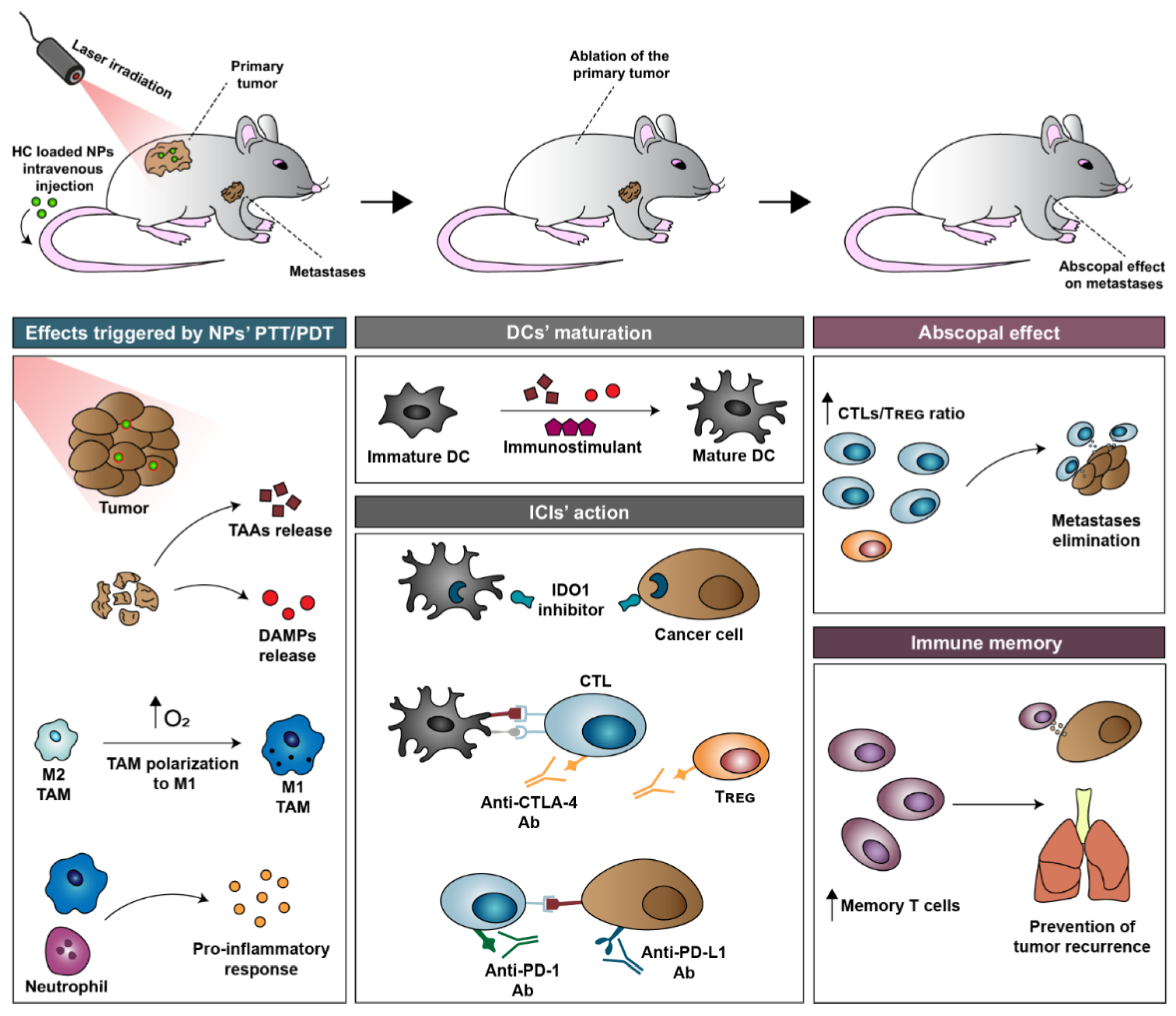
2. Overview of Nanomaterial-Mediated Immuno-PTT/PDT
3. ICG-Loaded Nanomaterials in Cancer Immuno-PTT/PDT
4. Prototypic HC-Loaded Nanomaterials in Cancer Immuno-PTT/PDT
5. Conclusions and Future Outlook
Author Contributions
Funding
Institutional Review Board Statement
Informed Consent Statement
Data Availability Statement
Conflicts of Interest
References
- Zhang, P.; Zhai, Y.; Cai, Y.; Zhao, Y.; Li, Y. Nanomedicine-based immunotherapy for the treatment of cancer metastasis. Adv. Mater. 2019, 31, 1904156. [Google Scholar] [CrossRef] [PubMed]
- Alix-Panabières, C.; Pantel, K. Challenges in circulating tumour cell research. Nat. Rev. Cancer 2014, 14, 623–631. [Google Scholar] [CrossRef] [PubMed]
- Farkona, S.; Diamandis, E.P.; Blasutig, I.M. Cancer immunotherapy: The beginning of the end of cancer? BMC Med. 2016, 14, 73. [Google Scholar] [CrossRef] [PubMed] [Green Version]
- Lima-Sousa, R.; Melo, B.L.; Alves, C.G.; Moreira, A.F.; Mendonça, A.G.; Correia, I.J.; de Melo-Diogo, D. Combining Photothermal-Photodynamic Therapy Mediated by Nanomaterials with Immune Checkpoint Blockade for Metastatic Cancer Treatment and Creation of Immune Memory. Adv. Funct. Mater. 2021, 31, 2010777. [Google Scholar] [CrossRef]
- Ng, C.W.; Li, J.; Pu, K. Recent progresses in phototherapy-synergized cancer immunotherapy. Adv. Funct. Mater. 2018, 28, 1804688. [Google Scholar] [CrossRef]
- Rajendrakumar, S.K.; Uthaman, S.; Cho, C.-S.; Park, I.-K. Nanoparticle-based phototriggered cancer immunotherapy and its domino effect in the tumor microenvironment. Biomacromolecules 2018, 19, 1869–1887. [Google Scholar] [CrossRef]
- Chen, Y.; Shen, X.; Han, S.; Wang, T.; Zhao, J.; He, Y.; Chen, S.; Deng, S.; Wang, C.; Wang, J. Irradiation pretreatment enhances the therapeutic efficacy of platelet-membrane-camouflaged antitumor nanoparticles. J. Nanobiotechnol. 2020, 18, 101. [Google Scholar] [CrossRef]
- Li, J.; Cui, D.; Huang, J.; He, S.; Yang, Z.; Zhang, Y.; Luo, Y.; Pu, K. Organic semiconducting pro-nanostimulants for near-infrared photoactivatable cancer immunotherapy. Angew. Chem. 2019, 131, 12810–12817. [Google Scholar] [CrossRef]
- Zhang, J.; Zhang, D.; Li, Q.; Jiang, Y.; Song, A.; Li, Z.; Luan, Y. Task-specific design of immune-augmented nanoplatform to enable high-efficiency tumor immunotherapy. ACS Appl. Mater. Interfaces 2019, 11, 42904–42916. [Google Scholar] [CrossRef]
- Alves, C.G.; Lima-Sousa, R.; de Melo-Diogo, D.; Louro, R.O.; Correia, I.J. IR780 based nanomaterials for cancer imaging and photothermal, photodynamic and combinatorial therapies. Int. J. Pharm. 2018, 542, 164–175. [Google Scholar] [CrossRef]
- Hou, Y.-J.; Yang, X.-X.; Liu, R.-Q.; Zhao, D.; Guo, C.-X.; Zhu, A.-C.; Wen, M.-N.; Liu, Z.; Qu, G.-F.; Meng, H.-X. Pathological Mechanism of Photodynamic Therapy and Photothermal Therapy Based on Nanoparticles. Int. J. Nanomed. 2020, 15, 6827–6838. [Google Scholar] [CrossRef] [PubMed]
- Agostinis, P.; Berg, K.; Cengel, K.A.; Foster, T.H.; Girotti, A.W.; Gollnick, S.O.; Hahn, S.M.; Hamblin, M.R.; Juzeniene, A.; Kessel, D. Photodynamic therapy of cancer: An update. Ca-Cancer J. Clin. 2011, 61, 250–281. [Google Scholar] [CrossRef] [PubMed]
- Juarranz, Á.; Jaén, P.; Sanz-Rodríguez, F.; Cuevas, J.; González, S. Photodynamic therapy of cancer. Basic principles and applications. Clin. Transl. Oncol. 2008, 10, 148–154. [Google Scholar] [CrossRef] [PubMed]
- Chen, D.; Xu, Q.; Wang, W.; Shao, J.; Huang, W.; Dong, X. Type I Photosensitizers Revitalizing Photodynamic Oncotherapy. Small 2021, 17, 2006742. [Google Scholar] [CrossRef] [PubMed]
- Zheng, B.-D.; Ye, J.; Zhang, X.-Q.; Zhang, N.; Xiao, M.-T. Recent advances in supramolecular activatable phthalocyanine-based photosensitizers for anti-cancer therapy. Coord. Chem. Rev. 2021, 447, 214155. [Google Scholar] [CrossRef]
- Chu, K.F.; Dupuy, D.E. Thermal ablation of tumours: Biological mechanisms and advances in therapy. Nat. Rev. Cancer 2014, 14, 199–208. [Google Scholar] [CrossRef]
- Zhang, L.; Jia, H.; Liu, X.; Zou, Y.; Sun, J.; Liu, M.; Jia, S.; Liu, N.; Li, Y.; Wang, Q. Heptamethine Cyanine–Based Application for Cancer Theranostics. Front. Pharmacol. 2022, 12, 3859. [Google Scholar] [CrossRef]
- Chiaviello, A.; Postiglione, I.; Palumbo, G. Targets and Mechanisms of Photodynamic Therapy in Lung Cancer Cells: A Brief Overview. Cancers 2011, 3, 1014–1041. [Google Scholar] [CrossRef] [Green Version]
- Fernandes, N.; Rodrigues, C.F.; Moreira, A.F.; Correia, I.J. Overview of the application of inorganic nanomaterials in cancer photothermal therapy. Biomater. Sci. 2020, 8, 2990–3020. [Google Scholar] [CrossRef]
- Liu, Y.; Bhattarai, P.; Dai, Z.; Chen, X. Photothermal therapy and photoacoustic imaging via nanotheranostics in fighting cancer. Chem. Soc. Rev. 2019, 48, 2053–2108. [Google Scholar] [CrossRef]
- Li, B.; Lin, L.; Lin, H.; Wilson, B.C. Photosensitized singlet oxygen generation and detection: Recent advances and future perspectives in cancer photodynamic therapy. J. Biophotonics 2016, 9, 1314–1325. [Google Scholar] [CrossRef] [PubMed] [Green Version]
- Zou, J.; Yin, Z.; Wang, P.; Chen, D.; Shao, J.; Zhang, Q.; Sun, L.; Huang, W.; Dong, X. Photosensitizer synergistic effects: D–A–D structured organic molecule with enhanced fluorescence and singlet oxygen quantum yield for photodynamic therapy. Chem. Sci. 2018, 9, 2188–2194. [Google Scholar] [CrossRef] [PubMed] [Green Version]
- Simões, A.; Eduardo, F.P.; Luiz, A.C.; Campos, L.; Sá, P.H.R.N.; Cristófaro, M.; Marques, M.M.; Eduardo, C.P. Laser phototherapy as topical prophylaxis against head and neck cancer radiotherapy-induced oral mucositis: Comparison between low and high/low power lasers. Lasers Surg. Med. 2009, 41, 264–270. [Google Scholar] [CrossRef] [PubMed]
- Kong, L.; Huang, Z.; Zhang, S.-S.; Song, J.; Zhang, Y.-Y.; Bai, X.-Y.; Yang, J.-X.; Li, L. A facile strategy to realize a single/double photon excitation-dependent photosensitizer for imaging-guided phototherapy against HeLa cancer cells at separate irradiation channels. Chem. Commun. 2020, 56, 571–574. [Google Scholar] [CrossRef] [PubMed]
- Tran, T.H.; Nguyen, H.T.; Pham, T.T.; Choi, J.Y.; Choi, H.-G.; Yong, C.S.; Kim, J.O. Development of a Graphene Oxide Nanocarrier for Dual-Drug Chemo-phototherapy to Overcome Drug Resistance in Cancer. ACS Appl. Mater. Interfaces 2015, 7, 28647–28655. [Google Scholar] [CrossRef]
- Campos, L.; Simões, A.; Sá, P.H.R.N.; Eduardo, C.D.P. Improvement in Quality of Life of An Oncological Patient by Laser Phototherapy. Photomed. Laser Surg. 2009, 27, 371–374. [Google Scholar] [CrossRef]
- Guo, R.; Peng, H.; Tian, Y.; Shen, S.; Yang, W. Mitochondria-Targeting Magnetic Composite Nanoparticles for Enhanced Phototherapy of Cancer. Small 2016, 12, 4541–4552. [Google Scholar] [CrossRef]
- Lin, J.; Wang, M.; Hu, H.; Yang, X.; Wen, B.; Wang, Z.; Jacobson, O.; Song, J.; Zhang, G.; Niu, G.; et al. Multimodal-Imaging-Guided Cancer Phototherapy by Versatile Biomimetic Theranostics with UV and γ-Irradiation Protection. Adv. Mater. 2016, 28, 3273–3279. [Google Scholar] [CrossRef]
- Bao, Y.-W.; Hua, X.-W.; Li, Y.-H.; Jia, H.-R.; Wu, F.-G. Endoplasmic reticulum-targeted phototherapy using one-step synthesized trace metal-doped carbon-dominated nanoparticles: Laser-triggered nucleolar delivery and increased tumor accumulation. Acta Biomater. 2019, 88, 462–476. [Google Scholar] [CrossRef]
- Huang, Y.; Mei, C.; Tian, Y.; Nie, T.; Liu, Z.; Chen, T. Bioinspired tumor-homing nanosystem for precise cancer therapy via reprogramming of tumor-associated macrophages. NPG Asia Mater. 2018, 10, 1002–1015. [Google Scholar] [CrossRef] [Green Version]
- Wu, K.; Zhao, H.; Sun, Z.; Wang, B.; Tang, X.; Dai, Y.; Li, M.; Shen, Q.; Zhang, H.; Fan, Q. Endogenous oxygen generating multifunctional theranostic nanoplatform for enhanced photodynamic-photothermal therapy and multimodal imaging. Theranostics 2019, 9, 7697–7713. [Google Scholar] [CrossRef] [PubMed]
- Zhao, H.; Xu, J.; Feng, C.; Ren, J.; Bao, L.; Zhao, Y.; Tao, W.; Zhao, Y.; Yang, X. Tailoring Aggregation Extent of Photosensitizer to Boost Phototherapy Potency for Eliciting Systemic Antitumor Immunity. Adv. Mater. 2021, 34, 2106390. [Google Scholar] [CrossRef] [PubMed]
- Tan, Y.-N.; Li, Y.-P.; Huang, J.-D.; Luo, M.; Li, S.-S.; Lee, A.W.-M.; Hu, F.-Q.; Guan, X.-Y. Thermal-sensitive lipid nanoparticles potentiate anti-PD therapy through enhancing drug penetration and T lymphocytes infiltration in metastatic tumor. Cancer Lett. 2021, 522, 238–254. [Google Scholar] [CrossRef] [PubMed]
- He, H.; Liu, L.; Liang, R.; Zhou, H.; Pan, H.; Zhang, S.; Cai, L. Tumor-targeted nanoplatform for in situ oxygenation-boosted immunogenic phototherapy of colorectal cancer. Acta Biomater. 2020, 104, 188–197. [Google Scholar] [CrossRef] [PubMed]
- Kang, M.W.C.; Liu, H.; Kah, J.C.Y. Innate immune activation by conditioned medium of cancer cells following combined phototherapy with photosensitizer-loaded gold nanorods. J. Mater. Chem. B 2020, 8, 10812–10824. [Google Scholar] [CrossRef] [PubMed]
- Bear, A.S.; Kennedy, L.C.; Young, J.K.; Perna, S.K.; Mattos Almeida, J.P.; Lin, A.Y.; Eckels, P.C.; Drezek, R.A.; Foster, A.E. Elimination of metastatic melanoma using gold nanoshell-enabled photothermal therapy and adoptive T cell transfer. PLoS ONE 2013, 8, e69073. [Google Scholar] [CrossRef] [Green Version]
- Zhang, H.; Zhang, J.; Li, Q.; Song, A.; Tian, H.; Wang, J.; Li, Z.; Luan, Y. Site-specific MOF-based immunotherapeutic nanoplatforms via synergistic tumor cells-targeted treatment and dendritic cells-targeted immunomodulation. Biomaterials 2020, 245, 119983. [Google Scholar] [CrossRef]
- Chen, Q.; Xu, L.; Liang, C.; Wang, C.; Peng, R.; Liu, Z. Photothermal therapy with immune-adjuvant nanoparticles together with checkpoint blockade for effective cancer immunotherapy. Nat. Commun. 2016, 7, 13193. [Google Scholar] [CrossRef]
- Zhang, L.; Jing, D.; Wang, L.; Sun, Y.; Li, J.J.; Hill, B.; Yang, F.; Li, Y.; Lam, K.S. Unique Photochemo-Immuno-Nanoplatform against Orthotopic Xenograft Oral Cancer and Metastatic Syngeneic Breast Cancer. Nano Lett. 2018, 18, 7092–7103. [Google Scholar] [CrossRef]
- Jain, P.K.; Lee, K.S.; El-Sayed, I.H.; El-Sayed, M.A. Calculated Absorption and Scattering Properties of Gold Nanoparticles of Different Size, Shape, and Composition: Applications in Biological Imaging and Biomedicine. J. Phys. Chem. B 2006, 110, 7238–7248. [Google Scholar] [CrossRef] [Green Version]
- Rodrigues, C.F.; Reis, C.A.; Moreira, A.F.; Ferreira, P.; Correia, I.J. Optimization of gold core-mesoporous silica shell functionalization with TPGS and PEI for cancer therapy. Microporous Mesoporous Mater. 2019, 285, 1–12. [Google Scholar] [CrossRef]
- Manikandan, M.; Hasan, N.; Wu, H.F. Platinum nanoparticles for the photothermal treatment of Neuro 2A cancer cells. Biomaterials 2013, 34, 5833–5842. [Google Scholar] [CrossRef] [PubMed]
- Gharibshahi, E.; Saion, E. Influence of dose on particle size and optical properties of colloidal platinum nanoparticles. Int. J. Mol. Sci. 2012, 13, 14723–14741. [Google Scholar] [CrossRef] [PubMed] [Green Version]
- Zhang, M.; Zhang, F.; Liu, T.; Shao, P.; Duan, L.; Yan, J.; Mu, X.; Jiang, J. Polydopamine Nanoparticles Camouflaged by Stem Cell Membranes for Synergistic Chemo-Photothermal Therapy of Malignant Bone Tumors. Int. J. Nanomed. 2020, 15, 10183–10197. [Google Scholar] [CrossRef]
- Ni, G.; Yang, G.; He, Y.; Li, X.; Du, T.; Xu, L.; Zhou, S. Uniformly sized hollow microspheres loaded with polydopamine nanoparticles and doxorubicin for local chemo-photothermal combination therapy. Chem. Eng. J. 2020, 379, 122317. [Google Scholar] [CrossRef]
- Lamch, Ł.; Kulbacka, J.; Pietkiewicz, J.; Rossowska, J.; Dubińska-Magiera, M.; Choromańska, A.; Wilk, K.A. Preparation and characterization of new zinc(II) phthalocyanine—Containing poly(l-lactide)-b-poly(ethylene glycol) copolymer micelles for photodynamic therapy. J. Photochem. Photobiol. B 2016, 160, 185–197. [Google Scholar] [CrossRef]
- Oluwole, D.O.; Sarı, F.A.; Prinsloo, E.; Dube, E.; Yuzer, A.; Nyokong, T.; Ince, M. Photophysicochemical properties and photodynamic therapy activity of highly water-soluble Zn(II) phthalocyanines. Spectrochim. Acta A Mol. Biomol. Spectrosc. 2018, 203, 236–243. [Google Scholar] [CrossRef]
- Chen, B.; Cao, J.; Zhang, K.; Zhang, Y.-N.; Lu, J.; Zubair Iqbal, M.; Zhang, Q.; Kong, X. Synergistic photodynamic and photothermal therapy of BODIPY-conjugated hyaluronic acid nanoparticles. J. Biomater. Sci. Polym. Ed. 2021, 32, 2028–2045. [Google Scholar] [CrossRef]
- Treekoon, J.; Chansaenpak, K.; Tumcharern, G.; Zaiman Zain, Z.S.; Lee, H.B.; Kue, C.S.; Kamkaew, A. Aza-BODIPY encapsulated polymeric nanoparticles as an effective nanodelivery system for photodynamic cancer treatment. Mater. Chem. Front. 2021, 5, 2283–2293. [Google Scholar] [CrossRef]
- Klfout, H.; Stewart, A.; Elkhalifa, M.; He, H. BODIPYs for Dye-Sensitized Solar Cells. ACS Appl. Mater. Interfaces 2017, 9, 39873–39889. [Google Scholar] [CrossRef]
- Yoo, J.; Jang, S.-y.; Park, C.; Lee, D.; Kwon, S.; Koo, H. Lowering glutathione level by buthionine sulfoximine enhances in vivo photodynamic therapy using chlorin e6-loaded nanoparticles. Dyes Pigm. 2020, 176, 108207. [Google Scholar] [CrossRef]
- Yang, J.; Teng, Y.; Fu, Y.; Zhang, C. Chlorins e6 loaded silica nanoparticles coated with gastric cancer cell membrane for tumor specific photodynamic therapy of gastric cancer. Int. J. Nanomed. 2019, 14, 5061–5071. [Google Scholar] [CrossRef] [PubMed] [Green Version]
- Losytskyy, M.Y.; Vretik, L.O.; Kutsevol, N.V.; Nikolaeva, O.A.; Yashchuk, V.M. Uptake of Chlorin e6 Photosensitizer by Polystyrene-Diphenyloxazole-Poly(N-Isopropylacrylamide) Hybrid Nanosystem Studied by Electronic Excitation Energy Transfer. Nanoscale Res. Lett. 2018, 13, 166. [Google Scholar] [CrossRef] [Green Version]
- Bhatta, A.; Krishnamoorthy, G.; Marimuthu, N.; Dihingia, A.; Manna, P.; Biswal, H.T.; Das, M.; Krishnamoorthy, G. Chlorin e6 decorated doxorubicin encapsulated chitosan nanoparticles for photo-controlled cancer drug delivery. Int. J. Biol. Macromol. 2019, 136, 951–961. [Google Scholar] [CrossRef] [PubMed]
- Choi, K.-H.; Nam, K.C.; Cho, G.; Jung, J.-S.; Park, B.J. Enhanced Photodynamic Anticancer Activities of Multifunctional Magnetic Nanoparticles (Fe3O4) Conjugated with Chlorin e6 and Folic Acid in Prostate and Breast Cancer Cells. Nanomaterials 2018, 8, 722. [Google Scholar] [CrossRef] [PubMed] [Green Version]
- Phuong, P.T.T.; Lee, S.; Lee, C.; Seo, B.; Park, S.; Oh, K.T.; Lee, E.S.; Choi, H.-G.; Shin, B.S.; Youn, Y.S. Beta-carotene-bound albumin nanoparticles modified with chlorin e6 for breast tumor ablation based on photodynamic therapy. Colloids Surf. B 2018, 171, 123–133. [Google Scholar] [CrossRef] [PubMed]
- Leitão, M.M.; Alves, C.G.; de Melo-Diogo, D.; Lima-Sousa, R.; Moreira, A.F.; Correia, I.J. Sulfobetaine methacrylate-functionalized graphene oxide-IR780 nanohybrids aimed at improving breast cancer phototherapy. RSC Adv. 2020, 10, 38621–38630. [Google Scholar] [CrossRef]
- Yan, M.; Liu, Y.; Zhu, X.; Wang, X.; Liu, L.; Sun, H.; Wang, C.; Kong, D.; Ma, G. Nanoscale Reduced Graphene Oxide-Mediated Photothermal Therapy Together with IDO Inhibition and PD-L1 Blockade Synergistically Promote Antitumor Immunity. ACS Appl. Mater. Interfaces 2019, 11, 1876–1885. [Google Scholar] [CrossRef]
- Rodrigues, C.F.; Fernandes, N.; de Melo-Diogo, D.; Ferreira, P.; Correia, I.J.; Moreira, A.F. HA/PEI-coated acridine orange-loaded gold-core silica shell nanorods for cancer-targeted photothermal and chemotherapy. Nanomedicine 2021, 16, 2569–2586. [Google Scholar] [CrossRef]
- Zhou, B.; Song, J.; Wang, M.; Wang, X.; Wang, J.; Howard, E.W.; Zhou, F.; Qu, J.; Chen, W.R. BSA-bioinspired gold nanorods loaded with immunoadjuvant for the treatment of melanoma by combined photothermal therapy and immunotherapy. Nanoscale 2018, 10, 21640–21647. [Google Scholar] [CrossRef]
- Alves, C.G.; de Melo-Diogo, D.; Lima-Sousa, R.; Costa, E.C.; Correia, I.J. Hyaluronic acid functionalized nanoparticles loaded with IR780 and DOX for cancer chemo-photothermal therapy. Eur. J. Pharm. Biopharm. 2019, 137, 86–94. [Google Scholar] [CrossRef] [PubMed]
- Alves, C.G.; de Melo-Diogo, D.; Lima-Sousa, R.; Correia, I.J. IR780 loaded sulfobetaine methacrylate-functionalized albumin nanoparticles aimed for enhanced breast cancer phototherapy. Int. J. Pharm. 2020, 582, 119346. [Google Scholar] [CrossRef] [PubMed]
- Ting, C.-W.; Chou, Y.-H.; Huang, S.-Y.; Chiang, W.-H. Indocyanine green-carrying polymeric nanoparticles with acid-triggered detachable PEG coating and drug release for boosting cancer photothermal therapy. Colloids Surf. B 2021, 208, 112048. [Google Scholar] [CrossRef] [PubMed]
- Zhang, D.; Zhang, J.; Li, Q.; Tian, H.; Zhang, N.; Li, Z.; Luan, Y. pH- and Enzyme-Sensitive IR820–Paclitaxel Conjugate Self-Assembled Nanovehicles for Near-Infrared Fluorescence Imaging-Guided Chemo–Photothermal Therapy. ACS Appl. Mater. Interfaces 2018, 10, 30092–30102. [Google Scholar] [CrossRef] [PubMed]
- Sun, Z.; Deng, G.; Peng, X.; Xu, X.; Liu, L.; Peng, J.; Ma, Y.; Zhang, P.; Wen, A.; Wang, Y.; et al. Intelligent photothermal dendritic cells restart the cancer immunity cycle through enhanced immunogenic cell death. Biomaterials 2021, 279, 121228. [Google Scholar] [CrossRef] [PubMed]
- Song, J.; Zhang, N.; Zhang, L.; Yi, H.; Liu, Y.; Li, Y.; Li, X.; Wu, M.; Hao, L.; Yang, Z.; et al. IR780-loaded folate-targeted nanoparticles for near-infrared fluorescence image-guided surgery and photothermal therapy in ovarian cancer. Int. J. Nanomed. 2019, 14, 2757–2772. [Google Scholar] [CrossRef] [Green Version]
- Jian, W.-H.; Yu, T.-W.; Chen, C.-J.; Huang, W.-C.; Chiu, H.-C.; Chiang, W.-H. Indocyanine Green-Encapsulated Hybrid Polymeric Nanomicelles for Photothermal Cancer Therapy. Langmuir 2015, 31, 6202–6210. [Google Scholar] [CrossRef]
- Leitão, M.M.; de Melo-Diogo, D.; Alves, C.G.; Lima-Sousa, R.; Correia, I.J. Prototypic Heptamethine Cyanine Incorporating Nanomaterials for Cancer Phototheragnostic. Adv. Healthc. Mater. 2020, 9, 1901665. [Google Scholar] [CrossRef]
- Zhang, F.; Lu, G.; Wen, X.; Li, F.; Ji, X.; Li, Q.; Wu, M.; Cheng, Q.; Yu, Y.; Tang, J. Magnetic nanoparticles coated with polyphenols for spatio-temporally controlled cancer photothermal/immunotherapy. J. Controlled Release 2020, 326, 131–139. [Google Scholar] [CrossRef]
- Huang, S.; Fong, C.I.; Xu, M.; Han, B.-n.; Yuan, Z.; Zhao, Q. Nano-loaded natural killer cells as carriers of indocyanine green for synergetic cancer immunotherapy and phototherapy. J. Innov. Opt. Health Sci. 2019, 12, 1941002. [Google Scholar] [CrossRef] [Green Version]
- Ou, W.; Jiang, L.; Thapa, R.K.; Soe, Z.C.; Poudel, K.; Chang, J.-H.; Ku, S.K.; Choi, H.-G.; Yong, C.S.; Kim, J.O. Combination of NIR therapy and regulatory T cell modulation using layer-by-layer hybrid nanoparticles for effective cancer photoimmunotherapy. Theranostics 2018, 8, 4574–4590. [Google Scholar] [CrossRef] [PubMed]
- Xiong, W.; Qi, L.; Jiang, N.; Zhao, Q.; Chen, L.; Jiang, X.; Li, Y.; Zhou, Z.; Shen, J. Metformin Liposome-Mediated PD-L1 Downregulation for Amplifying the Photodynamic Immunotherapy Efficacy. ACS Appl. Mater. Interfaces 2021, 13, 8026–8041. [Google Scholar] [CrossRef] [PubMed]
- Zhou, Y.; Liu, S.; Hu, C.; Cai, L.; Pang, M. A covalent organic framework as a nanocarrier for synergistic phototherapy and immunotherapy. J. Mater. Chem. B 2020, 8, 5451–5459. [Google Scholar] [CrossRef] [PubMed]
- Huang, J.; Zhang, L.; Zhou, W.; Wang, J.; Zhang, R.; Wang, Z.; Ran, H.; Li, P.; Li, R. Dual mitigation of immunosuppression combined with photothermal inhibition for highly effective primary tumor and metastases therapy. Biomaterials 2021, 274, 120856. [Google Scholar] [CrossRef] [PubMed]
- Zhang, D.; Zhang, J.; Li, Q.; Song, A.; Li, Z.; Luan, Y. Cold to hot: Rational design of a minimalist multifunctional photo-immunotherapy nanoplatform toward boosting immunotherapy capability. ACS Appl. Mater. Interfaces 2019, 11, 32633–32646. [Google Scholar] [CrossRef] [PubMed]
- Li, W.; Yang, J.; Luo, L.; Jiang, M.; Qin, B.; Yin, H.; Zhu, C.; Yuan, X.; Zhang, J.; Luo, Z.; et al. Targeting photodynamic and photothermal therapy to the endoplasmic reticulum enhances immunogenic cancer cell death. Nat. Commun. 2019, 10, 3349. [Google Scholar] [CrossRef] [PubMed] [Green Version]
- Bourquin, J.; Milosevic, A.; Hauser, D.; Lehner, R.; Blank, F.; Petri-Fink, A.; Rothen-Rutishauser, B. Biodistribution, Clearance, and Long-Term Fate of Clinically Relevant Nanomaterials. Adv. Mater. 2018, 30, 1704307. [Google Scholar] [CrossRef]
- Cheng, Z.; Li, M.; Dey, R.; Chen, Y. Nanomaterials for cancer therapy: Current progress and perspectives. J. Hematol. Oncol. 2021, 14, 85. [Google Scholar] [CrossRef]
- Matsumoto, Y.; Nichols, J.W.; Toh, K.; Nomoto, T.; Cabral, H.; Miura, Y.; Christie, R.J.; Yamada, N.; Ogura, T.; Kano, M.R.; et al. Vascular bursts enhance permeability of tumour blood vessels and improve nanoparticle delivery. Nat. Nanotechnol. 2016, 11, 533–538. [Google Scholar] [CrossRef]
- Zein, R.; Sharrouf, W.; Selting, K. Physical Properties of Nanoparticles That Result in Improved Cancer Targeting. J. Oncol. 2020, 2020, 5194780. [Google Scholar] [CrossRef]
- Li, B.; Lane, L.A. Probing the biological obstacles of nanomedicine with gold nanoparticles. Wiley Interdiscip. Rev. Nanomed. Nanobiotechnol. 2019, 11, e1542. [Google Scholar] [CrossRef] [PubMed]
- Baetke, S.C.; Lammers, T.; Kiessling, F. Applications of nanoparticles for diagnosis and therapy of cancer. Br. J. Radiol. 2015, 88, 20150207. [Google Scholar] [CrossRef] [PubMed]
- Rawal, M.; Singh, A.; Amiji, M.M. Quality-by-Design Concepts to Improve Nanotechnology-Based Drug Development. Pharm. Res. 2019, 36, 153. [Google Scholar] [CrossRef]
- De Melo-Diogo, D.; Pais-Silva, C.; Dias, D.R.; Moreira, A.F.; Correia, I.J. Strategies to Improve Cancer Photothermal Therapy Mediated by Nanomaterials. Adv. Healthc. Mater. 2017, 6, 28322514. [Google Scholar] [CrossRef]
- Liu, Y.; Lu, Y.; Zhu, X.; Li, C.; Yan, M.; Pan, J.; Ma, G. Tumor microenvironment-responsive prodrug nanoplatform via co-self-assembly of photothermal agent and IDO inhibitor for enhanced tumor penetration and cancer immunotherapy. Biomaterials 2020, 242, 119933. [Google Scholar] [CrossRef] [PubMed]
- Ge, R.; Liu, C.; Zhang, X.; Wang, W.; Li, B.; Liu, J.; Liu, Y.; Sun, H.; Zhang, D.; Hou, Y.; et al. Photothermal-Activatable Fe3O4 Superparticle Nanodrug Carriers with PD-L1 Immune Checkpoint Blockade for Anti-metastatic Cancer Immunotherapy. ACS Appl. Mater. Interfaces 2018, 10, 20342–20355. [Google Scholar] [CrossRef] [PubMed]
- Yan, S.; Zeng, X.; Tang, Y.a.; Liu, B.-F.; Wang, Y.; Liu, X. Activating Antitumor Immunity and Antimetastatic Effect Through Polydopamine-Encapsulated Core–Shell Upconversion Nanoparticles. Adv. Mater. 2019, 31, 1905825. [Google Scholar] [CrossRef]
- Zuo, H.; Hou, Y.; Yu, Y.; Li, Z.; Liu, H.; Liu, C.; He, J.; Miao, L. Circumventing Myeloid-Derived Suppressor Cell-Mediated Immunosuppression Using an Oxygen-Generated and-Economized Nanoplatform. ACS Appl. Mater. Interfaces 2020, 12, 55723–55736. [Google Scholar] [CrossRef]
- Song, X.; Xu, J.; Liang, C.; Chao, Y.; Jin, Q.; Wang, C.; Chen, M.; Liu, Z. Self-supplied tumor oxygenation through separated liposomal delivery of H2O2 and catalase for enhanced radio-immunotherapy of cancer. Nano Lett. 2018, 18, 6360–6368. [Google Scholar] [CrossRef]
- Shen, Z.; Xia, J.; Ma, Q.; Zhu, W.; Gao, Z.; Han, S.; Liang, Y.; Cao, J.; Sun, Y. Tumor microenvironment-triggered nanosystems as dual-relief tumor hypoxia immunomodulators for enhanced phototherapy. Theranostics 2020, 10, 9132–9152. [Google Scholar] [CrossRef]
- Li, M.; Xie, D.; Tang, X.; Yang, C.; Shen, Y.; Zhou, H.; Deng, W.; Liu, J.; Cai, S.; Bai, L. Phototherapy Facilitates Tumor Recruitment and Activation of Natural Killer T cells for Potent Cancer Immunotherapy. Nano Lett. 2021, 21, 6304–6313. [Google Scholar] [CrossRef] [PubMed]
- Diehn, M.; Cho, R.W.; Lobo, N.A.; Kalisky, T.; Dorie, M.J.; Kulp, A.N.; Qian, D.; Lam, J.S.; Ailles, L.E.; Wong, M. Association of reactive oxygen species levels and radioresistance in cancer stem cells. Nature 2009, 458, 780–783. [Google Scholar] [CrossRef] [PubMed]
- Kaufman, H.L.; Zloza, A.; Masopust, D.; Schenkel, J.M.; Rudra, J.S.; Snook, J.D.; Ruby, C.E.; Nabatiyan, A.; Poshepny, J.L.; Hill, G.E. NK cells and CD8+ T cells cooperate to improve therapeutic responses in melanoma treated with interleukin-2 (IL-2) and CTLA-4 blockade. J. Immunother. Cancer 2015, 3, 18. [Google Scholar] [CrossRef] [Green Version]
- Rahimi Kalateh Shah Mohammad, G.; Ghahremanloo, A.; Soltani, A.; Fathi, E.; Hashemy, S.I. Cytokines as potential combination agents with PD-1/PD-L1 blockade for cancer treatment. J. Cell. Physiol. 2020, 235, 5449–5460. [Google Scholar] [CrossRef]
- Wu, C.; Wang, L.; Tian, Y.; Guan, X.; Liu, Q.; Li, S.; Qin, X.; Yang, H.; Liu, Y. “Triple-Punch” anticancer strategy mediated by near-infrared photosensitizer/CpG oligonucleotides dual-dressed and mitochondria-targeted nanographene. ACS Appl. Mater. Interfaces 2018, 10, 6942–6955. [Google Scholar] [CrossRef]
- Zhang, L.-x.; Sun, X.-m.; Xu, Z.P.; Liu, R.-t. Development of multifunctional clay-based nanomedicine for elimination of primary invasive breast cancer and prevention of its lung metastasis and distant inoculation. ACS Appl. Mater. Interfaces 2019, 11, 35566–35576. [Google Scholar] [CrossRef]
- Luo, L.; Zhu, C.; Yin, H.; Jiang, M.; Zhang, J.; Qin, B.; Luo, Z.; Yuan, X.; Yang, J.; Li, W.; et al. Laser Immunotherapy in Combination with Perdurable PD-1 Blocking for the Treatment of Metastatic Tumors. ACS Nano 2018, 12, 7647–7662. [Google Scholar] [CrossRef]
- Chen, W.; Qin, M.; Chen, X.; Wang, Q.; Zhang, Z.; Sun, X. Combining photothermal therapy and immunotherapy against melanoma by polydopamine-coated Al2O3 nanoparticles. Theranostics 2018, 8, 2229–2241. [Google Scholar] [CrossRef]
- Wang, M.; Li, Y.; Wang, M.; Liu, K.; Hoover, A.R.; Li, M.; Towner, R.A.; Mukherjee, P.; Zhou, F.; Qu, J. Synergistic interventional photothermal therapy and immunotherapy using an iron oxide nanoplatform for the treatment of pancreatic cancer. Acta Biomater. 2021, 138, 453–462. [Google Scholar] [CrossRef]
- Dudek, A.M.; Martin, S.; Garg, A.D.; Agostinis, P. Immature, semi-mature, and fully mature dendritic cells: Toward a DC-cancer cells interface that augments anticancer immunity. Front. Immunol. 2013, 4, 438. [Google Scholar] [CrossRef] [Green Version]
- Suzuki, A.; Masuda, A.; Nagata, H.; Kameoka, S.; Kikawada, Y.; Yamakawa, M.; Kasajima, T. Mature dendritic cells make clusters with T cells in the invasive margin of colorectal carcinoma. J. Pathol. 2002, 196, 37–43. [Google Scholar] [CrossRef] [PubMed]
- Vilgelm, A.E.; Johnson, D.B.; Richmond, A. Combinatorial approach to cancer immunotherapy: Strength in numbers. J. Leukocyte Biol. 2016, 100, 275–290. [Google Scholar] [CrossRef] [PubMed] [Green Version]
- Xu, J.; Xu, L.; Wang, C.; Yang, R.; Zhuang, Q.; Han, X.; Dong, Z.; Zhu, W.; Peng, R.; Liu, Z. Near-infrared-triggered photodynamic therapy with multitasking upconversion nanoparticles in combination with checkpoint blockade for immunotherapy of colorectal cancer. ACS Nano 2017, 11, 4463–4474. [Google Scholar] [CrossRef] [PubMed]
- Chao, Y.; Xu, L.; Liang, C.; Feng, L.; Xu, J.; Dong, Z.; Tian, L.; Yi, X.; Yang, K.; Liu, Z. Combined local immunostimulatory radioisotope therapy and systemic immune checkpoint blockade imparts potent antitumour responses. Nat. Biomed. Eng. 2018, 2, 611–621. [Google Scholar] [CrossRef] [PubMed]
- Cano-Mejia, J.; Shukla, A.; Ledezma, D.K.; Palmer, E.; Villagra, A.; Fernandes, R. CpG-coated Prussian blue nanoparticles-based photothermal therapy combined with anti-CTLA-4 immune checkpoint blockade triggers a robust abscopal effect against neuroblastoma. Transl. Oncol. 2020, 13, 100823. [Google Scholar] [CrossRef]
- Peng, J.; Xiao, Y.; Li, W.; Yang, Q.; Tan, L.; Jia, Y.; Qu, Y.; Qian, Z. Photosensitizer micelles together with IDO inhibitor enhance cancer photothermal therapy and immunotherapy. Adv. Sci. 2018, 5, 1700891. [Google Scholar] [CrossRef]
- Wang, D.; Wang, T.; Yu, H.; Feng, B.; Zhou, L.; Zhou, F.; Hou, B.; Zhang, H.; Luo, M.; Li, Y. Engineering nanoparticles to locally activate T cells in the tumor microenvironment. Sci. Immunol. 2019, 4, eaau6584. [Google Scholar] [CrossRef]
- Yu, W.; Sun, J.; Liu, F.; Yu, S.; Hu, J.; Zhao, Y.; Wang, X.; Liu, X. Treating immunologically cold tumors by precise cancer photoimmunotherapy with an extendable nanoplatform. ACS Appl. Mater. Interfaces 2020, 12, 40002–40012. [Google Scholar] [CrossRef]
- Gao, Y.; Zhao, Q.; Xiao, M.; Huang, X.; Wu, X. A versatile photothermal vaccine based on acid-responsive glyco-nanoplatform for synergistic therapy of cancer. Biomaterials 2021, 273, 120792. [Google Scholar] [CrossRef]
- Zhou, Z.; Jiang, N.; Chen, J.; Zheng, C.; Guo, Y.; Ye, R.; Qi, R.; Shen, J. Selectively down-regulated PD-L1 by albumin-phenformin nanoparticles mediated mitochondrial dysfunction to stimulate tumor-specific immunological response for enhanced mild-temperature photothermal efficacy. J. Nanobiotechnol. 2021, 19, 375. [Google Scholar] [CrossRef]
- Sheng, Z.; Hu, D.; Zheng, M.; Zhao, P.; Liu, H.; Gao, D.; Gong, P.; Gao, G.; Zhang, P.; Ma, Y. Smart human serum albumin-indocyanine green nanoparticles generated by programmed assembly for dual-modal imaging-guided cancer synergistic phototherapy. ACS Nano 2014, 8, 12310–12322. [Google Scholar] [CrossRef] [PubMed]
- Qi, B.; Crawford, A.J.; Wojtynek, N.E.; Holmes, M.B.; Souchek, J.J.; Almeida-Porada, G.; Ly, Q.P.; Cohen, S.M.; Hollingsworth, M.A.; Mohs, A.M. Indocyanine green loaded hyaluronan-derived nanoparticles for fluorescence-enhanced surgical imaging of pancreatic cancer. Nanomed. Nanotechnol. Biol. Med. 2018, 14, 769–780. [Google Scholar] [CrossRef] [PubMed]
- Huang, T.-Y.; Huang, G.-L.; Zhang, C.-Y.; Zhuang, B.-W.; Liu, B.-X.; Su, L.-Y.; Ye, J.-Y.; Xu, M.; Kuang, M.; Xie, X.-Y. Supramolecular Photothermal Nanomedicine Mediated Distant Tumor Inhibition via PD-1 and TIM-3 Blockage. Front. Chem. 2020, 8, 2296–2646. [Google Scholar] [CrossRef]
- Wang, M.; Song, J.; Zhou, F.; Hoover, A.R.; Murray, C.; Zhou, B.; Wang, L.; Qu, J.; Chen, W.R. NIR-Triggered Phototherapy and Immunotherapy via an Antigen-Capturing Nanoplatform for Metastatic Cancer Treatment. Adv. Sci. 2019, 6, 1802157. [Google Scholar] [CrossRef] [PubMed] [Green Version]
- Hwang, J.; Zhang, W.; Park, H.-B.; Yadav, D.; Jeon, Y.H.; Jin, J.-O. Escherichia coli adhesin protein-conjugated thermal responsive hybrid nanoparticles for photothermal and immunotherapy against cancer and its metastasis. J. Immunother. Cancer 2021, 9, e002666. [Google Scholar] [CrossRef] [PubMed]
- Zhao, P.; Wang, M.; Chen, M.; Chen, Z.; Peng, X.; Zhou, F.; Song, J.; Qu, J. Programming cell pyroptosis with biomimetic nanoparticles for solid tumor immunotherapy. Biomaterials 2020, 254, 120142. [Google Scholar] [CrossRef]
- Sun, Q.; Yang, Z.; Lin, M.; Peng, Y.; Wang, R.; Du, Y.; Zhou, Y.; Li, J.; Qi, X. Phototherapy and anti-GITR antibody-based therapy synergistically reinvigorate immunogenic cell death and reject established cancers. Biomaterials 2021, 269, 120648. [Google Scholar] [CrossRef]
- Liu, Y.; Pan, Y.; Cao, W.; Xia, F.; Liu, B.; Niu, J.; Alfranca, G.; Sun, X.; Ma, L.; de la Fuente, J.M.; et al. A tumor microenvironment responsive biodegradable CaCO(3)/MnO(2)- based nanoplatform for the enhanced photodynamic therapy and improved PD-L1 immunotherapy. Theranostics 2019, 9, 6867–6884. [Google Scholar] [CrossRef]
- Chen, Q.; Huang, G.; Wu, W.; Wang, J.; Hu, J.; Mao, J.; Chu, P.K.; Bai, H.; Tang, G. A Hybrid Eukaryotic–Prokaryotic Nanoplatform with Photothermal Modality for Enhanced Antitumor Vaccination. Adv. Mater. 2020, 32, 1908185. [Google Scholar] [CrossRef]
- Fan, Z.; Liu, H.; Xue, Y.; Lin, J.; Fu, Y.; Xia, Z.; Pan, D.; Zhang, J.; Qiao, K.; Zhang, Z.; et al. Reversing cold tumors to hot: An immunoadjuvant-functionalized metal-organic framework for multimodal imaging-guided synergistic photo-immunotherapy. Bioact. Mater. 2021, 6, 312–325. [Google Scholar] [CrossRef]
- Xu, L.; Zhang, W.; Park, H.-B.; Kwak, M.; Oh, J.; Lee, P.C.W.; Jin, J.-O. Indocyanine green and poly I:C containing thermo-responsive liposomes used in immune-photothermal therapy prevent cancer growth and metastasis. J. Immunother. Cancer 2019, 7, 220. [Google Scholar] [CrossRef] [PubMed] [Green Version]
- Ma, B.; Sheng, J.; Wang, P.; Jiang, Z.; Borrathybay, E. Combinational phototherapy and hypoxia-activated chemotherapy favoring antitumor immune responses. Int. J. Nanomed. 2019, 14, 4541–4558. [Google Scholar] [CrossRef] [PubMed] [Green Version]
- Zitvogel, L.; Rusakiewicz, S.; Routy, B.; Ayyoub, M.; Kroemer, G. Immunological off-target effects of imatinib. Nat. Rev. Clin. Oncol. 2016, 13, 431–446. [Google Scholar] [CrossRef] [PubMed]
- Zhou, L.; Zhang, M.; Fu, Q.; Li, J.; Sun, H. Targeted near infrared hyperthermia combined with immune stimulation for optimized therapeutic efficacy in thyroid cancer treatment. Oncotarget 2016, 7, 6878–6890. [Google Scholar] [CrossRef] [Green Version]
- Gonçalves, A.S.C.; Rodrigues, C.F.; Fernandes, N.; de Melo-Diogo, D.; Ferreira, P.; Moreira, A.F.; Correia, I.J. IR780 loaded gelatin-PEG coated gold core silica shell nanorods for cancer-targeted photothermal/photodynamic therapy. Biotechnol. Bioeng. 2022, 119, 644–656. [Google Scholar] [CrossRef]
- Chauhan, D.S.; Kumawat, M.K.; Prasad, R.; Reddy, P.K.; Dhanka, M.; Mishra, S.K.; Bahadur, R.; Neekhra, S.; De, A.; Srivastava, R. Plasmonic carbon nanohybrids for repetitive and highly localized photothermal cancer therapy. Colloids Surf. B 2018, 172, 430–439. [Google Scholar] [CrossRef]
- Huang, C.; Hu, X.; Hou, Z.; Ji, J.; Li, Z.; Luan, Y. Tailored graphene oxide-doxorubicin nanovehicles via near-infrared dye-lactobionic acid conjugates for chemo-photothermal therapy. J. Colloid Interface Sci. 2019, 545, 172–183. [Google Scholar] [CrossRef]
- Dong, X.; Liang, J.; Yang, A.; Qian, Z.; Kong, D.; Lv, F. Fluorescence imaging guided CpG nanoparticles-loaded IR820-hydrogel for synergistic photothermal immunotherapy. Biomaterials 2019, 209, 111–125. [Google Scholar] [CrossRef]
- Qian, Y.; Lynch, J.H.; Guo, L.; Rhodes, D.; Morgan, J.A.; Dudareva, N. Completion of the cytosolic post-chorismate phenylalanine biosynthetic pathway in plants. Nat. Commun. 2019, 10, 15. [Google Scholar] [CrossRef]
- Wang, T.; Wang, D.; Yu, H.; Feng, B.; Zhou, F.; Zhang, H.; Zhou, L.; Jiao, S.; Li, Y. A cancer vaccine-mediated postoperative immunotherapy for recurrent and metastatic tumors. Nat. Commun. 2018, 9, 1532. [Google Scholar] [CrossRef] [Green Version]
- Chen, M.; Yang, D.; Sun, Y.; Liu, T.; Wang, W.; Fu, J.; Wang, Q.; Bai, X.; Quan, G.; Pan, X. In Situ Self-Assembly Nanomicelle Microneedles for Enhanced Photoimmunotherapy via Autophagy Regulation Strategy. ACS Nano 2021, 15, 3387–3401. [Google Scholar] [CrossRef] [PubMed]
- Yang, H.; Liu, H.-s.; Hou, W.; Gao, J.-x.; Duan, Y.; Wei, D.; Gong, X.-q.; Wang, H.-j.; Wu, X.-l.; Chang, J. An NIR-responsive mesoporous silica nanosystem for synergetic photothermal-immunoenhancement therapy of hepatocellular carcinoma. J. Mater. Chem. B 2020, 8, 251–259. [Google Scholar] [CrossRef] [PubMed]
- Peng, J.; Xiao, Y.; Yang, Q.; Liu, Q.; Chen, Y.; Shi, K.; Hao, Y.; Han, R.; Qian, Z. Intracellular aggregation of peptide-reprogrammed small molecule nanoassemblies enhances cancer chemotherapy and combinatorial immunotherapy. Acta Pharm. Sin. B 2021, 11, 1069–1082. [Google Scholar] [CrossRef] [PubMed]
- Abbas, M.; Zou, Q.; Li, S.; Yan, X. Self-Assembled Peptide- and Protein-Based Nanomaterials for Antitumor Photodynamic and Photothermal Therapy. Adv. Mater. 2017, 29, 1605021. [Google Scholar] [CrossRef] [PubMed]
- Liu, D.; Ma, L.; An, Y.; Li, Y.; Liu, Y.; Wang, L.; Guo, J.; Wang, J.; Zhou, J. Thermoresponsive Nanogel-Encapsulated PEDOT and HSP70 Inhibitor for Improving the Depth of the Photothermal Therapeutic Effect. Adv. Funct. Mater. 2016, 26, 4749–4759. [Google Scholar] [CrossRef]
- Shramova, E.I.; Kotlyar, A.B.; Lebedenko, E.N.; Deyev, S.M.; Proshkina, G.M. Near-Infrared Activated Cyanine Dyes As Agents for Photothermal Therapy and Diagnosis of Tumors. Acta Nat. 2020, 12, 102–113. [Google Scholar] [CrossRef]
- Gournaris, E.; Park, W.; Cho, S.; Bentrem, D.J.; Larson, A.C.; Kim, D.-H. Near-Infrared Fluorescent Endoscopic Image-Guided Photothermal Ablation Therapy of Colorectal Cancer Using Dual-Modal Gold Nanorods Targeting Tumor-Infiltrating Innate Immune Cells in a Transgenic TS4 CRE/APCloxΔ468 Mouse Model. ACS Appl. Mater. Interfaces 2019, 11, 21353–21359. [Google Scholar] [CrossRef]
- Wilhelm, S.; Tavares, A.J.; Dai, Q.; Ohta, S.; Audet, J.; Dvorak, H.F.; Chan, W.C.W. Analysis of nanoparticle delivery to tumours. Nat. Rev. Mater. 2016, 1, 16014. [Google Scholar] [CrossRef]
- Sindhwani, S.; Syed, A.M.; Ngai, J.; Kingston, B.R.; Maiorino, L.; Rothschild, J.; MacMillan, P.; Zhang, Y.; Rajesh, N.U.; Hoang, T.; et al. The entry of nanoparticles into solid tumours. Nat. Mater. 2020, 19, 566–575. [Google Scholar] [CrossRef]
- Izci, M.; Maksoudian, C.; Manshian, B.B.; Soenen, S.J. The Use of Alternative Strategies for Enhanced Nanoparticle Delivery to Solid Tumors. Chem. Rev. 2021, 121, 1746–1803. [Google Scholar] [CrossRef]
- Zhang, B.; Jiang, T.; Tuo, Y.; Jin, K.; Luo, Z.; Shi, W.; Mei, H.; Hu, Y.; Pang, Z.; Jiang, X. Captopril improves tumor nanomedicine delivery by increasing tumor blood perfusion and enlarging endothelial gaps in tumor blood vessels. Cancer Lett. 2017, 410, 12–19. [Google Scholar] [CrossRef] [PubMed]
- Batchelor, T.T.; Gerstner, E.R.; Emblem, K.E.; Duda, D.G.; Kalpathy-Cramer, J.; Snuderl, M.; Ancukiewicz, M.; Polaskova, P.; Pinho, M.C.; Jennings, D.; et al. Improved tumor oxygenation and survival in glioblastoma patients who show increased blood perfusion after cediranib and chemoradiation. Proc. Natl. Acad. Sci. USA 2013, 110, 19059–19064. [Google Scholar] [CrossRef] [PubMed] [Green Version]
- Satterlee, A.B.; Rojas, J.D.; Dayton, P.A.; Huang, L. Enhancing Nanoparticle Accumulation and Retention in Desmoplastic Tumors via Vascular Disruption for Internal Radiation Therapy. Theranostics 2017, 7, 253–269. [Google Scholar] [CrossRef] [PubMed]
- Chao, Y.; Chen, Q.; Liu, Z. Smart Injectable Hydrogels for Cancer Immunotherapy. Adv. Funct. Mater. 2020, 30, 1902785. [Google Scholar] [CrossRef]
- De Miguel, M.; Calvo, E. Clinical Challenges of Immune Checkpoint Inhibitors. Cancer Cell 2020, 38, 326–333. [Google Scholar] [CrossRef]
- Lei, Y.; Li, X.; Huang, Q.; Zheng, X.; Liu, M. Progress and Challenges of Predictive Biomarkers for Immune Checkpoint Blockade. Front. Oncol. 2021, 11, 609. [Google Scholar] [CrossRef]


| Formulation | Immuno Therapy Agent | PTT/ PDT Agent | Changes in the Levels of mDCs and T Cells | Ref |
|---|---|---|---|---|
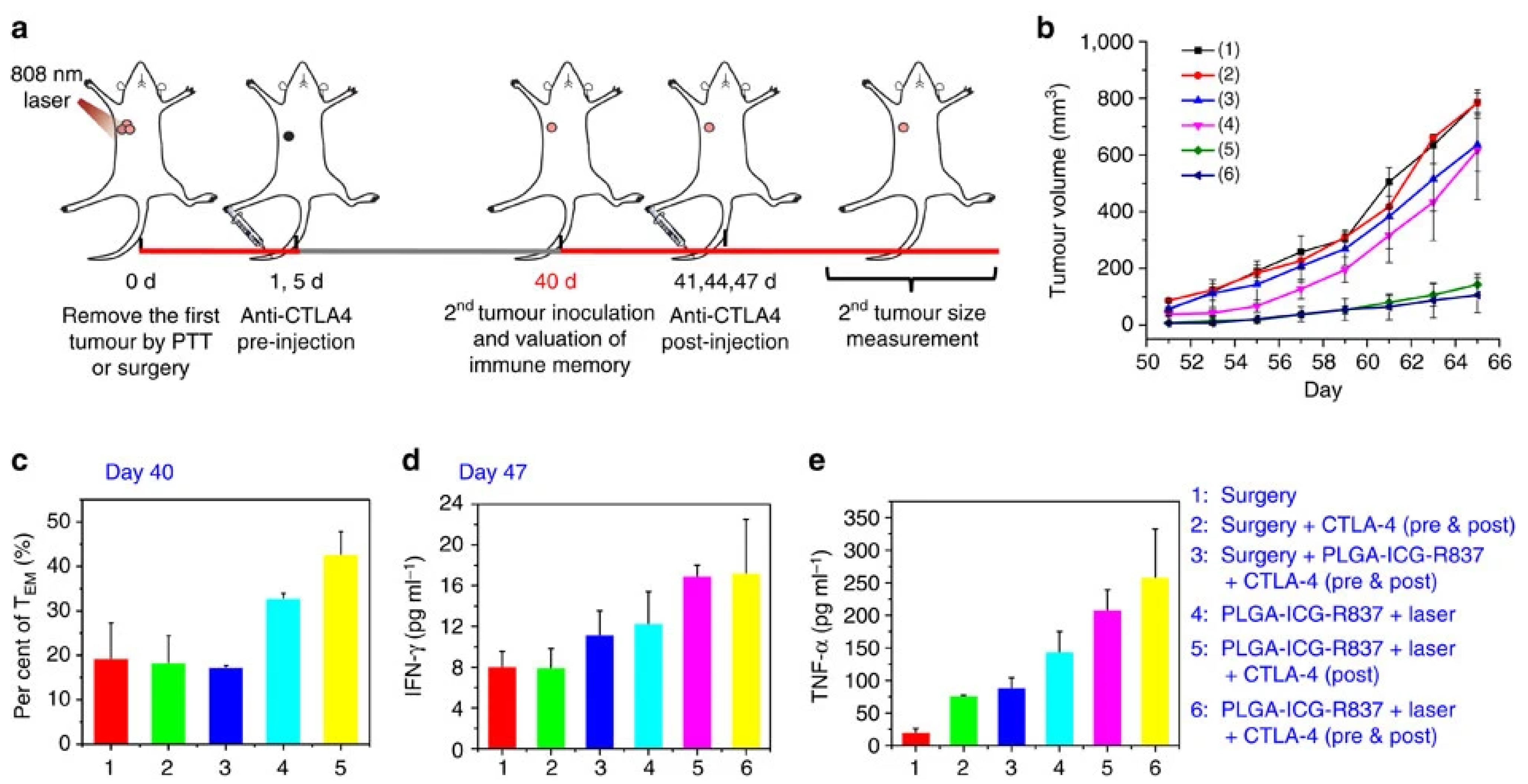
| R837- and ICG-loaded PLGA NPs | R837; Anti-CTLA-4 Ab (non-loaded) | ICG | R837- and ICG-loaded PLGA NPs + Laser induced 1.24 and 1.32 times higher mDC levels than the respective ICG-loaded PLGA NPs + Laser and R837- and ICG-loaded PLGA NPs (in the tumor-draining lymph nodes). | [38] |
| R837- and ICG-loaded PLGA NPs + Laser + anti-CTLA-4 Ab induced 1.40, 15.42, 10.15, 5.63, and 3.05 times higher CTLs/Treg ratios than surgery + R837- and ICG-loaded PLGA NPs + anti-CTLA-4 Ab, ICG-loaded PLGA NPs + Laser, surgery + R837- and ICG-loaded PLGA NPs, surgery + anti-CTLA-4 Ab, and surgery, respectively (in the secondary tumor). | ||||
| R837- and ICG-loaded PLGA NPs + Laser + anti-CTLA-4 Ab (post tumor reinoculation) resulted in 1.31, 2.50, 2.26, and 2.24 times higher TEM cells levels than R837- and ICG-loaded PLGA NPs + Laser, surgery + R837 and ICG-loaded PLGA NPs + anti-CTLA-4 Ab (pre and post tumor reinoculation), surgery + anti-CTLA-4 Ab (pre and post tumor reinoculation), and surgery, respectively (in the spleen). | ||||
| ICG and RB (a)-loaded DSPE (b)-PEG-mal (c) functionalized UCNPs (d) | - | ICG; RB | ICG- and RB-loaded DSPE-PEG-mal functionalized UCNPs + Laser induced 1.94 and 3.02 times higher mDC levels than ICG- and RB-loaded DSPE-PEG functionalized UCNPs + Laser, and the control + Laser, respectively (in the primary tumor). | [114] |
| ICG- and RB-loaded DSPE-PEG-mal functionalized UCNPs + Laser induced 3.10 and 5.69 times higher CTLs/Treg ratios than ICG- and RB-loaded DSPE-PEG functionalized UCNPs + Laser, and the control + Laser, respectively (in the secondary tumor); ICG- and RB-loaded DSPE-PEG-mal functionalized UCNPs + Laser induced 1.14 and 1.46 times higher CTLs levels than ICG- and RB-loaded DSPE-PEG functionalized UCNPs + Laser, and the control + Laser, respectively (in the spleen). | ||||
| Mg and ICG-loaded PES NPs | - | ICG | Mg and ICG-loaded PES NPs + Laser induced two times higher mDC levels than Mg and ICG-loaded PES NPs, and PES (in the primary tumor); Mg and ICG-loaded PES NPs + Laser induced 2.27 times higher mDC levels than the control (in the primary tumor); Mg and ICG-loaded PES NPs + Laser induced about two times higher mDC levels than Mg and ICG-loaded PES NPs, PES, and the control (in lymph nodes). | [32] |
| Mg and ICG-loaded PES NPs + Laser induced about 2.72 times higher CTLs levels than Mg and ICG-loaded PES NPs, PES, and the control (in the secondary tumor). | ||||
| ICG-loaded COF (e) coated with ovalbumin | Anti-PD-L1 Ab (non-loaded) | ICG; COF | ICG-loaded COF coated with ovalbumin + Laser + anti-PD-L1 Ab induced 1.31, 1.82, and 2.22 times higher mDC levels than ICG-loaded COF coated with ovalbumin + Laser, PBS + anti-PD-L1 Ab, and the control, respectively (in lymph nodes). | [73] |
| ICG-loaded COF coated with ovalbumin + Laser + anti-PD-L1 Ab induced 1.29, 2.05, and 2.51 times higher CTLs levels than ICG-loaded COF coated with ovalbumin + Laser, PBS + anti-PD-L1 Ab, and the control, respectively (in the primary tumor). | ||||
| ICG-loaded liposome (f) | - | ICG | ICG-loaded liposome + Laser induced 3.29 times higher CTLs/Treg ratios than the control (in the secondary tumor). | [113] |
| ICG-loaded PEG-Epacadostat conjugate NPs | Epacadostat; Anti-PD-L1 Ab (non-loaded) | ICG | ICG-loaded PEG-Epacadostat conjugate NPs + Laser induced 2.47, 2.27, and 3.83 times higher mDC levels than ICG-loaded PEG-Epacadostat conjugate NPs, PEG-Epacadostat conjugate NPs, and the control, respectively (in lymph nodes). | [85] |
| ICG-loaded PEG-Epacadostat conjugate NPs + Laser + anti-PD-L1 Ab induced 1.91, 2.10, 8.17, 6.10, and 6.81 times higher CTLs/Treg ratios than ICG-loaded PEG-Epacadostat conjugate NPs + anti-PD-L1 Ab, ICG-loaded PEG-Epacadostat conjugate NPs + Laser, ICG-loaded PEG-Epacadostat conjugate NPs, anti-PD-L1 Ab, and the control, respectively (in the secondary tumor). | ||||
| ICG-loaded lipid (g)-PLGA NPs decorated with FimH (h) | FimH | ICG | ICG-loaded lipid-PLGA NPs decorated with FimH + Laser, and FimH + Laser induced about three times higher mDC levels than ICG-loaded lipid-PLGA NPs + Laser, lipid-PLGA NPs + Laser, and the control, respectively (in lymph nodes). | [115] |
| ICG-loaded PLGA based NPs incorporated into decitabine, DSPE-PEG, and cell membranes based NPs | Decitabine | ICG | ICG-loaded PLGA based NPs incorporated in decitabine, DSPE-PEG, and cell membrane-based NPs + Laser induced 1.74, 3.28, 15.24, 7.38, and 12.63 times higher mDC levels than ICG-loaded PLGA based decitabine lipidic NPs + Laser, ICG + Decitabine + Laser, Decitabine + Laser, ICG + Laser, and the control, respectively (in the primary tumor); ICG-loaded PLGA-based NPs incorporated in decitabine, DSPE-PEG, and cell membrane-based NPs + Laser induced 1.73, 5.20, 8.27, 10.82, and 12.06 times higher mDC levels than ICG-loaded PLGA based decitabine lipidic NPs + Laser, ICG + Decitabine + Laser, Decitabine + Laser, ICG + Laser, and the control, respectively (in tumor-draining lymph nodes). | [116] |
| ICG-loaded PLGA-based NPs incorporated in decitabine, DSPE-PEG, and cell membrane-based NPs + Laser induced 2, 3.86, 6, 4.50, and 4.93 times higher CTLs levels than ICG-loaded PLGA based decitabine lipidic NPs + Laser, ICG + Decitabine + Laser, Decitabine + Laser, ICG + Laser, and the control, respectively (in the secondary tumor); ICG-loaded PLGA-based NPs incorporated in decitabine, DSPE-PEG, and cell membrane-based NPs + Laser induced 1.52, 2.56, 4.27, 3.37, and 5.49 times higher CTLs levels than ICG-loaded PLGA-based decitabine lipidic NPs + Laser, ICG + Decitabine + Laser, Decitabine + Laser, ICG + Laser, and the control, respectively (in the spleen). | ||||
| CAT (i), DTA-1 (j) and ICG functionalized PDA (k) NPs | CAT | ICG; PDA | CAT, DTA-1, and ICG functionalized PDA NPs + Laser induced 2.17, 2.48, 2.74, and 2.47 times higher mDC levels than ICG-functionalized PDA NPs + Laser, ICG + Laser, PDA + Laser, and the control, respectively (in spleen). | [117] |
| CAT, DTA-1, and ICG-functionalized PDA NPs + Laser induced 2.89, 2.03, 2.89, and 3.68 times higher CTLs/Treg ratios than ICG-functionalized PDA NPs + Laser, ICG + Laser, PDA + Laser, and the control, respectively (in the primary tumor). | ||||
| ICG-loaded Mn@CaCO3 NPs functionalized with siPD-L1 (l) and PAH (m) | siPD-L1 | ICG | ICG-loaded Mn@CaCO3 NPs functionalized with siPD-L1 and PAH + Laser induced 2.61, 4.50, 5.06, and 9 times higher mDC levels than ICG-loaded Mn@CaCO3 NPs + Laser, ICG-loaded Mn@CaCO3 NPs functionalized with siPD-L1 and PAH, ICG-loaded Mn@CaCO3 NPs, and the control, respectively (in the primary tumor). | [118] |
| ICG-loaded Mn@CaCO3 NPs functionalized with siPD-L1 and PAH + Laser induced 2, 20, 26.67, and 80 times higher CTLs levels than ICG-loaded Mn@CaCO3 NPs + Laser, ICG-loaded Mn@CaCO3 NPs functionalized with siPD-L1 and PAH, ICG-loaded Mn@CaCO3 NPs, and the control, respectively (in the primary tumor). | ||||
| Anti-PD-L1 Ab and ICG-loaded PEG-PLGLAG (n)-dEGCG (o) NPs | Anti-PD-L1 Ab | ICG | Anti-PD-L1 Ab and ICG-loaded PEG-PLGLAG-dEGCG NPs + Laser induced 2.18, 1.25, 2.95, 1.29, 3.25, 2.60 and 3.53 times higher mDC levels than Anti-PD-L1 Ab and ICG-loaded PEG-PLGLAG-dEGCG NPs, Anti-PD-L1 Ab, and ICG-loaded EGCG NPs + Laser, Anti-PD-L1 Ab, and ICG-loaded EGCG NPs, ICG-loaded PEG-PLGLAG-dEGCG NPs + Laser, ICG-loaded PEG-PLGLAG-dEGCG NPs, Anti-PD-L1 Ab, and the control, respectively (in the primary tumor); Anti-PD-L1 Ab and ICG-loaded PEG-PLGLAG-dEGCG NPs + Laser induced 2.15, 1.26, 1.44, 5.68, 2.79, and 7 times higher mDC levels than Anti-PD-L1 Ab and ICG-loaded PEG-PLGLAG-dEGCG NPs, Anti-PD-L1 Ab and ICG-loaded EGCG NPs + Laser, ICG-loaded PEG-PLGLAG-dEGCG NPs + Laser, ICG-loaded PEG-PLGLAG-dEGCG NPs, Anti-PD-L1 Ab, and the control, respectively (in lymph nodes of lymphatic metastases). | [107] |
| Anti-PD-L1 Ab and ICG-loaded PEG-PLGLAG-dEGCG NPs + Laser induced 2.77, 2.03, 6.91, 2.54, 10.46, 4.07, and 10.77 times higher CTLs/Treg ratio than Anti-PD-L1 Ab and ICG-loaded PEG-PLGLAG-dEGCG NPs, Anti-PD-L1 Ab, and ICG-loaded EGCG NPs + Laser, Anti-PD-L1 Ab and ICG-loaded EGCG NPs, ICG-loaded PEG-PLGLAG-dEGCG NPs + Laser, ICG-loaded PEG-PLGLAG-dEGCG NPs, Anti-PD-L1 Ab, and the control, respectively (in tumor-infiltrating lymphocytes); Anti-PD-L1 Ab and ICG-loaded PEG-PLGLAG-dEGCG NPs + Laser induced 2.15, 2.67, 2.26, 5.46, 2.84, and 4.40 times higher CTLs levels than Anti-PD-L1 Ab and ICG-loaded PEG-PLGLAG-dEGCG NPs, Anti-PD-L1 Ab, and ICG-loaded EGCG NPs + Laser, ICG-loaded PEG-PLGLAG-dEGCG NPs + Laser, ICG-loaded PEG-PLGLAG-dEGCG NPs, Anti-PD-L1 Ab, and the control, respectively (in lymph nodes). | ||||
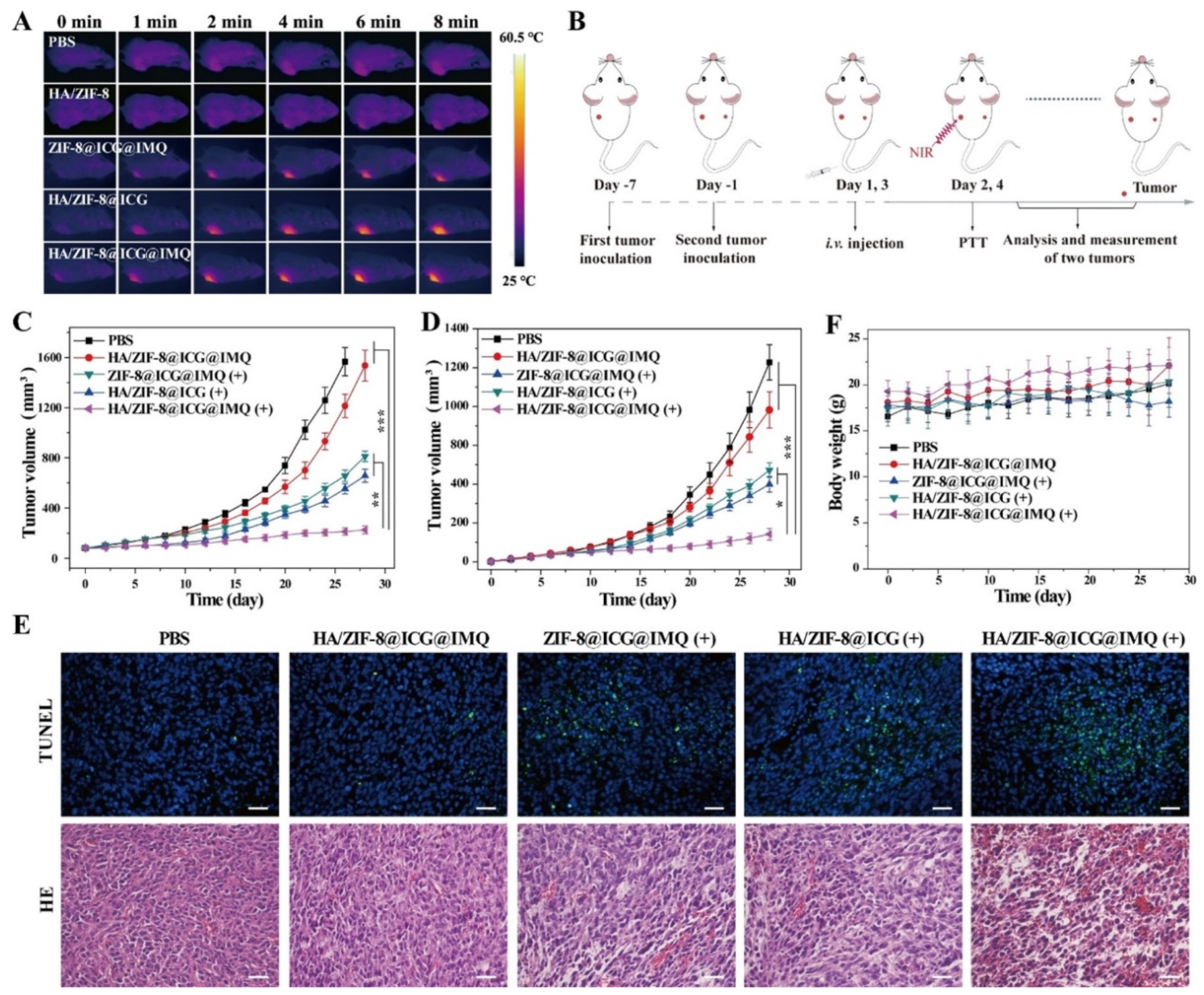
| ICG and R837-loaded HA functionalized ZIF-8 NPs | R837 | ICG | ICG- and R837-loaded HA-functionalized ZIF-8 NPs + Laser induced 1.44, 1.35, 1.31, and 1.82 times higher mDC levels than ICG-loaded HA-functionalized ZIF-8 NPs + Laser, ICG and R837-loaded ZIF-8 NPs + Laser, ICG- and R837-loaded HA-functionalized ZIF-8 NPs, and the control, respectively (in lymph nodes). | [108] |
| ICG- and R837-loaded HA functionalized ZIF-8 NPs + Laser induced about 1.50 times higher CTLs levels than ICG-loaded HA-functionalized ZIF-8 NPs + Laser, ICG- and R837-loaded ZIF-8 NPs + Laser, and ICG- and R837-loaded HA- functionalized ZIF-8 NPs (in the primary tumor); ICG- and R837-loaded HA-functionalized ZIF-8 NPs + Laser induced 3.11 times higher CTLs levels than control (in the primary tumor). | ||||
| ICG- and R837-loaded HA-functionalized ZIF-8 NPs + Laser generated 1.67, 1.69, 2.27, and 3.45 times higher memory T cells levels than ICG-loaded HA-functionalized ZIF-8 NPs + Laser, ICG- and R837-loaded ZIF-8 NPs + Laser, ICG- and R837--loaded HA-functionalized ZIF-8 NPs, and the control, respectively (in the spleen). | ||||
| ICG and R837-loaded PEG-polyphenols functionalized Fe3O4 based NPs | R837 | ICG | ICG- and R837-loaded PEG-polyphenol-functionalized Fe3O4-based NPs + Laser induced 1.37, 1.21, 2.06, and 2.00 times higher mDC levels than R837-loaded PEG-polyphenol-functionalized Fe3O4-based NPs + Laser, ICG-loaded PEG-polyphenol-functionalized Fe3O4-based NPs + Laser, PEG-polyphenol-functionalized Fe3O4-based NPs + Laser, and the control, respectively (in lymph nodes). | [69] |
| ICG- and R837-loaded PEG-polyphenol-functionalized Fe3O4-based NPs + Laser induced about 1.18 times higher CTLs levels than R837-loaded PEG-polyphenol-functionalized Fe3O4-based NPs + Laser, and ICG-loaded PEG-polyphenol-functionalized Fe3O4-based NPs + Laser (in the primary tumor); ICG- and R837-loaded PEG-polyphenol-functionalized Fe3O4-based NPs + Laser induced 1.41 and 4.13 times higher CTLs levels than PEG-polyphenol-functionalized Fe3O4-based NPs + Laser, and the control, respectively (in the primary tumor). | ||||
| ICG and PM (p)-loaded albumin MnO2 NPs | PM; MnO2 | ICG | ICG- and PM-loaded albumin MnO2 NPs + Laser induced 1.23, 1.93, and 2.73 times higher CTLs levels than ICG- and PM-loaded albumin MnO2 NPs, ICG-loaded albumin MnO2 NPs + Laser, and the control, respectively (in the primary tumor). | [110] |
| FAL (q)-PEG-TA (r) and PEI (s)-ICG functionalized AuNS (t) and Hb (u)-loaded FAL liposomes (v) | Hb | ICG; AuNS | FAL-PEG-TA- and PEI-ICG-functionalized AuNS and Hb-loaded FAL liposomes + Laser induced 1.25, 1.56, 4, 1.73, and 3.76 times higher mDC levels than FAL-PEG-TA- and PEI-ICG-functionalized AuNS + Laser, PEI-ICG-functionalized AuNS + Hb-loaded liposomes + Laser, PEI-ICG-functionalized AuNS + Laser, FAL-PEG-TA-functionalized AuNS + Laser, and the control, respectively (in lymph nodes). | [76] |
| FAL-PEG-TA- and PEI-ICG-functionalized AuNS and Hb-loaded FAL liposomes + Laser induced 1.56, 2, 2.19, 1.04, and 2.80 times higher CTLs levels than FAL-PEG-TA- and PEI-ICG-functionalized AuNS + Laser, PEI-ICG-functionalized AuNS + Hb-loaded liposomes + Laser, PEI-ICG-functionalized AuNS + Laser, FAL-PEG-TA-functionalized AuNS + Laser, and the control, respectively (in splenic lymphocytes); FAL-PEG-TA- and PEI-ICG-functionalized AuNS and Hb-loaded FAL liposomes + Laser induced 1.80 and 1.75 times lower Treg levels than PEI-ICG-functionalized AuNS + Hb-loaded liposomes + Laser, and the control, respectively (in spleens). | ||||
| ICG-loaded PLGA NPs incorporated in EPV (w) | EPV | ICG | ICG-loaded PLGA NPs incorporated in EPV + Laser induced 1.14, 1.22, 1.80, 1.23, 1.59, 1.62, 2, and 2.15 times higher CTLs levels than ICG-loaded PLGA NPs incorporated in Melanoma membrane vesicles + Laser, ICG-loaded PLGA NPs incorporated in Salmonella membrane vesicles + Laser, ICG-loaded PLGA NPs + Laser, ICG-loaded PLGA NPs incorporated in EPV, ICG-loaded PLGA NPs incorporated in Melanoma membrane vesicles, ICG-loaded PLGA NPs incorporated in Salmonella membrane vesicles, ICG-loaded PLGA NPs, and the control, respectively (in the primary tumor). | [119] |
| ICG, R837, and CTL-Ap (x)-loaded dextran NPs | R837; CTL-Ap | ICG | ICG-, R837-, and CTL-Ap-loaded dextran NPs + Laser induced 2.41 times higher CTLs levels than non-irradiated NPs (in the primary tumor). | [109] |
| ICG and R837-IONs (y)-loaded DSPE-PEG NPs | R837; IONs | ICG | ICG- and R837-ION-loaded DSPE-PEG NPs + Laser induced 1.80 times higher CTLs/Treg ratios than ICG- and ION-loaded DSPE-PEG NPs + Laser (in the primary tumor); ICG- and R837-ION-loaded DSPE-PEG NPs + Laser induced about 3.20 times higher CTLs/Treg ratios than ICG- and R837-ION-loaded DSPE-PEG NP-loaded DSPE-PEG NPs, control + Laser, and the control (in the primary tumor). | [99] |
| Formulation | Immuno Therapy Agent | PTT/ PDT Agent | Therapeutic Effect and Memory | Ref |
|---|---|---|---|---|
| R837- and ICG-loaded PLGA NPs | R837; Anti-CTLA-4 Ab (non-loaded) | ICG | R837- and ICG-loaded PLGA NPs + Laser + Anti-CTLA-4 Ab caused primary tumor eradication; R837- and ICG-loaded PLGA NPs + Laser + Anti-CTLA-4 Ab caused the greatest secondary tumor growth reduction; Metastases after R837- and ICG-loaded PLGA NPs + Anti-CTLA-4 Ab + Laser treatment decrease compared to control. | [38] |
| Tumor-bearing mice previously treated with R837- and ICG-loaded PLGA NPs + anti-CTLA-4 Ab + Laser have reinoculated tumors with the slowest growth. | ||||
| ICG- and RB-loaded DSPE-PEG-mal functionalized UCNPs | Anti-CTLA-4 Ab (non-loaded) | ICG; RB | ICG- and RB-loaded DSPE-PEG-mal functionalized UCNPs + Laser with and without Anti-CTLA-4 Ab caused primary tumor eradication while the other treatment groups only caused tumor growth reduction; ICG- and RB-loaded DSPE-PEG-mal functionalized UCNPs + Laser + Anti-CTLA-4 Ab caused the strongest secondary tumor growth reduction; Metastases decrease after ICG- and RB-loaded DSPE-PEG-mal functionalized UCNPs + Laser + Anti-CTLA-4 Ab treatment. | [114] |
| Tumor-bearing mice previously treated with ICG- and RB-loaded DSPE-PEG-mal functionalized UCNPs with Anti-CTLA-4 Ab + Laser have reinoculated tumors with the slowest growth. | ||||
| Mg and ICG-loaded PES NPs | - | ICG | Mg and ICG-loaded PES NPs + Laser caused tumor regression while the other treatment groups only caused tumor growth reduction; Mg and ICG-loaded PES NPs + Laser caused a great secondary tumor growth reduction compared to the other treatment groups; The number of metastatic nodules after Mg and ICG-loaded PES NPs + Laser treatment strongly decreases compared to control (3.39 vs. 41.53). | [32] |
| ICG-loaded COF coated with ovalbumin | Anti-PD-L1 Ab (non-loaded) | ICG; COF | ICG-loaded COF coated with ovalbumin + Laser, with and without Anti-PD-L1 Ab both caused primary tumor eradication; ICG-loaded COF coated with ovalbumin + Laser + Anti-PD-L1 Ab caused secondary tumor eradication while the other treatment groups only caused tumor growth reduction. | [73] |
| Metastases after ICG-loaded COF coated with ovalbumin + Laser + Anti-PD-L1 Ab do not occur in mice after tumor reinoculation. | ||||
| ICG-loaded liposome | Anti-PD-1 Ab (non-loaded); Anti-TIM-3 Ab (non-loaded) | ICG | ICG-loaded liposome + Laser caused primary tumor eradication; ICG-loaded liposome + Laser + anti-PD-1 Ab + anti-TIM-3 Ab caused the strongest secondary tumor growth inhibition while the other treatment groups only caused tumor growth reduction. | [113] |
| ICG-loaded PEG-Epacadostat conjugate NPs | Epacadostat; Anti-PD-L1 Ab (non-loaded) | ICG | ICG-loaded PEG-Epacadostat conjugate NPs + Laser + Anti-PD-L1 Ab caused primary tumor eradication while the other treatment groups only caused tumor growth reduction; ICG-loaded PEG-Epacadostat conjugate NPs + Laser + Anti-PD-L1 Ab caused the strongest secondary tumor growth reduction. | [85] |
| R837-loaded PEG-ICG Derivative (a)-Cholic Acid based NPs (b) | R837; Anti-PD-1 Ab (non-loaded) | ICG derivative | R837-loaded PEG-ICG Derivative-Cholic Acid and PEG-Cysteine-Lysine-Cholic Acid based NPs + Laser + Anti-PD-1 Ab caused primary and secondary tumor eradication while the other treatment groups only caused tumor growth reduction. | [39] |
| ICG-loaded lipid-PLGA NPs decorated with FimH | FimH | ICG | ICG-loaded lipid-PLGA NPs decorated with FimH + Laser caused primary tumor eradication while the other treatment groups only caused tumor growth reduction. | [115] |
| Metastases after ICG-loaded lipid-PLGA NPs decorated with FimH + Laser treatment do not occur in mice after tumor reinoculation. | ||||
| CpG ODNs-loaded ICG functionalized MOF | CpG ODNs | ICG | CpG-loaded ICG functionalized MOF + Laser caused primary tumor eradication while the other treatment groups only caused tumor growth reduction. | [120] |
| Metastases after CpG-loaded ICG functionalized MOF + Laser treatment decrease in mice after tumor reinoculation. | ||||
| ICG and poly I:C (c)-loaded liposomes (d) | poly I:C | ICG | ICG and poly I:C-loaded liposomes + Laser. and ICG-loaded liposomes + Laser caused primary tumor regression while the other treatment groups do not reduce tumor growth. | [121] |
| Metastases after ICG and poly I:C-loaded liposomes + Laser treatment strongly decrease compared to control in mice after tumor reinoculation. | ||||
| ICG-loaded PLGA based NPs incorporated into decitabine, DSPE-PEG, and cell membranes based NPs | Decitabine | ICG | ICG-loaded PLGA based NPs incorporated in decitabine, DSPE-PEG, and cell membranes based NPs + Laser caused primary tumor regression while the other treatment groups only reduce tumor growth; ICG-loaded PLGA based NPs incorporated in decitabine, DSPE-PEG, and cell membranes based NPs + Laser caused secondary tumor growth inhibition while the other treatment groups only caused tumor growth reduction. | [116] |
| CAT, DTA-1 and ICG functionalized PDA NPs | CAT | ICG; PDA | CAT, DTA-1, and ICG functionalized PDA NPs + Laser caused primary tumor regression while the other treatment groups only caused tumor growth reduction; CAT, DTA-1, and ICG functionalized PDA NPs + Laser caused the strongest secondary tumor growth reduction. | [117] |
| ICG-loaded Mn@CaCO3 NPs functionalized with siPD-L1 and PAH | siPD-L1 | ICG | ICG-loaded siPD-L1 and PAH functionalized Mn@CaCO3 NPs + Laser caused primary tumor regression while the other treatment groups only caused tumor growth reduction. | [118] |
| Anti-PD-L1 Ab and ICG-loaded PEG-PLGLAG-dEGCG NPs | Anti-PD-L1 Ab | ICG | Anti-PD-L1 Ab and ICG-loaded PEG-PLGLAG-dEGCG NPs + Laser caused primary tumor growth inhibition while the other treatment groups only caused tumor growth reduction; Metastases after Anti-PD-L1 Ab and ICG-loaded PEG-PLGLAG-dEGCG NPs + Laser treatment do not occur (nodules: 0 vs. 16.17, treated vs. control). | [107] |
| Tumor-bearing mice previously treated with Anti-PD-L1 Ab and ICG-loaded PEG-PLGLAG-dEGCG NPs have reinoculated tumors with the slowest growth. | ||||
| ICG and R837-loaded HA functionalized ZIF-8 NPs | R837 | ICG | ICG and R837-loaded HA functionalized ZIF-8 NPs + Laser caused the strongest primary and secondary tumor growth reduction. | [108] |
| Tumor-bearing mice previously treated with ICG and R837-loaded HA functionalized ZIF-8 NPs + Laser have reinoculated tumors with the slowest growth. | ||||
| ICG and R837-loaded PEG-polyphenols functionalized Fe3O4 based NPs | R837 | ICG | ICG and R837-loaded PEG-polyphenols functionalized Fe3O4 based NPs + Laser caused the strongest primary tumor growth reduction. | [69] |
| Tibia and lung metastases after ICG and R837-loaded PEG-polyphenols functionalized Fe3O4 based NPs + Laser treatment strongly decrease in mice after tumor reinoculation compared to the other treatment groups. | ||||
| ICG and PM-loaded albumin MnO2 NPs | PM; MnO2 | ICG | ICG and PM-loaded albumin MnO2 NPs caused the strongest primary and secondary tumor growth reduction. | [110] |
| FAL-PEG-TA and PEI-ICG functionalized AuNS and Hb-loaded FAL liposomes | Hb | ICG; AuNS | FAL-PEG-TA and PEI-ICG functionalized AuNS and Hb-loaded FAL liposomes + Laser caused the strongest primary tumor growth reduction. | [76] |
| ICG-loaded PLGA NPs incorporated in EPV | EPV | ICG | ICG-loaded PLGA NPs incorporated in EPV + Laser caused the strongest primary tumor growth reduction. | [119] |
| ICG, R837, and CTL-Ap-loaded dextran NPs | R837; CTL-ap | ICG | ICG, R837, and CTL-Ap-loaded dextran NPs + Laser caused the strongest primary tumor growth reduction. | [109] |
| ICG and R837-IONs-loaded DSPE-PEG NPs | R837; IONs | ICG | The number of metastatic nodules after ICG and R837-IONs-loaded DSPE-PEG NPs + Laser treatment severely decreases compared to control (3.46 vs. 22.30). | [99] |
| Formulation | Immuno Therapy Agent | PTT/ PDT Agent | Changes in the Levels of mDCs and T Cells | Ref |
|---|---|---|---|---|
| Met- (a) and IR775-loaded liposomes (b) | Met | IR775 | Met- and IR775-loaded liposomes + Laser induced 2.5, 2.21, 2.14, 3.96, and 4.49 times higher CTLs levels than Met- and IR775-loaded liposomes, Met-loaded liposomes, IR775-loaded liposomes + Laser, control + Laser, and the control, respectively (in the primary tumor); Met- and IR775-loaded liposomes + Laser induced 2.27, 4.17, 2.21, 4.69, and 4.69 times higher CTLs levels than Met and IR775-loaded liposomes, Met-loaded liposomes, IR775-loaded liposomes + Laser, control + Laser, and the control, respectively (in the secondary tumor). | [72] |
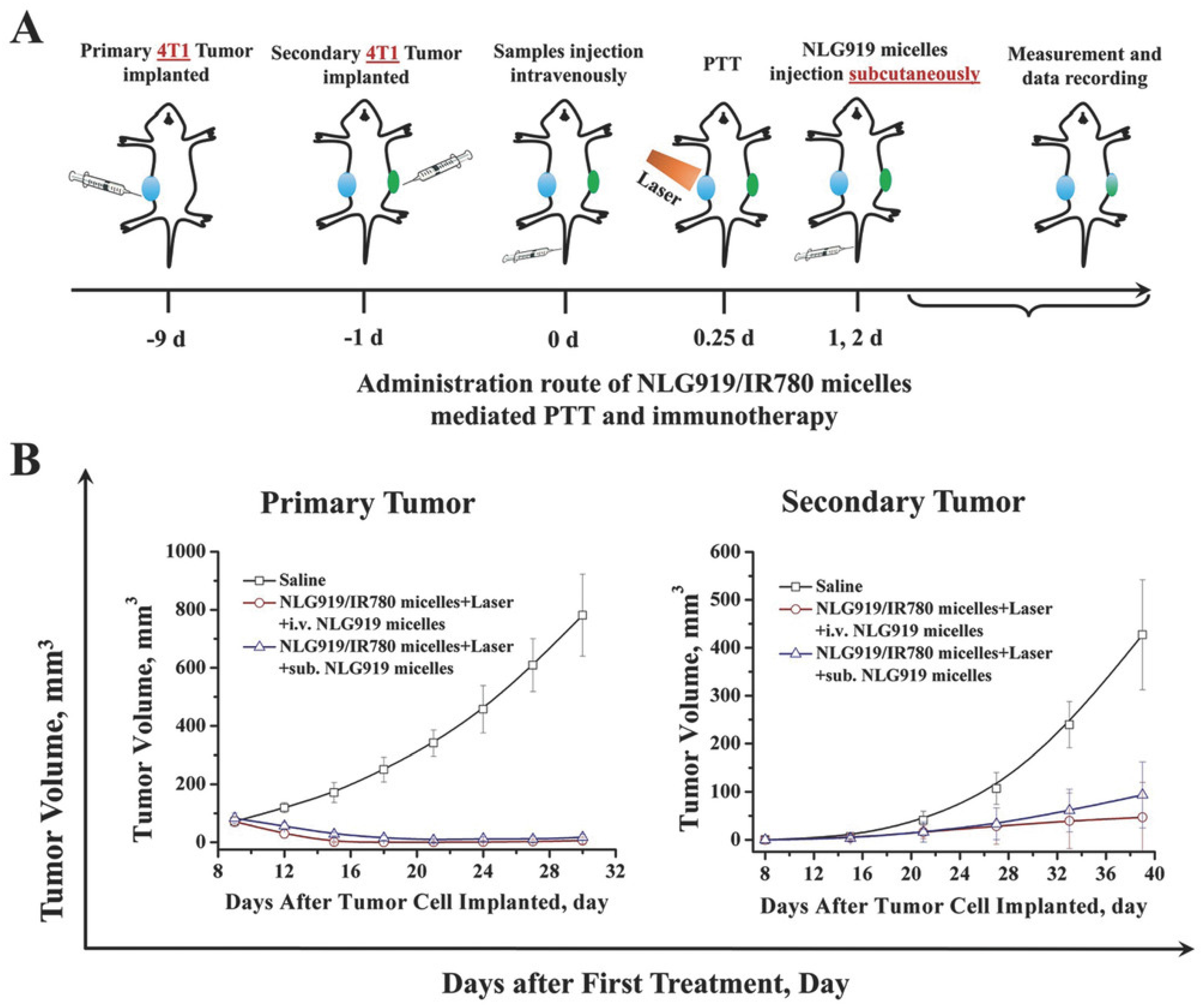
| NLG919- and IR780-loaded PEG-PCL micelles | NLG919 | IR780 | NLG919- and IR780-loaded PEG-PCL micelles + Laser induced 6.79, 33.12, 11.04, and 43.72 times higher CTLs/Treg ratio than NLG919 and IR780-loaded PEG-PCL micelles, IR780-loaded PEG-PCL micelles + Laser, NLG919-loaded PEG-PCL micelles, and the control, respectively (in spleen). | [106] |
| IR780 and Imatinib-loaded PEGylated GITR-functionalized PLGA based NPs | Imatinib | IR780 | IR780- and Imatinib-loaded PEGylated GITR-functionalized PLGA based NPs + Laser induced about two times higher mDC levels than IR780 + Laser, Imatinib, control + Laser, and the control, respectively (in the primary tumor). | [71] |
| IR780- and Imatinib-loaded PEGylated GITR-functionalized PLGA-based NPs + Laser induced 2.54, 2.34, 3.25, and 3.38 times lower Treg levels than IR780 + Laser, Imatinib, control + Laser, and the control, respectively (in the primary tumor). | ||||
| IR780- and Met-loaded CeO2-capped MSNs (c) | Met; CeO2 | IR780 | IR780- and Met-loaded CeO2-capped MSNs + Laser induced 1.06, 1.39, 2.10, and 1.58 times higher CTLs levels than IR780 and Met-loaded MSNs + Laser, IR780-loaded CeO2-capped MSNs + Laser, IR780-loaded MSNs + Laser, and the control, respectively (in the primary tumor). | [88] |
| BMS- (d)-loaded IR780-PEGylated lipidic (e) NPs | BMS | IR780 | BMS-loaded IR780-PEGylated lipidic NPs + Laser induced about 1.30 times higher mDC levels than IR780-PEGylated lipidic NPs + Laser, BMS-loaded lipidic NPs + Laser, and BMS + Laser (in lymph nodes); BMS-loaded IR780-PEGylated lipidic NPs + Laser induced 1.9 times higher mDC levels than the control (in lymph nodes). | [33] |
| BMS-loaded IR780-PEGylated lipidic NPs + Laser induced 1.69, 2, 2.34, and 3.58 times higher CTLs levels than IR780-PEGylated lipidic NPs + Laser, BMS-loaded lipidic NPs + Laser, BMS + Laser, and the control, respectively (in the primary tumor). | ||||
| IR780- and SB-505124-loaded liposomes (f) | SB-505124 | IR780 | IR780- and SB-loaded liposomes + Laser induced 1.61, 1.16, 1.76, and 2.24 times higher CTLs levels than IR780-loaded liposomes + Laser, IR780 and SB-loaded liposomes, SB, and the control, respectively (in the primary tumor); IR780- and SB-loaded liposomes + Laser induced 1.91, 1.22, 1.68, and 2.28 times lower Treg levels than IR780-loaded liposomes + Laser, IR780 and SB-loaded liposomes, SB, and the control, respectively (in the primary tumor). | [74] |
| IR780-loaded PEG-PCL NPs | - | IR780 | IR780-loaded PEG-PCL NPs + Laser induced 1.22 and 1.44 times higher mDC levels than IR780 + Laser, and the control, respectively (in the primary tumor). | [122] |
| IR780-loaded PEG-PCL NPs + Laser induced 1.44 and 2 times higher CTLs levels than IR780 + Laser, and the control, respectively (in the primary tumor). | ||||
| IR797-loaded DSPE-PEG NPs coated with mDCs membranes (g) | mDCs membranes | IR797 | IR797-loaded DSPE-PEG NPs coated with mDCs membranes + Laser induced 1.21 and 2.05 times higher mDC levels than IR797-loaded DSPE-PEG NPs coated with mDCs membranes and IR797-loaded DSPE-PEG NPs + Laser, respectively (in lymph nodes); IR797-loaded DSPE-PEG NPs coated with mDCs membranes + Laser induced about 2.60 times higher mDC levels than IR797-loaded DSPE-PEG NPs and the control (in lymph nodes). | [65] |
| IR797-loaded DSPE-PEG NPs coated with mDCs membranes + Laser induced 1.26 times higher CTLs levels than IR797-loaded DSPE-PEG NPs coated with mDCs membranes (in the primary tumor); IR797-loaded DSPE-PEG NPs coated with mDCs membranes + Laser induced about 4.8 times higher CTLs levels than IR797-loaded DSPE-PEG NPs + Laser, IR797-loaded DSPE-PEG NPs, and the control (in the primary tumor); IR797-loaded DSPE-PEG NPs coated with mDCs membranes + Laser induced 1.31, 2.48, 2.77, and 3.79 times higher CTLs levels than IR797-loaded DSPE-PEG NPs coated with mDCs membranes, IR797-loaded DSPE-PEG NPs + Laser, IR797-loaded DSPE-PEG NPs, and the control, respectively (in the secondary tumor). | ||||
| IR820-loaded HA functionalized MOF NPs (h), and R837 and 1MT-loaded mannan functionalized MOF NPs (h) | R837; 1MT | IR820 | IR820-loaded HA functionalized MOF NPs + R837 and 1MT-loaded mannan functionalized MOF NPs + Laser induced 2.32, 1.96, and 10.22 times higher CTLs/Treg ratios than IR820-loaded HA functionalized MOF NPs + Laser, R837 and 1MT-loaded mannan functionalized MOF NPs, and the control, respectively (in the primary tumor); IR820-loaded HA functionalized MOF NPs + R837 and 1MT-loaded mannan functionalized MOF NPs + Laser induced 3.84, 3.80, and 6.82 times higher CTLs/Treg ratios than IR820-loaded HA functionalized MOF NPs + Laser, R837 and 1MT-loaded mannan functionalized MOF NPs, and the control, respectively (in splenic lymphocytes); IR820-loaded HA functionalized MOF NPs + R837 and 1MT-loaded mannan functionalized MOF NPs + Laser induced 2.71, 2, and 10 times higher CTLs/Treg ratios than IR820-loaded HA functionalized MOF NPs + Laser, R837 and 1MT-loaded mannan functionalized MOF NPs, and the control, respectively (in the secondary tumor). | [37] |
| IR820-loaded HA functionalized MOF NPs + R837 and 1MT-loaded mannan functionalized MOF NPs + Laser generate 2.29, 1.75, and 4 times higher memory T cells levels than HA-functionalized MOF NPs + Laser, R837 and 1MT-loaded mannan-functionalized MOF NPs, and the control, respectively (in splenic lymphocytes). | ||||
| 1MT-IR820 NPs | 1MT; Anti-PD-L1 Ab (non-loaded) | IR820 | 1MT-IR820 NPs + Laser + Anti-PD-L1 Ab induced 1.18, 1.40, 1.51, 1.92, and 2.38 times higher mDC levels than 1MT-IR820 NPs + Laser, IR820 + Laser, Anti-PD-L1 Ab, 1MT, and the control, respectively (in lymph nodes). | [75] |
| 1MT-IR820 NPs + Laser + Anti-PD-L1 Ab induced 1.54, 1.97, 3.55, 4.73, and 6.45 times higher CTLs/Treg ratio than 1MT-IR820 NPs + Laser, IR820 + Laser, Anti-PD-L1 Ab, 1MT, and the control, respectively (in the primary tumor); 1MT-IR820 NPs + Laser + Anti-PD-L1 Ab induced 1.35, 1.64, 1.97, 2.42, and 3.38 times higher CTLs levels than 1MT-IR820 NPs + Laser, IR820 + Laser, Anti-PD-L1 Ab, 1MT, and the control, respectively (in the secondary tumor). | ||||
| 1MT-IR820 NPs + Laser + Anti-PD-L1 Ab generated 1.19, 1.36, 1.73, 1.93, and 2.24 times higher TEM cells levels than 1MT-IR820 NPs + Laser, IR820 + Laser, Anti-PD-L1 Ab, 1MT, and the control, respectively (in spleens). |
| Formulation | Immuno Therapy Agent | PTT/ PDT Agent | Therapeutic Effect and Memory | Ref |
|---|---|---|---|---|
| Met- and IR775-loaded liposomes | Met | IR775 | Met- and IR775-loaded liposomes + Laser caused the strongest primary and secondary tumor growth reduction. | [72] |
| NLG919- and IR780-loaded PEG-PCL micelles | NLG919 | IR780 | NLG919- and IR780-loaded PEG-PCL micelles + Laser caused primary tumor eradication while the other treatment groups only caused tumor growth reduction; NLG919- and IR780-loaded PEG-PCL micelles + Laser caused the strongest secondary tumor growth reduction; Metastases after NLG919- and IR780-loaded PEG-PCL micelles + Laser treatment decreased compared to the control. | [106] |
| IR780- and Imatinib-loaded PEGylated GITR-functionalized PLGA-based NPs | Imatinib | IR780 | IR780 and Imatinib-loaded PEGylated GITR-functionalized PLGA-based NPs caused primary tumor eradication while the other treatment groups only caused tumor growth reduction. | [71] |
| IR780- and Met-loaded CeO2-capped MSNs | Met; CeO2 | IR780 | IR780- and Met-loaded CeO2-capped MSNs + Laser caused primary tumor regression while the other treatment groups only caused tumor growth reduction; The number of metastatic nodules after IR780 and Met-loaded CeO2-capped MSNs + Laser treatment severely decreased compared to control (about 5 vs. 58). | [88] |
| BMS-loaded IR780-PEGylated lipidic NPs | BMS | IR780 | BMS-loaded IR780-PEG lipidic NPs + Laser caused primary tumor regression while other treatment groups only caused tumor growth reduction; The number of metastatic nodules after BMS-loaded IR780-PEG lipidic NPs + Laser treatment severely decreased compared to the control (about 4.60 vs. 52). | [33] |
| IR780- and SB-505124-loaded liposomes | SB-505124; Anti-PD-1 Ab (non-loaded) | IR780 | IR780- and SB-loaded liposomes + Laser + Anti-PD-1 caused primary tumor growth inhibition while the other treatment groups only caused tumor growth reduction; IR780- and SB-loaded liposomes + Laser + Anti-PD-1 caused the strongest secondary tumor growth reduction; The number of metastatic nodules after IR780 and SB-loaded liposomes + Laser + Anti-PD-1 treatment severely decreased compared to the control (about 2.80 vs. 38). | [74] |
| IR780-loaded PEG-PCL NPs | - | IR780 | IR780-loaded PEG-PCL NPs + Laser induced primary tumor growth reduction; Metastases after IR780-loaded PEG-PCL NPs + Laser treatment decreased compared to the control. | [122] |
| IR797-loaded DSPE-PEG NPs coated with mDC membranes | mDCs membranes | IR797 | IR797-loaded DSPE-PEG NPs coated with mDCs membranes + Laser caused primary tumor eradication while other treatment groups only caused tumor growth reduction; IR797-loaded DSPE-PEG NPs coated with mDCs membranes + Laser caused the strongest secondary tumor growth reduction. | [65] |
| IR820-loaded HA-functionalized MOF NPs, and R837- and 1MT-loaded mannan-functionalized MOF NPs | R837; 1MT | IR820 | IR820-loaded HA-functionalized MOF NPs + R837 and 1MT-loaded mannan-functionalized MOF NPs + Laser caused primary tumor regression while the other treatment groups only caused tumor growth reduction; IR820-loaded HA-functionalized MOF NPs + R837 and 1MT-loaded mannan-functionalized MOF NPs + Laser caused the strongest secondary tumor growth reduction. | [37] |
| Metastases after IR820-loaded HA-functionalized MOF NPs + R837 and 1MT-loaded mannan-functionalized MOF NPs + Laser treatment strongly decreased in mice after tumor reinoculation compared to the control. | ||||
| AG (a)-IR820 conjugate, Quercetin-loaded liposomes (b), and LPS (c) | LPS (non-loaded) | IR820 | AG-IR820 + Quercetin-loaded liposomes + Laser + LPS caused primary tumor regression while the other treatment groups only caused tumor growth reduction. | [124] |
| 1MT-IR820 NPs | 1MT; Anti-PD-L1 Ab (non-loaded) | IR820 | 1MT-IR820 NPs + Laser + Anti-PD-L1 Ab caused the strongest primary and secondary tumor growth reduction. | [75] |
| Metastases after 1MT-IR820 NPs + Laser + Anti-PD-L1 Ab treatment strongly decreased in mice after tumor reinoculation compared to the control. | ||||
| IR820-loaded DSPE-PEG-TPP (d) and DSPE-PEG-CpG ODNs-functionalized GO (e) | DSPE-PEG-CpG | GO; IR820 | IR820-loaded DSPE-PEG-TPP and DSPE-PEG-CpG ODNs-functionalized GO + Laser induced the strongest primary tumor growth reduction. | [95] |
Publisher’s Note: MDPI stays neutral with regard to jurisdictional claims in published maps and institutional affiliations. |
© 2022 by the authors. Licensee MDPI, Basel, Switzerland. This article is an open access article distributed under the terms and conditions of the Creative Commons Attribution (CC BY) license (https://creativecommons.org/licenses/by/4.0/).
Share and Cite
Alves, C.G.; Lima-Sousa, R.; Melo, B.L.; Moreira, A.F.; Correia, I.J.; de Melo-Diogo, D. Heptamethine Cyanine-Loaded Nanomaterials for Cancer Immuno-Photothermal/Photodynamic Therapy: A Review. Pharmaceutics 2022, 14, 1015. https://doi.org/10.3390/pharmaceutics14051015
Alves CG, Lima-Sousa R, Melo BL, Moreira AF, Correia IJ, de Melo-Diogo D. Heptamethine Cyanine-Loaded Nanomaterials for Cancer Immuno-Photothermal/Photodynamic Therapy: A Review. Pharmaceutics. 2022; 14(5):1015. https://doi.org/10.3390/pharmaceutics14051015
Chicago/Turabian StyleAlves, Cátia G., Rita Lima-Sousa, Bruna L. Melo, André F. Moreira, Ilídio J. Correia, and Duarte de Melo-Diogo. 2022. "Heptamethine Cyanine-Loaded Nanomaterials for Cancer Immuno-Photothermal/Photodynamic Therapy: A Review" Pharmaceutics 14, no. 5: 1015. https://doi.org/10.3390/pharmaceutics14051015
APA StyleAlves, C. G., Lima-Sousa, R., Melo, B. L., Moreira, A. F., Correia, I. J., & de Melo-Diogo, D. (2022). Heptamethine Cyanine-Loaded Nanomaterials for Cancer Immuno-Photothermal/Photodynamic Therapy: A Review. Pharmaceutics, 14(5), 1015. https://doi.org/10.3390/pharmaceutics14051015