Review

Potential Novel Food-Related and Biomedical Applications of Nanomaterials Combined with Bacteriocins

Department of Chemistry and Chemistry Institute for Functional Materials, Pusan National University, Busan 46241, Korea
*
Author to whom correspondence should be addressed.
Pharmaceutics 2021, 13(1), 86; https://doi.org/10.3390/pharmaceutics13010086
Submission received: 11 December 2020 / Revised: 8 January 2021 / Accepted: 8 January 2021 / Published: 11 January 2021
(This article belongs to the Special Issue Drug Delivery for Anti-Infective Agents)

Abstract

:
Bacteriocins are antimicrobial peptides or proteinaceous materials produced by bacteria against pathogens. These molecules have high efficiency and specificity and are equipped with many properties useful in food-related applications, such as food preservatives and additives, as well as biomedical applications, such as serving as alternatives to current antibacterial, antiviral, anticancer, and antibiofilm agents. Despite their advantages as alternative therapeutics over existing strategies, several limitations of bacteriocins, such as the high cost of isolation and purification, narrow spectrum of activity, low stability and solubility, and easy enzymatic degradation, need to be improved. Nanomaterials are promising agents in many biological applications. They are widely used in the conjugation or decoration of bacteriocins to augment the activity of bacteriocins or reduce problems related to their use in biomedical applications. Therefore, bacteriocins combined with nanomaterials have emerged as promising molecules that can be used in various biomedical applications. This review highlights the features of bacteriocins and their limitations in biomedical applications and provides a detailed overview of the uses of different nanomaterials in improving the limitations. Our review focuses on the potential applications of nanomaterials combined with bacteriocins as new designer molecules for use in future therapeutic strategies.

Graphical Abstract

1. Introduction

Bacteriocins are a group of ribosomally synthesized peptides that are secreted extracellularly by various gram-positive and gram-negative bacteria [1], although a majority of bacteriocins reported are produced by the former, especially lactic acid bacteria (LAB) [2,3]. Extensive studies have been carried out on bacteriocins owing to their excellent antibacterial activity, which is closely associated with strain producing species. In addition, bacteriocins have garnered considerable research attention in the field of biomedicine owing to their generally recognized as safe (GRAS) status, and because they are safe for human consumption due to their degradation by gastrointestinal proteases [4]. They are also being modified to improve the antibacterial spectrum. Most of the well-known bacteriocins are produced by gram-positive bacteria, whereas only a few from gram-negative bacteria have been characterized [5]. The activity of these small bacteriocins consisting of cationic molecules (30–60 amino acids) vary throughout the antibacterial spectrum, mainly due to their amphiphilic helices.
Bacteriocins are widely used as natural food preservatives—substances that delay the growth of microorganisms—in the food industry, because they are easily degraded by enzymes produced in the human gastrointestinal tract [4]. The high quality and safety profile of bacteriocins as natural food preservatives is possible without the use of chemical preservatives, which is strictly regulated by governmental agencies, such as the Food and Drug Administration (FDA) in the United States, owing to their safety issues. Generally, bacteriocins can be directly added to food or incorporated into food during cultivation with the help of bacteriocin-producing bacterial strains. More recently, bacteriocins have gained considerable attention in the healthcare industry as antibacterial and anticancer agents [6]. Some bacteriocins, such as nisin, have shown excellent and specific antibacterial activity against multi-drug resistant (MDR) strains of gram-positive bacteria, such as methicillin-resistant Staphylococcus aureus (MRSA) [7]. Moreover, gram-negative bacteria are naturally resistant to the action of bacteriocins produced by gram-positive bacteria, which are widely explored in foods. It is worthy to note that the bacteriocins produced by gram-positive bacteria can be combined with antibacterial nanoparticles to inhibit the growth of gram-negative bacteria such as P. aeruginosa. In this respect, Au-nisin successfully experimented for the bacterial growth inhibition of E. coli and P. aeruginosa [8]. Currently, researchers are investigating the biomedical properties of bacteriocins in several anticancer studies and have reported promising results [9]. Therefore, researchers are highly interested to use bacteriocins as safe materials in biomedical applications.
Despite their promising advantages, bacteriocins have certain limitations that hinder their applications in the field of biomedicine [10]. These limitations include the following: (i) easy degradation by proteolytic enzymes, (ii) constricted antibacterial spectrum, (iii) requirement of high dosage to kill MDR bacteria, (iv) high production cost, and (v) low natural production yield. Based on these limitations, only three FDA-approved bacteriocins (nisin, pediocin, and Micocin®) are available for food preservation and anti-spoilage processes [11]. The optimization of different production conditions, purification methods, and combinations with other antimicrobial agents or antibiotics has been assessed by various studies to overcome the aforementioned shortcomings in ensuring the broad application of bacteriocins in biomedicine. As adjuvant bioactive materials for bacteriocins, nanomaterials seem highly promising to overcome these challenges [12]. The major reasons for the use of nanomaterials can be attributed to their following characteristics: (i) broad antibacterial spectrum, (ii) stability in physiological solution, (iii) high surface area-to-volume ratio, (iv) easy to synthesize with low production cost, and (v) non-toxic nature at low concentrations. Bacteria are unable to generate rapid resistance against nanomaterials owing to their multi-dimensional approach compared to antibiotics, which targets a single cellular component [13]. Therefore, the combination of bacteriocins and nanomaterials offers new tactics to overcome the limitations posed by the sole use of bacteriocins in biomedical applications. In this review, we will focus on the advantages of bacteriocin–nanomaterial combinations in not only food-related applications, but also antibacterial and other biomedical applications.

2. Different Bacteriocins and Their Spectrum

It is necessary to determine the different types of bacteriocins based on their activity against closely related species. Generally, limited studies on bacteriocins from gram-negative bacteria have been conducted owing to their narrow spectrum of activity against closely related species [5]. Comparatively, the effect of gram-positive bacteriocins is much broader, and therefore, it has been studied extensively.
Gram-negative bacteriocins: One of the representative bacteriocins from gram-negative bacteria is colicin from Escherichia coli. It is a high molecular-weight protein that is widely used to control various gram-negative bacteria [14]. Another bacteriocin produced by E. coli is microcin, which is similar to gram-positive bacteriocins in terms of its pH and thermal stability along with protease resistance [15]. In addition, other gram-negative bacteria produce different bacteriocins (e.g., pyocins or aeruginocins from P. aeruginosa and klebicin from Klebsiella pneumonia) [5]. The limited spectrum of these gram-negative bacteriocins at times can be advantageous, since these bacteriocins can be used depending on a particular situation or for a specific purpose. However, this narrow antibacterial spectrum can be resolved with the use of nanomaterials, which will be discussed in later sections.
Gram-positive bacteriocins: Gram-positive bacteriocins possess broad antibacterial spectrum compared to gram-negative bacteriocins. The large spectrum is mainly attributed to the presence of a thick multilayered peptidoglycan wall instead of an outer membrane. This outer organization enables the penetration of small peptides without any receptor binding [5]. Gram-positive bacteriocins are largely produced by LAB. LAB bacteriocins are extensively used, because they have been accorded GRAS status by the U.S. FDA. Nisin and pediocin are well-known bacteriocins produced by gram-positive bacteria [16] and are already approved for commercial use by the FDA. Nisin, which is synthesized as a precursor peptide produced by Lactococcus lactis, contains 57 amino acids and has a molecular weight (MW) of 3354 daltons. Its mature form of 34 amino acids is further generated by post-translation modifications (Figure 1a). However, unlike nisin, pediocin PA-1 is produced as a mature form of 44 amino acids with MW of 4646.95 daltons and is not further processed by post-translation modification (Figure 1b).

3. Stability of Bacteriocins

Bacteriocins have potential biomedical applications considering their antibacterial activity in vitro [7,16,17,18,19]. They have a low in vivo stability and are vulnerable to proteolytic enzyme degradation, which hinders their clinical applicability [10]. Furthermore, bacteriocins are labile in tissues, serum, and organs (such as the liver and kidneys). They thus need to be modified or functionalized to ensure stability in physiological solutions. The stability of nisin, a frequently used variant for biomedical applications as an antibacterial agent or food preservative, is dependent on the environmental pH [20]. For instance, solubilized nisin is active and stable in acidic pH, whereas its solubility decreases in alkaline pH, which hinders its biological activity. Different functionalization methods have been reported to address stability for bacteriocins; cyclization by the incorporation of d-amino acids is one such process [21]. The stability of nisin (i.e., NisB and NisC) can also be increased by modifying unrelated peptides via the modification machinery used for the incorporation of lanthionine ring structures in nisin [22]. Peng et al. [23] enhanced the stability and bactericidal activity of nisin by conjugation with gellan gum. The resulting gellan–nisin conjugate exhibited excellent pH, heat, and chymotrypsin resistance compared with those of unmodified nisin. Similarly, Bagde et al. [24] reported enhanced bacteriocin stability via immobilization into cellulose nanocrystals. The bacteriocins extracted from Enterococcus faecium and immobilized on cellulose nanocrystals exhibited a 50% increase in stability. In another study, layered double hydroxide nanoparticles were used to improve the stability of the bacteriocin avicin A, which also enhanced its antibacterial activity [25]. Liposomes have also been used to encapsulate bacteriocins to make them more stable [26]. Malheiros et al. [27] reported the use of nanovesicles to encapsulate bacteriocin-like substances for enhanced stability and antilisterial activity. Chitosan nanoparticles are used to a considerable extent by many researchers to stabilize and enhance the biological activity of bacteriocins [28]. Nisin has also been encapsulated with poly-l-lactide (PLA) nanoparticles for potent and stable release of bacteriocins in food preservation [29]. Therefore, it is clear that future in vivo applications of bacteriocins are possible upon improvement in their stability via the use of nanomaterials.

4. Advantages and Limitations of Bacteriocins

Bacteriocins have several properties that render them advantageous for various biomedical applications; these include as antibacterial agents, drug delivery agents, and functional foods [6,21]. In terms of antibacterial activity, they present low toxicity to eukaryotic cells, stability in physiological solutions after functionalization, stability at high temperatures, and low minimum inhibitory concentrations against numerous bacterial strains. Bacteriocins also possess selective antimicrobial activity [30]. Thus, they are regarded as promising alternatives to [21] or synergistic materials for [31] currently used antibiotics. The synergistic property of bacteriocins allows lower usage of the antibiotic agent, thereby reducing in the risk of cytotoxic effects arising from its use. Furthermore, bacteriocins are generally protected from the acquisition of bacterial resistance. Therefore, it has the potential to reduce the development of MDR strains, as bacteriocins can be used as antibacterial agents either alone or together with nanomaterials instead of antibiotics. Bacteriocins capped with silver nanoparticles have reduced cytotoxicity [32]. Bacteriocins are also used for food preservation owing to their impressive in vitro and in situ effectiveness against various food-borne and human pathogens [4]. Three bacteriocins, nisin, pediocin, and Micocin®, have been approved by the FDA for use as food preservatives and antispoilage agents [11]. In terms of other biomedical applications, bacteriocins were effectively used for therapeutic drug delivery in anticancer treatment [6]. In this regard, Joo et al. [33] reported that nisin at low concentration (2.5%) effectively induces preferential apoptosis combined with cell-cycle arrest. It also reduces cell proliferation in head and neck squamous-cell carcinoma. Owing to these advantageous properties, bacteriocins have various biomedical applications and are used in catheter coatings [34], oral tablets [35], chewing gum [36], aquaculture dry sprays [37], hydrogels [38], scaffolds [39], and food packaging [40].
Despite the various potential biomedical applications of bacteriocins, their actual clinical use is limited. The primary issue is similar to that for antibiotics—bacteria can develop resistance against bacteriocins [41]. This needs to be resolved immediately and completely for its in vivo antibacterial applications to be feasible. The combination of nanoparticles with bacteriocins has the potential to resolve this issue, because bacteria do not develop rapid resistance against nanomaterials [13]. Another limitation of bacteriocins is their degradation by proteases [10]. Most reported bacteriocins are sensitive to proteases; they are rapidly degraded by proteases such as proteinase K and pepsin. This should be resolved for effective targeted drug delivery or use as antibacterial agents. The encapsulation of bacteriocins with nanomaterials is an effective way to resolve this issue [10,16]. Additionally, the toxicity of bacteriocins, specially nisin and pediocin, is debatable [42]. However, they are harmless for humans and animals when used as food preservatives. Overall, the limitations of bacteriocins need to be resolved to realize their immense potential for biomedical applications.

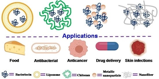
5. Bacteriocin–Nanomaterial Combination

As discussed in previous sections, certain limitations need to be resolved before bacteriocins can be used for various in vivo biomedical applications. These limitations include the following: (i) tendency towards degradation by proteolytic enzymes, (ii) limited antibacterial spectrum, (iii) failure to prevent the development of bacterial resistance, as with currently used antibiotics, and (iv) high production cost with low yield [10].
Nanomaterials have been recently used to potentially overcome such limitations [10,16]. For instance, researchers have already developed bacteriocin–nanomaterial complexes or bacteriocin–nanoconjugates for various biomedical applications of bacteriocins. Multiple advantages for the use of bacteriocin–nanoconjugates have been reported: (i) increased stability for long periods of use, (ii) protection from proteolytic enzyme degradation, and (iii) synergistic activity. The potential applicability of bacteriocin–nanoconjugates with different nanomaterials has been described in following sections.

5.1. Liposomes

Liposomes are spherical vesicles comprising single or multiple phospholipid bilayer membranes [43]. They are non-toxic and biodegradable agents as well as suitable encapsulating materials for both hydrophilic and hydrophobic substances. The size scale of liposomes varies from micrometers to nanometers, generated via sonication or functionalization [44]. Nano-sized liposomes—called nano-liposomes—are promising vehicles for the encapsulation and delivery of different bioactive compounds such as enzymes, vitamins, and food additives, as well as the delivery of therapeutic bacteriocins to target cells [16]. Liposome encapsulation protects bacteriocins (Figure 2) from degradation caused by physicochemical environment or protease action [45], susceptibility to which is a major limitation of bacteriocins. Various studies addressing the protection of bacteriocins from protease degradation have been reported [46,47]. Liposome encapsulation also confers other advantages, such as improved stability, reduced doses in therapeutic applications, and enhanced antibacterial spectrum [10,16]. Taylor et al. [48] showed that liposomes consisting of distearoylphosphatidylcholine (PC) and distearoylphosphatidylglycerol (PG) with trapped nisin can retain approximately 70–90% of the incorporated nisin with high stability under alkaline pH and elevated temperatures (25–75 °C). Similarly, Pinilla et al. [49] showed that nanoliposomes co-encapsulated with nisin and garlic extracts became broad-spectrum antimicrobial agents against L. monocytogenes, Salmonella enteritidis, E. coli, and S. aureus. This expanded antibacterial spectrum has shown that bacteriocin could be modulated to be active on both gram-positive and -negative bacteria. The successful application of liposome-encapsulated bacteriocins depends on appropriate phospholipid bacteriocin combinations, the avoidance of adverse liposome–bacteriocin interactions, and high purity of starting materials. Such liposome-encapsulated bacteriocins have been mainly used as antibacterial substances [49] and have food-related [50] applications.

5.2. Chitosan

Chitosan is another type of nanoparticle, which is widely used with bacteriocins in various biomedical applications. Chitosan is an ideal candidate for these applications due to its non-toxic, biocompatible, and biodegradable nature [51]. Moreover, its antibacterial and anticancer activity coupled with its ability to deliver drugs to their targets is well researched [51,52]. Many researchers have used chitosan with bacteriocins to obtain a material showing synergistic antibacterial activity. For instance, Namasivayam et al. [53] reported synergistic antibacterial activity of chitosan–nanoconjugates loaded with bacteriocins against Listeria monocytogenes; the activity levels of these nanoconjugates were higher than those of free bacteriocins. In another study, Alireza Alishahi [54] demonstrated excellent antibacterial activity by chitosan nanoparticles loaded with nisin against E. coli and S. aureus. Moreover, nanocomposite comprised of bacteriocin and chitosan have also been used in food packaging (Figure 3). For example, Divsalar et al. [55] formed a composite film containing chitosan, cellulose, and nisin for use in packaging of ultra-filtered cheese. The composite film showed better food packaging properties than chitosan and cellulose film alone. Similar studies have been conducted using bacteriocin–chitosan nanocomposites for drug delivery [56].

5.3. Metallic Nanoparticles

Currently, metallic nanoparticles such as zinc, copper, silver, and gold are being studied not only for their antibacterial activity, but also for their different potential biomedical applications [12,13,57,58,59,60]. The extensive use of these nanoparticles can be attributed to their large surface area along with their positive charge, which can interact with negatively charged bacterial cell surfaces [11]. Among bacteriocin–metallic nanocomposites, Ag and Au nanoparticles are the most studied materials, showing synergistic effects in biomedical applications [10,16]. The antibacterial activity of Ag and Au nanoparticles is well reported [12]. Therefore, it is easy to understand the rationale behind the combination of bacteriocins with Ag/Au nanoparticles. This combination will not only enlarge the antibacterial spectrum, but also reduce the toxicity of nanoparticles. In this respect, Sharma et al. [32] reported enterocin-coated silver nanoparticles, which not only showed broad-spectrum inhibition against various food-borne pathogenic bacteria but also admirable non-toxicity to red blood cells, emphasizing its biocompatible nature. Pandit et al. [61] also reported the antibacterial activity of Ag–nisin nanoconjugates against Listeria monocytogenes, S. aureus, Pseudomonas fluorescens, Aspergillus niger, and Fusarium moniliforme associated with food spoilage. These results reveal the potential uses of bacteriocin–metal nanoconjugates in food packaging. Preet et al. [62] also reported the use of Au nanoparticles in other biomedical applications such as the co-delivery of nisin and doxorubicin to treat murine skin cancer.

5.4. Nanofibers

Nanofiber technology is known for its application in wound care formulations, wherein nanofibers are loaded with antimicrobials and hemostatic agents for wound healing. Bacteriocins can also be loaded with nanofibers for the same purpose [10]. The large surface area, high physical stability, and excellent encapsulation ability along with small pore size have made nanofibers the perfect nanocarriers for target-specific drug delivery [63]. Nanofiber-based bacteriocin–nanoconjugates are used as antibacterial and antiviral substances. In a recent study, Torres et al. [64] explored the efficacy of a subtilosin-loaded poly(vinyl alcohol) (PVOH) nanofiber as an antiviral agent against Herpes simplex virus type 1 (HSV-1). Subtilosin with 200 µg mL-1 showed a remarkable virucidal and antiviral activity against HSV-1, although the mechanistic action has not been elucidated. The PVOH-based subtilosin nanofibers with a width of 278 nm has retained subtilosin’s antibacterial efficacy, increased the loading potential of subtilosin (2.4 mg subtilosin/g of fiber) with loading efficiency of 31.6%, and demonstrated non-toxicity to human epidermal tissues. Electrospinning-based bacteriocin-nanofibers have also been studied as a drug delivery system. For instance, Ahire et al. [65] determined the activity of nisin incorporated into nanofibers prepared from poly(D,L-lactide) (PDLLA) and poly (ethylene oxide) (PEO). This combination has shown enhanced antibiofilm activity against MRSA than that by nisin alone.

5.5. Other Nanoplatforms

Researchers are attempting to use nanoplatforms other than liposomes, chitosan, metallic nanoparticles, or nanofibers to encapsulate or conjugate bacteriocins with nanomaterials. Niaz et al. [66] used bacteriocin-loaded nanovesicles due to their excellent antimicrobial and antibiofilm activities against food-borne pathogens. Similarly, Breukink et al. [67] evaluated the binding of nisin Z to bilayer lipid vesicles. Yadav et al. [68] assessed the interaction between nisin and vesicles synthesized using different phospholipids. Another example of nanoplatforms is solid–lipid nanoparticles (SLN), which have been used to encapsulate bacteriocins for various biomedical applications [10]. Bacteriocin-loaded SLN not only protect bacteriocins from degradation but also extend their antibacterial activity for a long period of time [69]. Phytoglycogen nanoparticles are another type of nanomaterial used to carry bacteriocins to the target site [70].

6. Applications

Owing to the high potential of the combined use of bacteriocins and nanomaterials, researchers have now been screening for such combinations demonstrating antibacterial and anticancer activity, with potential applications in the food and biomedical industries. In this section, the applications of the bacteriocin–nanomaterial combination will be discussed.

6.1. Food Applications

Over the last decade, bacteriocins have received considerable attention from the scientific community owing to their applications in the food industry as bio-preservatives either alone or in combination with other methods [4,16]. Bacteriocins are used for chemical-free preservation, extension of shelf-life, and inhibition of food-borne pathogens during farming and food-processing stages [4]. Despite these advantages, the number of bacteriocins approved by the FDA as food preservatives and antispoilage agents is limited to three (nisin, pediocin, and Micocin®) [11]. The limited use of bacteriocins in the food industry can be attributed to its easy degradation, electrostatic repulsion, and uncontrolled interactions with various food components. To overcome these limitations, researchers have been exploring certain synergistic approaches to increase its applications. In this regard, nanomaterials have emerged as potential partners for bacteriocins, acting in combination with bacteriocins for excellent food-related applications (Table 1).
Nanoliposomes are well-known encapsulation systems for bacteriocins because they are biocompatible, possessing both hydrophilic and hydrophobic characteristics [4,16]. Encapsulation by nanoliposomes inhibits bacteriocins from undesirable interactions with food components, controls their release, and improves their antimicrobial activity. Recently, Niaz et al. [71] demonstrated the enhanced antimicrobial activity of nisin Z against MDR food-borne pathogens after its encapsulation within a nanoliposome. In a more practical approach, Pinilla et al. [50] successfully encapsulated nisin and garlic extract into phosphatidylcholine nanoliposomes and used the system to inhibit the microbial growth of L. monocytogenes, S. aureus, E. coli, and S. enteritidis in milk. Similarly, Zou et al. [72] reported that nisin-loaded liposomal carriers inhibit two main food-borne pathogens, L. monocytogenes and S. aureus. Liposomal encapsulation can prevent the degradation of bacteriocin by the proteases in food. Recently, researchers have also attempted to use this combination in food packaging after its success as a bio-preservative. The main challenges, such as sensitivity to environmental stimuli and uncontrolled release of the antimicrobial materials in food packaging, can be overcome or minimized by using nanocarrier-mediated bacteriocin in food packaging. Boelter et al. [73] and Imran et al. [74] encapsulated nisin into nanoliposomes to produce bio-nanocomposite films for use in active food packaging. Both reports showed effective antimicrobial activity against L. monocytogenes. However, further studies are required to investigate the more potent food-related applications of nanoliposomes.
Apart from liposomes, chitosan is another material used in many food-related applications in combination with bacteriocins. It is used for its properties such as biodegradability, biocompatibility, and antibacterial activity [51,52]. For example, Khan et al. [75] evaluated nisin-loaded chitosan-monomethyl fumaric acid nanoparticles as a food additive. The nanocomposite significantly reduced bacterial growth in orange juice compared to the other tested samples after 48 h of incubation. Similarly, Hui et al. [76] reported better preservation of large yellow croaker with combined use of chitosan and nisin than with chitosan alone. Moreover, synergistic antimicrobial activity can be seen for nisin, lysozyme, EDTA nanoparticles, and/or ZnO nanoparticles against food-borne pathogens in minced beef [77]. In another study, Chopra et al. [83] used chitosan/carrageenan nanocapsules with nisin for enhanced antimicrobial activity. These nisin-loaded nanocarriers demonstrated excellent controlled release and synergistic antimicrobial activity in tomato juice.
Metallic nanoparticles have been used with bacteriocins in food packaging owing to their broad antibacterial spectrum and non-toxic nature at low concentration. In this regard, Song et al. [78] evaluated iron oxide nanoparticles functionalized with nisin for antimicrobial activity of Alicyclobacillus spp. The nanocomposite demonstrated excellent antimicrobial activity against the experimented food-spoiling microbes. Similarly, Thirumurugan et al. [79] reported that the combination of bacteriocins and gold nanoparticles displayed antimicrobial activity against food-spoiling microorganisms. In this case, antibacterial activity of the bacteriocin in combination with Au nanoparticles was better than that of the bacteriocin alone.
Nanofibers are additional nanomaterial used to encapsulate bacteriocin in food [10]. High stability and large surface area with excellent encapsulation capacity have made nanofibers good candidates for encapsulating bacteriocins. Saini et al. [80] evaluated nisin-anchored cellulose nanofibers for food packaging applications. Similarly, Soto et al. [81] encapsulated nisin into amaranth-protein-isolate:pullulan (API:PUL) nanofibers for sustained release and enhanced antimicrobial activity against S. Typhimurium, L. monocytogenes, and L. mesenteroides in fresh cheese and apple juice. Cui et al. [82] effectively incorporated nisin into nanofibers to prevent the growth of L. monocytogenes in packaged cheese.

6.2. Antibacterial Activity

As discussed in previous sections, the main drawbacks in using bacteriocins for antibacterial activity are their limited antibacterial spectrum and the bacterial resistance against them [10,16]. However, these problems could be resolved with the help of nanomaterials. Nanomaterials have a broad antibacterial spectrum, and the chance of bacteria gaining resistance against them is low. Therefore, bacteriocin–nanomaterial combination can be the solution to the long-standing issues with bacteriocins. Liposomes, chitosan, nanofibers, and metallic nanomaterials are mainly used in conjugation with bacteriocins for different biomedical applications (Table 2), including antibacterial activity.
In this respect, Gruskiene et al. [92] displayed antibacterial activity of iron oxide magnetic nanoparticles functionalized by nisin against gram-positive bacteria. In a more recent study, Wang et al. [84] demonstrated antibacterial activity and cytotoxicity of nisin@PEGylated MoS2. The nanocomposite showed antibacterial activity against both gram-positive and -negative bacteria with the mechanism of ROS production and membrane disruption strategy. The toxicity of the nanocomposite was very low, which emphasize its biomedical potential. Many researchers have encapsulated bacteriocins with liposomes to protect bacteriocins from proteases. Malheiros et al. [85] showed excellent stability and antibacterial activity of liposome-encapsulated bacteriocins against the growth of L. monocytogenes. Prevention of dental caries is also possible using liposome-encapsulated nisin [45]. Similarly, García-Toledo et al. [86] investigated the anti-listerial activity of pediocin encapsulated with liposome with excellent synergistic activity.
Another widely used nanomaterial that shows synergistic activities with bacteriocins is chitosan nanoparticles. Zohri et al. [87] successfully investigated the synergistic antibacterial activity of chitosan nanoparticles loaded with nisin against S. aureus; they exhibited two-fold higher antimicrobial activity than nisin alone. In a similar study, Namasivayam et al. [53] showed synergistic antibacterial activity of chitosan–nanoconjugates loaded with bacteriocins against L. monocytogenes, where a larger zone of inhibition was observed for nanoconjugates than for free bacteriocins. Hu et al. [93] demonstrated the synergistic effect of chitosan and bacteriocins against E. coli AW1.7 and S. Typhimurium in lean beef.
Metallic nanoparticles are also used with bacteriocins against bacterial infections owing to their excellent antibacterial activity [10,16]. They are commonly used because of their large surface area and positive charge, which is already discussed in previous section. Ag nanoparticles are an excellent antibacterial agent with broad antibacterial spectrum. Therefore, the combination of Ag nanoparticles and bacteriocin will not only increase antibacterial spectrum of bacteriocin but also enhance the antibacterial activity of nisin. Several researchers have followed this principle for bacteriocin–Ag nanoparticle combination toward antibacterial activity [32,61]. Au nanoparticles also showed synergistic antibacterial activity with nisin [62]. Thirumurugan et al. [79] showed the combined effect of bacteriocins and Au nanoparticles against food-spoiling microbes where the combination worked better against the microorganisms than bacteriocin alone.
In addition to their ability to inhibit bacterial growth, nanomaterials have also been used as a vehicle for targeted drug delivery. Moreover, sustained antimicrobial activity can also be achieved through this process where the effect would be highly beneficial for one week or more in many applications, such as protective textiles, medicinal products, and food packaging. Encapsulation of bacteriocin in nanofiber may give sustained delivery of bacteriocin over an extended period of time. In this regard, nanofibers are often used for targeted delivery of bacteriocin in biomedical applications. Heunis et al. [39] effectively electrospun plantaricin 423 and bacteriocin ST4SA into nanofibers, which were made from various combinations of PDLLA and PEO. Such a combination retained 88% of its original antimicrobial activity at 37 °C. The efficient capacity of the carrier matrix for bacteriocins has demonstrated by these nanofibers and therefore, the carrier maxtrix can be further used for controlled antimicrobial delivery systems. Recently, Han et al. [88] showed that the nisin released from electrospun triaxial fiber membranes showed an antimicrobial activity against S. aureus for up to seven days. Furthermore, the triaxial fiber membranes provided comparatively robust and more sustained antimicrobial activity than that of other forms of electrospun membranes.

6.3. Anticancer Activity

Beyond their typical antibacterial activities, bacteriocins show anticancer activity in combination with nanomaterials [89]. Studies have shown that bacteriocins can impact tumor growth and exhibit selective cytotoxicity toward cancer cells [94]. Nisin is a normally used bacteriocin for its anticancer activity; however, its degradation when in contact with proteases hinders its application as an in vivo anticancer agent [10]. Its safety profile is another issue to be resolved. Along with these drawbacks, the limited knowledge available on bacteriocin pharmacokinetics and pharmacodynamics, which are the major requirements of any anticancer drug delivery system, is an obstacle to their use as anticancer agents. In recent times, nanotechnology can be used to overcome these drawbacks. Goudarzi et al. [89] showed the cytotoxic effects of nisin-loaded PLA-PEG-PLA nanoparticles on different cancer cell lines. The results revealed that, compared to free nisin, nisin-loaded nanoparticles had synergistic cytotoxic effects on all the cancer cell lines tested. The loading efficiency of the nanoparticles was calculated as 85–90%. In another study, Au nanoparticles were used for the co-delivery of nisin and doxorubicin for their anticancer activity against murine skin cancer [62]. After evaluating the in vivo therapeutic effects of nanoconjugates in DMBA (7,12-Dimethylbenz(a)anthracene) induced skin carcinogenesis, mean tumor volume(s) (26.3–52.6%) reduction can be seen at the end of therapy period (~7 weeks). Moreover, all tested cytokines (TNF-α {17.39–47.89%}; TNF-β {24.52–69.88%}; NF-κβ {19.74–27.33%}; {IL-1 25.37–52.59%} and IL-10 {31.91–62.76%}) seemed to be in decline in serum levels, whereas significant increase in tissue reactive oxygen species (ROS), lipid peroxides, and superoxide dismutase activities could be seen in various treatment groups by comparing with an untreated tumor group. Along with its biocompatible nature coupled with low doses, nisin could emerge as new ray of hope as an anticancer material.

6.4. Other Biomedical Applications

Similar to the aforementioned applications, bacteriocins have the potential for other biomedical applications. For example, a nisin-eluting nanofiber scaffold was effectively used to treat S. aureus-induced skin infections in mice with no adverse effects [90]. Similarly, Au nanoparticles formulated with bacteriocins exhibited potent activity against intestinal microsporidiosis in immunocompromised mice [91]. Further studies are required to realize the full potential of nano-combinations with bacteriocins for other biomedical applications.

7. Conclusions and Future Perspectives

The early results of experiments examining the interface between and combination of nanomaterials and bacteriocins seem promising. The various studies discussed here have emphasized the potential biomedical applications of this platform. The problems faced with the use of bacteriocins such as protease sensitivity, limited antibacterial spectrum, high dose, and toxicity issues have been thoroughly researched, and positive results have been obtained with the nanomaterial–bacteriocin combination. However, at present, the increased use of bacteriocins in the healthcare and pharmaceutical industries is not encouraging owing to the unavailability of proper in vivo experimental information or clinical study data. Therefore, more in vivo studies are required to ensure the practicality of the nanomaterial–bacteriocin combination. Further studies are also required on the combination of other antibacterial nanomaterials such as ZnO and CuO and typical Au and Ag nanoparticles along with some polymers with bacteriocins for biomedical applications. Another area that needs more attention is the toxic nature of the combinations. Hence, researchers should focus on strategies to eradicate these limitations so that the true potential of nanomaterial–bacteriocin combinations is realized. Therefore, this nanomaterial–bacteriocin platform is in its nascent stage, but it holds promise for biomedical applications.

Author Contributions

A.N.: conceptualization, investigation, data collection, formal analysis, writing—original draft; K.-s.K.: conceptualization, supervision, writing—reviewing and editing. All authors have read and agreed to the published version of the manuscript.

Funding

This work was supported by the National Research Foundation of Korea (NRF) grant funded by the Korean government (MSIT) [grant number 2018R1D1A1B07040941].

Conflicts of Interest

The authors declare no conflict of interest.

References

  1. Cotter, P.D.; Hill, C.; Ross, R.P. Bacteriocins: Developing innate immunity for food. Nat. Rev. Microbiol. 2005, 3, 777–788. [Google Scholar] [CrossRef] [PubMed]
  2. Hassan, M.; Kjos, M.; Nes, I.F.; Diep, D.B.; Lotfipour, F. Natural antimicrobial peptides from bacteria: Characteristics and potential applications to fight against antibiotic resistance. J. Appl. Microbiol. 2012, 113, 723–736. [Google Scholar] [CrossRef] [PubMed]
  3. Klaenhammer, T.R. Bacteriocins of lactic acid bacteria. Biochimie 1988, 70, 337–349. [Google Scholar] [CrossRef]
  4. Silva, C.C.G.; Silva, S.P.M.; Ribeiro, S.C. Application of bacteriocins and protective cultures in dairy food preservation. Front. Microbiol. 2018, 9, 594. [Google Scholar] [CrossRef] [PubMed]
  5. Ansari, A. Bacteriocin from LAB for medical and health applications. In Beneficial Microorganisms in Medical and Health Applications. Microbiology Monographs; Liong, M.T., Ed.; Springer: Cham, Switzerland, 2015; Volume 28, pp. 199–221. [Google Scholar]
  6. Shin, J.M.; Gwak, J.W.; Kamarajan, P.; Fenno, J.C.; Rickard, A.H.; Kapila, Y.L. Biomedical applications of nisin. J. Appl. Microbiol. 2016, 120, 1449–1465. [Google Scholar] [CrossRef] [PubMed] [Green Version]
  7. Field, D.; O’Connor, R.; Cotter, P.D.; Ross, R.P.; Hill, C. In Vitro activities of nisin and nisin derivatives alone and in combination with antibiotics against Staphylococcus biofilms. Front. Microbiol. 2016, 7, 508. [Google Scholar] [CrossRef] [PubMed] [Green Version]
  8. Vukomanović, M.; Žunič, V.; Kunej, Š.; Jančar, B.; Jeverica, S.; Podlipec, R.; Suvorov, D. Nano-engineering the antimicrobial spectrum of lantibiotics: Activity of nisin against gram negative bacteria. Sci. Rep. 2017, 7, 4324. [Google Scholar] [CrossRef]
  9. Baindara, P.; Korpole, S.; Grover, V. Bacteriocins: Perspective for the development of novel anticancer drugs. Appl. Microbiol. Biotechnol. 2018, 102, 10393–10408. [Google Scholar] [CrossRef]
  10. Fahim, H.A.; Khairalla, A.S.; El-Gendy, A.O. Nanotechnology: A valuable strategy to improve bacteriocin formulations. Front. Microbiol. 2016, 7, 1385. [Google Scholar] [CrossRef] [Green Version]
  11. Radaic, A.; de Jesus, M.B.; Kapila, Y.L. Bacterial anti-microbial peptides and nano-sized drug delivery systems: The state of the art toward improved bacteriocins. J. Control. Release 2020, 321, 100–118. [Google Scholar] [CrossRef]
  12. Naskar, A.; Kim, K.-s. Nanomaterials as delivery vehicles and components of new strategies to combat bacterial infections: Advantages and limitations. Microorganisms 2019, 7, 356. [Google Scholar] [CrossRef] [PubMed] [Green Version]
  13. Naskar, A.; Lee, S.; Lee, Y.; Kim, S.; Kim, K.-s. A new nano-platform of erythromycin combined with Ag nano-particle ZnO nano-structure against methicillin-resistant Staphylococcus aureus. Pharmaceutics 2020, 12, 841. [Google Scholar] [CrossRef] [PubMed]
  14. Micenková, L.; Bosák, J.; Kucera, J.; Hrala, M.; Dolejšová, T.; Šedo, O.; Linke, D.; Fišer, R.; Šmajs, D. Colicin Z, a structurally and functionally novel colicin type that selectively kills enteroinvasive Escherichia coli and Shigella strains. Sci. Rep. 2019, 9, 11127. [Google Scholar] [CrossRef] [PubMed]
  15. Eberhart, L.J.; Deringer, J.R.; Brayton, K.A.; Sawant, A.A.; Besser, T.E.; Call, D.R. Characterization of a novel microcin that kills enterohemorrhagic Escherichia coli O157:H7 and O26. Appl. Environ. Microbiol. 2012, 78, 6592–6599. [Google Scholar] [CrossRef] [Green Version]
  16. Sidhu, P.K.; Nehra, K. Bacteriocin-nanoconjugates as emerging compounds for enhancing antimicrobial activity of bacteriocins. J. King Saud Univ. Sci. 2019, 31, 758–767. [Google Scholar] [CrossRef]
  17. Yang, S.C.; Lin, C.H.; Sung, C.T.; Fang, J.Y. Antibacterial activities of bacteriocins: Application in foods and pharmaceuticals. Front Microbiol. 2014, 5, 241. [Google Scholar]
  18. Li, Q.; Montalban-Lopez, M.; Kuipers, O.P. Increasing the antimicrobial activity of nisin-based Lantibiotics against Gram-Negative pathogens. Appl. Environ. Microbial. 2018, 84, e00052-18. [Google Scholar] [CrossRef] [Green Version]
  19. Weishaupt, R.; Heuberger, L.; Siqueira, G.; Gutt, B.; Zimmermann, T.; Maniura-Weber, K.; Salentinig, S.; Faccio, G. Enhanced antimicrobial activity and structural transitions of a nanofibrillated cellulose-nisin biocomposite suspension. ACS Appl. Mater. Interfaces 2018, 10, 20170–20181. [Google Scholar] [CrossRef]
  20. Holcapkova, P.; Raskova, Z.K.; Hrabalikova, M.; Salakova, A.; Drbohlav, J.; Sedlarik, V. Isolation and thermal stabilization of bacteriocin nisin derived from whey for antimicrobial modifications of polymers. Int. J. Polym. Sci. 2017, 2017, 3072582. [Google Scholar] [CrossRef] [Green Version]
  21. Wang, G. Post-translational modifications of natural antimicrobial peptides and strategies for peptide engineering. Curr. Biotechnol. 2012, 1, 72–79. [Google Scholar] [CrossRef]
  22. Lagedroste, M.; Reiners, J.; Smits, S.H.J.; Schmitt, L. Impact of the nisin modification machinery on the transport kinetics of NisT. Sci. Rep. 2020, 10, 12295. [Google Scholar] [CrossRef] [PubMed]
  23. Peng, X.; Zhu, L.; Wang, Z.; Zhan, X. Enhanced stability of the bactericidal activity of nisin through conjugation with gellan gum. Int. J. Biol. Macromol. 2020, 148, 525–532. [Google Scholar] [CrossRef] [PubMed]
  24. Bagde, P.; Vigneshwaran, N. Improving the stability of bacteriocin extracted from Enterococcus faecium by immobilization onto cellulose nanocrystals. Carbohydr. Polym. 2019, 209, 172–180. [Google Scholar] [CrossRef]
  25. Fahim, H.A.; El Rouby, W.M.A.; El-Gendy, A.O.; Khairalla, A.S.; Naguib, I.A.; Farghali, A.A. Enhancement of the productivity of the potent bacteriocin avicin A and improvement of its stability using nanotechnology approaches. Sci. Rep. 2017, 7, 10604. [Google Scholar] [CrossRef] [Green Version]
  26. Da Silva Malheiros, P.; Daroit, D.J.; Brandelli, A. Food applications of liposome-encapsulated antimicrobial peptides. Trends Food Sci. Technol. 2010, 21, 284–292. [Google Scholar] [CrossRef]
  27. Da Silva Malheiros, P.; Sant’Anna, V.; Utpott, M.; Brandelli, A. Antilisterial activity and stability of nanovesicle-encapsulated antimicrobial peptide P34 in milk. Food Control 2012, 23, 42–47. [Google Scholar] [CrossRef] [Green Version]
  28. Lee, E.H.; Khan, I.; Oh, D.H. Evaluation of the efficacy of nisin-loaded chitosan nanoparticles against foodborne pathogens in orange juice. J. Food Sci. Technol. 2018, 55, 1127–1133. [Google Scholar] [CrossRef]
  29. Salmaso, S.; Elvassore, N.; Bertucco, A.; Lante, A.; Caliceti, P. Nisin-loaded poly-L-lactide nano-particles produced by CO2 anti-solvent precipitation for sustained antimicrobial activity. Int. J. Pharm. 2004, 287, 163–173. [Google Scholar] [CrossRef]
  30. Simons, A.; Alhanout, K.; Duval, R.E. Bacteriocins, antimicrobial peptides from bacterial origin: Overview of their biology and their impact against multidrug-resistant bacteria. Microorganisms 2020, 8, 639. [Google Scholar] [CrossRef]
  31. Mathur, H.; Field, D.; Rea, M.C.; Cotter, P.D.; Hill, C.; Ross, R.P. Bacteriocin-antimicrobial synergy: A medical and food perspective. Front. Microbiol. 2017, 8, 1205. [Google Scholar] [CrossRef] [Green Version]
  32. Sharma, T.K.; Sapra, M.; Chopra, A.; Sharma, R.; Patil, S.D.; Malik, R.K.; Pathania, R.; Navani, N.K. Interaction of bacteriocin-capped slver nanoparticles with food pathogens and their antibacterial effect. Int. J. Green Nanotechnol. 2012, 4, 93–110. [Google Scholar] [CrossRef]
  33. Joo, N.E.; Ritchie, K.; Kamarajan, P.; Miao, D.; Kapila, Y.L. Nisin, an apoptogenic bacteriocin and food preservative, attenuates HNSCC tumorigenesis via CHAC1. Cancer Med. 2012, 1, 295–305. [Google Scholar] [CrossRef]
  34. Ray Mohapatra, A.; Jeevaratnam, K. Inhibiting bacterial colonization on catheters: Antibacterial and antibiofilm activities of bacteriocins from Lactobacillus plantarum SJ33. J. Glob. Antimicrob. Resist. 2019, 19, 85–92. [Google Scholar] [CrossRef]
  35. Hegarty, J.W.; Guinane, C.M.; Ross, R.P.; Hill, C.; Cotter, P.D. Bacteriocin production: A relatively unharnessed probiotic trait? F1000Research 2016, 5, 2587. [Google Scholar] [CrossRef]
  36. Gillor, O.; Etzion, A.; Riley, M.A. The dual role of bacteriocins as anti- and probiotics. Appl. Microbiol. Biotechnol. 2008, 81, 591–606. [Google Scholar] [CrossRef] [PubMed] [Green Version]
  37. Chauhan, A.; Singh, R. Probiotics in aquaculture: A promising emerging alternative approach. Symbiosis 2019, 77, 99–113. [Google Scholar] [CrossRef]
  38. Flynn, J.; Durack, E.; Collins, M.N.; Hudson, S.P. Tuning the strength and swelling of an injectable polysaccharide hydrogel and the subsequent release of a broad spectrum bacteriocin, nisin A. J. Mater. Chem. B. 2020, 8, 4029–4038. [Google Scholar] [CrossRef] [PubMed]
  39. Heunis, T.; Bshena, O.; Klumperman, B.; Dicks, L. Release of bacteriocins from nanofibers prepared with combinations of poly(d,l-lactide) (PDLLA) and poly(ethylene oxide) (PEO). Int. J. Mol. Sci. 2011, 12, 2158–2173. [Google Scholar] [CrossRef]
  40. Woraprayote, W.; Pumpuang, L.; Tosukhowong, A.; Zendo, T.; Sonomoto, K.; Benjakul, S.; Visessanguan, W. Antimicrobial biodegradable food packaging impregnated with Bacteriocin 7293 for control of pathogenic bacteria in pangasius fish fillets. LWT 2018, 89, 427–433. [Google Scholar] [CrossRef]
  41. Bastos Mdo, C.; Coelho, M.L.; Santos, O.C. Resistance to bacteriocins produced by Gram-positive bacteria. Microbiology 2015, 161, 683–700. [Google Scholar] [CrossRef] [Green Version]
  42. Dicks, L.M.T.; van Staden, A.D.P.; Klumperman, B. Bacteriocins and nanotechnology. In Functionalized Nanomaterials for the Management of Microbial Infection; Boukherroub, R., Szunerits, S., Drider, D., Eds.; Elsevier: Amsterdam, The Netherlands, 2017; pp. 267–288. [Google Scholar]
  43. Panahi, Y.; Farshbaf, M.; Mohammadhosseini, M.; Mirahadi, M.; Khalilov, R.; Saghfi, S.; Akbarzadeh, A. Recent advances on liposomal nanoparticles: Synthesis, characterization and biomedical applications. Artif. Cells Nanomed. Biotechnol. 2017, 45, 788–799. [Google Scholar] [CrossRef] [PubMed] [Green Version]
  44. Lujan, H.; Griffin, W.C.; Taube, J.H.; Sayes, C.M. Synthesis and characterization of nanometer-sized liposomes for encapsulation and microRNA transfer to breast cancer cells. Int. J. Nanomed. 2019, 14, 5159–5173. [Google Scholar] [CrossRef] [PubMed] [Green Version]
  45. Tsumori, H.; Shimizu, Y.; Nagatoshi, K.; Sakurai, Y.; Yamakami, K. Prospects for liposome-encapsulated nisin in the prevention of dental caries. In Interface Oral Health Science 2014; Sasaki, K., Suzuki, O., Takahashi, N., Eds.; Springer: Tokyo, Japan, 2015. [Google Scholar]
  46. Benech, R.O.; Kheadr, E.E.; Lacroix, C.; Fliss, I. Antibacterial activities of nisin Z encapsulated in liposomes or produced in situ by mixed culture during cheddar cheese ripening. Appl. Environ. Microbiol. 2002, 68, 5607–5619. [Google Scholar] [CrossRef] [PubMed] [Green Version]
  47. Were, L.M.; Bruce, B.; Davidson, P.M.; Weiss, J. Encapsulation of nisin and lysozyme in liposomes enhances efficacy against Listeria monocytogenes. J. Food Prot. 2004, 67, 922–927. [Google Scholar] [CrossRef] [PubMed]
  48. Taylor, T.M.; Gaysinsky, S.; Davidson, P.M.; Bruce, B.D.; Weiss, J. Characterization of antimicrobial-bearing liposomes by ζ-potential, vesicle size, and encapsulation efficiency. Food Biophys. 2007, 2, 1–9. [Google Scholar] [CrossRef]
  49. Pinilla, C.M.B.; Brandelli, A. Antimicrobial activity of nanoliposomes co-encapsulating nisin and garlic extract against Gram-positive and Gram-negative bacteria in milk. Innov. Food Sci. Emerg. Technol. 2016, 36, 287–293. [Google Scholar] [CrossRef]
  50. Bahrami, A.; Delshadi, R.; Jafari, S.M.; Williams, L. Nanoencapsulated nisin: An engineered natural antimicrobial system for the food industry. Trends Food Sci. Technol. 2019, 94, 20–31. [Google Scholar] [CrossRef]
  51. Zhao, D.; Yu, S.; Sun, B.; Gao, S.; Guo, S.; Zhao, K. Biomedical applications of chitosan and its derivative nanoparticles. Polymers 2018, 10, 462. [Google Scholar] [CrossRef] [Green Version]
  52. Ojeda-Hernández, D.D.; Canales-Aguirre, A.A.; Matias-Guiu, J.; Gomez-Pinedo, U.; Mateos-Díaz, J.C. Potential of chitosan and its derivatives for biomedical applications in the central nervous system. Front. Bioeng. Biotechnol. 2020, 8, 389. [Google Scholar] [CrossRef]
  53. Namasivayam, S.K.R.; Krishnappa, S.; Debnath, S.; Jayaprakash, C. Biocompatible chitosan nanoparticles incorporated bacteriocin (CSNps-B) preparation for the controlled release and improved anti-bacterial activity against food borne pathogenic bacteria Listeria monocytogenes. Res. J. Pharm. Biol. Chem. Sci. 2015, 6, 625–631. [Google Scholar]
  54. Alishahi, A. Antibacterial effect of chitosan nanoparticle loaded with nisin for the prolonged effect. J. Food Saf. 2014, 34, 111–118. [Google Scholar] [CrossRef]
  55. Divsalar, E.; Tajik, H.; Moradi, M.; Forough, M.; Lotfi, M.; Kuswandi, B. Characterization of cellulosic paper coated with chitosan-zinc oxide nanocomposite containing nisin and its application in packaging of UF cheese. Int. J. Biol. Macromol. 2018, 109, 1311–1318. [Google Scholar] [CrossRef] [PubMed]
  56. Imran, M.; Klouj, A.; Revol-Junelles, A.M.; Desobry, S. Controlled release of nisin from HPMC, sodium caseinate, poly-lactic acid and chitosan for active packaging applications. J. Food Eng. 2014, 143, 178–185. [Google Scholar] [CrossRef]
  57. Naskar, A.; Kim, K.S. Recent advances in nanomaterial-based wound-healing therapeutics. Pharmaceutics 2020, 12, 499. [Google Scholar] [CrossRef]
  58. Naskar, A.; Lee, S.; Kim, K.-s. Antibacterial potential of Ni-doped zinc oxide nanostructure: Comparatively more effective against Gram-negative bacteria including multidrug resistant strains. RSC Adv. 2020, 10, 1232–1242. [Google Scholar] [CrossRef] [Green Version]
  59. Naskar, A.; Kim, S.; Kim, K.-s. A nontoxic biocompatible nanocomposite comprising black phosphorus with Au–ɣ-Fe2O3 nanoparticles. RSC Adv. 2020, 10, 16162–16167. [Google Scholar] [CrossRef]
  60. Naskar, A.; Lee, S.; Kim, K.-s. Easy one-pot low-temperature synthesized Ag-ZnO nanoparticles and their activity against clinical isolates of methicillin-resistant Staphylococcus aureus. Front. Bioeng. Biotechnol. 2020, 8, 216. [Google Scholar] [CrossRef] [Green Version]
  61. Pandit, R.; Rai, M.; Santos, C.A. Enhanced antimicrobial activity of the food-protecting nisin peptide by bioconjugation with silver nanoparticles. Environ. Chem. Lett. 2017, 15, 443–452. [Google Scholar] [CrossRef]
  62. Preet, S.; Pandey, S.K.; Kaur, K.; Chauhan, S.; Saini, A. Gold nanoparticles assisted co-delivery of nisin and doxorubicin against murine skin cancer. J. Drug Deliv. Sci. Technol. 2019, 53, 101147. [Google Scholar] [CrossRef]
  63. Torres-Martinez, E.J.; Cornejo Bravo, J.M.; Serrano Medina, A.; Pérez González, G.L.; Villarreal Gómez, L.J. A summary of electrospun nanofibers as drug delivery system: Drugs loaded and biopolymers used as matrices. Curr. Drug Deliv. 2018, 15, 1360–1374. [Google Scholar] [CrossRef]
  64. Torres, N.I.; Noll, K.S.; Xu, S.; Li, J.; Huang, Q.; Sinko, P.J.; Wachsman, M.B.; Chikindas, M.L. Safety, formulation, and in vitro antiviral activity of the antimicrobial peptide subtilosin against herpes simplex virus type 1. Probiotics Antimicrob. Proteins 2013, 5, 26–35. [Google Scholar] [CrossRef] [Green Version]
  65. Ahire, J.J.; Dicks, L.M. Nisin incorporated with 2,3-Dihydroxybenzoic acid in nanofibers inhibits biofilm formation by a methicillin-resistant strain of Staphylococcus aureus. Probiotics Antimicrob. Proteins. 2015, 7, 52–59. [Google Scholar] [CrossRef] [PubMed]
  66. Niaz, T.; Shabbir, S.; Noor, T.; Imran, M. Antimicrobial and antibiofilm potential of bacteriocin loaded nano-vesicles functionalized with rhamnolipids against foodborne pathogens. LWT. 2019, 116, 108583. [Google Scholar] [CrossRef]
  67. Breukink, E.; Ganz, P.; de Kruijff, B.; Seelig, J. Binding of Nisin Z to bilayer vesicles as determined with isothermal titration calorimetry. Biochemistry 2000, 39, 10247–10254. [Google Scholar] [CrossRef] [PubMed]
  68. Yadav, M.K.; Kumar, V.; Singh, B.; Tiwari, S.K. Phospholipid/Polydiacetylene vesicle-based colorimetric assay for high-throughput screening of bacteriocins and halocins. Appl. Biochem. Biotechnol. 2017, 182, 142–154. [Google Scholar] [CrossRef]
  69. Prombutara, P.; Kulwatthanasal, Y.; Supaka, N.; Sramala, I.; Chareonpornwattana, S. Production of nisin-loaded solid lipid nanoparticles for sustained antimicrobial activity. Food Control. 2012, 24, 184–190. [Google Scholar] [CrossRef]
  70. Bi, L.; Yang, L.; Narsimhan, G.; Bhunia, A.K.; Yao, Y. Designing carbohydrate nanoparticles for prolonged efficacy of antimicrobial peptide. J. Control Release 2011, 150, 150–156. [Google Scholar] [CrossRef]
  71. Niaz, T.; Shabbir, S.; Noor, T.; Rahman, A.; Bokhari, H.; Imran, M. Potential of polymer stabilized nano-liposomes to enhance antimicrobial activity of nisin Z against foodborne pathogens. LWT 2018, 96, 98–110. [Google Scholar] [CrossRef]
  72. Zou, Y.; Lee, H.Y.; Seo, Y.C.; Ahn, J. Enhanced antimicrobial activity of nisin-loaded liposomal nanoparticles against foodborne pathogens. J. Food Sci. 2012, 77, M165–M170. [Google Scholar] [CrossRef]
  73. Boelter, J.F.; Brandelli, A. Innovative bionanocomposite films of edible proteins containing liposome-encapsulated nisin and halloysite nanoclay. Colloids Surf. B. Biointerfaces 2016, 145, 740–747. [Google Scholar] [CrossRef]
  74. Imran, M.; Revol-Junelles, A.-M.; Paris, C.; Guedon, E.; Linder, M.; Desobry, S. Liposomal nanodelivery systems using soy and marine lecithin to encapsulate food biopreservative nisin. LWT Food Sci. Technol. 2015, 62, 341–349. [Google Scholar] [CrossRef]
  75. Khan, I.; Tango, C.N.; Miskeen, S.; Oh, D.-H. Evaluation of nisin-loaded chitosan-monomethyl fumaric acid nanoparticles as a direct food additive. Carbohydr. Polym. 2018, 184, 100–107. [Google Scholar] [CrossRef] [PubMed]
  76. Hui, G.; Liu, W.; Feng, H.; Li, J.; Gao, Y. Effects of chitosan combined with nisin treatment on storage quality of large yellow croaker (Pseudosciaena crocea). Food Chem. 2016, 203, 276–282. [Google Scholar] [CrossRef] [PubMed]
  77. Morsy, M.K.; Elsabagh, R.; Trinetta, V. Evaluation of novel synergistic antimicrobial activity of nisin, lysozyme, EDTA nanoparticles, and/or ZnO nanoparticles to control foodborne pathogens on minced beef. Food Control. 2018, 92, 249–254. [Google Scholar] [CrossRef]
  78. Song, Z.; Yuan, Y.; Niu, C.; Dai, L.; Wei, J.; Yue, T. Iron oxide nanoparticles functionalized with nisin for rapid inhibition and separation of Alicyclobacillus spp. RSC Adv. 2017, 7, 6712–6719. [Google Scholar] [CrossRef] [Green Version]
  79. Thirumurugan, A.; Ramachandran, S.; Shiamala, G.A. Combined effect of bacteriocin with gold nanoparticles against food spoiling bacteria—An approach for food packaging material preparation. Int. Food Res. J. 2013, 20, 1909–1912. [Google Scholar]
  80. Saini, S.; Sillard, C.; Belgacem, M.N.; Bras, J. Nisin anchored cellulose nanofibers for long term antimicrobial active food packaging. RSC Adv. 2016, 6, 12422–12430. [Google Scholar] [CrossRef]
  81. Soto, K.M.; Hernández-Iturriaga, M.; Loarca-Piña, G.; Luna-Bárcenas, G.; Mendoza, S. Antimicrobial effect of nisin electrospun amaranth: Pullulan nanofibers in apple juice and fresh cheese. Int. J. Food Microbiol. 2019, 295, 25–32. [Google Scholar] [CrossRef]
  82. Cui, H.; Changzhu, L.; Juan, W.; Lin, L. Improving anti-listeria activity of cheese packaging via nanofiber containing nisin-loaded nanoparticles. LWT-Food Sci. Technol. 2017, 81, 233–242. [Google Scholar] [CrossRef]
  83. Chopra, M.; Kaur, P.; Bernela, M.; Thakur, R. Surfactant assisted nisin loaded chitosan-carageenan nanocapsule synthesis for controlling food pathogens. Food Control 2014, 37, 158–164. [Google Scholar] [CrossRef]
  84. Wang, P.; Wang, H.; Zhao, X.; Li, L.; Chen, M.; Cheng, J.; Liu, J.; Li, X. Antibacterial activity and cytotoxicity of novel silkworm-like nisin@PEGylated MoS2. Colloids Surf. B. Biointerfaces 2019, 183, 110491. [Google Scholar] [CrossRef] [PubMed]
  85. Malheiros, P.d.S.; Sant’Anna, V.; Barbosa, M.d.S.; Brandelli, A.; Franco, B.D. Effect of liposome-encapsulated nisin and bacteriocin-like substance P34 on Listeria monocytogenes growth in Minas frescal cheese. Int. J. Food. Microbiol. 2012, 156, 272–277. [Google Scholar] [CrossRef] [PubMed]
  86. García-Toledo, J.A.; Torrestiana-Sánchez, B.; Martínez-Sánchez, C.E.; Tejero-Andrade, J.M.; García-Bórquez, A.; Mendoza-García, P.G. Nanoencapsulation of a bacteriocin from Pediococcus acidilactici ITV26 by microfluidization. Food. Bioprocess Technol. 2019, 12, 88–97. [Google Scholar] [CrossRef]
  87. Zohri, M.; Alavidjeh, M.S.; Haririan, I.; Ardestani, M.S.; Ebrahimi, S.E.; Sani, H.T.; Sadjadi, S.K. A comparative study between the antibacterial effect of nisin and nisin-loaded chitosan/alginate nanoparticles on the growth of Staphylococcus aureus in raw and pasteurized milk samples. Probiotics Antimicrob. Proteins. 2010, 2, 258–266. [Google Scholar] [CrossRef] [PubMed]
  88. Han, D.; Sherman, S.; Filocamo, S.; Steckl, A.J. Long-term antimicrobial effect of nisin released from electrospun triaxial fiber membranes. Acta Biomater. 2017, 53, 242–249. [Google Scholar] [CrossRef]
  89. Goudarzi, F.; Asadi, A.; Afsharpour, M.; Jamadi, R.H. In Vitro characterization and evaluation of the cytotoxicity effects of nisin and nisin-loaded PLA-PEG-PLA nanoparticles on Gastrointestinal (AGS and KYSE-30), Hepatic (HepG2) and Blood (K562) Cancer Cell Lines. AAPS PharmSciTech 2018, 19, 1554–1566. [Google Scholar] [CrossRef]
  90. Heunis, T.D.; Smith, C.; Dicks, L.M. Evaluation of a nisin-eluting nanofiber scaffold to treat Staphylococcus aureus-induced skin infections in mice. Antimicrob. Agents Chemother. 2013, 57, 3928–3935. [Google Scholar] [CrossRef] [Green Version]
  91. Mossallam, S.F.; Amer, E.I.; Diab, R.G. Potentiated anti-microsporidial activity of Lactobacillus acidophilus CH1 bacteriocin using gold nanoparticles. Exp. Parasitol. 2014, 144, 14–21. [Google Scholar] [CrossRef]
  92. Gruskiene, R.; Krivorotova, T.; Staneviciene, R.; Ratautas, D.; Serviene, E.; Sereikaite, J. Preparation and characterization of iron oxide magnetic nanoparticles functionalized by nisin. Colloids Surf. B. Biointerfaces 2018, 169, 126–134. [Google Scholar] [CrossRef]
  93. Hu, Z.Y.; Balay, D.; Hu, Y.; McMullen, L.M.; Gänzle, M.G. Effect of chitosan, and bacteriocin—Producing Carnobacterium maltaromaticum on survival of Escherichia coli and Salmonella Typhimurium on beef. Int. J. Food Microbiol. 2019, 290, 68–75. [Google Scholar] [CrossRef]
  94. Kaur, S.; Kaur, S. Bacteriocins as potential anticancer agents. Front. Pharmacol. 2015, 6, 272. [Google Scholar] [CrossRef] [PubMed] [Green Version]
Figure 1. Primary structure of (a) nisin and (b) pediocin divided into N-terminal and C-terminal. Reproduced with. permission from Ref. [16], copyright 2019, Elsevier.
Pharmaceutics 13 00086 g001
Figure 2. Schematic representation of bacteriocin encapsulation with liposome for antibacterial activity.
Pharmaceutics 13 00086 g002
Figure 3. Schematic representation of improved food packaging with bacteriocin.
Pharmaceutics 13 00086 g003
Table 1. Various food related applications of nano-combination with bacteriocin.
Nano-Combination with BacteriocinEffectReference
Nisin Z with nanoliposomeSynergistically inhibited the growth of food-borne pathogens[71]
Nisin into phosphatidylcholine nanoliposomesInhibited the microbial growth in milk[49]
Nisin-loaded liposomal nanoparticlesInhibition of two main food-borne pathogens, L. monocytogenes and S. aureus[72]
Liposome-encapsulated nisinActive food packaging[73]
Liposome-encapsulated nisinSynergistically act as excellent bio-preservative[74]
Nisin-loaded chitosan-monomethyl fumaric acid nanoparticlesExcellent as a nanocarrier for food-grade antimicrobial agents and potential alternative food preservative in beverages [75]
Chitosan with nisinBetter preservation of large yellow croaker[76]
Nisin, lysozyme, EDTA nanoparticles, and/or ZnOInhibited the growth of food-borne pathogens in minced beef[77]
Iron oxide nanoparticles functionalized with nisinInhibited the growth of food borne pathogens[78]
Bacteriocin with gold nanoparticlesSynergistic growth inhibition against food spoiling microorganisms[79]
Nisin-anchored cellulose nanofibersActive food packaging[80]
Nisin into amaranth-protein-isolate:pullulan (API:PUL) nanofibersSustained release and enhanced antimicrobial activity against food-borne pathogens in fresh cheese and apple juice[81]
Nisin into nanofibersPrevent the growth of L. monocytogenes in packaged cheese[82]
Table 2. Biomedical applications of nano-combination with bacteriocins.
Nano-Combination with BacteriocinEffectReference
Nisin@PEGylated MoS2Antibacterial activity against both gram-positive and -negative bacteria with low toxicity[84]
Liposome-encapsulated nisinExcellent stability and antimicrobial activity L. monocytogenes[85]
Liposome-encapsulated nisinPrevention of dental caries[45]
Pediocin-encapsulated liposomesAntilisterial activity[86]
Chitosan nanoparticles loaded with nisinTwo-fold higher antimicrobial activity than that of nisin alone[87]
Chitosan nanoconjugates loaded with bacteriocinsSynergistic antimicrobial activity against L. monocytogenes[53]
Bacteriocin-Ag nanoparticlesSynergistic antimicrobial activity[32,61]
Bacteriocin-Au nanoparticlesSynergistic antimicrobial activity[79]
Plantaricin 423 and bacteriocin ST4SA into nanofibersThe combination retained its 88% original antimicrobial activity at 37 °C[60]
Nisin electrospun into triaxial fiber membranesSustained release of nisin for antimicrobial activity[88]
Nisin-loaded PLA-PEG-PLA nanoparticlesShowed cytotoxic effects on different cancer cell lines[89]
Au nanoparticles with nisinDelivery of the drug with anticancer activity against murine skin cancer[62]
Nisin-eluting nanofiber scaffoldExcellent wound healing activity against S. aureus-induced skin infections in mice[90]
Bacteriocin with gold nanoparticlesShowed excellent antimicrosporidial activity[91]
Publisher’s Note: MDPI stays neutral with regard to jurisdictional claims in published maps and institutional affiliations.

