
Strategies That Facilitate Extraction-Free SARS-CoV-2 Nucleic Acid Amplification Tests

 , , and
Abstract
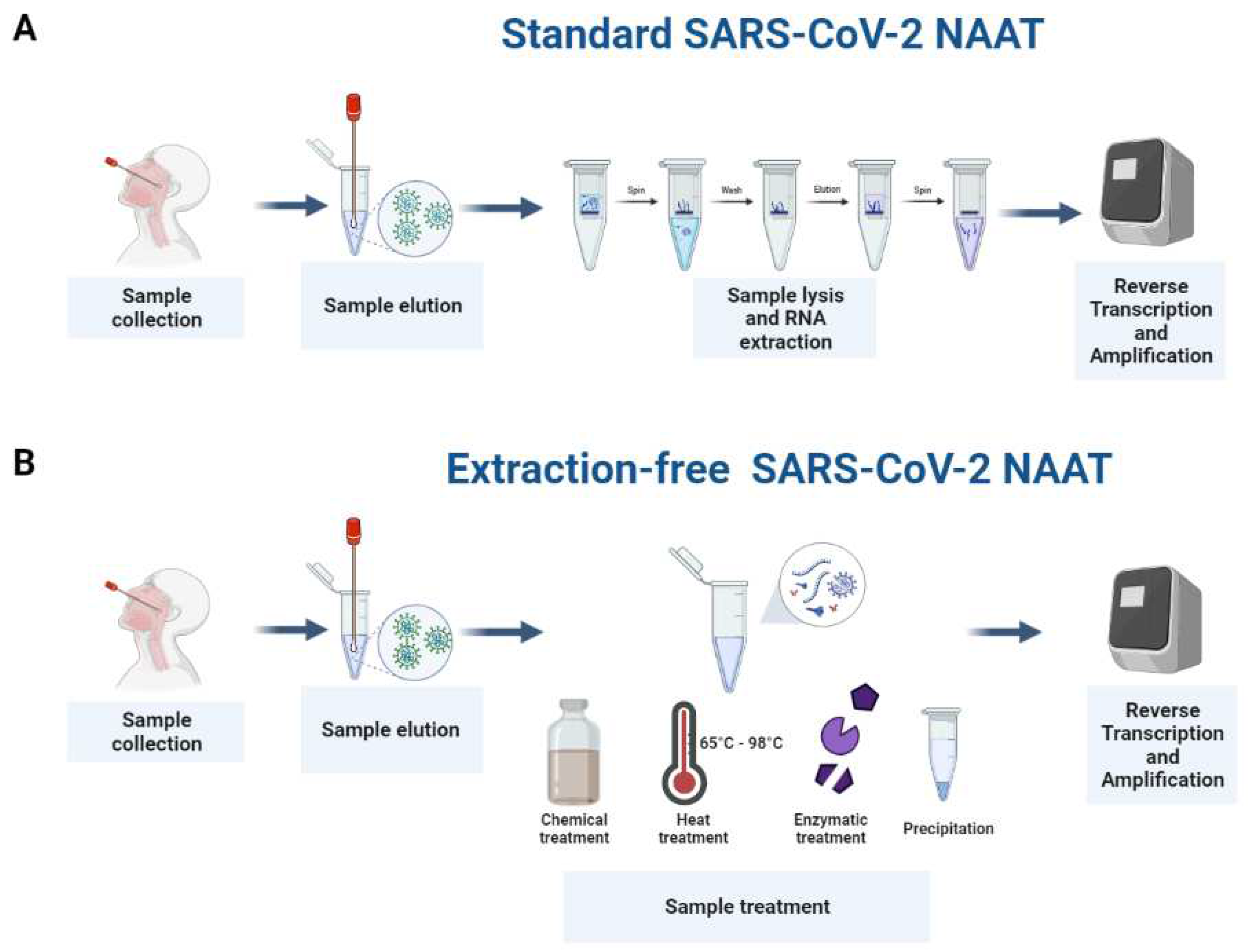
:1. Introduction
2. Extraction-Free Strategies for the Detection of SARS-CoV-2 by NAATs
2.1. Enrichment of Template
2.1.1. Adjusting the Proportion of Sample in the Reaction Mixture
2.1.2. Precipitation and Concentration of Nucleic Acid
2.2. Dilution and Removal of Contaminants That May Interfere with NAAT
2.3. Prevention of Template Degradation
2.3.1. Chemical Treatment of Respiratory Samples
2.3.2. Enzymatic Treatment of Respiratory Samples
2.3.3. Heat Treatment of Respiratory Samples
2.3.4. Optimization of Amplification Conditions
3. Discussion
4. Conclusions
Supplementary Materials
Author Contributions
Funding
Institutional Review Board Statement
Informed Consent Statement
Data Availability Statement
Acknowledgments
Conflicts of Interest
References
- Zhu, N.; Zhang, D.; Wang, W.; Li, X.; Yang, B.; Song, J.; Zhao, X.; Huang, B.; Shi, W.; Lu, R.; et al. A novel coronavirus from patients with pneumonia in China, 2019. N. Engl. J. Med. 2020, 382, 727–733. [Google Scholar] [CrossRef] [PubMed]
- Kim, D.; Lee, J.-Y.; Yang, J.-S.; Kim, J.W.; Kim, V.N.; Chang, H. The architecture of SARS-CoV-2 transcriptome. Cell 2020, 181, 914–921. [Google Scholar] [CrossRef] [PubMed]
- Yang, H.; Rao, Z. Structural biology of SARS-CoV-2 and implications for therapeutic development. Nat. Rev. Microbiol. 2021, 19, 685–700. [Google Scholar] [CrossRef] [PubMed]
- World Health Organization. Diagnostic Testing for SARS-CoV-2: Interim Guidance, 11 September 2020; World Health Organization: Geneva, Switzerland, 2020.
- Barra, G.B.; Santa Rita, T.H.; Mesquita, P.G.; Jácomo, R.H.; Nery, L.F.A. Overcoming supply shortage for SARS-CoV-2 detection by RT-qPCR. Genes 2021, 12, 90. [Google Scholar] [CrossRef] [PubMed]
- Sakthivel, D.; Delgado-Diaz, D.; McArthur, L.; Hopper, W.; Richards, J.S.; Narh, C.A. Point-of-Care diagnostic tools for surveillance of SARS-CoV-2 infections. Front. Public Health 2021, 9, 766871. [Google Scholar] [CrossRef]
- Kriegova, E.; Fillerova, R.; Kvapil, P. Direct-RT-qPCR Detection of SARS-CoV-2 without RNA Extraction as Part of a COVID-19 Testing Strategy: From Sample to Result in One Hour. Diagnostics 2020, 10, 605. [Google Scholar] [CrossRef]
- Brown, J.R.; Atkinson, L.; Shah, D.; Harris, K. Validation of an extraction-free RT-PCR protocol for detection of SARS-CoV2 RNA. medRxiv 2020. [Google Scholar] [CrossRef]
- Hedman, J.; Rådström, P. Overcoming inhibition in real-time diagnostic PCR. In PCR Detection of Microbial Pathogens; Springer: Berlin/Heidelberg, Germany, 2013; pp. 17–48. [Google Scholar]
- Ulloa, S.; Bravo, C.; Parra, B.; Ramirez, E.; Acevedo, A.; Fasce, R.; Fernandez, J. A simple method for SARS-CoV-2 detection by rRT-PCR without the use of a commercial RNA extraction kit. J. Virol. Methods 2020, 285, 113960. [Google Scholar] [CrossRef]
- Graham, T.G.; Dugast-Darzacq, C.; Dailey, G.M.; Nguyenla, X.H.; van Dis, E.; Esbin, M.N.; Abidi, A.; Stanley, S.A.; Darzacq, X.; Tjian, R. Open-source RNA extraction and RT-qPCR methods for SARS-CoV-2 detection. PLoS ONE 2021, 16, e0246647. [Google Scholar] [CrossRef]
- Lillehoj, E.P.; Kim, K.C. Airway mucus: Its components and function. Arch. Pharm. Res. 2002, 25, 770–780. [Google Scholar] [CrossRef]
- Smyrlaki, I.; Ekman, M.; Lentini, A.; Rufino de Sousa, N.; Papanicolaou, N.; Vondracek, M.; Aarum, J.; Safari, H.; Muradrasoli, S.; Rothfuchs, A.G.; et al. Massive and rapid COVID-19 testing is feasible by extraction-free SARS-CoV-2 RT-PCR. Nat. Commun. 2020, 11, 4812. [Google Scholar] [CrossRef] [PubMed]
- Grant, P.R.; Turner, M.A.; Shin, G.Y.; Nastouli, E.; Levett, L.J. Extraction-free COVID-19 (SARS-CoV-2) diagnosis by RT-PCR to increase capacity for national testing programmes during a pandemic. bioRxiv 2020. [Google Scholar] [CrossRef]
- Wee, S.K.; Sivalingam, S.P.; Yap, E.P.H. Rapid direct nucleic acid amplification test without RNA extraction for SARS-CoV-2 using a portable PCR thermocycler. Genes 2020, 11, 664. [Google Scholar] [CrossRef] [PubMed]
- Lee, J.Y.H.; Best, N.; McAuley, J.; Porter, J.L.; Seemann, T.; Schultz, M.B.; Sait, M.; Orlando, N.; Mercoulia, K.; Ballard, S.A.; et al. Validation of a single-step, single-tube reverse transcription loop-mediated isothermal amplification assay for rapid detection of SARS-CoV-2 RNA. J. Med. Microbiol. 2020, 69, 1169–1178. [Google Scholar] [CrossRef]
- Morecchiato, F.; Coppi, M.; Baccani, I.; Maggini, N.; Ciccone, N.; Antonelli, A.; Rossolini, G.M. Evaluation of extraction-free RT-PCR methods for faster and cheaper detection of SARS-CoV-2 using two commercial systems. Int. J. Infect. Dis. 2021, 112, 264–268. [Google Scholar] [PubMed]
- Guan, B.; Frank, K.M.; Maldonado, J.O.; Beach, M.; Pelayo, E.; Warner, B.M.; Hufnagel, R.B. Sensitive extraction-free SARS-CoV-2 RNA virus detection using a chelating resin. Iscience 2021, 24, 102960. [Google Scholar] [CrossRef] [PubMed]
- Rajh, E.; Šket, T.; Praznik, A.; Sušjan, P.; Šmid, A.; Urbančič, D.; Mlinarič-Raščan, I.; Kogovšek, P.; Demšar, T.; Milavec, M. Robust Saliva-Based RNA Extraction-Free One-Step Nucleic Acid Amplification Test for Mass SARS-CoV-2 Monitoring. Molecules 2021, 26, 6617. [Google Scholar] [CrossRef]
- Howson, E.L.; Kidd, S.P.; Armson, B.; Goring, A.; Sawyer, J.; Cassar, C.; Cross, D.; Lewis, T.; Hockey, J.; Rivers, S. Preliminary optimisation of a simplified sample preparation method to permit direct detection of SARS-CoV-2 within saliva samples using reverse-transcription loop-mediated isothermal amplification (RT-LAMP). J. Virol. Methods 2021, 289, 114048. [Google Scholar] [CrossRef]
- Lalli, M.A.; Langmade, J.S.; Chen, X.; Fronick, C.C.; Sawyer, C.S.; Burcea, L.C.; Wilkinson, M.N.; Fulton, R.S.; Heinz, M.; Buchser, W.J. Rapid and extraction-free detection of SARS-CoV-2 from saliva by colorimetric reverse-transcription loop-mediated isothermal amplification. Clin. Chem. 2020, 67, 415–424. [Google Scholar] [CrossRef]
- Wei, S.; Suryawanshi, H.; Djandji, A.; Kohl, E.; Morgan, S.; Hod, E.A.; Whittier, S.; Roth, K.; Yeh, R.; Alejaldre, J.C. Field-deployable, rapid diagnostic testing of saliva for SARS-CoV-2. Sci. Rep. 2021, 11, 5448. [Google Scholar] [CrossRef]
- Rabe, B.A.; Cepko, C. SARS-CoV-2 detection using isothermal amplification and a rapid, inexpensive protocol for sample inactivation and purification. Proc. Natl. Acad. Sci. USA 2020, 117, 24450–24458. [Google Scholar] [CrossRef] [PubMed]
- Mallmann, L.; Hermann, B.; Schallenberger, K.; Demoliner, M.; Eisen, A.; Heldt, F.; Gularte, J.; Hansen, A.; de Almeida, P.; Weber, M. Proteinase K treatment in absence of RNA isolation classical procedures is a quick and cheaper alternative for SARS-CoV-2 molecular detection. J. Virol. Methods 2021, 293, 114131. [Google Scholar] [CrossRef]
- De Oliveira, C.M.; Brochi, L.; Scarpelli, L.C.; Lopes, A.C.W.; Levi, J.E. SARS-CoV-2 saliva testing is a useful tool for COVID-19 diagnosis. J. Virol. Methods 2021, 296, 114241. [Google Scholar] [CrossRef]
- Mio, C.; Cifù, A.; Marzinotto, S.; Bergamin, N.; Caldana, C.; Cattarossi, S.; Cmet, S.; Cussigh, A.; Martinella, R.; Zucco, J. A streamlined approach to rapidly detect SARS-CoV-2 infection avoiding RNA extraction: Workflow validation. Dis. Markers 2020, 2020, 8869424. [Google Scholar] [CrossRef] [PubMed]
- Genoud, V.; Stortz, M.; Waisman, A.; Berardino, B.G.; Verneri, P.; Dansey, V.; Salvatori, M.; Remes Lenicov, F.; Levi, V. Extraction-free protocol combining proteinase K and heat inactivation for detection of SARS-CoV-2 by RT-qPCR. PLoS ONE 2021, 16, e0247792. [Google Scholar] [CrossRef] [PubMed]
- Ñique, A.M.; Coronado-Marquina, F.; Mendez Rico, J.A.; García Mendoza, M.P.; Rojas-Serrano, N.; Simas, P.V.M.; Cabezas Sanchez, C.; Drexler, J.F. A faster and less costly alternative for RNA extraction of SARS-CoV-2 using proteinase k treatment followed by thermal shock. PLoS ONE 2021, 16, e0248885. [Google Scholar] [CrossRef]
- Thi, V.L.D.; Herbst, K.; Boerner, K.; Meurer, M.; Kremer, L.P.; Kirrmaier, D.; Freistaedter, A.; Papagiannidis, D.; Galmozzi, C.; Stanifer, M.L. A colorimetric RT-LAMP assay and LAMP-sequencing for detecting SARS-CoV-2 RNA in clinical samples. Sci. Transl. Med. 2020, 12, eabc7075. [Google Scholar]
- Hasan, M.R.; Mirza, F.; Al-Hail, H.; Sundararaju, S.; Xaba, T.; Iqbal, M.; Alhussain, H.; Yassine, H.M.; Perez-Lopez, A.; Tang, P. Detection of SARS-CoV-2 RNA by direct RT-qPCR on nasopharyngeal specimens without extraction of viral RNA. PLoS ONE. 2020, 15, e0236564. [Google Scholar]
- Mancini, F.; Barbanti, F.; Scaturro, M.; Errico, G.; Iacobino, A.; Bella, A.; Riccardo, F.; Marsili, G.; Stefanelli, P.; Pezzotti, P. Laboratory management for SARS-CoV-2 detection: A user-friendly combination of the heat treatment approach and rt-Real-time PCR testing. Emerg. Microbes Infect. 2020, 9, 1393–1396. [Google Scholar] [CrossRef]
- Ranoa, D.; Holland, R.; Alnaji, F.G.; Green, K.; Wang, L.; Brooke, C.; Burke, M.; Fan, T.; Hergenrother, P.J. Saliva-based molecular testing for SARS-CoV-2 that bypasses RNA extraction. bioRxiv 2020. [Google Scholar] [CrossRef]
- Merindol, N.; Pépin, G.; Marchand, C.; Rheault, M.; Peterson, C.; Poirier, A.; Houle, C.; Germain, H.; Danylo, A. SARS-CoV-2 detection by direct rRT-PCR without RNA extraction. J. Clin. Virol. 2020, 128, 104423. [Google Scholar] [CrossRef]
- Fomsgaard, A.S.; Rosenstierne, M.W. An alternative workflow for molecular detection of SARS-CoV-2–escape from the NA extraction kit-shortage, Copenhagen, Denmark, March 2020. Eurosurveillance 2020, 25, 2000398. [Google Scholar] [CrossRef] [PubMed] [Green Version]
- Burton, J.; Love, H.; Richards, K.; Burton, C.; Summers, S.; Pitman, J.; Easterbrook, L.; Davies, K.; Spencer, P.; Killip, M. The effect of heat-treatment on SARS-CoV-2 viability and detection. J. Virol. Methods 2021, 290, 114087. [Google Scholar] [CrossRef] [PubMed]
- Blairon, L.; Piteüs, S.; Beukinga, I.; Tré-Hardy, M. Development and implementation of a RT-qPCR extraction-free protocol for the detection of SARS-CoV-2 and impact on the turn-around-time. J. Med. Virol. 2021, 93, 2538–2542. [Google Scholar] [CrossRef]
- Bruce, E.A.; Huang, M.-L.; Perchetti, G.A.; Tighe, S.; Laaguiby, P.; Hoffman, J.J.; Gerrard, D.L.; Nalla, A.K.; Wei, Y.; Greninger, A.L.; et al. Direct RT-qPCR detection of SARS-CoV-2 RNA from patient nasopharyngeal swabs without an RNA extraction step. PLoS Biol. 2020, 18, e3000896. [Google Scholar] [CrossRef] [PubMed]
- Lownik, J.C.; Way, G.W.; Farrar, J.S.; Martin, R.K. Extraction-Free Rapid Cycle Quantitative RT-PCR and Extreme RT-PCR for SARS-CoV-2 Virus Detection. J. Mol. Diagn. 2021, 23, 1671–1679. [Google Scholar] [CrossRef]
- Butler, D.; Mozsary, C.; Meydan, C.; Foox, J.; Rosiene, J.; Shaiber, A.; Danko, D.; Afshinnekoo, E.; MacKay, M.; Sedlazeck, F.J.; et al. Shotgun transcriptome, spatial omics, and isothermal profiling of SARS-CoV-2 infection reveals unique host responses, viral diversification, and drug interactions. Nat. Commun. 2021, 12, 1660. [Google Scholar] [CrossRef] [PubMed]
- Meyerson, N.R.; Yang, Q.; Clark, S.K.; Paige, C.L.; Fattor, W.T.; Gilchrist, A.R.; Barbachano-Guerrero, A.; Sawyer, S.L. A community-deployable sars-cov-2 screening test using raw saliva with 45 minutes sample-to-results turnaround. medRxiv 2020. [Google Scholar] [CrossRef]
- Kellner, M.J.; Ross, J.J.; Schnabl, J.; Dekens, M.P.S.; Heinen, R.; Grishkovskaya, I.; Bauer, B.; Stadlmann, J.; Menéndez-Arias, L.; Fritsche-Polanz, R.; et al. A rapid, highly sensitive and open-access SARS-CoV-2 detection assay for laboratory and home testing. Front. Mol. Biosci. 2022, 9, 801309. [Google Scholar] [CrossRef]
- Centers for Disease Control and Prevention. Interim Laboratory Biosafety Guidelines for Handling and Processing Specimens Associated with Coronavirus Disease 2019 (COVID-19); Centers for Disease Control and Prevention: Atlanta, GA, USA, 2021. [Google Scholar]
- Centers for Disease Control and Prevention. Guidelines for SARS-CoV-2 Rapid Testing Performed in Point-of-Care Settings; Centers for Disease Control and Prevention: Atlanta, GA, USA, 2022. [Google Scholar]

| Sample Type | NAAT/Detection Method | Sample Preparation Strategy | Ease of Implementation | Reference |
|---|---|---|---|---|
| Nasopharyngeal and nasal swab in UTM | RT-PCR | Increase in sample input combined with the use of enzymes with high tolerability to inhibitors * | Very easy to implement as no sample treatment is required. | [7] |
| Nasal and throat swabs suspended in nuclease-free water | RT-PCR, RT-LAMP | Increased input volume of swabs eluted in nuclease-free water or saline * | Very easy to implement as no sample treatment is required. | [8,39] |
| NP swabs in UTM, PBS, Hanks medium, DNA/RNA shield | RT-PCR | Precipitation of sample with PEG/NaCl combined with heat treatment at 70 °C for 30 min. | Laborious methodology as precipitation involves more than one step. A heating source is required for this method. | [10] |
| Heat-inactivated nasopharyngeal swab-UTM eluates | RT-PCR | Precipitation of template with 1.1 volumes of isopropanol, incubation at −20 °C for 30 min and centrifugation, ethanol addition, and centrifugation. | This method involves several steps, including centrifugation and a freezer. | [11] |
| Swab in viral transport medium | RT-PCR | Reduced input volume of swabs eluted in viral transport medium * | Very easy to implement as no sample treatment is required. | [14] |
| Nasopharyngeal swab in UTM | Fluorescence RT-LAMP, RT-PCR | Dilution of sample in RNase-free water * | Very easy to implement as no sample treatment is required. | [16,17] |
| Nasopharyngeal swabs and saliva | RT-PCR and RT-ddPCR | Elution of swabs into Chelex-TED buffer (50% Chelex-100, TE buffer, DMSO) or addition to saliva, followed by heat treatment at 98 °C for 5 min and centrifugation. | Difficult to implement as this method involves several steps, and relies on a centrifuge and a heating source. | [18] |
| Sputum and nasal exudate | Portable RT-PCR | Treatment of sample with sputasol and the RNAse inhibitor RNAseOUT™ * | Easy to implement as the sample can be treated in one step. | [15] |
| Saliva | Colorimetric RT-LAMP | Combination of proteinase K treatment, heat inactivation, and RNAsecure treatment. | Challenging to implement as several sample preparation methods are involved and Proteinase K treatment requires a final step to denature the enzyme. | [21] |
| Saliva and swabs | Colorimetric RT-LAMP | Addition of carrier nucleic acid, treatment with RNase inhibitors, and increase in the reaction volume * | Easy to implement as treatment of the sample can be done in one step. | [22] |
| Saliva and Nasopharyngeal swabs | RT-PCR and RT-LAMP | Elution of swab or mixing of saliva with RNA stabilization buffer (TCEP, EDTA, Chelex, and RNasecure in Tris buffer) followed by 95 °C 15 min heat inactivation and cooling. | Although several sample preparation methods are involved, this strategy can be done in two steps. | [19] |
| Saliva | RT-LAMP | 1:1 dilution in Mucolyse (DTT), followed by dilution in 10% (w/v) chelex 100 resin and 98 °C heat treatment for 2 min. | Several sample preparation methods and steps are involved making it challenging to implement. | [20] |
| Saliva or Nasopharyngeal swab eluted in saline or PBS | Colorimetric RT-LAMP | Combination of treatment with a reducing agent (TCEP/EDTA) and heat treatment at 95 °C for 5 min. | Relatively easy to implement as treatment of the sample involves a two-step process. However, a heating source is required. | [23] |
| Saliva, nasopharyngeal and oropharyngeal swabs eluted in saline or UTM | RT-PCR | Proteinase K followed by heat inactivation at 95–98 °C for 5 min. | Moderately easy to implement; however, denaturing Proteinase K at high temperature is essential. | [24,25,26,27,28] |
| Nasopharyngeal, oropharyngeal swab in transport medium, saline, PBS, or water. Saliva | RT-PCR and RT-LAMP | Several heating conditions from 65 °C to 98 °C for periods of 5 to 30 min. | Relatively easy to implement; however, this strategy requires a heating source and optimization of the heating conditions. | [10,13,23,29,30,31,32,33,34,37,40,41] |
| Nasopharyngeal swabs | RT-PCR | Thermal shock of the sample at 95 °C for 5 min followed by 4 °C for 10 min. | Relatively easy to implement; however, this strategy requires both a heating source and an active cooling source. | [36] |
| Nasopharyngeal swabs in universal transport media | RT-PCR | Combination of heat treatment (65 °C for 10 min) and increase in sample input volume. | Relatively easy to implement; however, this strategy requires a heating source. | [30] |
| Saliva | RT-PCR | Lysis in TBE buffer and tween-20 combined with heat treatment at 95 °C for 30 min. | Relatively easy to implement; however, this strategy requires a heating source. | [32] |
| Nasopharyngeal swabs and gargle lavage | Fluorescence and Colorimetric RT-LAMP | Combination of Quickextract and heat treatment at 95 °C for 5 min, supplemented with carboxylated magnetic beads to enrich target RNA. | Several sample preparation methods and steps are involved making it challenging to implement. | [41] |
Publisher’s Note: MDPI stays neutral with regard to jurisdictional claims in published maps and institutional affiliations. |
© 2022 by the authors. Licensee MDPI, Basel, Switzerland. This article is an open access article distributed under the terms and conditions of the Creative Commons Attribution (CC BY) license (https://creativecommons.org/licenses/by/4.0/).
Share and Cite
Delgado-Diaz, D.J.; Sakthivel, D.; Nguyen, H.H.T.; Farrokzhad, K.; Hopper, W.; Narh, C.A.; Richards, J.S. Strategies That Facilitate Extraction-Free SARS-CoV-2 Nucleic Acid Amplification Tests. Viruses 2022, 14, 1311. https://doi.org/10.3390/v14061311
Delgado-Diaz DJ, Sakthivel D, Nguyen HHT, Farrokzhad K, Hopper W, Narh CA, Richards JS. Strategies That Facilitate Extraction-Free SARS-CoV-2 Nucleic Acid Amplification Tests. Viruses. 2022; 14(6):1311. https://doi.org/10.3390/v14061311
Chicago/Turabian StyleDelgado-Diaz, David J., Dhanasekaran Sakthivel, Hanh H. T. Nguyen, Khashayar Farrokzhad, William Hopper, Charles A. Narh, and Jack S. Richards. 2022. "Strategies That Facilitate Extraction-Free SARS-CoV-2 Nucleic Acid Amplification Tests" Viruses 14, no. 6: 1311. https://doi.org/10.3390/v14061311
APA StyleDelgado-Diaz, D. J., Sakthivel, D., Nguyen, H. H. T., Farrokzhad, K., Hopper, W., Narh, C. A., & Richards, J. S. (2022). Strategies That Facilitate Extraction-Free SARS-CoV-2 Nucleic Acid Amplification Tests. Viruses, 14(6), 1311. https://doi.org/10.3390/v14061311