Article

Entomopathogenic Fungi-Mediated AgNPs: Synthesis and Insecticidal Effect against Plutella xylostella (Lepidoptera: Plutellidae)

by
Tárcio S. Santos
1,
Camila de Souza Varize
2,
Elena Sanchez-Lopez
3,4,5,
Sona A. Jain
1,6,
Eliana B. Souto
3,7,8,*,
Patrícia Severino
1,6,* and
Marcelo da Costa Mendonça
1,2,6
1
Post-graduate Program in Industrial Biotechnology (PBI), University Tiradentes (Unit), Av. Murilo Dantas, 300, Aracaju 49010-390, Brazil
2
Sergipe Agricultural Development Company (Emdagro), Av. Carlos Rodrigues da Cruz s/n, Aracaju 49081-015, Brazil
3
Department of Pharmacy, Pharmaceutical Technology and Physical Chemistry, Faculty of Pharmacy and Food Sciences, University of Barcelona, 08028 Barcelona, Spain
4
Institute of Nanoscience and Nanotechnology (IN2UB), University of Barcelona, 08028 Barcelona, Spain
5
Unit of Synthesis and Biomedical Applications of Peptides, IQAC-CSIC, 08034 Barcelona, Spain
6
Institute of Technology and Research (ITP), Av. Murilo Dantas, 300, Aracaju 49010-390, Brazil
7
Department of Pharmaceutical Technology, Faculty of Pharmacy, University of Porto, Rua de Jorge Viterbo Ferreira, 228, 4050-313 Porto, Portugal
8
REQUIMTE/UCIBIO, Faculty of Pharmacy, University of Porto, Rua de Jorge Viterbo Ferreira, 228, 4050-313 Porto, Portugal
*
Authors to whom correspondence should be addressed.
Materials 2022, 15(21), 7596; https://doi.org/10.3390/ma15217596
Submission received: 1 October 2022 / Revised: 22 October 2022 / Accepted: 26 October 2022 / Published: 28 October 2022
(This article belongs to the Special Issue Functional Nanomaterials for a Better Life)

Abstract

:
The insect Plutella xylostella is known worldwide to cause severe damage to brassica plantations because of its resistance against several groups of chemicals and pesticides. Efforts have been conducted to overcome the barrier of P. xylostella genetic resistance. Because of their easy production and effective insecticidal activity against different insect orders, silver nanoparticles are proposed as an alternative for agricultural pest control. The use of entomopathogenic fungi for nanoparticle production may offer additional advantages since fungal biomolecules may synergistically improve the nanoparticle’s effectiveness. The present study aimed to synthesize silver nanoparticles using aqueous extracts of Beauveria bassiana, Metarhizium anisopliae, and Isaria fumosorosea isolates and to evaluate their insecticidal activity against P. xylostella, as innovative nano-ecofriendly pest control. The produced silver nanoparticles were evaluated by measuring the UV–vis spectrum and the mean particle size by dynamic light scattering (DLS). I. fumosorosea aqueous extract with 3-mM silver nitrate solution showed the most promising results (86-nm mean diameter and 0.37 of polydispersity). Scanning electron microscopy showed spherical nanoparticles and Fourier-Transform Infrared Spectroscopy revealed the presence of amine and amide groups, possibly responsible for nanoparticles’ reduction and stabilization. The CL50 value of 0.691 mg mL−1 was determined at 72-h for the second-instar larvae of the P. xylostella, promoting a 78% of cumulative mortality rate after the entire larval stage. From our results, the synthesis of silver nanoparticles mediated by entomopathogenic fungi was successful in obtaining an efficient product for insect pest control. The I. fumosorosea was the most suitable isolate for the synthesis of silver nanoparticles contributing to the development of a green nanoproduct and the potential control of P. xylostella.

1. Introduction

The diamondback moth Plutella xylostella (Lepidoptera: Plutellidae) is a microlepidopteran, popularly known as the cabbage moth. This species is one of the most studied insect pests in the world and is reported to cause extensive damage to vegetable crops of the Brassicaceae family, including cabbage, cauliflower, mustard, and rapeseed, among others [1]. The damage caused by this insect to the host plant is related to the feeding habits in the larval stage. The caterpillars tend to scrape the parenchyma tissue, making holes in the leaf surface and reducing the photosynthetically active area. Besides this, its high rate of reproduction (e.g., more than 20 generations per year in tropical regions) increases plant damage quite rapidly, making the entire crop harvesting unfeasible, if no containment measures are adopted. In addition, the low incidence of natural enemies and the high genetic variability of this insect facilitate/promotes the development of chemical pesticide resistance [2,3].
The control of P. xylostella costs a minimum of USD 1.4 billion per year globally, reaching USD 5 billion when considering crop productivity loss [2]. Therefore, the main challenge is to overcome the high resistance against chemical pesticides since this moth is one of the most resistant and destructive pests worldwide. Due to the high rate of reproduction, the intense and fast infestation caused by this plague increases the need to overcome these obstacles [4]. Nanotechnology is providing significant benefits to the agrifood sector, including the delivery of nutraceuticals and nanopesticides, increasing productivity, and favoring plant management and development, to ultimately increase food safety [5]. Among available alternatives, silver nanoparticles emerge as a potential product for use in insect pest control, since they are easy to synthesize and handle, and also present insecticidal/antimicrobial actions [6,7,8,9]. However, the main concern involving the production of nanomaterials is the generation of toxic waste. Thus, the development of these products requires more sustainable, green ways to reach higher profits and safer synthesis. The synthesis of silver nanoparticles by biological process is attracting increasingly interest because it represents a simple, sustainable, and low-cost method [6,7].
Silver nanoparticles synthesized by a biological process have toxicity against insects, being the application mainly directed against mosquitoes [10]. The use of algae extracts to produce silver nanoparticles was already reported to control the insect P. xylostella [11], however, there is no evidence for the effectiveness of silver nanoparticle synthesis mediated by entomopathogenic fungi for the same purpose.
Therefore, this work aims to synthesize silver nanoparticles with an aqueous extract of entomopathogenic fungi and to evaluate their insecticidal action against Plutella xylostella. We report the synthesis of silver nanoparticles using different species of entomopathogenic fungi. The influence of the silver nitrate concentration on nanoparticle formation is also presented. The nanoparticles were characterized by UV-Vis spectroscopy, Dynamic Light Scattering (DLS), Fourier Transform Infrared Spectroscopy (FTIR), and Scanning Electron Microscopy (SEM). Finally, from the bioassays using the nanomaterial selected, we showed the sublethal concentrations and the effect of the LC50 in the life-cycle stages of P. xylostella.

2. Materials and Methods

The experimental procedure followed the sequence described in Figure 1.

2.1. Entomopathogenic Fungal Isolates

The four fungal isolates used to produce silver nanoparticles belong to the species Beauveria bassiana (Bals.—Criv.) Vuill. (Hypocreales: Cordycipitaceae) strains SE109 and 1260, Metarhizium anisopliae (Metschn.) Sorokīn (Hypocreales: Clavicipitaceae) SE202, and Isaria fumosorosea Wize (Hypocreales: Cordycipitaceae) SE301 (Table 1). The fungi are kept in colonized potato dextrose agar (PDA) disks (5-mm-diameter), and stored in cryogenic tubes containing 10% of a glycerol solution, at −20 °C.

2.2. Extracellular Synthesis of Silver Nanoparticles

The isolates were grown on a PDA medium at a temperature of 25 ± 2 °C, for 7 days. For their use, colonized PDA disks (5-mm-diameter) were inoculated into potato dextrose broth (PDB) and incubated for 7 days at 25 ± 2 °C on an orbital shaker (100 rpm). Next, the fungal biomass was filtered through filter paper (Whatman No. 1, London) and washed three times with ultrapure water. Then 10 g of the washed biomass was added to an Erlenmeyer flask containing 100 mL of ultrapure water, maintained for 72 h at 25 ± 2 °C on an orbital shaker (100 rpm). Then, 90 mL of the liquid extracted (aqueous extract) from a new filtering was mixed with 10 mL of 1, 3, and 5 mM of AgNO3 solution. This mixture was incubated in the dark for 96 h at 25 ± 2 °C on an orbital shaker (100 rpm) [6,12].

2.3. Silver Nanoparticles Characterization

The formation of silver nanoparticles was monitored by UV-Visible spectroscopy (DR 5000 spectrophotometer, Hexis Científica, Jundiaí, São Paulo, Brazil,) in the spectral region between 300 and 800 nm. The absorbance values were plotted in graphs using the Origin Pro 2019b software (OriginLab Coorporation, Northampton, MA, USA, 2019). The nanoparticle diameter and Polydispersity Index (PDI) were determined by Dynamic Light Scattering (DLS) analysis using the Zetasizer Nano Instrument (Malvern, Australia, model Nano-S). The selected nanoparticles were characterized by Scanning Electron Microscopy (SEM) (JEOL microscope, model JSM-IT200, Tokyo, Japan). The functional groups present in the silver nanoparticles were analyzed by Attenuated Total Reflection-Fourier Transform Infrared (ATR-FTIR) spectroscopy (Agilent Technologies, Agilent Cary 630, Santa Clara, CA, USA), equipped with a ZnSe-diamond composite crystal accessory. The spectra were collected over a wavenumber range from 4000–600 cm−1 with a spectral resolution of <2 cm−1. Agilent MicroLab PC and Origin Pro 2019b software were used for data gathering and transmittance graph plotting, respectively (OriginLab, 2019).

2.4. Sublethal Concentrations of Biogenic Silver Nanoparticles against P. xylostella

Cabbage leaf discs (Brassica oleracea L.) (8-cm-diameter) were submerged in the suspension of silver nanoparticles (previously dispersed in an ultrasonic bath for 10 min) at concentrations of 0.1; 0.3; 0.7; 0.9 and 1.2 mg mL−1 and maintained at room temperature (26 ± 2 °C) till complete drying. Then, one cabbage leaf disk was transferred to a Petri dish (90 × 10 mm), which received 25 second-instar larvae of P. xylostella (emerged at 72 h). The Petri dishes were sealed and maintained in an acclimatized room (T = 26 ± 2 °C, RH = 60 ± 10%, and photophase = 12 h) for a 72-h period to assess the mortality rate. The commercial pesticide Deltamethrin (Decis 25 EC) was also evaluated at concentrations of 0.0075, 0.0225, 0.0525, 0.5, and 0.75 mg mL−1, applying the same method as mentioned above. Five replicates were tested for the silver nanoparticles and Deltamethrin treatments. Since Tween® 80 0.05% was used to homogenize nanoparticle suspension used in the bioassay prior to the application of silver nanoparticles in the insect, the same solution of Tween® 80 0.05% was used as a negative control.

2.5. Survival, Viability, and Longevity Analysis of P. xylostella Larvae Exposed to LC50 of Silver Nanoparticles

Cabbage leaf discs (8-cm-diameter) were submerged into a suspension of silver nanoparticles corresponding to the CL50, following the same method/conditions of the sublethal concentrations test above described. Each Petri dish (90 × 10 mm) received one cabbage leaf disk and 25 second-instar larvae of P. xylostella (emerged at 72 h), also maintained in an acclimatized room (T = 26 ± 2 °C, RH = 60 ± 10% and photophase = 12 h). The larvae were transferred to a new Petri dish containing one untreated cabbage leaf disc every 72 h, during the entire evaluation. The caterpillar mortality was evaluated daily until it reaches 100% or enter the pupal stage. The percentage of viable insects and the time taken in each phase of the biological cycle were also determined, until the pupal stage. In total, 10 replicates were tested for this bioassay and a solution of Tween 80® 0.05% was used as a negative control.

2.6. Statistical Analysis

The cumulative mortality of the insects after the third day of exposure to the treatment was submitted to the Probit analysis to estimate the lethal concentration (LC). The daily mortality values were used to estimate the lethal time (LT) and to construct a survival curve by the Kaplan–Meier method (Log-rank—Mantel-Cox). The number of insects that passed to the pupal phase as well as the duration time of each biological cycle phase was quantified. The parameters of larval viability, larval duration, pupal viability, and pupal duration after exposure to LC50 of silver nanoparticles were analyzed. The results were submitted to the t-test. The statistical analyzes were performed using SPSS software version 23 (IBM Corp., Armonk, NY, USA, 2015). The graphs of LC were made by the GraphPad Prism 8.0.1. software (GraphPad 8.0.1. Software, 2018).

3. Results and Discussion

3.1. Synthesis of Silver Nanoparticles

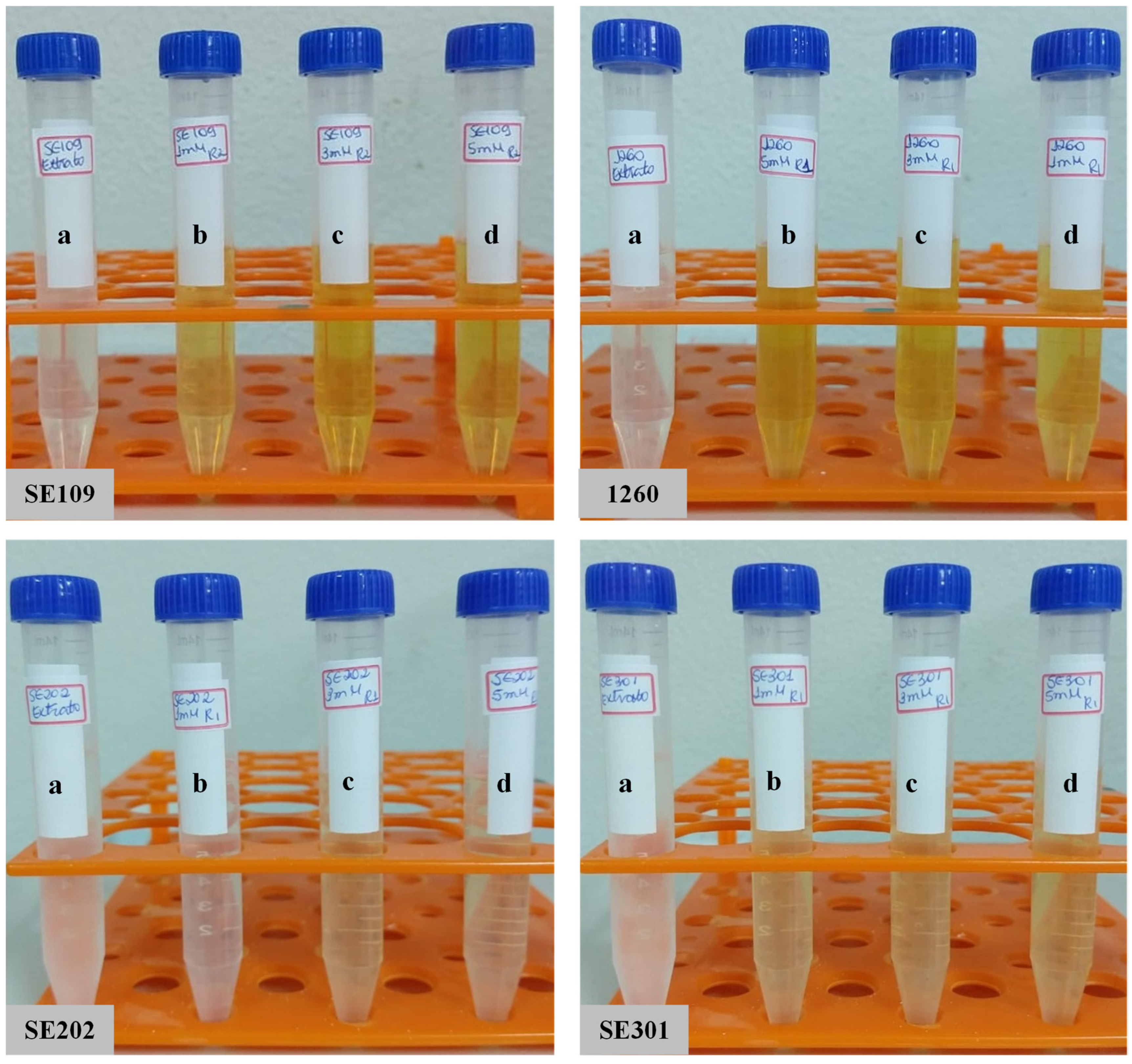
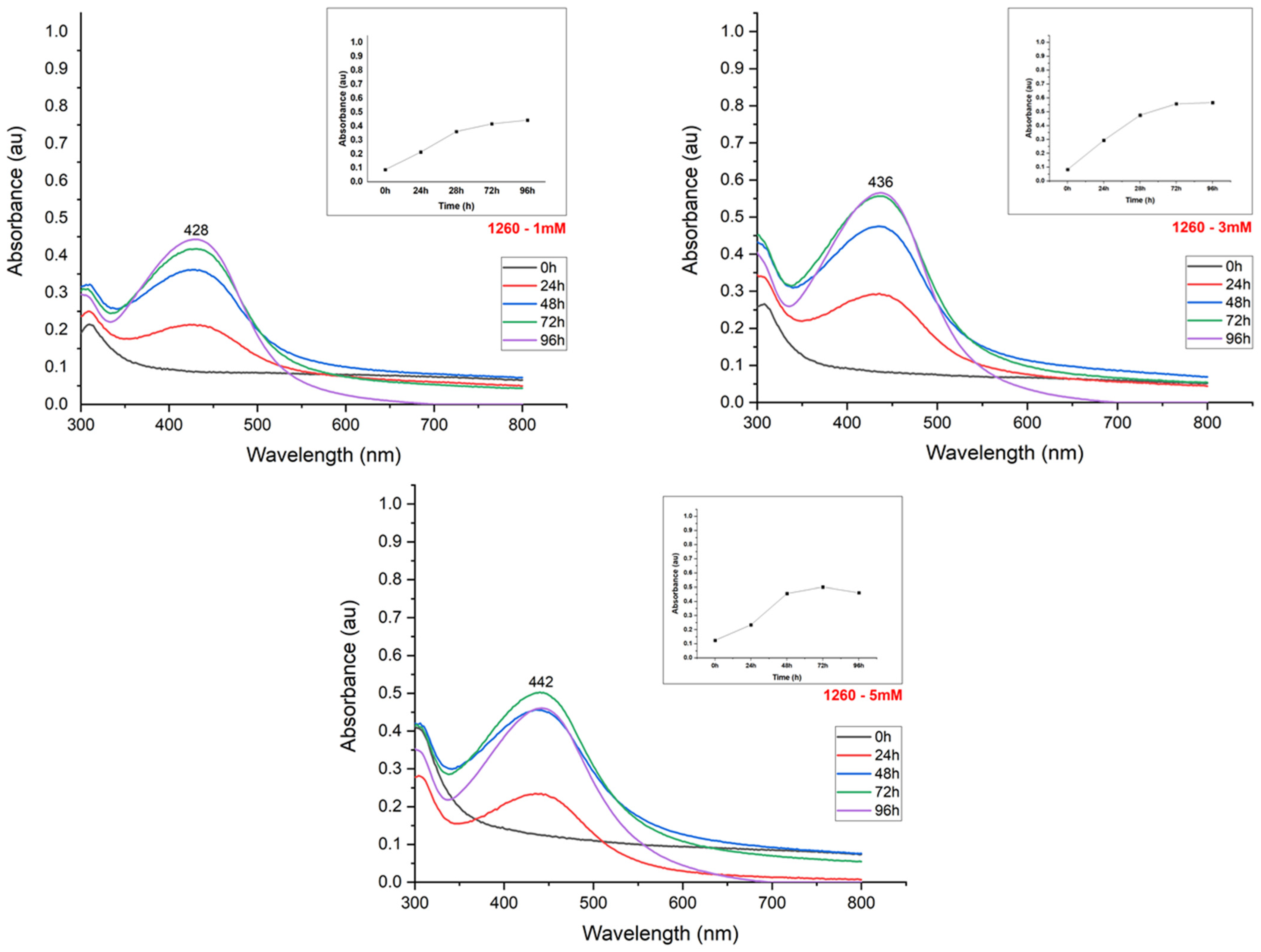
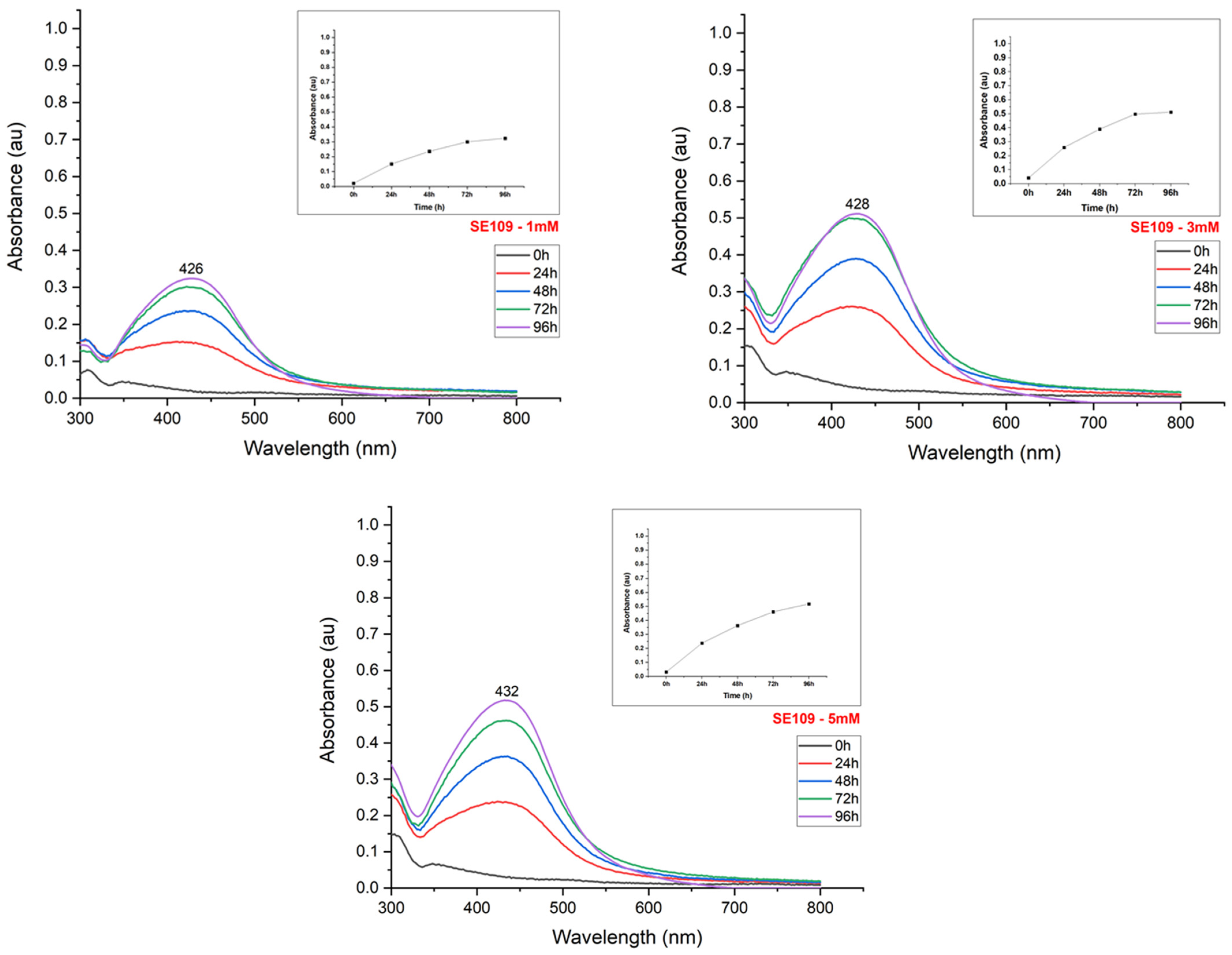
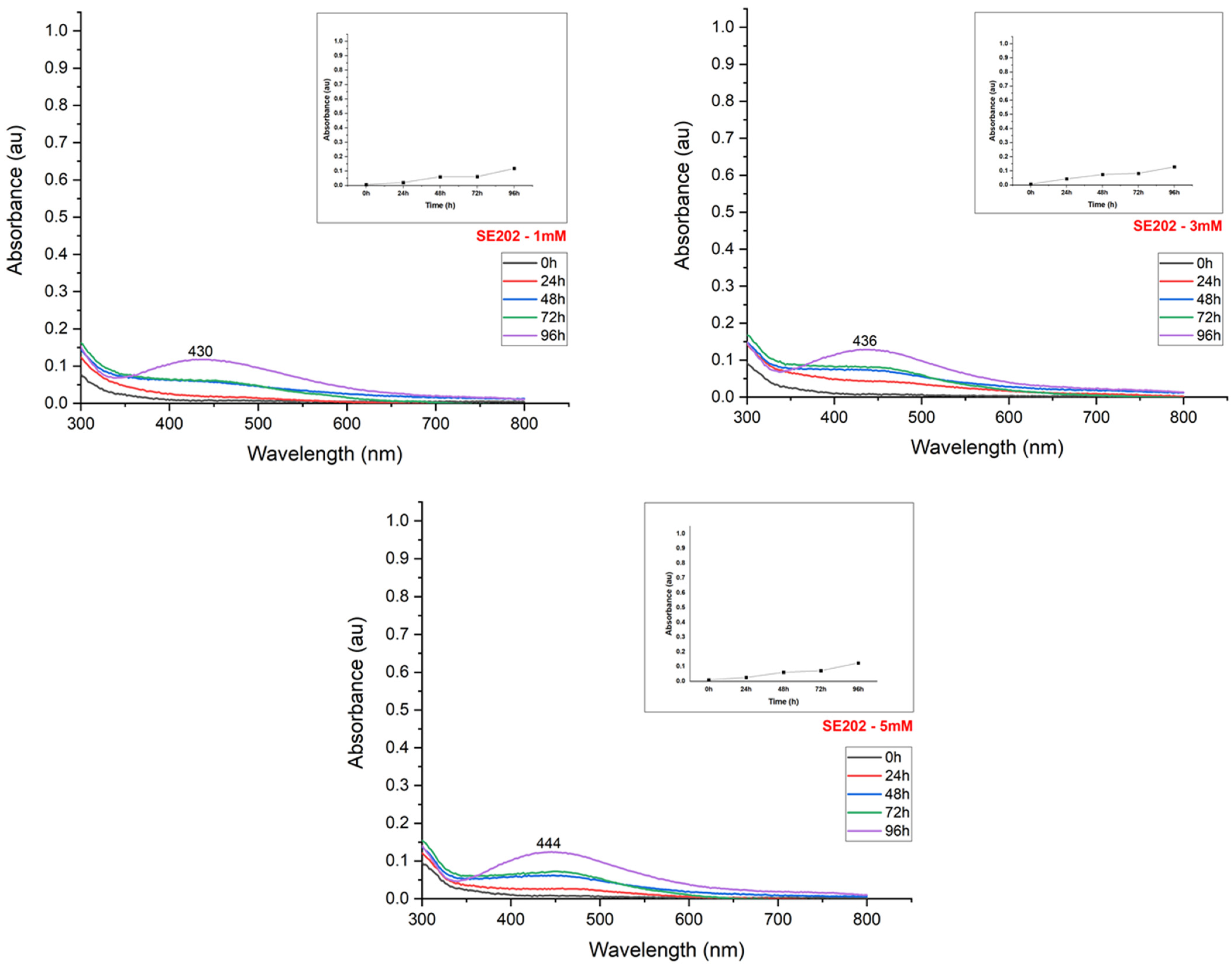
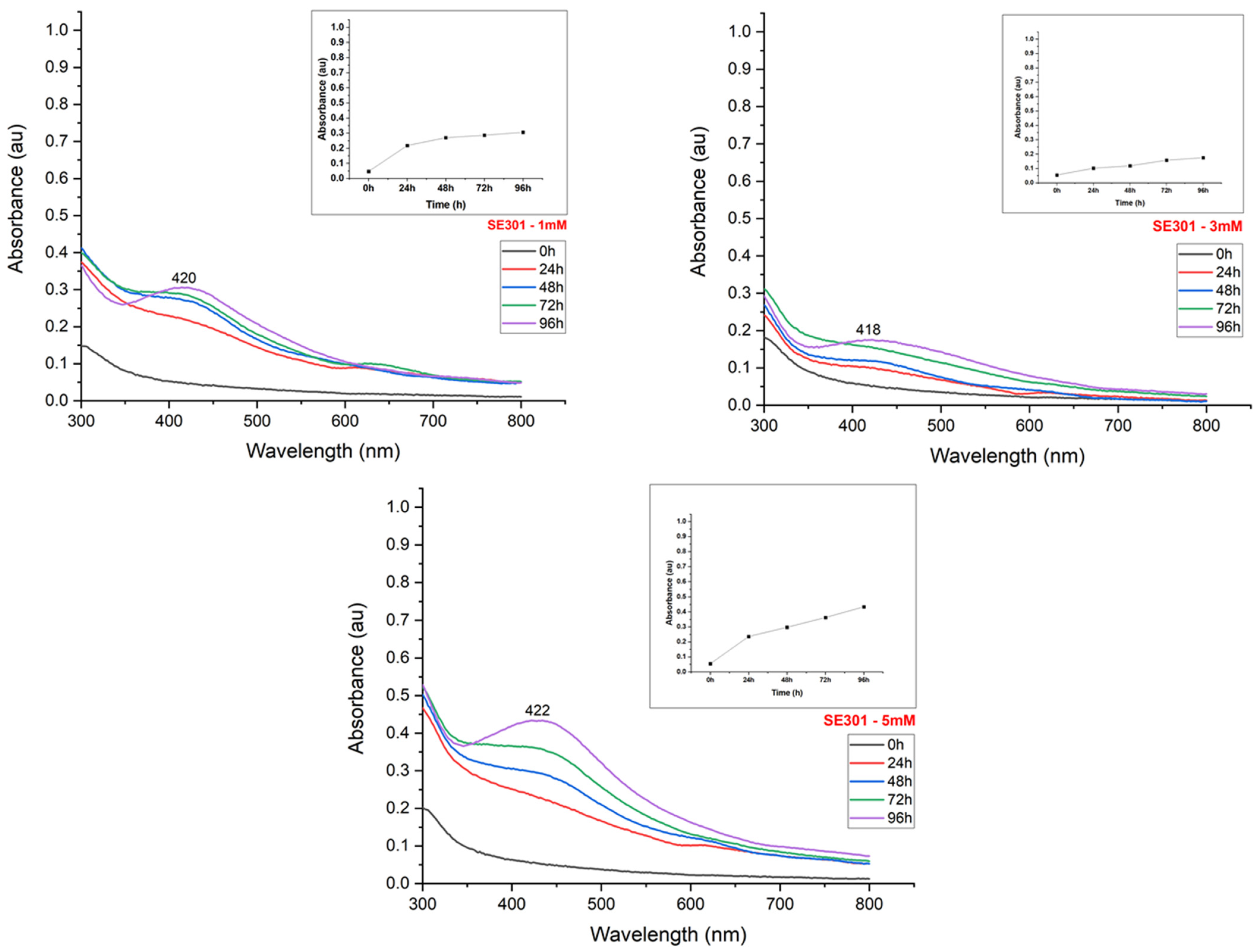
The synthesis reaction caused a color change from light to dark yellow in the suspensions, according to the silver nitrate content used for nanoparticle production (Figure 2). The UV-Visible spectroscopy exhibited peaks between 400–500 nm, confirming the reduction of the silver salt and the effective formation of nanoparticles (Figure 3, Figure 4, Figure 5 and Figure 6). The absorption peaks increased in concomitance with the higher concentrations of AgNO3, observed in 1260, SE109, and SE301 aqueous extract (Figure 3, Figure 4 and Figure 6), being this relationship not observed in SE202 (Figure 4). Previously, it has been reported that the silver ions content affects the reaction time of the nanoparticle synthesis. A previous study used the Aspergillus oryzae extract in the synthesis of the silver nanoparticles and reported that although the speed of the reaction increases at higher concentrations of AgNO3, the size of particles can also increase (9 and 10 mM) [13]. The optimization of the synthesis rate was achieved when the silver nitrate content was increased from 1 mM to 1.5 mM, using Aspergillus fumigatus extract in the nanoparticle synthesis reaction [14]. Therefore, it is suggested that the higher availability of silver ions affords the improvement of the nanoparticle formation in different aspects, such as the reaction time and synthesis rate. On the other hand, the excess of silver ions induces a delay in nanoparticle formation and affects the particle size. The silver nitrate above 8 mM was reported to cause a reduction in nanoparticle formation, using the fungus Talaromyces purpurogenus (Eurotiales: Trichocomaceae) extract [15].
The reduction in the nanoparticle synthesis was also observed at 96 h in the presence of 5 mM silver nitrate using isolate 1260 (Figure 3). The broad peak in the absorption spectrum and the decrease in absorbance indicate the formation of agglomerates, aggregates, or even nanoparticle dissolution. The imbalance between the concentration of the reducing agent and silver ions suggests the denaturation of biomolecules or the inefficiency of the reducing agents to form new nanoparticles [16].
The maximum absorbance evaluation (UV-Visible absorption spectrum) allowed us to estimate the nanoparticle synthesis rate according to the incubation time (Figure 3, Figure 4, Figure 5 and Figure 6). The nanoparticle formation stabilized at 96 h in the presence of AgNO3 3 mM and 1260, SE109, and SE301 extracts, allowing sufficient time for the silver ions depletion. The most suitable incubation time was observed at 72 h in the presence of AgNO3 5 mM and 1260 aqueous extract for the nanoparticle synthesis. The nanoparticle synthesis rate depends mainly on the characteristics of the metabolite responsible for the reduction of silver. A study reported the absorbance stabilization peak at 8 min of incubation using AgNO3 1 mM in the presence of Ocimum sanctum extract in the nanoparticle synthesis [17]. Another study showed an increase in nanoparticle formation at 48 h using the extract of cyanobacteria Oscillatoria limnetica [18]. Differences in the incubation time are expected for the biological synthesis of silver nanoparticles once extracts from different organisms present quantitative and qualitative differences in their compounds.

3.2. Silver Nanoparticle Characterization

The silver nanoparticles formed in the aqueous extracts of 1260 (B. bassiana) and SE202 (M. anisopliae) showed larger diameters, according to the increase of AgNO3. The opposite was observed in the SE109 (B. bassiana), presenting a smaller diameter of nanoparticles in a higher concentration of AgNO3. The AgNO3 3 mM in the presence of the SE301 (I. fumosorosea) aqueous extract formed the smallest size nanoparticles of 86.26 nm diameter (Table 2).
The polydispersity index was not affected by the different concentrations of AgNO3. The silver nanoparticles synthesized with 1 mM + SE202 and 3 mM + SE301 showed the lowest polydispersity index, with values of 0.27 and 0.37, respectively. (Table 2). Therefore, these combinations (1 mM + SE202 and 3 mM + SE301) produced the most homogeneous particles size. The other reactions produced nanoparticles with a polydispersity index ranging from 0.41 to 054, presenting medium values of polydispersity [19].
The production and stabilization of silver nanoparticles occur through the interaction between metabolites present in the biological extract and silver ions. The extract concentration and the type of molecule involved in the nanoparticle synthesis (protein, pigment, toxin, among others) affect the number of functional groups available to promote the metal ions reduction [13,20]. However, there is no consensus on which biomolecule is responsible for the synthesis process, and there may be different enzymes involved in the biosynthesis of metallic nanoparticles depending on the species or fungal isolate used. Studies on the mechanism of formation of silver nanoparticles by fungal metabolites report the influence of functional groups of proteins and amino acid residues in the synthesis of silver nanoparticles by B. bassiana and I. fumosorosea [21,22], oxidoreductases enzymes during the synthesis of these materials by Metarhizium robertsii [23] and the action of nitrate reductase enzyme and NADPH-dependent enzymes and quinones in the mediated synthesis by Fusarium oxysporum [24]. Thus, the imbalance between the functional groups and metal ions reduces the reaction efficiency, forming larger particles, as observed in the 1260 and SE202 extracts. The SE109 extract showed particle size reduction even in the higher concentrations of AgNO3, indicating the presence of more efficient metabolites for nanoparticle formation. The presence of 3 mM AgNO3 in the SE301 extract promoted the smallest nanoparticles formation and also more homogeneous particle size, it may be related to the better distribution between the reducing agent and silver ions in the reaction, once these aspects were not observed in other concentrations of AgNO3. Therefore, the nanoparticles synthesized in the presence of AgNO3 3 mM + entomopathogenic fungi I. fumosorosea (SE301) aqueous extract were used in the next stages of the work, for their characterization (FTIR and SEM) and toxicity evaluation against P. xylostella.
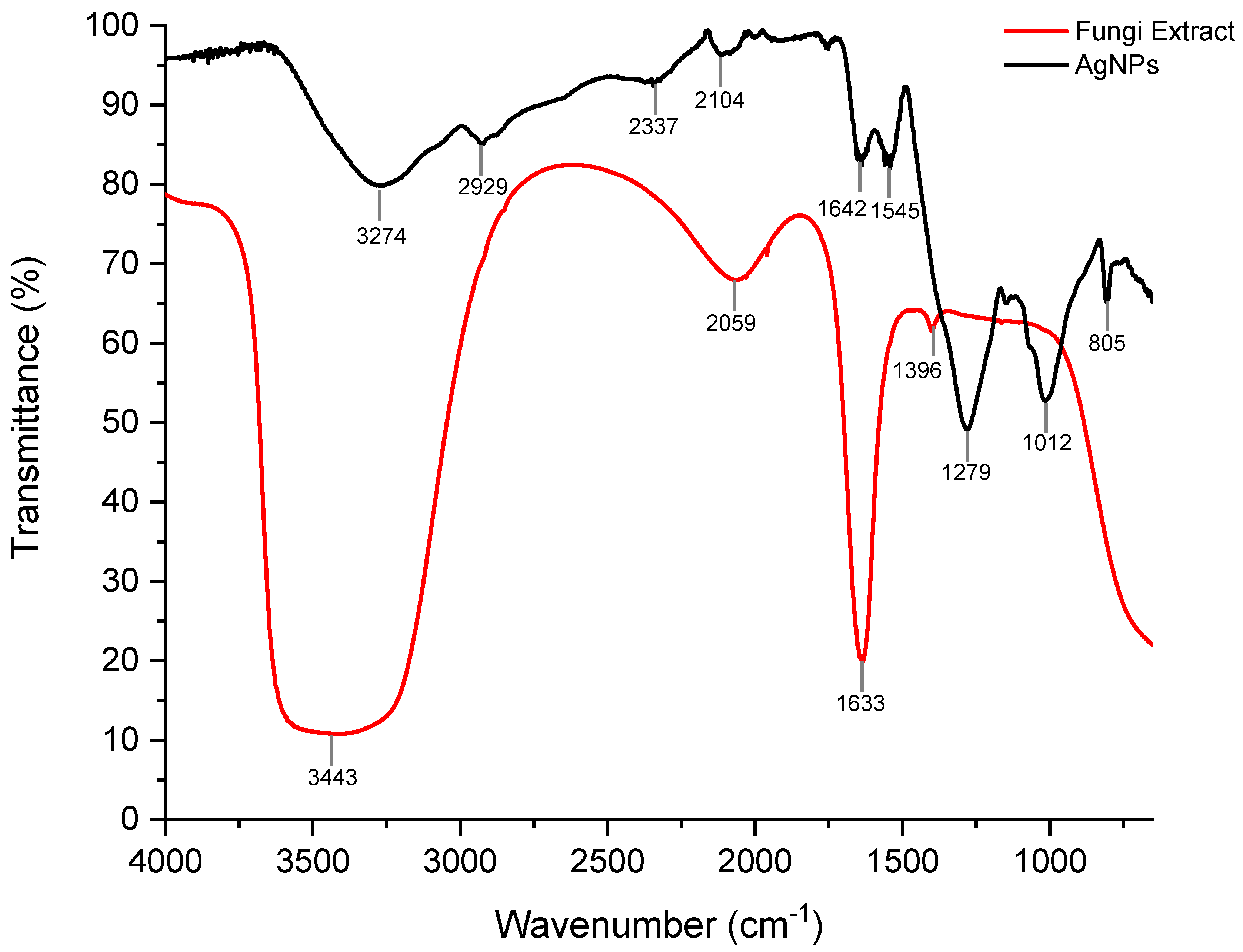
The FTIR analysis of the silver nanoparticles (formed in the combined presence of I. fumosorosea aqueous extract + AgNO3 3mM) showed characteristic bands in the spectral regions of 3400–2400, 2260–2100, 1680–1630, 1450–1375, 1350–1000 and 805 cm−1 (Figure 7), indicating the presence of N-H, C-N, C=O, C-H, and C≡C functional groups, which may be related to the presence of proteins or amino acid residues present in the fungal extract—being these molecules possibly responsible for the reduction of silver ions and the formation of nanoparticles [22,25,26].
The band shift of the fungal extract spectrum at 3443 and 1633 cm−1 regions compared to the silver nanoparticles spectrum can be attributed to the bond breaking of amine and amide groups, which are associated with the interaction in the silver nanoparticle formation. The interaction between amine/amide groups and silver nanoparticles in the biological route was previously reported, using plants and fungal extracts [10,27,28,29]. In this context, we suggest that the amine and amide groups present in the I. fumosorosea extract may be the precursors of the silver ions reduction, supporting the synthesis of the silver nanoparticles.
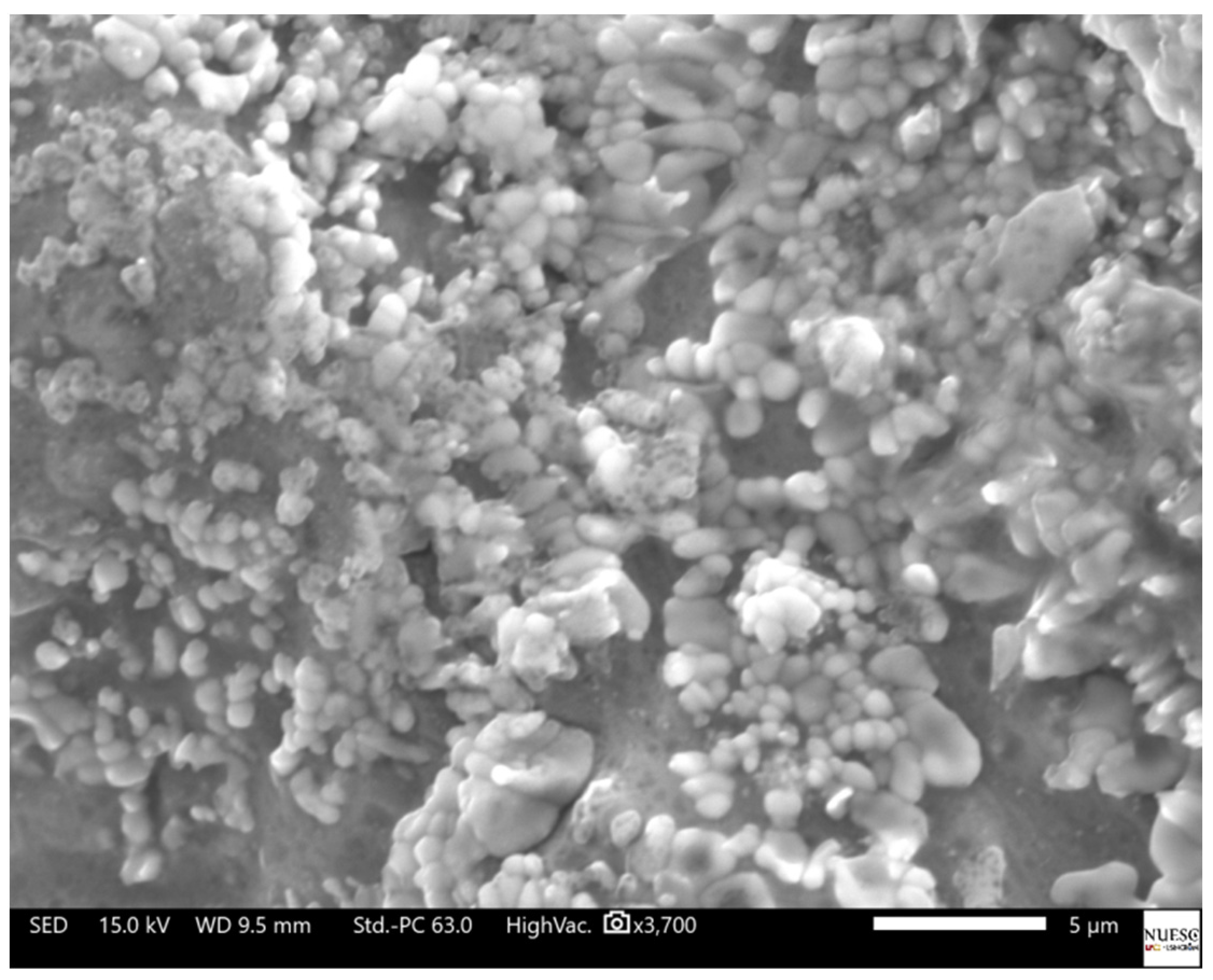
The SEM analysis of the silver nanoparticles allowed the visualization of circular morphology predominance, showing varied particle sizes adhered to the matrix of the lyophilized fungal extract (Figure 8). The morphology and diameter variation of the silver nanoparticle is influenced by the extract concentration of the microorganism used in the reaction (i.e., reducing agent), as well as the proportion of silver ions available in the reaction [29,30].

3.3. The Lethal Concentration of Biogenic Silver Nanoparticles against P. xylostella

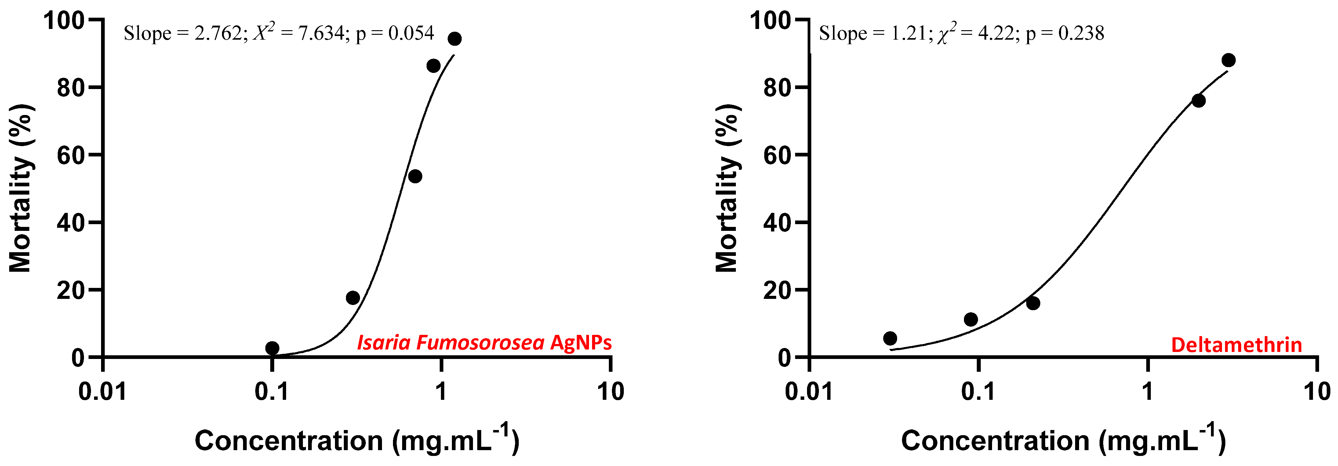
Silver nanoparticles at different concentrations were toxic against P. xylostella, showing a significant variation in the results (F4.20 = 53.967; p = 0.000). Thus, it was possible to estimate the sublethal concentrations using a probit procedure. (Figure 9). The LCs estimated values used for the silver nanoparticles action against P. xylostella were LC30 = 0.144 mg mL−1, LC50 = 0.691 mg mL−1, and LC90 = 2.011 mg mL−1, considering an angular coefficient (line slope) of 2.762 (Table 3).
Some studies reported the lethal concentration of the silver nanoparticles for P. xylostella. The silver nanoparticles synthesized using a marine macro red algae Hypnea muciformis showed high toxicity on P. xylostella in the larval and pupal stages, presenting LC50 = 26.47 mg L−1 for second instar larvae at 96 h of exposure [11]. The silver nanoparticles produced from ethanolic extracts of ginger (Zingiber officinale), Indian neem (Azadirachta indica), fig tree (Datura stramonium), bitter melon (Momordica charantia), clove (Syzygium aromaticum), cinnamon (Melia azedarach), eucalyptus (Eucalyptus camaldulensis) and garlic (Allium sativum) also presented toxic effects on P. xylostella third instar larvae, showing LC50 ranging from 0.337 mg mL−1 (fig tree) to 0.729 mg mL−1 (garlic) at 72 h of exposure [31]. The LCs values of silver nanoparticles synthesized using I. fumosorosea extract have not yet been reported on for the different life cycle stages of P. xylostella. The silver nanoparticle LCs variation previously reported may occur due to the different nanoparticle physicochemical characteristics and also because of the molecules involved in the nanoparticle synthesis and capping. The molecules from extracts or even adhered to the biogenic nanoparticle can directly influence the biological action, affecting the toxicity [32,33]. The LC50 value (0.691 mg mL−1) of the silver nanoparticles was higher compared to Roni et al. (2015) (26.47 mg L−1) [11], and within the range compared to Ali et al., (2019) [31] (0.337–0.729 mg mL−1).
The P. xylostella populations have high genetic variability and toxicity resistance to several chemical compounds [34]. The toxic effect of the commercial insecticide Deltamethrin (Decis 25 EC) on P. xylostella was estimated in order to compare the LCs results of the silver nanoparticles toxicity tests. The different concentrations of the Deltamethrin showed significant difference in the mortality of P. xylostella (F4.20 = 56.864; p = 0.000), presenting LC30 = 0.009 mg mL−1; LC50 = 0.301 mg mL−1, and LC90 = 3.427 mg mL−1, considering an angular coefficient (line slope) of 1.214 (Figure 9; Table 3). Deltamethrin is an insecticide belonging to the pyrethroid family, indicated in the control of P. xylostella at a concentration of 0.0075 mg mL−1. The CL30 value (0.009 mg mL1) estimated in our study is approximate to the concentration indicated for Deltamethrin application. Other study presented the values of LC50 = 0.332 mg mL−1 (0.321–0.345) and LC90 = 0.436 mg mL−1 (0.411–0.473) to control P. xylostella using Deltamethrin [35], that is, approximate value of LC50 presented in our study, but different comparing to the CL90. The present study presented a slower action of Deltamethrin LC90 on P. xylostella, possibly due to adaptation and better resistance to this chemical. The difference in the toxicity results from the application of silver nanoparticles (from AgNO3 3 mM + entomopathogenic fungi I. fumosorosea) and Deltamethrin may occur because of their different modes of action. Deltamethrin acts against insects via ingestion and direct contact, promoting neurotoxicity through the interaction with sodium channels, present in the neurological cells. The frequent use of Deltamethrin promotes mutations in insect structures, contributing to its resistance [36,37]. Silver nanoparticles can also act against insects via ingestion and direct contact, presenting toxicity through different routes, promoting cuticle damage, and oxidative stress and reducing enzymatic activity (acetylcholinesterase and Cu-dependent enzymes). These different routes of toxicity can act simultaneously, favoring the action of the silver nanoparticles against the insect and reducing the possibility of insect resistance. Therefore, the use of silver nanoparticles is an advantageous alternative in the control of insect pests, such as P. xylostella, which is resistant to several chemical pesticides [38,39,40,41,42,43]. In addition, Deltamethrin is highly toxic to humans and other animals and may cause the development of cellular anomalies. In contrast, silver nanoparticles can be synthesized by environmentally sustainable techniques, such as synthesis using fungal extracts, which improve their biocompatibility [44,45]. However, the interaction of AgNPs with plant tissues, their post-harvest preservation, and also their interaction with the human gastrointestinal tract, must be considered to ensure the safe use of such materials [46,47].

3.4. Survival, Viability, and Longevity Analysis of P. xylostella Larvae Exposed to LC50 of Silver Nanoparticles

The survival of P. xylostella caterpillars treated with silver nanoparticles (from AgNO3 3 mM + entomopathogenic fungi I. fumosorosea) at LC50 (0.691 mg mL−1) showed different results compared to the control group (Log-rank Mantel-Cox: X2 = 129.814; p = 0.000). The insects treated with silver nanoparticles presented a median lethal time (LT50) of 4.624 days (3.821–5.427) and 78.25 ± 11.04% (F1.8 = 192.627; p = 0.000) of cumulative mortality over the entire larval period (Figure 10).
The silver nanoparticles (from AgNO3 3 mM + entomopathogenic fungi I. fumosorosea) showed effective toxicity against P. xylostella caterpillars. Significant insecticidal action was previously reported by the exposure of biogenic silver nanoparticles on P. xylostella (96 h) [11]. The time required for the establishment of toxicity is variable, as it depends on the nanomaterial characteristics and insect physiology [40].
The viability of P. xylostella caterpillars treated with silver nanoparticles showed a significative difference (T = 13.879; p = 0.000), However, significative differences were not observed in the duration of larval and pupal stages (days) and in the pupal viability (%), compared to the control (Table 4).
The studies reporting the insect life cycle in the toxicity analyses provide knowledge about the total effect of a particular substance [48]. The larval viability is a parameter directly proportional to caterpillar mortality, thus these results can be correlated (Figure 9).
Several factors can affect insect survival and development, such as temperature, humidity, photoperiod, food, and exposure to toxic agents. These factors can promote sublethal effects, including the non-hatching of eggs, morphological structural anomalies, non-emergence of adults, and fecundity reduction. Silver nanoparticles can affect insect physiology, reducing fertility and survival, according to previously observed in the Drosophila sp. (Diptera: Drosophilidae) [1,49,50]. The results presented a modification of both the life cycle and larval survival of P. xylostella submitted to silver nanoparticles. The chronic effect was not observed after the change from the larval to the pupal phase.

4. Conclusions

In this study, a sustainable green synthesis approach of silver nanoparticles mediated by entomopathogenic fungi was described to be applicable to insect control. This work highlighted the potential action of entomopathogenic fungi extracts in the reduction and stabilization of silver nanoparticles. The insecticidal efficacy of silver nanoparticles synthesized in the presence of I. fumosorosea against P. xylostella was also demonstrated. Silver nanoparticles presented toxicity against P. xylostella caterpillars, showing close values compared to a commercial chemical pesticide; it may thus be considered an advantageous alternative in the control of P. xylostella populations.

Author Contributions

T.S.S., C.d.S.V., S.A.J., P.S. and M.d.C.M. contributed to the conceptualization, methodology, validation, formal analysis, and investigation and to the writing—original draft preparation; E.S.-L., S.A.J., E.B.S., P.S. and M.d.C.M. contributed to the supervision, writing the second version, editing the revision, project administration, resources, and funding acquisition. All authors have read and agreed to the published version of the manuscript.

Funding

This research was funded by the Banco do Nordeste (Convênio BNB FUNDECI 2017.0016 - Project: “Desenvolvimento de nanobioinseticida preparada a partir de óleo essencial de laranja com aplicação no controle de insetos na agricultura”), Coordenação Aperfeiçoamento de Pessoal de Nivel Superior (CAPES), Fundação de Amparo à Pesquisa do Estado de Sergipe (FAPITEC), and Conselho Nacional de Desenvolvimento Científico e Tecnológico (CNPq).

Institutional Review Board Statement

Not applicable.

Informed Consent Statement

Not applicable.

Data Availability Statement

Not applicable.

Acknowledgments

We would like to thank the Sergipe Agricultural Development Company (Emdagro), Sergipe Technological Park (SergipeTec), Institute of Technology and Research (ITP/UNIT), and the Industrial Biotechnology Program, University Tiradentes, for assistance during the research.

Conflicts of Interest

The authors declare no conflict of interest.

References

  1. De Bortoli, S.; Polanczyk, R.; Vacari, A.; De Bortoli, C.; Duarte, R. Plutella xylostella (Linnaeus, 1758)(Lepidoptera: Plutellidae): Tactics for integrated pest management in Brassicaceae. Weed Pest Control.-Conv. New Chall. Rij. InTechOpen 2013, 31–51. [Google Scholar] [CrossRef] [Green Version]
  2. Reddy, G.V. Integrated Management of Insect Pests on Canola and Other Brassica Oilseed Crops; CABI: Wallingford, UK, 2017. [Google Scholar]
  3. Gautam, M.; Singh, H.; Kumar, S.; Kumar, V.; Singh, G.; Singh, S. Diamondback moth, Plutella xylostella (Linnaeus)(Insecta: Lepidoptera: Plutellidae) a major insect of cabbage in India: A review. J. Entomol. Zool. Stud. 2018, 6, 1394–1399. [Google Scholar]
  4. Li, Z.; Furlong, M.; Yonow, T.; Kriticos, D.; Bao, H.-L.; Yin, F.; Lin, Q.; Feng, X.; Zalucki, M. Management and population dynamics of diamondback moth (Plutella xylostella): Planting regimes, crop hygiene, biological control and timing of interventions. Bull. Entomol. Res. 2019, 109, 257–265. [Google Scholar] [CrossRef]
  5. Chaud, M.; Souto, E.B.; Zielinska, A.; Severino, P.; Batain, F.; Oliveira-Junior, J.; Alves, T. Nanopesticides in Agriculture: Benefits and Challenge in Agricultural Productivity, Toxicological Risks to Human Health and Environment. Toxics 2021, 9, 131. [Google Scholar] [CrossRef]
  6. Santos, T.S.; Silva, T.M.; Cardoso, J.C.; Albuquerque-Júnior, R.L.D.; Zielinska, A.; Souto, E.B.; Severino, P.; Mendonca, M.D.C. Biosynthesis of Silver Nanoparticles Mediated by Entomopathogenic Fungi: Antimicrobial Resistance, Nanopesticides, and Toxicity. Antibiotics 2021, 10, 852. [Google Scholar] [CrossRef]
  7. de Oliveira, D.M.; Menezes, D.B.; Andrade, L.R.; Lima, F.D.C.; Hollanda, L.; Zielinska, A.; Sanchez-Lopez, E.; Souto, E.B.; Severino, P. Silver nanoparticles obtained from Brazilian pepper extracts with synergistic anti-microbial effect: Production, characterization, hydrogel formulation, cell viability, and in vitro efficacy. Pharm. Dev. Technol. 2021, 26, 539–548. [Google Scholar] [CrossRef]
  8. Sanchez-Lopez, E.; Gomes, D.; Esteruelas, G.; Bonilla, L.; Lopez-Machado, A.L.; Galindo, R.; Cano, A.; Espina, M.; Ettcheto, M.; Camins, A.; et al. Metal-Based Nanoparticles as Antimicrobial Agents: An Overview. Nanomaterials 2020, 10, 292. [Google Scholar] [CrossRef] [PubMed] [Green Version]
  9. Garcia-Torra, V.; Cano, A.; Espina, M.; Ettcheto, M.; Camins, A.; Barroso, E.; Vazquez-Carrera, M.; Garcia, M.L.; Sanchez-Lopez, E.; Souto, E.B. State of the Art on Toxicological Mechanisms of Metal and Metal Oxide Nanoparticles and Strategies to Reduce Toxicological Risks. Toxics 2021, 9, 195. [Google Scholar] [CrossRef] [PubMed]
  10. Ahadian, S.; Yamada, S.; Ramón-Azcón, J.; Estili, M.; Liang, X.; Nakajima, K.; Shiku, H.; Khademhosseini, A.; Matsue, T. Hybrid hydrogel-aligned carbon nanotube scaffolds to enhance cardiac differentiation of embryoid bodies. Acta Biomater. 2016, 31, 134–143. [Google Scholar] [CrossRef] [PubMed]
  11. Roni, M.; Murugan, K.; Panneerselvam, C.; Subramaniam, J.; Nicoletti, M.; Madhiyazhagan, P.; Dinesh, D.; Suresh, U.; Khater, H.F.; Wei, H. Characterization and biotoxicity of Hypnea musciformis-synthesized silver nanoparticles as potential eco-friendly control tool against Aedes aegypti and Plutella xylostella. Ecotoxicol. Environ. Saf. 2015, 121, 31–38. [Google Scholar] [CrossRef]
  12. Santos, T.S.; Passos, E.M.d.; Seabra, M.G.d.J.; Souto, E.B.; Severino, P.; Mendonça, M.D. Entomopathogenic Fungi Biomass Production and Extracellular Biosynthesis of Silver Nanoparticles for Bioinsecticide Action. Appl. Sci. 2021, 11, 2465. [Google Scholar] [CrossRef]
  13. Phanjom, P.; Ahmed, G. Effect of different physicochemical conditions on the synthesis of silver nanoparticles using fungal cell filtrate of Aspergillus oryzae (MTCC No. 1846) and their antibacterial effect. Adv. Nat. Sci. Nanosci. Nanotechnol. 2017, 8, 045016. [Google Scholar] [CrossRef] [Green Version]
  14. Othman, A.M.; Elsayed, M.A.; Al-Balakocy, N.G.; Hassan, M.M.; Elshafei, A.M. Biosynthesis and characterization of silver nanoparticles induced by fungal proteins and its application in different biological activities. J. Genet. Eng. Biotechnol. 2019, 17, 1–13. [Google Scholar] [CrossRef] [Green Version]
  15. Bhatnagar, S.; Kobori, T.; Ganesh, D.; Ogawa, K.; Aoyagi, H. Biosynthesis of silver nanoparticles mediated by extracellular pigment from Talaromyces purpurogenus and their biomedical applications. Nanomaterials 2019, 9, 1042. [Google Scholar] [CrossRef] [Green Version]
  16. Sampaio, S.; Viana, J.C. Production of silver nanoparticles by green synthesis using artichoke (Cynara scolymus L.) aqueous extract and measurement of their electrical conductivity. Adv. Nat. Sci. Nanosci. Nanotechnol. 2018, 9, 045002. [Google Scholar] [CrossRef]
  17. Singhal, G.; Bhavesh, R.; Kasariya, K.; Sharma, A.R.; Singh, R.P. Biosynthesis of silver nanoparticles using Ocimum sanctum (Tulsi) leaf extract and screening its antimicrobial activity. J. Nanoparticle Res. 2011, 13, 2981–2988. [Google Scholar] [CrossRef]
  18. Hamouda, R.A.; Hussein, M.H.; Abo-Elmagd, R.A.; Bawazir, S.S. Synthesis and biological characterization of silver nanoparticles derived from the cyanobacterium Oscillatoria limnetica. Sci. Rep. 2019, 9, 13071. [Google Scholar] [CrossRef] [Green Version]
  19. Sadeghi, R.; Etemad, S.G.; Keshavarzi, E.; Haghshenasfard, M. Investigation of alumina nanofluid stability by UV–vis spectrum. Microfluid. Nanofluidics 2015, 18, 1023–1030. [Google Scholar] [CrossRef]
  20. Hadjidemetriou, M.; Kostarelos, K. Evolution of the nanoparticle corona. Nat. Nanotechnol. 2017, 12, 288–290. [Google Scholar] [CrossRef]
  21. Banu, A.N.; Balasubramanian, C. Myco-synthesis of silver nanoparticles using Beauveria bassiana against dengue vector, Aedes aegypti (Diptera: Culicidae). Parasitol. Res. 2014, 113, 2869–2877. [Google Scholar] [CrossRef]
  22. Banu, A.N.; Balasubramanian, C. Optimization and synthesis of silver nanoparticles using Isaria fumosorosea against human vector mosquitoes. Parasitol. Res. 2014, 113, 3843–3851. [Google Scholar] [CrossRef] [PubMed]
  23. Różalska, S.; Soliwoda, K.; Długoński, J. Synthesis of silver nanoparticles from Metarhizium robertsii waste biomass extract after nonylphenol degradation, and their antimicrobial and catalytic potential. RSC Adv. 2016, 6, 21475–21485. [Google Scholar] [CrossRef]
  24. Zhao, X.; Zhou, L.; Riaz Rajoka, M.S.; Yan, L.; Jiang, C.; Shao, D.; Zhu, J.; Shi, J.; Huang, Q.; Yang, H.; et al. Fungal silver nanoparticles: Synthesis, application and challenges. Crit. Rev. Biotechnol. 2018, 38, 817–835. [Google Scholar] [CrossRef]
  25. Mistry, H.; Thakor, R.; Patil, C.; Trivedi, J.; Bariya, H. Biogenically proficient synthesis and characterization of silver nanoparticles employing marine procured fungi Aspergillus brunneoviolaceus along with their antibacterial and antioxidative potency. Biotechnol. Lett. 2021, 43, 307–316. [Google Scholar] [CrossRef] [PubMed]
  26. Gupta, K.; Chundawat, T.S.; Malek, N. Antibacterial, antifungal, photocatalytic activities and seed germination effect of mycosynthesized silver nanoparticles using Fusarium oxysporum. Biointerface Res. Appl. Chem. 2020, 11, 12082–12091. [Google Scholar]
  27. Qayyum, S.; Oves, M.; Khan, A.U. Obliteration of bacterial growth and biofilm through ROS generation by facilely synthesized green silver nanoparticles. PLoS ONE 2017, 12, e0181363. [Google Scholar] [CrossRef] [Green Version]
  28. Justus, B.; Arana, A.F.M.; Gonçalves, M.M.; Wohnrath, K.; Boscardin, P.M.D.; Kanunfre, C.C.; Budel, J.M.; Farago, P.V.; de Paula, J.d.F.P. Characterization and cytotoxic evaluation of silver and gold nanoparticles produced with aqueous extract of Lavandula dentata L. in relation to K-562 cell line. Braz. Arch. Biol. Technol. 2019, 62. [Google Scholar] [CrossRef]
  29. Lotfy, W.A.; Alkersh, B.M.; Sabry, S.A.; Ghozlan, H.A. Biosynthesis of silver nanoparticles by Aspergillus terreus: Characterization, optimization, and biological activities. Front. Bioeng. Biotechnol. 2021, 265. [Google Scholar] [CrossRef]
  30. Rose, G.K.; Soni, R.; Rishi, P.; Soni, S.K. Optimization of the biological synthesis of silver nanoparticles using Penicillium oxalicum GRS-1 and their antimicrobial effects against common food-borne pathogens. Green Process. Synth. 2019, 8, 144–156. [Google Scholar] [CrossRef] [Green Version]
  31. Ali, A.; Mohammad, S.; Khan, M.A.; Raja, N.I.; Arif, M.; Kamil, A.; Mashwani, Z.U. Silver nanoparticles elicited in vitro callus cultures for accumulation of biomass and secondary metabolites in Caralluma tuberculata. Artif. Cells Nanomed. Biotechnol. 2019, 47, 715–724. [Google Scholar] [CrossRef] [Green Version]
  32. Bhargava, A.; Dev, A.; Mohanbhai, S.J.; Pareek, V.; Jain, N.; Choudhury, S.R.; Panwar, J.; Karmakar, S. Pre-coating of protein modulate patterns of corona formation, physiological stability and cytotoxicity of silver nanoparticles. Sci. Total Environ. 2021, 772, 144797. [Google Scholar] [CrossRef] [PubMed]
  33. Rónavári, A.; Igaz, N.; Adamecz, D.I.; Szerencsés, B.; Molnar, C.; Kónya, Z.; Pfeiffer, I.; Kiricsi, M. Green silver and gold nanoparticles: Biological synthesis approaches and potentials for biomedical applications. Molecules 2021, 26, 844. [Google Scholar] [CrossRef]
  34. Tamilselvan, R.; Kennedy, J.; Suganthi, A. Monitoring the resistance and baseline susceptibility of Plutella xylostella (L.)(Lepidoptera: Plutellidae) against spinetoram in Tamil Nadu, India. Crop Prot. 2021, 142, 105491. [Google Scholar] [CrossRef]
  35. Costa, Â.C.F.; Cavalcanti, S.C.H.; Santana, A.S.; Lima, A.P.S.; Brito, T.B.; Oliveira, R.R.B.; Macêdo, N.A.; Cristaldo, P.F.; Araújo, A.P.A.; Bacci, L. Insecticidal activity of indole derivatives against Plutella xylostella and selectivity to four non-target organisms. Ecotoxicology 2019, 28, 973–982. [Google Scholar] [CrossRef] [PubMed]
  36. Endersby, N.; Viduka, K.; Baxter, S.; Saw, J.; Heckel, D.G.; McKechnie, S. Widespread pyrethroid resistance in Australian diamondback moth, Plutella xylostella (L.), is related to multiple mutations in the para sodium channel gene. Bull. Entomol. Res. 2011, 101, 393–405. [Google Scholar] [CrossRef]
  37. Wang, X.L.; Su, W.; Zhang, J.H.; Yang, Y.H.; Dong, K.; Wu, Y.D. Two novel sodium channel mutations associated with resistance to indoxacarb and metaflumizone in the diamondback moth, Plutella xylostella. Insect Sci. 2016, 23, 50–58. [Google Scholar] [CrossRef] [Green Version]
  38. Kumari, R.; Singh, D. Silver nanoparticle in agroecosystem: Applicability on plant and risk-benefit assessment. In Plant Responses to Xenobiotics; Springer: Singapore, 2016; pp. 293–305. [Google Scholar]
  39. Benelli, G.; Caselli, A.; Canale, A. Nanoparticles for mosquito control: Challenges and constraints. J. King Saud Univ.-Sci. 2017, 29, 424–435. [Google Scholar]
  40. Benelli, G. Mode of action of nanoparticles against insects. Environ. Sci. Pollut. Res. 2018, 25, 12329–12341. [Google Scholar] [CrossRef] [PubMed]
  41. Amarasinghe, L.; Wickramarachchi, P.; Aberathna, A.; Sithara, W.; De Silva, C. Comparative study on larvicidal activity of green synthesized silver nanoparticles and Annona glabra (Annonaceae) aqueous extract to control Aedes aegypti and Aedes albopictus (Diptera: Culicidae). Heliyon 2020, 6, e04322. [Google Scholar] [CrossRef] [PubMed]
  42. Jamtsho, T.; Banu, N.; Kinley, C. Critical Review on Past, Present and Future Scope of Diamondback Moth Management. Plant Arch. 2021, 21, 1199–1210. [Google Scholar] [CrossRef]
  43. Shahzad, K.; Manzoor, F. Nanoformulations and their mode of action in insects: A review of biological interactions. Drug Chem. Toxicol. 2021, 44, 1–11. [Google Scholar] [CrossRef] [PubMed]
  44. Esan, V.; Mahboob, S.; Al-Ghanim, K.A.; Elanchezhiyan, C.; Al-Misned, F.; Ahmed, Z.; Govindarajan, M. Novel biogenic synthesis of silver nanoparticles using Alstonia venenata leaf extract: An enhanced mosquito larvicidal agent with negligible impact on important eco-biological fish and insects. J. Clust. Sci. 2021, 32, 489–497. [Google Scholar] [CrossRef]
  45. Medeiros, J.F.D.; Acayaba, R.D.A.; Montagner, C.C. The chemistry in the human health risk assessment due to pesticides exposure. Química Nova 2021, 44, 584–598. [Google Scholar]
  46. Jasrotia, P.; Nagpal, M.; Mishra, C.N.; Sharma, A.K.; Kumar, S.; Kamble, U.; Bhardwaj, A.K.; Kashyap, P.L.; Kumar, S.; Singh, G.P. Nanomaterials for Postharvest Management of Insect Pests: Current State and Future Perspectives. Front. Nanotechnol. 2022, 3, 811056. [Google Scholar] [CrossRef]
  47. Ferdous, Z.; Nemmar, A. Health Impact of Silver Nanoparticles: A Review of the Biodistribution and Toxicity Following Various Routes of Exposure. Int. J. Mol. Sci. 2020, 21, 2375. [Google Scholar] [CrossRef] [Green Version]
  48. Lemes, A.A.F.; Sipriano-Nascimento, P.T.; Vieira, N.F.; Cardoso, C.P.; Vacari, A.M.; Bortoli, S.A.D. Acute and Chronic Toxicity of Indoxacarb in Two Populations of Plutella xylostella (Lepidoptera: Plutellidae). J. Econ. Entomol. 2021, 114, 298–306. [Google Scholar] [CrossRef]
  49. Rodrigues, W.C. Fatores que influenciam no desenvolvimento dos insetos. Info Insetos 2004, 1, 1–4. [Google Scholar]
  50. Raj, A.; Shah, P.; Agrawal, N. Dose-dependent effect of silver nanoparticles (AgNPs) on fertility and survival of Drosophila: An in-vivo study. PLoS ONE 2017, 12, e0178051. [Google Scholar] [CrossRef]
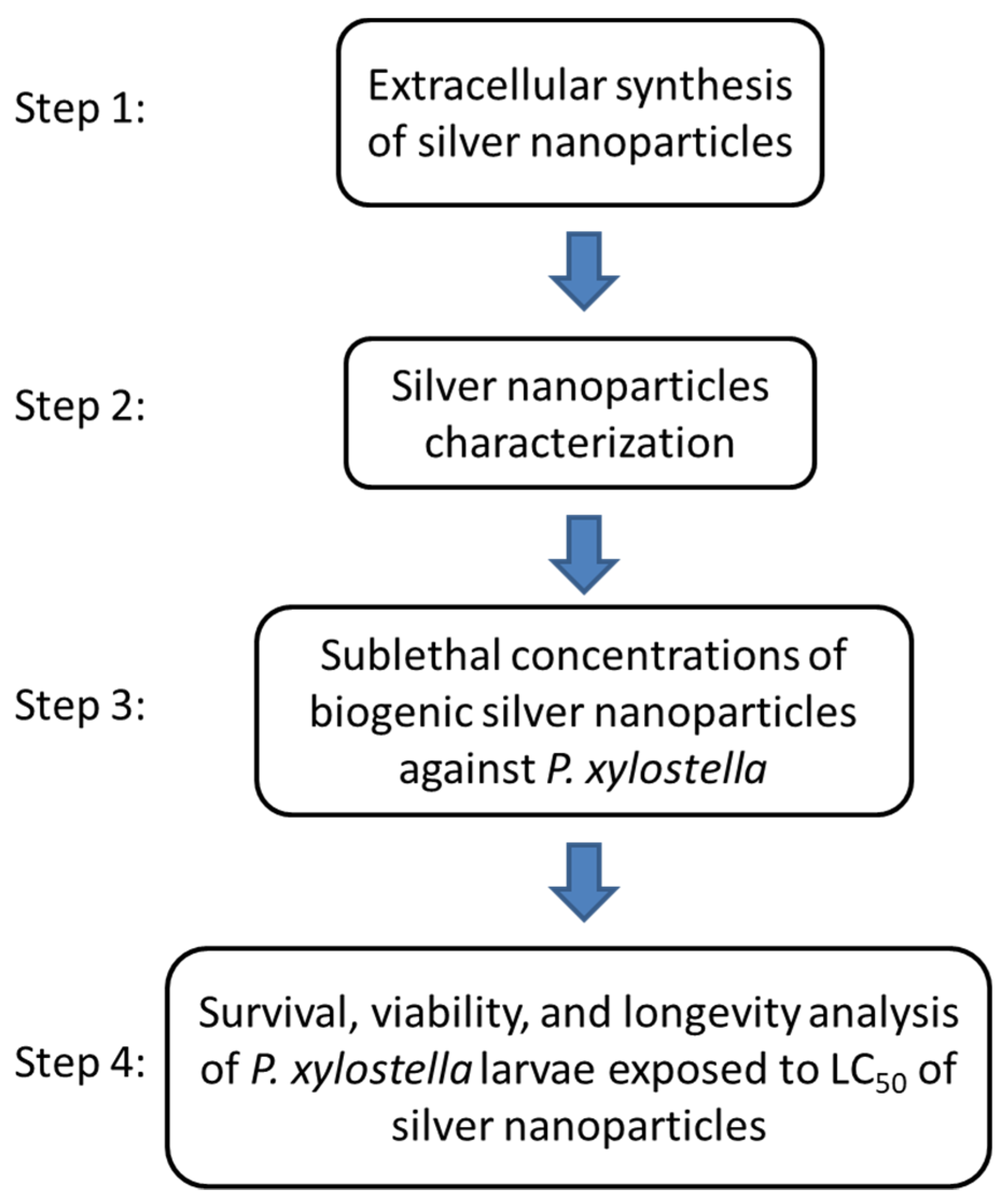
Figure 1. Experimental procedure flowchart.
Materials 15 07596 g001
Figure 2. Synthesis reaction of silver nanoparticles with aqueous extract of entomopathogenic fungi isolates and variation in silver nitrate concentration. SE109 e 1260 (B. bassiana), SE202 (M. anisopliae) and SE301 (I. fumosorosea). (a) Pure extract, (b) AgNO3 1 mM, (c) AgNO3 3 mM, (d) AgNO3 5 mM.
Materials 15 07596 g002
Figure 3. UV-Visible absorption spectrum of the evaluation of the effect of silver nitrate concentration on the synthesis of silver nanoparticles using an extract of the fungus B. bassiana (isolate 1260).
Materials 15 07596 g003
Figure 4. UV-Visible absorption spectrum of the evaluation of the effect of silver nitrate concentration on the synthesis of silver nanoparticles using an extract of the fungus B. bassiana (isolate SE109).
Materials 15 07596 g004
Figure 5. UV-Visible absorption spectrum of the evaluation of the effect of silver nitrate concentration on the synthesis of silver nanoparticles using an extract of the fungus M. anisopliae (isolate SE202).
Materials 15 07596 g005
Figure 6. UV-Visible absorption spectrum of the evaluation of the effect of silver nitrate concentration on the synthesis of silver nanoparticles using an extract of the fungus I. fumosorosea (isolate SE301).
Materials 15 07596 g006
Figure 7. Fourier Transform Infrared Vibrational Spectroscopy (FTIR) spectrum showing band patterns of the aqueous extract of the fungus I. fumosorosea (red) and of AgNPs synthesized with the fungal extract (black).
Materials 15 07596 g007
Figure 8. Scanning electron microscopy of silver nanoparticles synthesized with I. fumosorosea extract and 3 mM silver nitrate.
Materials 15 07596 g008
Figure 9. Probit curve of Plutella xylostella exposed to silver nanoparticles synthesized with the entomopathogenic fungus Isaria fumosorosea (left) and Deltamethrin (right). Residual effect after 72 h.
Materials 15 07596 g009
Figure 10. Survival curve of Plutella xylostella caterpillars treated with Isaria fumosorosea AgNPs.
Materials 15 07596 g010
Table 1. Entomopathogenic fungi isolates used in this study.
IsolateSpecieOriginCollection Site/Host
1260Beauveria bassianaLaboratory of Pathology of Insects (USP), Piracicaba, (SP-Brazil)Insect
Leptopharsa larvae
SE109Laboratory of Biotechnological Pest Control (Sergipetec), São Cristovão (SE-Brazil)Soil
SE202Metarhizium anisopliaeLaboratory of Biotechnological Pest Control (Sergipetec), São Cristovão (SE-Brazil)Soil
SE301Isaria fumosoroseaLaboratory of Biotechnological Pest Control (Sergipetec), São Cristovão (SE-Brazil)Soil
Table 2. Effect of silver nitrate (AgNO3) concentration on the average diameter and polydispersity of silver nanoparticles synthesized with aqueous extracts of entomopathogenic fungi.
IsolateAgNO3Diameter (nm) *PI *
12601 mM202.60 ± 20.250.54 ± 0.08
3 mM224.83 ± 61.870.52 ± 0.04
5 mM257.07 ± 17.860.55 ± 0.06
SE1091 mM172.77 ± 59.730.54 ± 0.07
3 mM121.37 ± 20.680.50 ± 0.05
5 mM119.48 ± 6.110.47 ± 0.04
SE2021 mM103.97 ± 6.300.27 ± 0.05
3 mM122.44 ± 4.340.41 ± 0.03
5 mM135.32 ± 23.390.54 ± 0.07
SE3011 mM129.03 ± 30.510.55 ± 0.09
3 mM86.26 ± 8.190.37 ± 0.05
5 mM119.39 ± 26.690.54 ± 0.06
* Mean values with standard error. PI = Polydispersity index.
Table 3. Estimation of lethal concentration (LC) of silver nanoparticles synthesized with the fungus Isaria fumosorosea on Plutella xylostella (AgNPs) and Deltamethrin.
TreatmentNLC30LC50LC90Slopeχ2P
AgNPs7410.144
(0.104–0.182)
0.691
(0.627–0.762)
2.011
(1.684–2.550)
2.762
(±0.231)
7.6340.054
Deltamethrin7500.009
(0.005–0,013)
0.301
(0.238–0.391)
3.427
(2.211–6.033)
1.214
(±0.088)
4.2220.238
Lethal concentration values in mg/mL, followed by lower and upper limit (p < 0.05); Slope value, followed by standard error; N: Total insects used in the analysis; χ2: Chi-square test; P: Significance of χ2.
Table 4. Analysis of the impact of silver nanoparticles synthesized with the fungus Isaria fumosorosea (LC50) on the development of Plutella xylostella.
AgNPs Concentration (mg/mL)Life Cycle Phase
LarvaePupae
Duration (Days)Viability (%)Duration (Days)Viability (%)
0.010.8 ± 1.4491.14 ± 1.768.6 ± 1.3479.56 ± 7.06
0.710.8 ± 1.0421.74 ± 11.047.2 ± 1.0977.95 ± 14.90
T0.00013.8791.8070.219
p10.0000.1080.832
Average values of duration and viability, followed by standard error; Analysis of variance (ANOVA) for comparing values in the column (p < 0.05); T = Student test; p = Calculated significance.
Publisher’s Note: MDPI stays neutral with regard to jurisdictional claims in published maps and institutional affiliations.