Abstract
To investigate the effect of co-doping on the optical properties of Eu(2+/3+) doped in Ba0.98SiO3:0.02Eu, the series of Ba0.96SiO3:0.02Eu, 0.02R+/3+ (R+ = Li+, K+ or Na+, R3+ = La3+ or Y3+) phosphors were synthesized using a solid-state reaction method. The excitation efficiency due to the charge transfer band (CTB) was enhanced via co-doping of R+ and the emission intensity due to Eu3+ was thus increased by 3.7 times compared with that of the single-doped Ba0.98SiO3:0.02Eu3+. However, the co-doping of R+ does not increase the emission intensity of Eu3+ via the direct 7F0→5L6 excitation of Eu3+, but rather decreases it. On the other hand, the emission intensities due to Eu2+ were decreased via the co-doping of R+ but increased via the co-doping of La3+. The present work reveals that the optical properties of Eu3+ or Eu2+ doped in BaSiO3 depend not on the charge state (+ or 3+) of the co-doped ions, but on the co-doped element itself.
1. Introduction
The photoluminescent properties of rare-earth (RE) ions have attracted considerable interest because of their potential applications as phosphors, white light-emitting diodes (WLEDs), and lasers, etc. When doped in inorganic host materials, RE ions energetically prefer the trivalent state in hosts. Some RE ions, such as Eu, Sm, and Yb, where Yb2+ is more stable than Yb3+, are doped with divalent atoms in some hosts. Specifically, Eu2+-doped phosphors exhibit a broad emission band that varies from ultra-violet to red, depending on the host lattice, and some of Eu3+-doped phosphors are used as a typical red phosphor. Thus, europium-based luminescence materials are extensively used in solid-state lighting, solar cells, red phosphors, and WLEDs.
Silicates are widely used as phosphor hosts due to their merits, such as their high thermal stability, the high energy efficiency of the silicate phosphors, and the inexpensive nature of the raw materials. Most often, the europium ions in a host are substituted into alkaline earth metals such as Ba2+, Sr2+, or Ca2+. When Eu3+ ions are doped in silicate hosts such as BaSiO3, Ca2SiO4, and Sr2SiO4 through heat-treatment in air, there should be a charge balance mechanism involved in replacing A2+(A2+ = Ba2+, Ca2+, and Sr2+) with Eu3+. In the case of BaSiO3, in which Eu3+ replaces the Ba2+ site based on the ionic radius, negatively charged barium ion vacancies (Vba) are created for charge compensation. It is known that the creation of such defects leads to a deterioration in the optical performance of the synthesized phosphors. Jiang et al. have investigated the influence of Li+ co-doping on the photoluminescence of Eu3+ doped in an SrWO4 host [1]. Li et al. also studied the effect of Li+, Na+, and K+ on the PL enhancement of Tb3+ doped in CaMoO4 [2]. In many cases, it has been reported that the co-doping of alkali-metal ions or Y3+ or La3+ enhances the emission intensity of RE ions doped in host materials [3,4,5,6,7]. However, it has also been reported that the emission intensity decreased in ZnTa2O6:Pr3+ co-doped with alkali-metal ions [8]. The Eu3+ ions doped in a BaSiO3 host will be located along with the Ba2+ site because the ionic radius of Eu3+ is similar to that of Ba2+. Meanwhile, the BaSiO3:Eu2+ phosphors are mostly prepared via the heat-treatment of BaSiO3:Eu3+ in a reducing atmosphere at high temperature. In this reduction process, the Vba defect created for charge compensation in BaSiO3:Eu3+ will lose its charge compensation function. To date, to the best of the authors’ knowledge, there have been no reports on the effect of Vba on the optical properties of Eu2+ doped in BaSiO3.
In this article, we report on the co-doping effects of alkali metal ions and La3+ (or Y3+) on the optical properties of BaSiO3:Eu(2+/3+) fabricated via a solid-state reaction (SSR) method. The effects of Li+, K+, Na+, La3+, and Y3+ co-doped with Eu(2+/3+) on the crystalline phase and the optical properties of BaSiO3:Eu(2+/3+) are discussed. The results showed that the doping of Li+, K+, Na+, La3+, and Y3+ can significantly affect the optical properties of Eu(2+/3+) but the element used for the co-doping ion has a greater effect on the optical properties of Eu(2+/3+) than the charge state (+ or 3+) of the co-doping ion.
2. Materials and Methods
A series of Ba0.98SiO3:0.02Eu, Ba0.96SiO3:0.02Eu, and 0.02R (R = Li, K, Na, La, and Y) phosphors were prepared using a conventional solid-state reaction. The BaCO3 (99+%), SiO2 (particle size 5~20 nm, 99.5%), Eu2O3 (99.99%), K2CO3 (99+%), Li2CO3 (99+%), Li2O (97%), Na2CO3 (99+%), LaCl3 (99.99%), and Y2O3 (99.99%), purchased from Merck, were used as a raw materials but SiO2 was used 20% more to obtain a single-phase BaSiO3 sample. The raw materials were thoroughly mixed in an agate mortar and subsequently fired in air at 1100 °C for 4 h to synthesize the Eu3+-doped BaSiO3. The BaSiO3:Eu2+ samples were prepared by sintering the BaSiO3:Eu3+ samples again under a reduction atmosphere (95% N2 + 5% H2) at 1200 °C for 6 h. The specific formulations of the studied samples are shown in Table 1.

Table 1.
Studied samples.
The crystal phase of the obtained samples was identified using an X-ray diffractometer (X’Pert PRO MPD-3040, Malvern Panalytical, Malvern, UK, CuKα1, λ = 1.5406 Å) operating at 40 kV and 30 mA with a scan speed of 0.02°/sec. The photoluminescence excitation (PLE) and emission (PL) spectra were measured using a fluorescent spectrofluorometer (JASCO FP-8500, JASCO, Tokyo, Japan) equipped with an integrating sphere (ISF-834, JASCO, Tokyo, Japan). All measurements were performed at room temperature.
3. Results and Discussion
Figure 1 shows the X-ray powder diffraction (XRD) patterns of the studied samples. The XRD patterns of all samples were relatively well matched with pure BaSiO3 (JCPDS-702112) within the accuracy of our XRD equipment, indicating that the studied samples were single-phased BaSiO3, which crystallizes in an orthorhombic structure and has a space group of P212121 with lattice constants of a = 0.4580 nm, b = 0.5611 nm, and c = 1.2431 nm, α = β = γ = 90° [7]. This also means that the activator Eu(2+/3+) ions and co-doping ions R+/3+ which were small-doped in the samples, did not cause any significant changes in the crystal phase of BaSiO3. Miscellaneous minor peaks were marked with a plus (+) and an asterisk (*). The XRD patterns of samples 8–14, prepared under a reducing atmosphere, were identical to those of samples 1–7 because there was no change in crystallinity even if the secondary heat treatment was performed under a reducing atmosphere. The XRD patterns of the samples prepared under a reducing atmosphere are thus not presented in Figure 1.
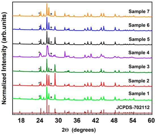
Figure 1.
XRD patterns of Ba0.98SiO3:0.02Eu3+, 0.02R (R = Li+, K+ and Na+).
Based on the data provided by the Cambridge Crystallographic Data Center, Si4+ ions are located at the center of a tetrahedron coordinated with four O2− ions. The [SiO4]2− tetrahedrons form a chain running parallel to the c-axis by sharing corners and the Ba2+ ions are accommodated in the tunnels between chains [9,10].
In our previous work [11], the crystal structure of BaSiO3 was found to be highly dependent on the particle size of the SiO2 used as a raw material. Even though this result was not presented in this paper, it was not possible to fabricate samples not having the minor second phase with SiO2, the particle size of which was greater than 500 nm. This means that the characteristics of raw materials having the same molecular formula can highly affect the crystal structures of the samples prepared under the same preparation conditions. Specially, the optical properties of BaSiO3:Eu2+ are highly sensitive to the presence or absence of the minor second phase in the crystal structure, even though this is not true of other materials. The main reason that previously reported results are slightly different may be due to the difficulties involved in achieving single-phase BaSiO3:Eu2+ fabrication without having some minor peaks [10,12,13].
The crystal structure of BaSiO3 has been detailed in other works [10,14], where a Ba site and six oxygen atoms bond together in the BaSiO3 unit cell. Since Ba2+ ions (0.135 nm) are closer to Eu3+ ions (0.0947 nm) than Si4+ ions (0.0400 nm) in terms of their ionic radius, Ba2+ is expected to be replaced by Eu3+ [7]. Assuming that Ba2+ is expected to be replaced by Eu3+, an imbalance of charges is generated in the sample, resulting in the creation of a negatively charged defect, VBa. To solve this problem, charge compensators such as Li+, K+, or Na+ were co-doped with Eu3+ and the effect of charge compensation on the emission of Eu3+ has been reported [7]. Yang et al. reported that doping with Na+ and K+ ions has a drastic effect on the improvement of the excitation and emission of Eu3+ [7]. However, through this work, we found that doping with Y3+ ions also has the same effect as that obtained with Na+ and K+ ions. This result means that the improvement of the excitation and emission of Eu3+ could be determined by the species of co-doping ions, rather than the charge state of the co-doping ions, and also shows that this process may not be explained with charge compensation alone, as described so far.
Figure 2 illustrates the excitation and emission spectra of Ba0.96SiO3:0.02Eu3+, 0.02R+/3+ samples. The PLE, measured by monitoring the strongest Eu3+ emission at 612 nm, is shown in Figure 2a. When Eu2O3, which is an optical active material, was mixed with other host materials and then heat-treated in air, the Eu ions were doped into Eu3+ in the host material due to its chemical stability. The PLE spectra exhibit a wide band known as a charge transfer band (CTB), where the electrons come from the entirely filled 2p orbitals of O2- ions and are transferred to the partially filled 4f orbitals of Eu3+. The CTB ranged from 200 nm to 290 nm, with the highest peak centered around 228 nm. Lingxiang Yang et al. reported that the CTB peaked around 246 nm [14]. This 18 nm difference in the CTB peak position may be attributed to the crystallinity of the studied samples. The sample studied by Lingxiang Yang et al. exhibited relatively many miscellaneous peaks, which were ascribed to the BaSi2O5 phase (JCPDS#26-0176), compared with our samples.
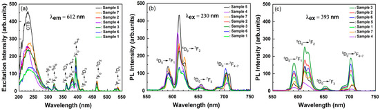
Figure 2.
Excitation spectra (a) obtained by monitoring the emission at 612 nm, emission spectra at an excitation wavelength of (b) 230 nm and (c) 393 nm for Ba0.96SiO3:0.02Eu3+, 0.02R+/3+ phosphors (samples 1–7).
Based on our previous study, we knew that the optical properties of Eu(2+/3+) doped in BaSiO3 were more highly dependent upon the crystallinity of the host materials, compared to other host materials. Several narrow bands from 300 nm to 550 nm, due to the intra-4f transitions of Eu3+ ions, were also observed. Since the transitions of Eu3+ were very stable, and are almost certainly not affected by the lattice structure or neighboring anion ligands of Eu3+, the excitation peaks were almost same for all samples. However, in the case of the CTB, things were quite different. It has been reported in the literature that the CTB depends greatly on the lattice structure of the matrix in which Eu3+ was introduced [15]. For example, the CTB peak position for Sr2SiO4:Eu3+ is 293 nm and it was 250 nm for the Gd2O3 [16]. It is thus reasonable to assume that the 18 nm difference in the CTB peak position may be attributed to the crystallinity of the studied samples.
One interesting aspect of our results is that the excitation efficiency due to the CTB in sample 5, co-doped with Na+ ions, was enhanced by 3.7 times compared with that of the single-doped sample 1. The CTB excitation efficiency of sample 7, co-doped with Y3+, was two times higher than that of sample 6, co-doped with La3+. Furthermore, The CTB excitation efficiency of samples co-doped with Li+ or K+ was twice as high as that of the single-doped sample 1. These results mean that the co-doped R+/3+ ions act as a sensitizer and can thus contribute to the enhancement of the emission intensity due to Eu3+. It is thus relatively reasonable to conclude that the CTB excitation efficiency does not depend greatly on the charge state of the co-doping element, but rather on the species of co-doping ions.
Upon the excitation of CTB’s 230 nm region, as shown in Figure 2a, the 5D0→7F2 maximum transition intensity of Eu3+ for sample 5, co-doped with Na+, was enhanced by a factor of 3.7 times compared with sample 1. In the case of samples 2 and 3, which were co-doped with the same Li+, the 5D0→7F2 transition intensity of sample 2 was stronger than that of sample 3. We have thus shown through this study, for the first time, that the emission efficiency of Eu3+ is dependent upon the raw material of the co-doping ions, rather than the charge state of the co-doping ions. Figure 2c shows the emission spectra obtained at an excitation wavelength of 393 nm, which represents a 7F0→5L6 excitation of Eu3+. The emission intensities of samples 2, 3, and 6, co-doped with Li+ or La3+ ions, were stronger than that of sample 1 but that of sample 4, co-doped with K+, Na+ and K+, was much weaker than that of sample 1.
Figure 3 shows the strongest Eu3+ emission intensity that was obtained at 612 nm when excited by 230 nm and 393 nm, and it also shows the difference in the excitation efficiency through the CTB and the direct 7F0→5L6 excitation of Eu3+. When excited by CTB’s 230 nm region, the results clearly showed that the excitation efficiencies of sample 5, co-doped with Na+, and sample 4, co-doped with K+, were much better than those of other samples. However, the excitation efficiency of sample 4, excited by 393 nm, which represents 7F0→5L6 excitation of Eu3+, was the lowest among the studied samples.
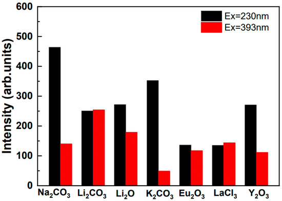
Figure 3.
Effect of co-doping on the PL intensity of Eu3+ ions excited by 230 nm and 393 nm, respectively.
Figure 4a–d shows the PLE and PL spectra of the studied BaSiO3:Eu2+ phosphors (samples 8–14). The PLE spectra, measured by monitoring the 577 nm emission intensity, are shown in Figure 4a. The PLE spectra cover a very broad region, from 200 to 450 nm, with two peaks of 280 nm and 340 nm. The broad excitation covered the UV/vis range widely, which matches well with commercial LED chip emissions, so this phosphor is suitable for solid-state lighting based on an n-UV LED chip. The broad excitation band is commonly ascribed to the crystal-field split 4f65d1 configuration of Eu2+ ions. It is well known that the 4f65d1 electron state is not shielded from anion ligands in the compounds. It has thus been suggested that the 4f65d1 electron state has a strong interaction with neighboring anion ligands in the BaSiO3 host, which results in a broad excitation band, as shown in Figure 4a. Since samples 8–14 were actually the same as samples 1–7, which were heat-treated in air, except for the fact that samples 8–14 were heat-treated in a reducing atmosphere, and the ionic radius of Eu3+ (0.0947 nm) was smaller than that of Ba (0.135 nm), it was expected that Eu2+ ions in sample 8–14 would replace the Ba sites of BaSiO3. The excitation spectrum shown in Figure 4a has two peaks at 280 nm and 340 nm, which corresponds to the crystal-field splitting of the 5d levels in the excited 4f65d1 configuration of the Eu2+. The excitation spectra for samples 8–14 show a similar pattern, even though the relative excitation intensity over the whole excitation band is slightly different.
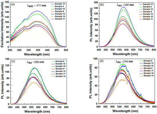
Figure 4.
Excitation (a) and emission (b–d) spectra of the Ba0.96SiO3:0.02Eu2+, 0.02R+/3+ phosphors (samples 8–14).
Figure 4b shows the room-temperature PL spectra of samples 8–14 excited at 340 nm, in which the PL features of Eu2+ showed slight changes in peak position depending on the co-doping ions. Since only one Ba site exists crystallographically in BaSiO3, a single broad emission band from 450 nm to 750 nm with the peak location at 577 nm was expected, which was attributed to the typical 4f65d1→4f7 transition. This single and symmetrical broad emission band indicated that the Eu2+ ions were replaced into the only Ba2+ site in BaSiO3, as expected. The full width at half maximum (FWHM) of the emission spectrum was about 131 nm, which is broader than the common values of FWHM (50–100 nm) of Eu2+ in most phosphors [3,12,17] and is also broader than that (119 nm) of the well-known yellow-emitting Y3Al5O12:Ce3+ phosphor [18]. This broad emission may indicate that the interaction of Eu2+ with the ligands of the BaSiO3 host was strong and thus it may also be suitable as a warm white light source using an n-UV Led chip.
We also found the emission due to Eu3+ ions when samples 8–14 were excited at 280 nm was efficient in exciting Eu3+ ions. This implies that not the all Eu3+ ions present in samples 1–7 were completely reduced into Eu2+ via heat treatment under the mixed nitrogen (90%) and hydrogen (10%) atmosphere. Co-doping ions such as Li+, K+, and Na+ were effective in enhancing the emission of Eu3+ via the excitation of the CTB (refer to Figure 2b) but they decreased the emission intensity of Eu2+ ions, as shown in Figure 4b. Based on the radii of the compound elements, all Eu3+ ions were expected to replace the Ba2+ sites of BaSiO3 due to the occurrence of the charge imbalance. To solve this charge imbalance effect, charge compensators such as Li+, K+, and Na+ ions were co-doped with Eu3+ ions. When Eu3+ and R+ (R+ = Li+, K+ and Na+) were co-doped, two kinds of defects were created, since one Eu3+ and one R+ occupied two Ba2+ sites in BaSiO3:Eu3+, R+ (samples 2–5). Considering that the ionic sizes of cations varied with their coordination numbers (CN), we expected that Eu3+ and R+ ions would prefer to replace the Ba2+ (CN = 8, r = 1.42Å) in BaSiO3 because the ionic radius of Eu3+ (CN = 6, r = 1.07~1.087 Å) was closer to that of Ba2+ and the Si4+ sites with an ionic radius of 0.42 Å were too small for Eu3+ to occupy [7,9,19]. The ionic radii of Li+, Na+, and K+ ions are also known to be 0.9, 1.16, and 1.52 Å, respectively [19,20].
Ba0.96SiO3:0.02Eu2+, 0.02R+/3+ (samples 8–14) phosphors were prepared by carrying out a second heat-treatment of Ba0.96SiO3:0.02Eu3+, 0.02R+/3+ samples (samples 1–7) in a mixed nitrogen (90%) and hydrogen (10%) atmosphere to reduce Eu3+ into Eu2+. When Ba0.96SiO3:0.02Eu3+, 0.02R+/3+ phosphors were heated in the mixed nitrogen (90%) and hydrogen (10%) atmosphere, hydroxyl bonds were formed, which seemed to play an important role in the reduction of Eu3+ ions. We assume that these hydroxyl bonds may have reacted with the negatively charged barium ion vacancies (Vba) which were generated due to the charge imbalance when Eu3+ replaced the Ba2+ site in Ba0.96SiO3:0.02Eu3+, 0.02R+/3+, resulting in the decreased PL intensity of Eu2. However, more experimental and theoretical studies are needed to reveal the mechanism underlying the decrease in PL intensity due to Eu2+.
Figure 4d shows the room-temperature PL spectra of samples 8–14 excited at 254 nm, which belonged to the CTB and was also efficient to excite Eu3+ ions. As shown in Figure 4c, the PL intensity of sample 13, co-doped with La3+, was stronger than that of samples 9–12, co-doped with R+. Furthermore, the PL intensity of sample 13 was stronger than that of sample 14, which was co-doped with Y3+. Figure 2 shows that the PL intensity of the 5D0→7F2 transition of Eu3+ for sample 6, co-doped with La3+, was weaker than that of the 5D0→7F4–7 transition but the PL intensity of the 5D0→7F2 transition of Eu3+ for samples 1–7 (except for sample 6) was stronger than that of the 5D0→7F4–7 transition. In general, it is known that the emission intensity due to the 5D0→7F4–7 transition is weaker than the emission intensity caused by the 5D0→7F2 transition because Ω4 is much smaller than Ω2 according to the Judd–Ofelt theory [20]. However, depending on the symmetry of Eu3+, it has been reported that the luminescence intensity due to the 5D0→7F4 transition is stronger than that of 5D0→7F2 [21]. The results shown in Figure 2 and Figure 4d may indicate that the PL intensity of Eu2+ or Eu3+ doped in BaSiO3 depends on the co-doped element or the environment around the Eu2+ or Eu3+, rather than the charge state of the co-doped elements such as R+, La3+, and Y3+.
4. Conclusions
In summary, Ba0.98+0.02RSiO3: 0.02Eu(2+/3+), R+/3+(R = Li+, Na+, K+, La3+ and Y3+) phosphors were prepared by means of a solid-state reaction method and studies were carried out on the effect of co-doping with ions such as Li+, Na+, K+, La3+, and Y3+ on the optical properties of the Ba0.98SiO3: 0.02Eu(2+/3+) phosphors.
One notable result of this work is that the excitation efficiency due to CTB was enhanced by co-doping of R+/3+, which acted as a kind of sensitizer, and thus increased the emission intensity due to Eu3+ by 3.7 times. However, the co-doped R+/3+ ions did not increase the emission intensity of Eu3+ via direct 7F0→5L6 excitation of Eu3+, but rather decreased it. On the other hand, the co-doped R+ ions did not enhance the emission intensity of Eu2+ via the excitation of the CTB or through a direct excitation of Eu2+, but the co-doped La3+ did achieve this effect. The present work thus reveals for the first time that the optical properties of Eu2+ or Eu3+ doped in BaSiO3 depend on the co-doped element itself, rather than the charge state of the co-doped elements such as R+, La3+, and Y3+.
Author Contributions
P.N.: synthesis of compounds, XRD, and luminescence measurements. K.J.: the idea of work and its planning, interpretation of results, and preparation of the paper. All authors have read and agreed to the published version of the manuscript.
Funding
This work was financially supported by the National Research Foundation of Korea funded by the Ministry of Education, Science and Technology (NRF-2018R1D1A1B07043099).
Informed Consent Statement
Not applicable.
Conflicts of Interest
The authors declare no conflict of interest.
References
- Jiang, P.; Gao, W.; Cong, R.; Yang, T. Structural investigation of the A-site vacancy in scheelite and the luminescence behavior of two continuous solid solutions A1−1.5xEux□0.5xWO4 and A0.64−0.5yEu0.24Liy□0.12−0.5yWO4 (A = Ca, Sr, □ = vacancy). Dalton Trans. 2015, 44, 6175–6183. [Google Scholar] [CrossRef] [PubMed]
- Li, X.; Yang, Z.; Guan, L.; Guo, J.; Wang, Y.; Guo, Q. Synthesis and luminescent properties of CaMoO4:Tb3+, R+ (Li+, Na+, K+). J. Alloys Compd. 2009, 478, 684–686. [Google Scholar] [CrossRef]
- Zhong, J.; Zhao, W.; Lan, L.; Wang, J.; Chen, J.; Wang, N. Enahnced emission from Li2CaSiO4:Eu2+ phosphors by doping Y3+. J. Alloys Compd. 2014, 592, 213–219. [Google Scholar] [CrossRef]
- Xiaoye, H.U.; Zhenhua, L.I.; Xin, X.U.; Yongxiu, L.I. Enhancement of photoluminescence of Ba2SiO4:Eu2+ by co-doping of La3+ or Y3+. J. Rare Earths 2009, 27, 47–49. [Google Scholar]
- Gupta, S.K.; Sudarshan, K.; Yadav, A.K.; Gupta, R.; Bhattacharyya, D.; Jha, S.N.; Kadam, R.M. Deciphering the Role of Charge Compensator in Optical Properties of SrWO4:Eu3+:A (A = Li+, Na+, K+): Spectroscopic Insight Using Photoluminescence, Positron Annihilation, and X-ray Absorption. Inorg. Chem. 2018, 57, 821–832. [Google Scholar] [CrossRef] [PubMed]
- Jiayue, S.; Jinli, L.; Jianfeng, S.; Haiyan, D. Luminescence properties of a new red emitting Eu3+-doped alkaline-earth fluoborate phosphor: BaCa(1−2x)BO3F:xEu3+, xM+ (M = Li, Na, K). J. Rare Earths 2011, 29, 321–325. [Google Scholar]
- Yang, Z.; Yang, L.; Pu, Y.; Zhu, D. The effect and mechanism of different charge compensation on the luminescent properties of Eu-doped BaSiO3 phosphor calcined in air with self-reduction. Opt. Mater. 2021, 114, 110981. [Google Scholar] [CrossRef]
- Noto, L.L.; Ntwaeaborwa, O.M.; Yagoub, M.Y.; Swart, H.C. Enhancement of persistent luminescence of ZnTa2O6:Pr3+ by addition Li+, Na+, K+ and Cs+ ions. Mater. Res. Bull. 2015, 70, 545–552. [Google Scholar] [CrossRef]
- Guo, C.; Xu, Y.; Ren, Z.; Bai, J. Blue-White-Yellow Tunable Emission from Ce3+ and Eu2+ Co-Doped BaSiO3 Phosphors. J. Electrochem. Soc. 2011, 158, J373–J376. [Google Scholar] [CrossRef]
- Pan, Y.X.; Liu, G.K. Enhancement of Eu2+ Luminescence in BaO-SiO2 Compounds Through Composition Modification, Spectroscopy. Letters 2011, 44, 1–7. [Google Scholar]
- Namkhai, P.; Jang, K. Optical Properties of Eu(2+/3+) on the Fabrication Method for BaSiO3:Eu(2+/3+) Phosphors. New Phys. Sae Mulli 2021, 71, 1010–1017. [Google Scholar] [CrossRef]
- Cho, I.S.; Yim, D.K.; Kwak, C.H.; An, J.S.; Roh, H.S.; Hong, K.S. Investigation of crystal/electronic structure effects on the photoluminescence properties in the BaO–SiO2:Eu2+ systems. J. Lumin. 2012, 132, 375–380. [Google Scholar]
- Xu, J.; Zhao, Y.; Chen, I.J.; Mao, Z.; Yang, Y.; Wang, D. Insights into the discrepant luminescence for BaSiO3:Eu2+ phosphors prepared by solid-state reaction and precipitation reaction methods. Luminescence 2017, 32, 957–963. [Google Scholar] [CrossRef] [PubMed]
- Yang, L.; Zhu, D.; Liu, S.; Wang, J.; Zhao, C.; Pu, Y. Photoluminescence properties and crystal structure of BaSi03:xEu3+, yBi3+ red phosphor synthesized by co-precipitation method. Phys. B Condens. Matter 2019, 556, 6–11. [Google Scholar] [CrossRef]
- Nag, A.; Kutty, T.R.N. The light induced valence change of europium in Sr2SiO4:Eu involing transient crystal structure. J. Mater. Chem. 2004, 14, 1598–1604. [Google Scholar] [CrossRef]
- Chang, C.; Kimura, F.; Kimura, T.; Wada, H. Preparation and characterization of rod-like Eu:Gd2O3 phosphor through a hydrothermal routine. Mater. Lett. 2005, 59, 1037–1041. [Google Scholar] [CrossRef]
- Song, W.; Kim, H.; Kim, Y.; Yang, H. Synthesis of Ba2SiO3O8:Eu2+ Phosphor for Fabrication of White Light-Emitting Diodes Assisted by ZnCdSe/ZnSe Quantum. J. Electrochem. Soc. 2010, 157, J319–J323. [Google Scholar] [CrossRef]
- Song, W.; Kim, Y.; Yang, H. Yellow-emitting phosphor of Sr3B2O6:Eu2+ for application to white light-emitting diodes. Mater. Chem. Phys. 2009, 117, 500–503. [Google Scholar] [CrossRef]
- Mukherjee, S.; Sudarsan, V.; Vatsa, R.K.; Tyagi, A.K. Luminescence studies on lanthanide ions (Eu3+, Dy3+ and Tb3+) doped YAG:Ce nano-phosphors. J. Lumin. 2009, 129, 69–72. [Google Scholar] [CrossRef]
- Chan, W.L.; Liu, Z.; Lu, S.; Tanner, P.A.; Wong, K.L. The reported anomalous emission intensity of the 5D0 →7F4 transition of Eu3+ in a molybdate double perovskite. J. Mater. Chem. C 2015, 3, 960–963. [Google Scholar] [CrossRef]
- Blasse, G. Luminescence from the Eu3+ Ion in D4d Symmetry. Inorg. Chim. Acta 1988, 142, 153–154. [Google Scholar] [CrossRef]
Publisher’s Note: MDPI stays neutral with regard to jurisdictional claims in published maps and institutional affiliations. |
© 2022 by the authors. Licensee MDPI, Basel, Switzerland. This article is an open access article distributed under the terms and conditions of the Creative Commons Attribution (CC BY) license (https://creativecommons.org/licenses/by/4.0/).