Article

Colored Tattoo Ink Screening Method with Optical Tissue Phantoms and Raman Spectroscopy
Department of Metrology and Optoelectronics, Faculty of Electronics, Telecommunications and Informatics, Gdańsk University of Technology, G. Narutowicza 11/12, 80-233 Gdańsk, Poland
*
Author to whom correspondence should be addressed.
Materials 2021, 14(12), 3147; https://doi.org/10.3390/ma14123147
Submission received: 10 May 2021 / Revised: 1 June 2021 / Accepted: 3 June 2021 / Published: 8 June 2021
(This article belongs to the Special Issue Advanced Materials for Biophotonics Applications)

Abstract

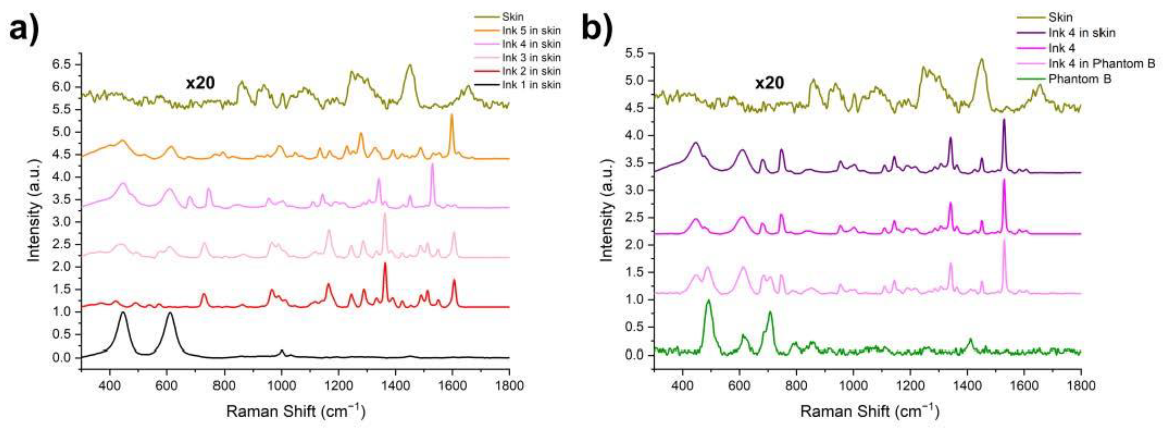
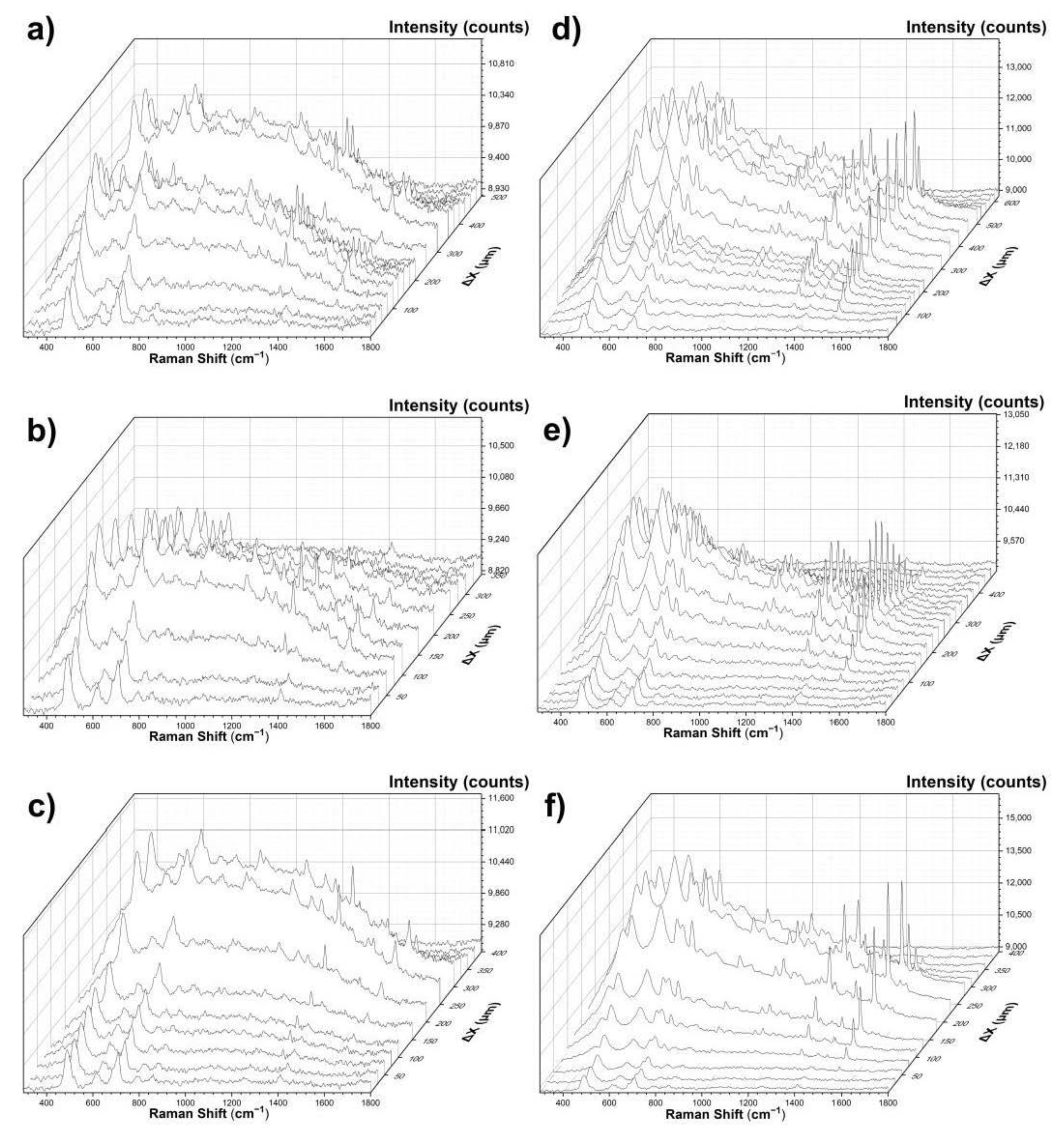
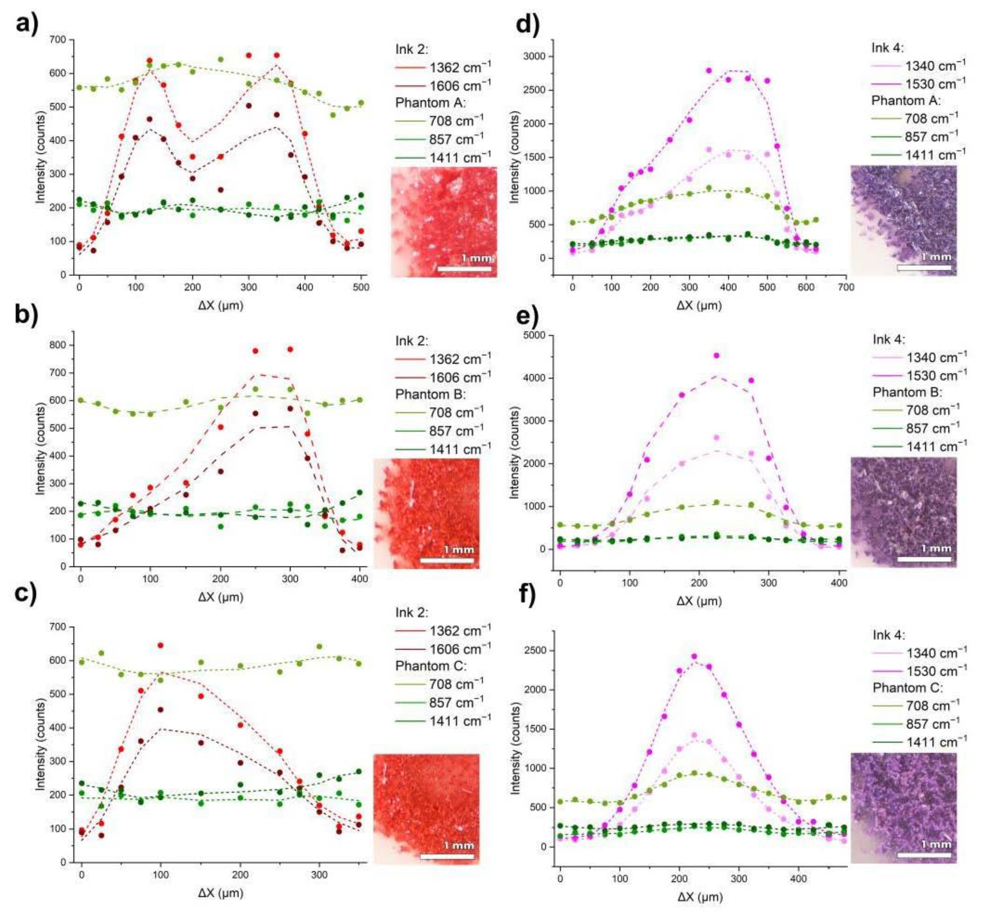
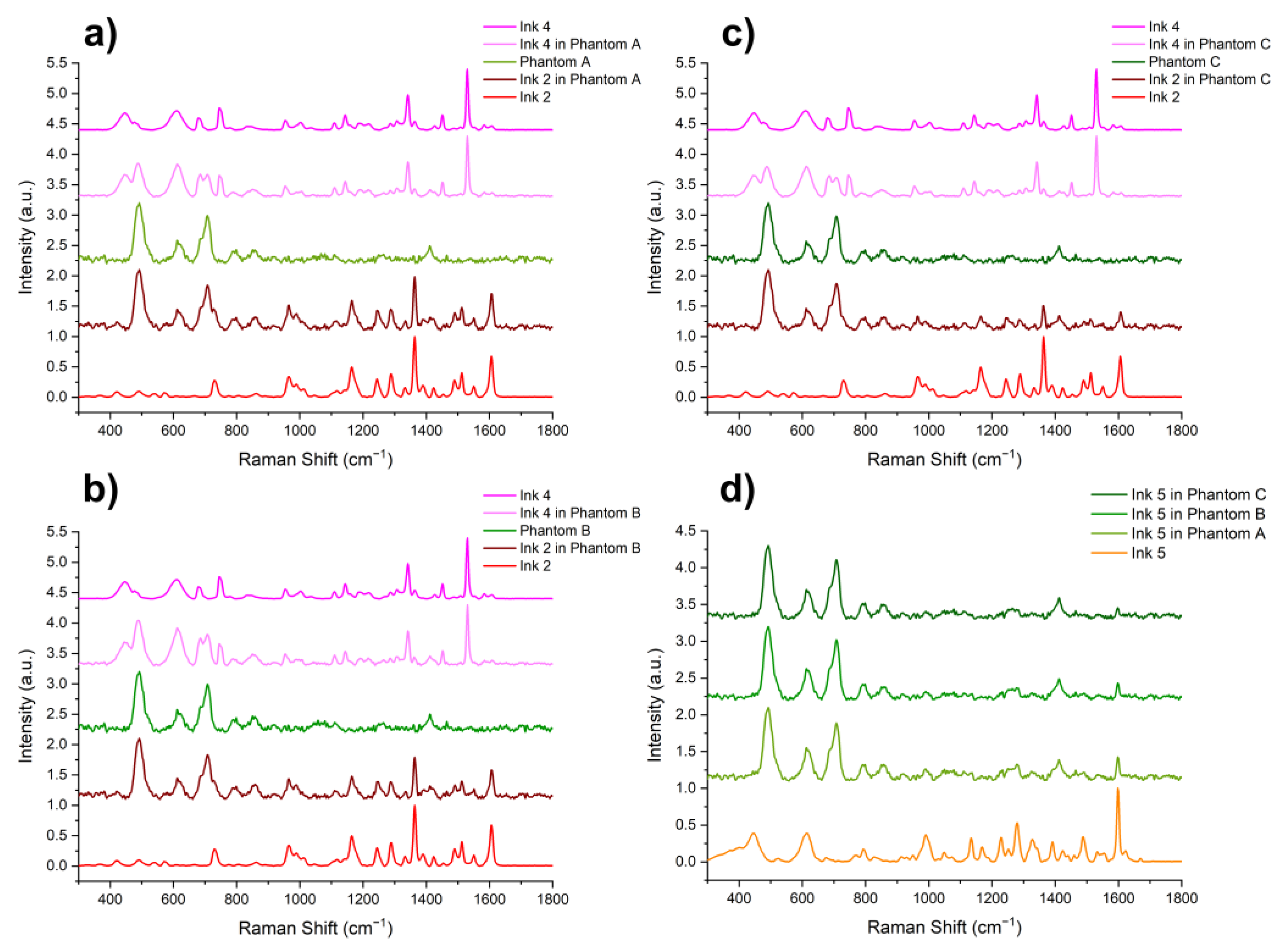
Due to the increasing popularity of tattoos among the general population, to ensure their safety and quality, there is a need to develop reliable and rapid methods for the analysis of the composition of tattoo inks, both in the ink itself and in already existing tattoos. This paper presents the possibility of using Raman spectroscopy to examine tattoo inks in biological materials. We have developed optical tissue phantoms mimicking the optical scattering coefficient typical for human dermis as a substitute for an in vivo study. The material employed herein allows for mimicking the tattoo-making procedure. We investigated the effect of the scattering coefficient of the matrix in which the ink is located, as well as its chemical compositions on the spectra. Raman surface line scanning has been carried out for each ink in the skin phantom to establish the spatial gradient of ink concentration distribution. This ensures the ability to detect miniature concentrations for a tattoo margin assessment. An analysis and comparison of the spectra of the inks and the tattooed inks in the phantoms are presented. We recommend the utilization of Raman spectroscopy as a screening method to enforce the tattoo ink safety legislations as well as an early medical diagnostic screening tool.
Keywords: Raman spectroscopy; tattoo inks; optical tissue phantoms Raman spectroscopy; tattoo inks; optical tissue phantoms