Next Article in Journal
New Management in Bilateral Masseter Muscle Hypertrophy
Previous Article in Journal
Porcine Urinary Bladder Extracellular Matrix for the Salvage of Fibula Free Flap Skin Paddle: Technical Note and Description of a Case
 
 
Craniomaxillofacial Trauma & Reconstruction is published by MDPI from Volume 18 Issue 1 (2025). Previous articles were published by another publisher in Open Access under a CC-BY (or CC-BY-NC-ND) licence, and they are hosted by MDPI on mdpi.com as a courtesy and upon agreement with Sage.
Technical Note

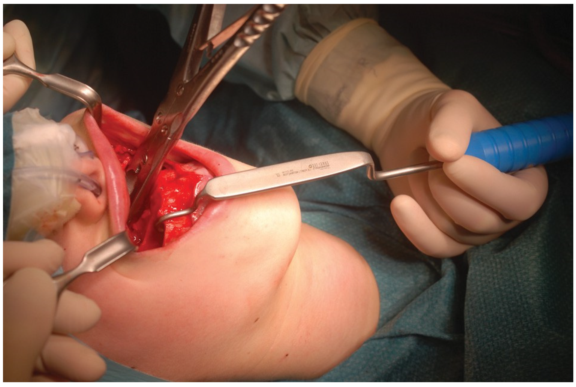
Le Fort I–Type Osteotomy Retractor

by
Maurice Yves Mommaerts
1,2
1
European Face Centre, Universitair Ziekenhuis Brussel, VUB, Brussels, Belgium
2
Orthoface Clinic, St. Martens-Latem, Oost-Vlaanderen, Belgium
Craniomaxillofac. Trauma Reconstr. 2017, 10(4), 323-324; https://doi.org/10.1055/s-0036-1592097
Submission received: 8 May 2016 / Revised: 30 May 2016 / Accepted: 19 June 2016 / Published: 23 August 2016

Abstract

:
A new instrument for retracting the maxilla after mobilization is described. It does neither compress nor inadvertently pierce the lower lip and it does neither obliterate the view nor hinder access to bone removing instruments.

The Le Fort I segment comprises part of the maxillary and palatine bones. Before and after mobilization, trimming of interferences is required for proper repositioning. Cartilaginous septum, conchal bone and mucosa, and bony nasal and facial wall have to be recontoured using rongeurs and rotary instruments. To have access for this work, surgeons use Langenbeck (Obwegeser) type retractors for the labial and cheek tissues, Rowe forceps [1], a J stripper [2], a Tessier blunt zygomatic hook [3], a Volkmann 1–pronged retractor, a Horch zygoma hook, or a Ginestet hook elevator to pull the Le Fort I segment down. Some use their fingers.
The inconvenience of manual retraction is the obliteration of the view to the cavity. The disadvantage of using a hook-like instrument is the undue pressure on the lower lip and the difficult stabilization of the instrument by the assistant surgeon.
The author designed a Le Fort I osteotomy retractor that will not impinge on the lower lip and can be more comfortably held by the assistant surgeon (Figure 1). In contrast to the above-mentioned hook-like instruments, the presented retractor has an anatomical design with a recess for the lower lip and chin, a flat sheet where exceptionally soft-tissue contact may occur, and a blunt tip that will not hook the lower lip when slipping from the mobilized maxillary segment. The assistant surgeon holds the retractor with a vertical fist on the chest of the patient. The tip of the instrument is located at the anterior nasal floor, or in the frontal recess of the maxillary sinus. It will not tear the lower lip, when inadvertently slipping from the segment, because the tip is blunt (Figure 2). It has been found useful over the past 11 years.4 The instrument is available from Surgi-Tec NV, Ghent, Belgium (www.surgi-tec.com).

Institutional Review Board Statement

Ethical approval was not required.

References

  1. Shelton, D.W.; Irby, W.B.; Volume, V. Current Advances in Oral and Maxillofacial Surgery: Orthognathic Surgery; The C.V. Mosby Company: St. Louis, MO, 1986; p. 101. [Google Scholar]
  2. Frost, D.E. Orthognathic surgical techniques. In Maxillofacial Surgery; Ward Booth, P., Schendel, S.A., Hausamen, J.-E., Eds.; Elsevier-Churchill-Livingstone, 2006; Volume 2, p. 1278. [Google Scholar]
  3. Posnick, J.C. Orthognathic Surgery: Principles and Practice; Elsevier-Saunders: St. Louis, MO, 2014; p. 452. [Google Scholar]
  4. Mommaerts, M.Y. The surgical art of facial makeover. In The Le Fort-I Osteotomy; Orthoface R&D: Sint-Martens-Latem, Belgium, 2013; Volume I, pp. 151–171. [Google Scholar]
Figure 1. Instrument (with blue handle) in use: downward retraction of the mobilized maxilla.
Cmtr 10 00051 g001
Figure 2. Anatomical design and blunt tip.
Cmtr 10 00051 g002

