Identification of Early Biochemical Recurrence Predictors in High-Risk Prostate Cancer Patients Treated with Carbon-Ion Radiotherapy and Androgen Deprivation Therapy

 ,
, 
 , , ,
, , ,
Abstract
:1. Introduction
2. Materials and Methods
2.1. Patients
2.2. CIRT
2.3. ADT
2.4. Assessments
2.5. Statistical Analysis
3. Results
4. Discussion
5. Conclusions
Author Contributions
Funding
Institutional Review Board Statement
Informed Consent Statement
Data Availability Statement
Conflicts of Interest
References
- Sung, H.; Ferlay, J.; Siegel, R.L.; Laversanne, M.; Soerjomataram, I.; Jemal, A.; Bray, F. Global cancer statistics 2020: GLOBOCAN estimates of incidence and mortality worldwide for 36 cancers in 185 countries. CA Cancer J. Clin. 2021, 71, 209–249. [Google Scholar] [CrossRef] [PubMed]
- Siegel, R.L.; Miller, K.D.; Jemal, A. Cancer statistics, 2019. CA Cancer J. Clin. 2019, 69, 7–34. [Google Scholar] [CrossRef] [PubMed]
- Teoh, J.Y.C.; Hirai, H.W.; Ho, J.M.W.; Chan, F.C.; Tsoi, K.K.; Ng, C.F. Global incidence of prostate cancer in developing and developed countries with changing age structures. PLoS ONE 2019, 14, e0221775. [Google Scholar] [CrossRef] [PubMed]
- Bergengren, O.; Pekala, K.R.; Matsoukas, K.; Fainberg, J.; Mungovan, S.F.; Bratt, O.; Bray, F.; Brawley, O.; Luckenbaugh, A.N.; Mucci, L.; et al. 2022 Update on prostate cancer epidemiology and risk factors-a systematic review. Eur. Urol. 2023, 84, 191–206. [Google Scholar] [CrossRef] [PubMed]
- D’Amico, A.V.; Whittington, R.; Malkowicz, S.B.; Schultz, D.; Blank, K.; Broderick, G.A.; Tomaszewski, J.E.; Renshaw, A.A.; Kaplan, I.; Beard, C.J.; et al. Biochemical outcome after radical prostatectomy, external beam radiation therapy, or interstitial radiation therapy for clinically localized prostate cancer. JAMA 1998, 280, 969–974. [Google Scholar] [CrossRef] [PubMed]
- Schaeffer, E.M.; Srinivas, S.; Adra, N.; An, Y.; Barocas, D.; Bitting, R.; Bryce, A.; Chapin, B.; Cheng, H.H.; D’Amico, A.V.; et al. NCCN Guidelines Version 4.2023 Prostate Cancer. Available online: https://www.nccn.org/home/ (accessed on 1 July 2023).
- Gómez-Aparicio, M.A.; López-Campos, F.; José Lozano, A.J.; Maldonado, X.; Caballero, B.; Zafra, J.; Suarez, V.; Moreno, E.; Arcangeli, S.; Scorsetti, M.; et al. Novel approaches in the systemic management of high-risk prostate cancer. Clin. Genitourin. Cancer 2023. [Google Scholar] [CrossRef]
- Wang, Z.; Ni, Y.; Chen, J.; Sun, G.; Zhang, X.; Zhao, J.; Zhu, X.; Zhang, H.; Zhu, S.; Dai, J.; et al. The efficacy and safety of radical prostatectomy and radiotherapy in high-risk prostate cancer: A systematic review and meta-analysis. World J. Surg. Oncol. 2020, 18, 42. [Google Scholar] [CrossRef]
- Vu, E.; Pratsinis, M.; Plasswilm, L.; Schmid, H.P.; Panje, C.; Betschart, P. Radiotherapy or surgery? Comparative, qualitative assessment of online patient education materials on prostate cancer. Curr. Oncol. 2021, 28, 3420–3429. [Google Scholar] [CrossRef]
- Zumsteg, Z.S.; Spratt, D.E.; Romesser, P.B.; Pei, X.; Zhang, Z.; Polkinghorn, W.; McBride, S.; Kollmeier, M.; Yamada, Y.; Zelefsky, M.J. The natural history and predictors of outcome following biochemical relapse in the dose escalation era for prostate cancer patients undergoing definitive external beam radiotherapy. Eur. Urol. 2015, 67, 1009–1016. [Google Scholar] [CrossRef]
- Parker, C.; Castro, E.; Fizazi, K.; Heidenreich, A.; Ost, P.; Procopio, G.; Tombal, B.; Gillessen, S. Prostate cancer: ESMO Clinical Practice Guidelines for diagnosis, treatment and follow-up. Ann. Oncol. 2020, 31, 1119–1134. [Google Scholar] [CrossRef]
- Akakura, K.; Tsuji, H.; Morita, S.; Tsuji, H.; Yagishita, T.; Isaka, S.; Akaza, H.; Hata, M.; Fujime, M.; Harada, M.; et al. Phase I/II clinical trials of carbon ion therapy for prostate cancer. Prostate 2004, 58, 252–258. [Google Scholar] [CrossRef] [PubMed]
- Kasuya, G.; Ishikawa, H.; Tsuji, H.; Haruyama, Y.; Kobashi, G.; Ebner, D.K.; Akakura, K.; Suzuki, H.; Ichikawa, T.; Shimazaki, J.; et al. Cancer-specific mortality of high-risk prostate cancer after carbon-ion radiotherapy plus long-term androgen deprivation therapy. Cancer Sci. 2017, 108, 2422–2429. [Google Scholar] [CrossRef]
- Kasuya, G.; Ishikawa, H.; Tsuji, H.; Nomiya, T.; Makishima, H.; Kamada, T.; Akakura, K.; Suzuki, H.; Shimazaki, J.; Haruyama, Y.; et al. Significant impact of biochemical recurrence on overall mortality in patients with high-risk prostate cancer after carbon-ion radiotherapy combined with androgen deprivation therapy. Cancer 2016, 122, 3225–3231. [Google Scholar] [CrossRef] [PubMed]
- Ishikawa, H.; Tsuji, H.; Murayama, S.; Sugimoto, M.; Shinohara, N.; Maruyama, S.; Murakami, M.; Shirato, H.; Sakurai, H. Particle therapy for prostate cancer: The past, present and future. Int. J. Urol. 2019, 26, 971–979. [Google Scholar] [CrossRef] [PubMed]
- Waltz, J.; Chun, F.K.H.; Klein, E.A.; Reuther, A.; Saad, F.; Graefen, M.; Huland, H.; Karakiewicz, P.I. Nomogram predicting the probability of early recurrence after radical prostatectomy for prostate cancer. J. Urol. 2009, 181, 601–607. [Google Scholar] [CrossRef] [PubMed]
- Buyyounouski, M.; Hanlon, A.L.; Horwitz, E.M.; Pollack, A. Interval to biochemical failure highly prognostic for distant metastasis and prostate cancer-specific mortality after radiotherapy. Int. J. Radiat. Oncol. Biol. Phys. 2008, 70, 59–66. [Google Scholar] [CrossRef]
- Broeck, T.V.D.; Bergh, R.C.v.D.; Arfi, N.; Gross, T.; Moris, L.; Briers, E.; Cumberbatch, M.; De Santis, M.; Tilki, D.; Fanti, S.; et al. Prognostic value of biochemical recurrence following treatment with curative intent for prostate cancer: A systematic review. Eur. Urol. 2019, 75, 967–987. [Google Scholar] [CrossRef]
- Baas, D.J.H.; Schilham, M.; Hermsen, R.; de Baaij, J.M.S.; Vrijhof, H.J.E.J.; Hoekstra, R.J.; Sedelaar, J.P.M.; Küsters-Vandevelde, H.V.N.; Gotthardt, M.; Wijers, C.H.W.; et al. Preoperative PSMA-PET/CT as a predictor of biochemical persistence and early recurrence following radical prostatectomy with lymph node dissection. Prostate Cancer Prostatic Dis. 2022, 25, 65–70. [Google Scholar] [CrossRef]
- Amiel, T.; Würnschimmel, C.; Heck, M.; Horn, T.; Nguyen, N.; Budäus, L.; Knipper, S.; Wenzel, M.; Rauscher, I.; Eiber, M.; et al. Regional lymph node metastasis on prostate specific membrane antigen positron emission tomography correlates with decreased biochemical recurrence-free and therapy-free survival after radical prostatectomy: A retrospective single-center single-arm observational study. J. Urol. 2021, 205, 1663–1670. [Google Scholar]
- Attard, G.; Murphy, L.; Clarke, N.W.; Cross, W.; Jones, R.J.; Parker, C.C.; Gillessen, S.; Cook, A.; Brawley, C.; Amos, C.L.; et al. Abiraterone acetate and prednisolone with or without enzalutamide for high-risk non-metastatic prostate cancer: A meta-analysis of primary results from two randomised controlled phase 3 trials of the STAMPEDE platform protocol. Lancet 2022, 399, 34–44. [Google Scholar] [CrossRef]
- Brierley, J.D.; Gospodarowics, M.K.; Wittekind, C. TNM Classification of Malignant Tumours, 8th ed.; Wiley-Blackwell Inc.: New York, NY, USA, 2017. [Google Scholar]
- Roach, M.; Hanks, G.; Thames, H.; Schellhammer, P.; Shipley, W.U.; Sokol, G.H.; Sandler, H. Defining biochemical failure following radiotherapy with or without hormonal therapy in men with clinically localized prostate cancer: Recommendations of the RTOG-ASTRO Phoenix Consensus Conference. Int. J. Radiat. Oncol. Biol. Phys. 2006, 65, 965–974. [Google Scholar] [CrossRef] [PubMed]
- Kanda, Y. Investigation of the freely available easy-to-use software “EZR” for medical statistics. Bone Marrow Transpl. 2013, 48, 452–458. [Google Scholar] [CrossRef] [PubMed]
- Ray, M.E.; Thames, H.D.; Levy, L.B.; Horwitz, E.M.; Kupelian, P.A.; Martinez, A.A.; Michalski, J.M.; Pisansky, T.M.; Shipley, W.U.; Zelefsky, M.J.; et al. PSAnadir predicts biochemical and distant failures after external beam radiotherapy for prostate cancer: A multi-institutional analysis. Int. J. Radiat. Oncol. Biol. Phys. 2005, 64, 1140–1150. [Google Scholar] [CrossRef]
- Nishimura, S.; Ohashi, T.; Momma, T.; Sakayori, M.; Eriguchi, T.; Tanaka, T.; Yamashita, S.; Kosaka, T.; Oya, M.; Shigematsu, N. Prostate-specific antigen nadir within 12 months as an early surrogate marker of biochemical failure and distant metastasis after low-dose-rate brachytherapy or external beam radiotherapy for localized prostate cancer. Cancer Med. 2018, 7, 1794–1801. [Google Scholar] [CrossRef] [PubMed]
- Fernandes, C.D.; Simoes, R.; Ghobadi, G.; Heijmink, S.W.; Schoots, I.G.; de Jong, J.; Walraven, I.; van der Poel, H.G.; van Houdt, P.J.; Smolic, M.; et al. Multiparametric MRI tumor probability model for the detection of locally recurrent prostate cancer after radiation therapy: Pathologic validation and comparison with manual tumor delineations. Int. J. Radiat. Oncol. Biol. Phys. 2019, 105, 140–148. [Google Scholar] [CrossRef] [PubMed]
- Zamboglou, C.; Eiber, M.; Fassbender, T.R.; Eder, M.; Kirste, S.; Bock, M.; Schilling, O.; Reichel, K.; van der Heide, U.A.; Grosu, A.L. Multimodal imaging for radiation therapy planning in patients with primary prostate cancer. Phys. Imaging Radiat. Oncol. 2018, 8, 8–16. [Google Scholar] [CrossRef]
- Pucar, D.; Hricak, H.; Shukla-Dave, A.; Kuroiwa, K.; Drobnjak, M.; Eastham, J.; Scardino, P.T.; Zelefsky, M.J. Clinically significant prostate cancer local recurrence after radiation therapy occurs at the site of primary tumor: Magnetic resonance imaging and step-section pathology evidence. Int. J. Radiat. Oncol. Biol. Phys. 2007, 69, 62–69. [Google Scholar] [CrossRef]
- Poon, D.M.C.; Yuan, J.; Yang, B.; Kerkmeijer, L.G.; Kishan, A.U.; Murthy, V.; Tree, A.; Zapatero, A.; Wong, O.L. Magnetic resonance imaging–guided focal boost to intraprostatic lesions using external beam radiotherapy for localized prostate cancer: A systematic review and meta-analysis. Eur. Urol. Oncol. 2023, 6, 116–127. [Google Scholar] [CrossRef]
- Utsumi, T.; Suzuki, H.; Ishikawa, H.; Hiroshima, Y.; Wakatsuki, M.; Harada, M.; Ichikawa, T.; Akakura, K.; Tsuji, H. External validation of the Candiolo nomogram for high-risk prostate cancer patients treated with carbon ion radiotherapy plus androgen deprivation therapy: A retrospective cohort study. Jpn. J. Clin. Oncol. 2022, 52, 950–953. [Google Scholar] [CrossRef]
- Dudderidge, T.; Payne, H.; Emberton, M. An algorithm for managing the failure of external beam radiotherapy in prostate cancer. BJU Int. 2007, 100, 518–527. [Google Scholar] [CrossRef]
- Mottet, N.; Cornford, P.; van den Bergh, R.C.N.; Briers, E.; Van den Broeck, T.; Cumberbatch, M.G.; De Santis, M.; Fanti, S.; Fossati, N.; Gandaglia, G.; et al. EAU-EANM-ESTRO-ESUR-ISUP-SIOG guidelines on prostate cancer update. Eur. Urol. 2022, 79, 243–262. [Google Scholar] [CrossRef] [PubMed]
- Gillessen, S.; Bossi, A.; Davis, I.D.; de Bono, J.; Fizazi, K.; James, N.D.; Mottet, N.; Shore, N.; Small, E.; Smith, M.; et al. Management of patients with advanced prostate cancer. Part I: Intermediate-/high-risk and locally advanced disease, biochemical relapse, and side effects of hormonal treatment: Report of the Advanced Prostate Cancer Consensus Conference 2022. Eur. Urol. 2023, 83, 267–293. [Google Scholar] [CrossRef] [PubMed]
- Gaudreault, M.; Chang, D.; Hardcastle, N.; Jackson, P.; Kron, T.; Hofman, M.S.; Siva, S. Feasibility of biology-guided radiotherapy using PSMA-PET to boost to dominant intraprostatic tumour. Clin. Transl. Radiat. Oncol. 2022, 35, 84–89. [Google Scholar] [CrossRef] [PubMed]


| Variable | n = 670 |
|---|---|
| Age (years), mean (SD) | 68.1 (6.9) |
| PSA level (ng/mL), median (IQR) | 12.7 (7.2–24.3) |
| cT stage | cT1: 110 cT2a: 132, cT2b: 91, cT2c: 37 cT3a: 239, cT3b: 61 |
| Gleason score | 3 + 3: 22 3 + 4: 93, 4 + 3: 103 4 + 4: 135, 3 + 5: 25 4 + 5: 255, 5 + 4: 34 5 + 5: 3 |
| %PC (%), median (IQR) | 41.7 (25.0–60.0) |
| ADT duration (months), median (IQR) | 25.0 (24.0–27.0) |
| CIRT dose | 63 Gy(RBE)/20 fr: 77 57.6Gy(RBE)/16 fr: 298 51.6 Gy(RBE)/12 fr: 295 |
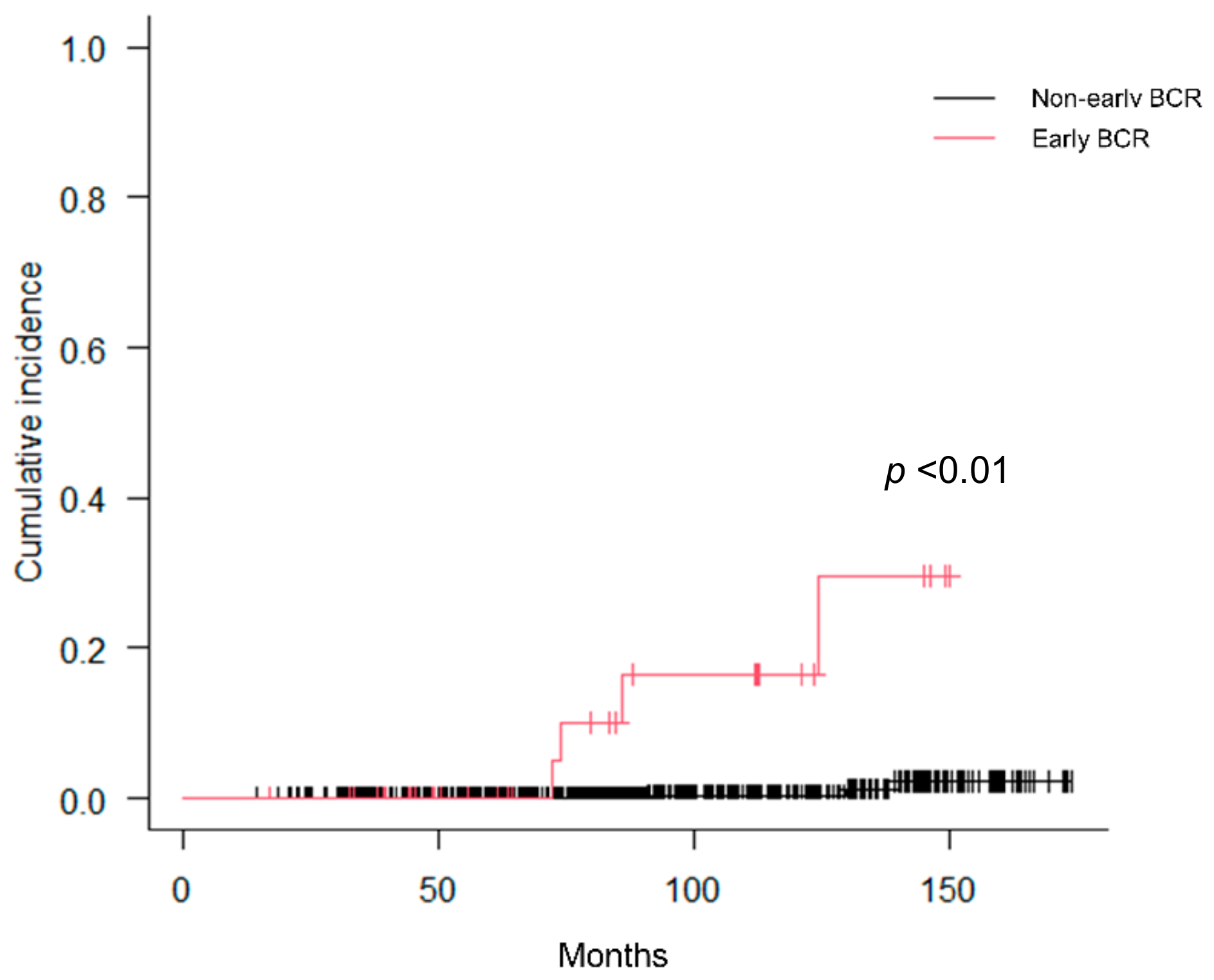
| Early BCR | 36 (5.4%) |
| Time to early BCR (months), median (IQR) | 42.8 (29.0–45.3) |
| Variables | |
|---|---|
| STAMPEDE | |
| Risk = 0 | 58 (8.7%) |
| Risk = 1 | 432 (64.5%) |
| Risk = 2 | 142 (21.2%) |
| Risk = 3 | 38 (5.6%) |
| STAMPEDE risk factors | |
| PSA level ≥ 40 ng/mL | 78 (11.6%) |
| cT stage ≥ 3 | 300 (44.8%) |
| Gleason score ≥ 8 | 452 (67.5%) |
| Variables | Early BCR (n = 36) | Non-Early BCR (n = 634) | p Value |
|---|---|---|---|
| Age (years), mean (SD) | 68.5 (6.3) | 68.1 (7.0) | 0.69 |
| PSA level (ng/mL), median (IQR) | 18.6 (10.8–42.5) | 12.5 (7.1–24.1) | 0.01 |
| cT stage | cT1: 4 cT2a: 5, cT2b: 5, cT2c: 2 cT3a: 10, cT3b: 10 | cT1: 106 cT2a: 127, cT2b: 86, cT2c: 35 cT3a: 229, cT3b: 51 | 0.03 |
| Gleason score | 3 + 4: 4, 4 + 3: 6 4 + 4: 4, 3 + 5: 2 | 3 + 3: 22 3 + 4: 89, 4 + 3: 97 4 + 4: 131, 3 + 5: 23 5 + 5: 3 | 0.52 |
| %PC (%), median (IQR) | 50.0 (35.1–83.3) | 40.0 (25.0–27.0) | <0.01 |
| ADT duration (months), median (IQR) | 24.9 (23.7–29.1) | 25.0 (24.0–27.0) | 0.48 |
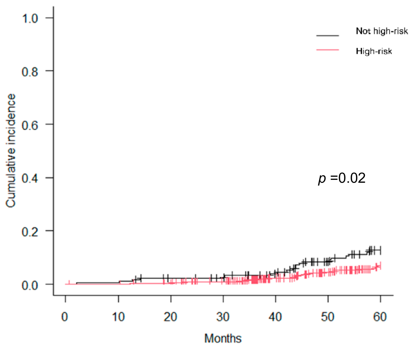
| STAMPEDE high-risk (%) | 16 (44.4%) | 168 (25.9%) | 0.02 |
| Variables | Univariate | p Value | Multivariate | p Value | ||
|---|---|---|---|---|---|---|
| Early BCR (%) | Non-Early BCR (%) | HR | 95%CI | |||
| Age ≥ 70 vs. < 70 (years) | 4.9 | 5.7 | 0.77 | |||
| PSA ≥ 50 vs. < 50 (ng/mL) | 12.5 | 4.7 | 0.02 | - | 0.29 | |
| cT3b vs. cT3a-cT1 | 16.4 | 4.3 | <0.01 | 3.63 | 1.76–7.49 | <0.01 |
| GS ≥ 9 and 3 + 5 vs. GS ≤ 4 + 4 | 6.9 | 4.0 | 0.07 | - | 0.41 | |
| %PC ≥ 75% vs. < 75% | 14.6 | 3.7 | <0.01 | 3.51 | 1.84–6.69 | <0.01 |
Disclaimer/Publisher’s Note: The statements, opinions and data contained in all publications are solely those of the individual author(s) and contributor(s) and not of MDPI and/or the editor(s). MDPI and/or the editor(s) disclaim responsibility for any injury to people or property resulting from any ideas, methods, instructions or products referred to in the content. |
© 2023 by the authors. Licensee MDPI, Basel, Switzerland. This article is an open access article distributed under the terms and conditions of the Creative Commons Attribution (CC BY) license (https://creativecommons.org/licenses/by/4.0/).
Share and Cite
Utsumi, T.; Suzuki, H.; Ishikawa, H.; Wakatsuki, M.; Okonogi, N.; Harada, M.; Ichikawa, T.; Akakura, K.; Murakami, Y.; Tsuji, H.; et al. Identification of Early Biochemical Recurrence Predictors in High-Risk Prostate Cancer Patients Treated with Carbon-Ion Radiotherapy and Androgen Deprivation Therapy. Curr. Oncol. 2023, 30, 8815-8825. https://doi.org/10.3390/curroncol30100636
Utsumi T, Suzuki H, Ishikawa H, Wakatsuki M, Okonogi N, Harada M, Ichikawa T, Akakura K, Murakami Y, Tsuji H, et al. Identification of Early Biochemical Recurrence Predictors in High-Risk Prostate Cancer Patients Treated with Carbon-Ion Radiotherapy and Androgen Deprivation Therapy. Current Oncology. 2023; 30(10):8815-8825. https://doi.org/10.3390/curroncol30100636
Chicago/Turabian StyleUtsumi, Takanobu, Hiroyoshi Suzuki, Hitoshi Ishikawa, Masaru Wakatsuki, Noriyuki Okonogi, Masaoki Harada, Tomohiko Ichikawa, Koichiro Akakura, Yoshitaka Murakami, Hiroshi Tsuji, and et al. 2023. "Identification of Early Biochemical Recurrence Predictors in High-Risk Prostate Cancer Patients Treated with Carbon-Ion Radiotherapy and Androgen Deprivation Therapy" Current Oncology 30, no. 10: 8815-8825. https://doi.org/10.3390/curroncol30100636
APA StyleUtsumi, T., Suzuki, H., Ishikawa, H., Wakatsuki, M., Okonogi, N., Harada, M., Ichikawa, T., Akakura, K., Murakami, Y., Tsuji, H., & Yamada, S. (2023). Identification of Early Biochemical Recurrence Predictors in High-Risk Prostate Cancer Patients Treated with Carbon-Ion Radiotherapy and Androgen Deprivation Therapy. Current Oncology, 30(10), 8815-8825. https://doi.org/10.3390/curroncol30100636