Abstract
In most cases, dyspnoea, chest pain and negative T waves found on ECG represent myocardial ischaemia, pulmonary embolism, left ventricular hypertrophy or pericarditis. In some cases, the cause is unusual. We discuss here the case of a 76-year-old woman presenting with chest pain and dyspnoea as symptoms of a metastatic hepatocellular carcinoma located in her right ventricular septum and incidentally found on coronary angiography.
Introduction
Metastatic cardiac tumours are rare (0.7%–3.5% of autopsy cases), but are more common in patients suffering from cancer (1%–14% of autopsy series) [,,]. Spread to any part of the heart is possible, but involvement of the pericardium and epicardium is common, whereas spread to the myocardium and the endocardium is extremely atypical [,]. In decreasing order of frequency, lung tumours, breast tumours and haematological malignancies are the most frequent cancers to have secondary cardiac metastases [,,]. Melanoma has a unique propensity to metastasise to the heart. Metastases reach the heart by three potential paths: direct growth, through the bloodstream, or via the lymphatic system. Clinical presentations are diverse, varying from a serendipitous asymptomatic finding to constitutional symptoms, embolism or symptoms directly related to mass infiltration or obstruction.
Case report
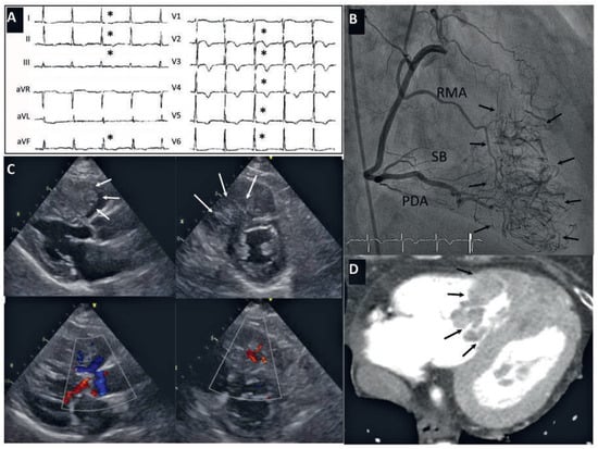
A 76-year-old woman was referred for invasive coronary angiography because of new onset of exercise-induced chest pain and dyspnoea together with diffuse negative T wave inversion (asterisk) on a 12-lead ECG (Figure panel A). She had a history of hepatocellular carcinoma that was diagnosed 3 years before and was apparently cured with right hemihepatectomy, chemoembolisation, and systemic chemotherapy with sorafenib, a multikinase inhibitor. Recently, the patient had experienced a local recurrence and one solitary metastasis in the right inferior pulmonary lobe was discovered. Her quality of life was good and the oncologist in-charge predicted a 2-year life expectancy. Physical examination revealed right pleural effusion. The coronary angiogram, depicted in panel B of the figure, showed normal coronary arteries but a dense network of septal neovessels (arrows). Considering the likelihood of a myocardial mass, we next performed transthoracic echocardiography, which delineated a large vascularised mass in the right ventricular septal apex, with no outflow tract obstruction (Figure panel C). This cardiac mass was later demonstrated with chest computed tomography (panel D – arrows). Conservative treatment was considered.

Figure.
Panel A. The 12-lead ECG showing diffuse alterations of the repolarisation with inverted T waves (asterisk). Panel B. The coronary angiogram of the right coronary artery (here LAO 30°) demonstrated a dense network of neovessels (tumor stain is delineated by arrows) tributaries from right marginal branches (RMAs) and septal branches (SB) from the posterior descending artery (PDA). Panel C. Echocardiogram showing the tumour (arrows) in the right ventricle in parasternal long (PLAX-left) and short (PSAXright) axis in 2D (upper) and its vascularisation with colour Doppler studies (lower). Panel D. Chest CT showing the tumour (arrows) in the right ventricle.
Discussion
Cardiac metastases of hepatocellular carcinoma in the right ventricle are very rare [,]. Hepatomas usually spread to the endocardium from the inferior vena cava system and that is probably what occurred in our patient. The prognosis is poor. Surgical resection has been performed to improve prognosis, or to control symptoms. Transcoronary chemoembolisation has been reported to effectively control symptoms []. As an alternative, although currently never reported, we considered metastasis embolisation by neovessel coiling, but refrained because of the risk of massive pulmonary tumour embolism during the process of necrosis.
Disclosures
No financial support and no other potential conflict of interest relevant to this article was reported.
References
- Goldberg, A.D.; Blankstein, R.; Padera, R.F. Tumors metastatic to the heart. Circulation. 2013, 128, 1790–1794. [Google Scholar] [CrossRef] [PubMed]
- Bussani, R.; De Giorgio, F.; Abbate, A.; Silvestri, F. Cardiac metastases. J Clin Pathol. 2007, 60, 27–34. [Google Scholar] [CrossRef] [PubMed]
- Amano, J.; Nakayama, J.; Yoshimura, Y.; Ikeda, U. Clinical classification of cardiovascular tumors and tumor like lesions, and its incidences. Gen Thorac Cardiovasc Surg. 2013, 61, 435–447. [Google Scholar] [CrossRef]
- Liu, W.C.; Lui, K.W.; Ho, M.C.; Fan, S.Z.; Chao, A. Right ventricular exclusion for hepatocellular carcinoma metastatic to the heart. J Cardiothorac Surg. 2010, 5, 95. [Google Scholar] [CrossRef]
- Kotani, E.; Kiuchi, K.; Takayama, M.; Takano, T.; Tabata, M.; Aramaki, T.; et al. Effectiveness of transcoronary chemoembolization for metastatic right ventricular tumor derived from hepatocellular carcinoma. Chest 2000, 117, 287–289. [Google Scholar] [CrossRef]
© 2015 by the author. Attribution - Non-Commercial - NoDerivatives 4.0.