Case description
A 71-year-old woman with long-standing hypertension was referred for cardiological evaluation because of dyspnea on exertion, orthopnea, and orthostatic dizziness. On physical examination, a 3/6 systolic murmur was heard at the apex of the heart. She had no clinical signs of heart failure. There were no pulmonary rales.
Transthoracic echocardiography showed pronounced concentric hypertrophy of the left ventricle with normal ejection fraction. Additionally, a large tumor attached to the posterior mitral annulus causing impairment of transmitral left ventricular inflow was visible. This tumor moved the mitral leaflet apparatus more anteriorly toward the left ventricular outflow tract resulting in systolic anterior motion (SAM) of the mitral valve leaflets with variable left ventricular outflow tract obstruction of up to 50 mm Hg. The diagnosis of caseous calcification of the mitral annulus was made (Figure 1).
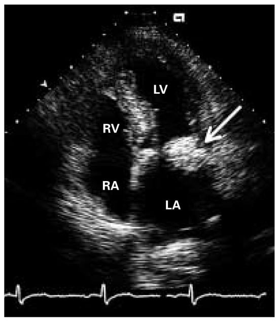
Figure 1.
Apical four chamber view with a round tumor attached to the posterior mitral annulus (arrow) causing minimal acoustic shadowing artifact. LV = left ventricle; LA = left atrium; RV = right ventricle; RA = right atrium.
The patient underwent surgical resection of the tumor. The wall of the tumor was incised, a toothpaste-like mass was evacuated from a slightly calcified coat (Figure 2A), and the wall of the collapsed tumor was adapted with a running suture. The normal morphology and competence of the mitral valve could be preserved.
Grossly, the lesion consisted of gray-white to yellow necrotic debris, resembling caseous necrosis seen in granulomas caused by M. tuberculosis (Figure 2B).

Figure 2.
(A) Intraoperative picture of the calcified coat of the tumor attached to the posterior mitral annulus. (B) Evacuated caseous mass.

Figure 3.
Histologically, one sees in a H&E-stained section amorphous basophilic granular debris, nodular calcifications and degenerating blood cells (macrophages, eosinophils).
Discussion
Caseous calcification of the mitral annulus presents as an echodense round mass with a characteristic central echolucent zone mostly located at the posterior mitral valve annulus [1]. It can obstruct the transmitral left ventricular inflow tract or lead to prolapse of a mitral leaflet causing mitral regurgitation. Most commonly, however, no haemodynamic effect is caused. The prevalence of caseous calcification of the mitral annulus ranges from 0.06–0.07% in patients with mitral annular calcification [2]. Most likely the diagnosis is greatly unrecognised. Compared to mitral annulus calcification which presents with a posterior echo shadow, caseous calcification of the mitral valve shows a minimal acoustic shadowing artifact.
Further differential diagnosis of echogenic structures in mitral valve position include mitral annular abscess, benign tumors (lymph node hyperplasia, cysts) and rarely malignant tumors (lymphoma, metastatic disease) [3].
Correct diagnosis of caseous calcification of the mitral annulus is essential and may prevent unnecessary explorative thoracotomy.
References
- Harpaz, D.; Auerbach, I.; Vered, Z.; Motro, M.; Tobar, A.; Rosenblatt, S. Caseous calcification of the mitral annulus: a neglected, unrecognized diagnosis. J Am Soc Echocardiogr 2001, 14, 825–831. [Google Scholar] [CrossRef] [PubMed]
- Novaro, G.M.; Griffin, B.P.; Hammer, D.F. Caseous calcification of the mitral annulus: an underappreciated variant. Heart 2004, 90, 388. [Google Scholar] [CrossRef] [PubMed]
- Zuber, M.; Oechslin, E.; Jenni, R. Echogenic structures in the left atrioventricular groove: diagnostic pitfalls. J Am Soc Echocardiogr 1998, 11, 381–386. [Google Scholar] [CrossRef] [PubMed]
Publisher’s Note: MDPI stays neutral with regard to jurisdictional claims in published maps and institutional affiliations. |
© 2005 by the author. Attribution - Non-Commercial - NoDerivatives 4.0.