Case report
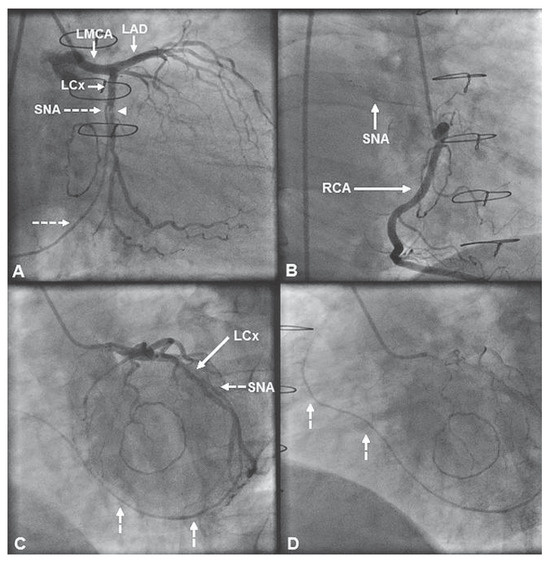
A 79-year-old male patient with chronic atrial fibrillation and a history of mitral valve repair including ring annuloplasty several years ago underwent coronary angiography (Figure 1) for acute coronary syndrome, which showed the culprit lesion in the proximal left circumflex (LCx) artery. Angiography also showed a branch that arose just proximal to the culprit lesion, coursed on the left and then on the posterior atrioventricular groove at the crux cordis and followed thereafter an anterior and superior course toward the sinus node area; it was recognised as a retrocardiac sinus node artery (SNA). A second SNA arising from the right coronary artery (RCA) was also seen. A 3.0 mm × 12 mm bare metal stent was uneventfully implantated across the culprit lesion and the ostium of the SNA.

Figure 1.
Coronary angiographic views. (A) The sinus node artery (SNA; dashed arrows) arising from the left circumflex (LCx) artery immediately proximal to the culprit lesion (arrowhead) is seen coursing on the left atrioventricular (AV) groove. (B) Right coronary artery (RCA) angiogram showing a second SNA (arrow). (C,D) (Late frame) Post-angioplasty views showing the LCx-derived sinus node artery (SNA) (dashed arrows) following a retrocardiac course on the posterior atrioventricular groove, eventually ramifying at the sinus node area. LAD = left anterior descending artery; LMCA = left main coronary artery.
The origin, course and distribution of the SNA has been well investigated, and a dual SNA distribution comprising a RCA-connected SNA and a left posterior SNA originating from the posterolateral LCx artery and coursing across the dome of the left atrium has been reported [1]. Knowledge of the variant anatomy of the SNA is important in cardiac operations involving atrial incisions and catheter ablation procedures for atrial fibrillation (AF), in order to preserve its integrity and thereby avoid ischaemic sinus node dysfunction. For example, a posterior SNA may be transected during the popular superior septal approach to the mitral valve or be injured during circumferential pulmonary vein and mitral isthmus catheter ablation [2]. Furthermore, a left SNA has been associated with unsuccessful left atrial roof catheter ablation in patients with persistent AF owing to failure to achieve complete conduction block probably because of convective heat loss to the blood flowing in the SNA that impedes adequate heating of the myocardium and thereby hinders transmural lesion formation [3]. Therefore, proper identification of a left SNA on preprocedural imaging may be important for planning the ablation strategy. Occlusion of the SNA is increasingly recognised as a clinically relevant complication of percutaneous coronary interventions (PCI), potentially leading to atrial infarction and arrhythmic sequelae [4]. Thus, knowledge of SNA anatomy is important to prevent or treat such complications. This is the first report describing a retrocardiac LCx artery-derived SNA; this branch did not supply right ventricular branches to account for a second RCA [5]. Here, the SNA remained patent after the stent had been implanted across its ostium, probably because of its large (>1 mm) diameter and absent ostial disease. Awareness of the SNA retrocardiac variant is important for correct interpretation of coronary angiograms and for a tailored PCI strategy that preserves blood supply to the sinus node, particularly in the case of a single SNA.
Funding/potential competing interests
No financial support and no other potential conflict of interest relevant to this article was reported.
References
- Saremi F, Channual S, Abolhoda A, Gurudevan SV, Narula J, Milliken JC. MDCT of the S-shaped sinoatrial node artery. AJR Am J Roentgenol. 2008;190(6):1569–75. [CrossRef]
- Andreou AY, Georgiou GM, Avraamides PC. Right posterior sinoatrial node artery showing a pericaval course: a previously undescribed mode of termination. Surg Radiol Anat. 2010;32(6):609–12. [CrossRef]
- Yokokawa M, Sundaram B, Oral H, Morady F, Chugh A. The course of the sinus node artery and its impact on achieving linear block at the left atrial roof in patients with persistent atrial fibrillation. Heart Rhythm. 2012;9(9):1395–402. [CrossRef]
- Alvarez-García J, Vives-Borrás M, Ferrero A, Aizpurua DA, Peñaranda AS, Cinca J. Atrial coronary artery occlusion during elective percutaneous coronary angioplasty. Cardiovasc Revasc Med. 2013;14(5):270– 4. [CrossRef][Green Version]
- Angelini, P. Coronary artery anomalies: an entity in search of an identity. Circulation. 2007;115(10):1296–305. [CrossRef]
© 2014 by the author. Attribution - Non-Commercial - NoDerivatives 4.0.