
Pyogenic Liver Abscess Complicating Acute Cholecystitis: Different Management Options


 ,
,  , , and
Abstract
1. Introduction
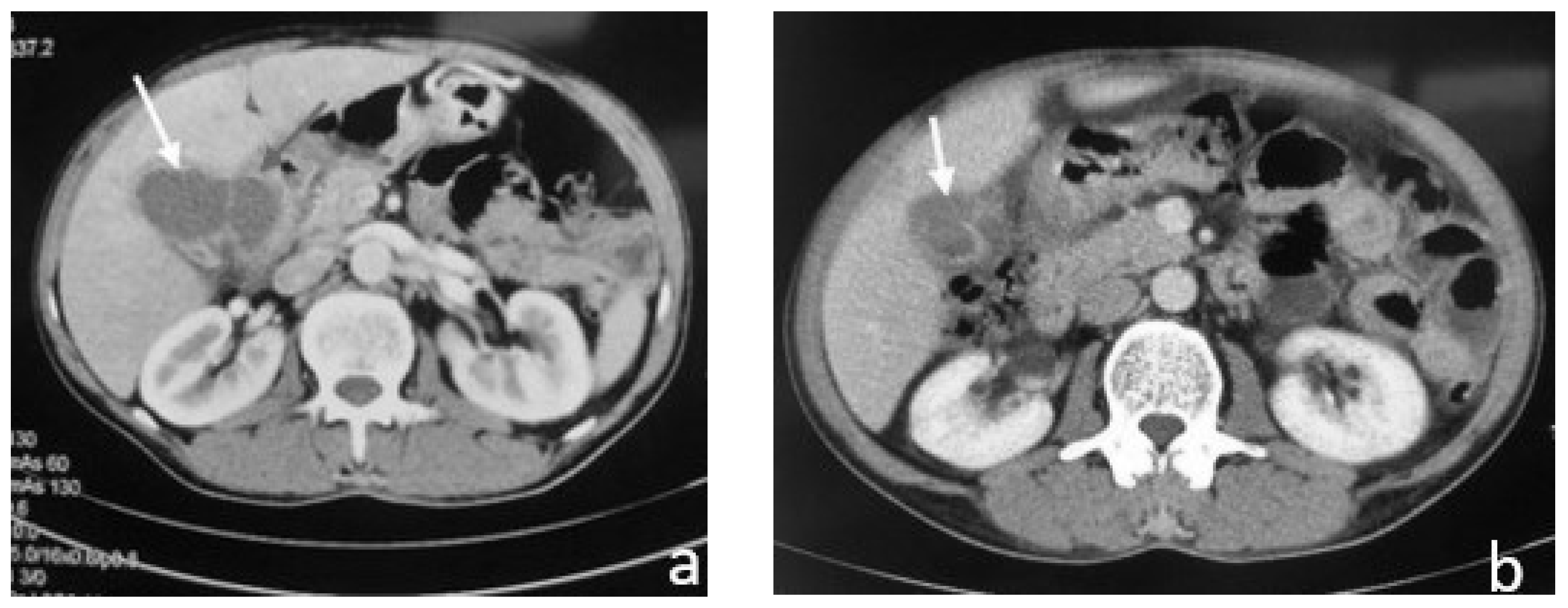
2. Case Reports
2.1. Case 1
2.2. Case 2
2.3. Case 3
3. Discussion
4. Conclusions
Author Contributions
Funding
Institutional Review Board Statement
Informed Consent Statement
Data Availability Statement
Conflicts of Interest
References
- Costi, R.; Le Bian, A.Z.; Cauchy, F.; Diop, P.S.; Carloni, A.; Catherine, L.; Smadja, C. Synchronous pyogenic liver abscess and acute cholecystitis: How to recognize it and what to do (emergency cholecystostomy followed by delayed laparoscopic cholecystectomy). Surg. Endosc. 2011, 26, 205–213. [Google Scholar] [CrossRef] [PubMed]
- Chou, F.-F.; Sheen-Chen, S.-M.; Chen, Y.-S.; Chen, M.-C. Single and Multiple Pyogenic Liver Abscesses: Clinical Course, Etiology, and Results of Treatment. World J. Surg. 1997, 21, 384–389. [Google Scholar] [CrossRef] [PubMed]
- Kochar, K.; Vallance, K.; Mathew, G.; Jadhav, V. Intrahepatic perforation of the gall bladder presenting as liver abscess: Case report, review of literature and Niemeier’s classification. Eur. J. Gastroenterol. Hepatol. 2008, 20, 240–244. [Google Scholar] [CrossRef] [PubMed]
- Niemeier, O.W. Acute free perforation of the gall bladder. Annu. Surg. 1934, 99, 922–924. [Google Scholar] [CrossRef]
- Izadi, K.; Moser, F.G.; Haker, K.; Satey, S. Gallstone Liver Abscess Secondary to Gallbladder Perforation. Radiol. Case Rep. 2009, 4, 280. [Google Scholar] [CrossRef]
- Masood, M.R.; Ali, M.; Burgaul, R.; Smith, A. Hepatic abscess secondary to gallbladder perforation: Case report and literature review. Scott. Med. J. 2008, 53, 1–6. [Google Scholar] [CrossRef]
- Gabriel, D.S.; Eduardo, V.; Eduardo, O.P. Cholecystitis and Synchronous Liver Abscess: Percutaneous Treatment Results. Acta Sci. Gastrointest. Disord. 2019, 2, 16–21. [Google Scholar] [CrossRef]
- Hatzidakis, A.A.; Prassopoulos, P.; Petinarakis, I.; Sanidas, E.; Chrysos, E.; Chalkiadakis, G.; Tsiftsis, D.; Gourtsoyiannis, N.C. Acute cholecystitis in high-risk patients: Percutaneous cholecystostomy vs conservative treatment. Eur. Radiol. 2002, 12, 1778–1784. [Google Scholar] [CrossRef]
- Escartín, A.; González, M.; Cuello, E.; Pinillos, A.; Muriel, P.; Merichal, M.; Palacios, V.; Escoll, J.; Gas, C.; Olsina, J.-J. Acute Cholecystitis in Very Elderly Patients: Disease Management, Outcomes, and Risk Factors for Complications. Surg. Res. Pract. 2019, 2019, e9709242. [Google Scholar] [CrossRef]
- Agresta, F.; Campanile, F.C.; Vettoretto, N.; Silecchia, G.; Bergamini, C.; Maida, P.; Lombari, P.; Narilli, P.; Marchi, D.; Carrara, A.; et al. Laparoscopic cholecystectomy: Consensus conference-based guidelines. Langenbecks Arch. Surg. 2015, 400, 429–453. [Google Scholar] [CrossRef]
- Campanile, F.C.; Pisano, M.; Coccolini, F.; Catena, F.; Agresta, F.; Ansaloni, L. Acute cholecystitis: WSES position statement. World J. Emerg. Surg. 2014, 9, 58. [Google Scholar] [CrossRef] [PubMed]
- Amirthalingam, V.; Low, J.K.; Woon, W.; Shelat, V. Tokyo Guidelines 2013 may be too restrictive and patients with moderate and severe acute cholecystitis can be managed by early cholecystectomy too. Surg. Endosc. 2016, 31, 2892–2900. [Google Scholar] [CrossRef] [PubMed]
- Wakabayashi, G.; Iwashita, Y.; Hibi, T.; Takada, T.; Strasberg, S.M.; Asbun, H.J.; Endo, I.; Umezawa, A.; Asai, K.; Suzuki, K.; et al. Tokyo Guidelines 2018: Surgical management of acute cholecystitis: Safe steps in laparoscopic cholecystectomy for acute cholecystitis (with videos). J. Hepato-Biliary-Pancreat. Sci. 2017, 25, 73–86. [Google Scholar] [CrossRef] [PubMed]
- Gurusamy, K.S.; Davidson, C.; Gluud, C.; Davidson, B.R. Early versus delayed laparoscopic cholecystectomy for people with acute cholecystitis. Cochrane Database Syst. Rev. 2013, 6, CD005440. [Google Scholar] [CrossRef] [PubMed]
- Serban, D.; Socea, B.; Balasescu, S.; Badiu, C.; Tudor, C.; Dascalu, A.; Vancea, G.; Spataru, R.; Sabau, A.; Sabau, D.; et al. Safety of Laparoscopic Cholecystectomy for Acute Cholecystitis in the Elderly: A Multivariate Analysis of Risk Factors for Intra and Postoperative Complications. Medicina 2021, 57, 230. [Google Scholar] [CrossRef]
- Lasithiotakis, K.; Petrakis, J.; Venianaki, M.; Georgiades, G.; Koutsomanolis, D.; Andreou, A.; Zoras, O.; Chalkiadakis, G. Frailty predicts outcome of elective laparoscopic cholecystectomy in geriatric patients. Surg. Endosc. 2012, 27, 1144–1150. [Google Scholar] [CrossRef]
- Lorenzon, L.; Costa, G.; Massa, G.; Frezza, B.; Stella, F.; Balducci, G. The impact of frailty syndrome and risk scores on emergency cholecystectomy patients. Surg. Today 2016, 47, 74–83. [Google Scholar] [CrossRef]
- Evers, B.M.; Townsend, C.M.; Thompson, J.C. Organ Physiology of Aging. Surg. Clin. N. Am. 1994, 74, 23–39. [Google Scholar] [CrossRef]
- Shin, M.S.; Park, S.H. Clinical outcomes of laparoscopic cholecystectomy in elderly patients after preoperative assessment and optimization of comorbidities. Ann. Hepato-Biliary-Pancreat. Surg. 2018, 22, 374–379. [Google Scholar] [CrossRef]
- Łącka, M.; Obłój, P.; Spychalski, P.; Łaski, D.; Rostkowska, O.; Wieszczy, P.; Kobiela, J. Clinical presentation and outcomes of cholecystectomy for acute cholecystitis in patients with diabetes—A matched pair analysis. A pilot study. Adv. Med. Sci. 2020, 65, 409–414. [Google Scholar] [CrossRef]
- Serban, D.; Balasescu, S.A.; Alius, C.; Balalau, C.; Sabau, A.D.; Badiu, C.D.; Socea, B.; Trotea, A.M.; Dascalu, A.M.; Motofei, I.; et al. Clinical and therapeutic features of acute cholecystitis in diabetic patients. Exp. Ther. Med. 2021, 22, 758. [Google Scholar] [CrossRef] [PubMed]
- Jafar, N.; Edriss, H.; Nugent, K. The Effect of Short-Term Hyperglycemia on the Innate Immune System. Am. J. Med. Sci. 2016, 351, 201–211. [Google Scholar] [CrossRef] [PubMed]
- Alves, C.; Casqueiro, J.; Casqueiro, J. Infections in patients with diabetes mellitus: A review of pathogenesis. Indian J. Endocrinol. Metab. 2012, 16, 27–36. [Google Scholar] [CrossRef] [PubMed]
- Xiu, F.; Stanojcic, M.; Diao, L.; Jeschke, M.G. Stress Hyperglycemia, Insulin Treatment, and Innate Immune Cells. Int. J. Endocrinol. 2014, 2014, 486403. [Google Scholar] [CrossRef]
- Bouassida, M.; Chtourou, M.; Charrada, H.; Zribi, S.; Hamzaoui, L.; Mighri, M.; Touinsi, H. The severity grading of acute cholecystitis following the Tokyo Guidelines is the most powerful predictive factor for conversion from laparoscopic cholecystectomy to open cholecystectomy. J. Visc. Surg. 2017, 154, 239–243. [Google Scholar] [CrossRef]
- Paajanen, H.; Suuronen, S.; Nordstrom, P.; Miettinen, P.; Niskanen, L. Laparoscopic versus open cholecystectomy in diabetic patients and postoperative outcome. Surg. Endosc. 2010, 25, 764–770. [Google Scholar] [CrossRef]
- Spampinato, S.F.; Caruso, G.I.; De Pasquale, R.; Sortino, M.A.; Merlo, S. The Treatment of Impaired Wound Healing in Diabetes: Looking among Old Drugs. Pharmaceuticals 2020, 13, 60. [Google Scholar] [CrossRef]
- Patel, S.; Srivastava, S.; Singh, M.R.; Singh, D. Mechanistic insight into diabetic wounds: Pathogenesis, molecular targets and treatment strategies to pace wound healing. Biomed. Pharmacother. 2019, 112, 108615. [Google Scholar] [CrossRef]
- Liu, C.-M.; Chung, C.-L.; Hsu, C.-T.; Song, M.-Z.; Chen, C.-C.; Li, C.-Y. Impact of diabetes mellitus on cholecystectomy rate: A population-based follow-up study. Formos. J. Surg. 2015, 48, 157–162. [Google Scholar] [CrossRef]
- Duncan, J. Femoral hernia, gangrene of the gall bladder: Extravasation of bile: Peritonitis: Death. N. J. Med. 1844, 2, 151–153. [Google Scholar]
- Aljiffry, M.; Walsh, M.; Peltekian, K.; Molinari, M. Type II Gall Bladder Perforation with Abdominal Wall Abscess in a Cirrhotic Patient: Case Report and Review of the Literature. J. Surg. Educ. 2008, 65, 367–371. [Google Scholar] [CrossRef] [PubMed]
- Donati, M.; Biondi, A.; Basile, F.; Gruttadauria, S. An atypical presentation of intrahepatic perforated cholecystitis: A modern indication to open cholecystectomy. Report of a case. BMC Surg. 2014, 14, 6. [Google Scholar] [CrossRef] [PubMed]
- Fagan, S.P.; Awad, S.S.; Rahwan, K.; Hira, K.; Aoki, N.; Itani, K.M.; Berger, D.H. Prognostic factors for the development of gangrenous cholecystitis. Am. J. Surg. 2003, 186, 481–485. [Google Scholar] [CrossRef] [PubMed]
- Tonolini, M.; Ravelli, A.; Villa, C.; Bianco, R. Urgent MRI with MR cholangiopancreatography (MRCP) of acute cholecystitis and related complications: Diagnostic role and spectrum of imaging findings. Emerg. Radiol. 2012, 19, 341–348. [Google Scholar] [CrossRef]
- Soroken, C.; Samaras, N.; Samaras, D.; Huber, P. An Unusual Case of Cholecystitis and Liver Abscesses in an Older Adult. J. Am. Geriatr. Soc. 2012, 60, 160–161. [Google Scholar] [CrossRef][Green Version]
- Hosaka, A.; Nagayoshi, M.; Sugizaki, K.; Masaki, Y. Gallbladder perforation associated with carcinoma of the duodenal papilla: A case report. World J. Surg. Oncol. 2010, 8, 41. [Google Scholar] [CrossRef]
- Derici, H.; Kara, C.; Bozdag, A.D.; Nazli, O.; Tansug, T.; Akca, E. Diagnosis and treatment of gallbladder perforation. World J. Gastroenterol. 2006, 12, 7832–7836. [Google Scholar] [CrossRef]
- Fletcher, A.; Ravdin, I. Perforation of the gallbladder. Am. J. Surg. 1951, 81, 178–185. [Google Scholar] [CrossRef]
- Tang, S.; Wang, Y.; Wang, Y. Contrast-enhanced ultrasonography to diagnose gallbladder perforation. Am. J. Emerg. Med. 2013, 31, 1240–1243. [Google Scholar] [CrossRef]
- Anderson, B.B.; Nazem, A. Perforations of the gallbladder and cholecystobiliary fistulae: A review of management and a new classification. J. Natl. Med. Assoc. 1987, 79, 393–399. [Google Scholar]
- Gilly, F.N.; Roche, M.; Vital-Durand, D.; Braillon, G.; Levrat, R.; Chatelard, P. Liver abscess caused by gallbladder perforation during pyocholecystitis: Apropos of a case. J. Chir. 1989, 126, 544–545. [Google Scholar]
- Cristian, D.; Grama, F.; Burcoş, T. Laparoscopic Treatment of a Hepatic Subcapsular Abscess Secondary to Gallbladder Per-foration: Case Report. Chirurgia 2014, 109, 132–135. [Google Scholar] [PubMed]
- Singal, R.; Mittal, A.; Gupta, S.; Singh, B.; Jain, P. Management of Gall Bladder Perforation Evaluation on Ultrasonography: Report of Six Rare Cases with Review of Literature. J. Med. Life 2011, 4, 364–371. [Google Scholar] [PubMed]
- Göbel, T.; Kubitz, R.; Blondin, D.; Häussinger, D. Intrahepatic type II gall bladder perforation by a gall stone in a CAPD patient. Eur. J. Med. Res. 2011, 16, 213–216. [Google Scholar] [CrossRef]
- Alessiani, M.; Peloso, A.; Tramelli, P.; Magnani, E. Successful non-operative management of spontaneous type II gallbladder perforation in a patient with Alzheimer’s disease. BMJ Case Rep. 2014, 2014, bcr2014204337. [Google Scholar] [CrossRef]
- Zerman, G.; Bonfiglio, M.; Borzellino, G.; Guglielmi, A.; Tasselli, S.; Valloncini, E.; Di Leo, A.; De Manzoni, G. Liver abscess due to acute cholecystitis. Report of five cases. Chir. Ital. 2003, 55, 195–198. [Google Scholar]


Publisher’s Note: MDPI stays neutral with regard to jurisdictional claims in published maps and institutional affiliations. |
© 2022 by the authors. Licensee MDPI, Basel, Switzerland. This article is an open access article distributed under the terms and conditions of the Creative Commons Attribution (CC BY) license (https://creativecommons.org/licenses/by/4.0/).
Share and Cite
Paramythiotis, D.; Karakatsanis, A.; Karlafti, E.; Bareka, S.; Psoma, E.; Hatzidakis, A.A.; Michalopoulos, A. Pyogenic Liver Abscess Complicating Acute Cholecystitis: Different Management Options. Medicina 2022, 58, 782. https://doi.org/10.3390/medicina58060782
Paramythiotis D, Karakatsanis A, Karlafti E, Bareka S, Psoma E, Hatzidakis AA, Michalopoulos A. Pyogenic Liver Abscess Complicating Acute Cholecystitis: Different Management Options. Medicina. 2022; 58(6):782. https://doi.org/10.3390/medicina58060782
Chicago/Turabian StyleParamythiotis, Daniel, Anestis Karakatsanis, Eleni Karlafti, Stella Bareka, Elizabeth Psoma, Adam A. Hatzidakis, and Antonios Michalopoulos. 2022. "Pyogenic Liver Abscess Complicating Acute Cholecystitis: Different Management Options" Medicina 58, no. 6: 782. https://doi.org/10.3390/medicina58060782
APA StyleParamythiotis, D., Karakatsanis, A., Karlafti, E., Bareka, S., Psoma, E., Hatzidakis, A. A., & Michalopoulos, A. (2022). Pyogenic Liver Abscess Complicating Acute Cholecystitis: Different Management Options. Medicina, 58(6), 782. https://doi.org/10.3390/medicina58060782