Multicentric Reticulohistiocytosis Exhibiting Positive HLA-B*07 and HLA-B*08: A Case Report


Abstract
1. Introduction
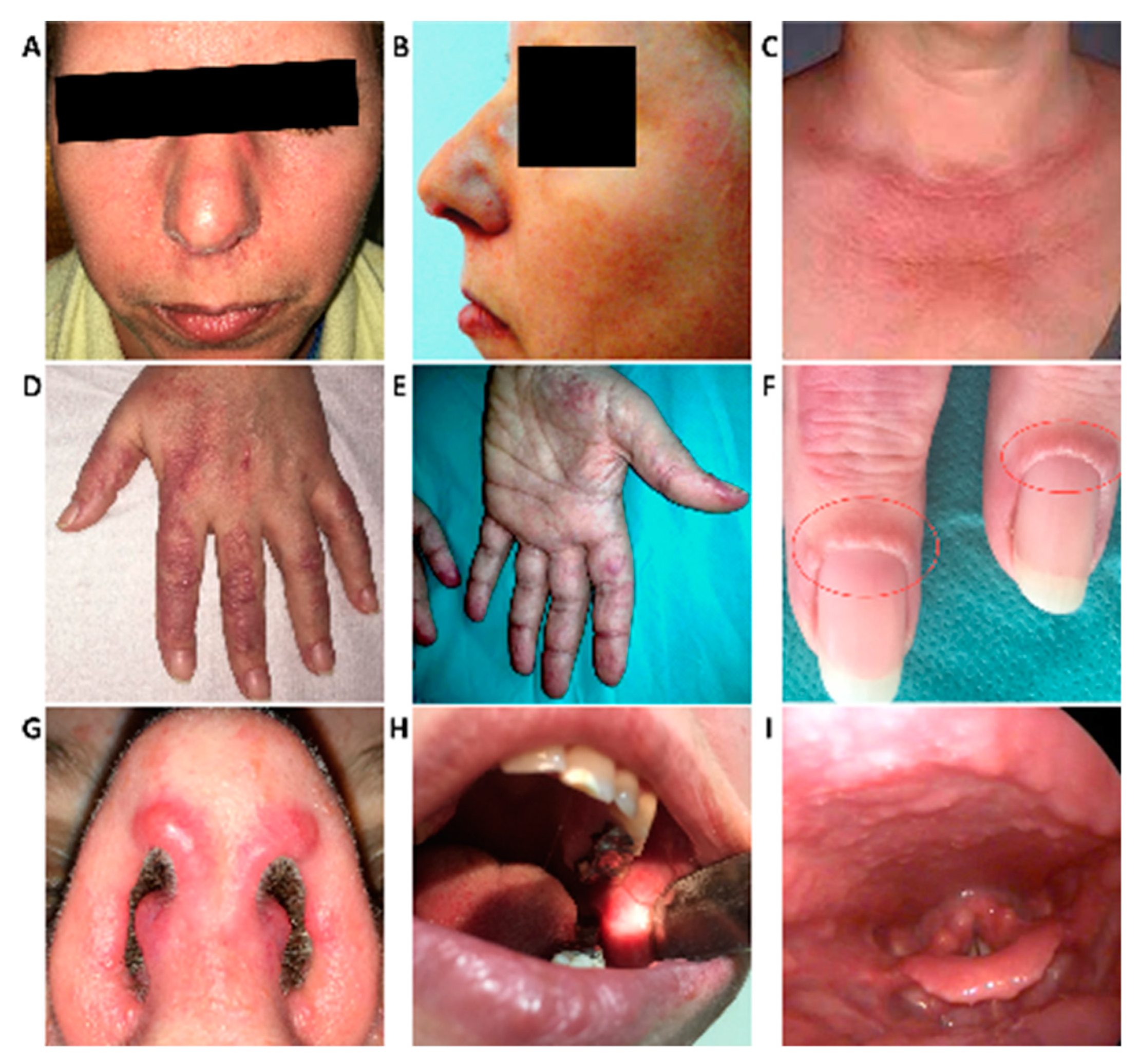
2. Case Report
3. Discussion
4. Conclusions
Supplementary Materials
Author Contributions
Funding
Acknowledgments
Conflicts of Interest
References
- Baek, I.W.; Yoo, S.H.; Yang, H.; Park, J.; Kim, K.J.; Cho, C.S. A case of multicentric reticulohistiocytosis. Mod. Rheumatol. 2017, 27, 165–168. [Google Scholar] [CrossRef] [PubMed]
- Emile, J.F.; Abla, O.; Fraitag, S.; Horne, A.; Haroche, J.; Donadieu, J.; Requena-Caballero, L.; Jordan, M.B.; Abdel-Wahab, O.; Allen, C.E.; et al. Revised classification of histiocytoses and neoplasms of the macrophage-dendritic cell lineages. Blood 2016, 127, 2672–2681. [Google Scholar] [CrossRef]
- Tariq, S.; Hugenberg, S.T.; Hirano-Ali, S.A.; Tariq, H. Multicentric reticulohistiocytosis (MRH): Case report with review of literature between 1991 and 2014 with in depth analysis of various treatment regimens and outcomes. Springer Plus 2016, 5, 180. [Google Scholar] [CrossRef]
- Murakami, N.; Sakai, T.; Arai, E.; Muramatsu, H.; Ichikawa, D.; Asai, S.; Shimoyama, Y.; Ishiguro, N.; Takahashi, Y.; Okuno, Y.; et al. Targetable driver mutations in multicentric reticulohistiocytosis. Haematologica 2020, 105, e61. [Google Scholar] [CrossRef] [PubMed]
- Bonometti, A.; Berti, E. AssociazioneItalianaRicercaIstiocitosiOnlus.Reticulohistiocytoses: A revision of the full spectrum. J. Eur. Acad. Dermatol. 2020. [Google Scholar] [CrossRef] [PubMed]
- Kumar, B.; Singh, N.; Rahnama-Moghadam, S.; Wanat, K.A.; Ijdo, J.W.; Werth, V.P. Multicentric reticulohistiocytosis: A multicenter case series and review of literature. J. Clin. Rheumatol. 2018, 24, 45–49. [Google Scholar] [CrossRef] [PubMed]
- Sanchez-Alvarez, C.; Sandhu, A.S.; Crowson, C.S.; Wetter, D.A.; McKenzie, G.A.; Lehman, J.S.; Makol, A. Multicentric reticulohistiocytosis: The Mayo Clinic experience (1980–2017). Rheumatology 2020, 59, 1898–1905. [Google Scholar] [CrossRef]
- Laza, I.M.; Ventades, N.G.; Hervella, M.; De-la-Rúa, C. Contribution of ancient human remains analysis to the understanding of the variability in HLA-B gene variants in relation to the diagnosis of spondyloarthropathies. J. Autoimmun. 2018, 94, 70–82. [Google Scholar] [CrossRef] [PubMed]
- Mead, M.; Baes, T.; Dass, G. Multicentric Reticulohistiocytosis: Elective Excision of Symptomatic Hand Nodules With 1-Year Follow-Up. J. Hand Surg. Am. 2020, 45, 457. [Google Scholar] [CrossRef]
- El-Gabalawy, H.S.; Goldbach-Mansky, R.; Smith, D.; Arayssi, T.; Bale, S.; Gulko, P.; Yarboro, C.; Wilder, R.L.; Klippel, J.H.; Schumacher, H.R. Association of HLA alleles and clinical features in patients with synovitis of recent onset. Arthritis Rheum. 1999, 42, 1696–1705. [Google Scholar] [CrossRef]
- Toz, B.; Büyükbabani, N.; İnanç, M. Multicentric reticulohistiocytosis: Rheumatology perspective. Best Pract. Res. Clin. Rheumatol. 2016, 30, 250–260. [Google Scholar] [CrossRef] [PubMed]
- Niklas, K.; Niklas, A.; Puszczewicz, M. Multicentric reticulohistiocytosisin the course of undifferentiated connective tissue disease. Postepy Dermatol. Alergol. 2019, 36, 646–647. [Google Scholar] [CrossRef]
- Farokhi, A.; Van Vugt, R.M.; Hoekzema, R.; Nurmohamed, M.T. Multicentric reticulohistiocytosis: A case report. BMC Res. Notes 2018, 11, 1–4. [Google Scholar] [CrossRef] [PubMed]
- Camargo, K.; Pinkston, O.; Abril, A.; Sluzevich, J.C. Xanthomatous multicentric reticulohistiocytosis: An underrecognized variant. J. Clin. Rheumatol. 2018, 24, 285–287. [Google Scholar] [CrossRef] [PubMed]
- Gkoutzourelas, A.; Liaskos, C.; Mytilinaiou, M.G.; Simopoulou, T.; Katsiari, C.; Tsirogianni, A.; Daoussis, D.; Scheper, T.; Meyer, W.; Bogdanos, D.P.; et al. Anti-Ro60 seropositivity determines Anti-Ro52 epitope mapping in patients with systemic sclerosis. Front. Immunol. 2018, 9, 2835. [Google Scholar] [CrossRef]
- Sclafani, A.; D’Silva, K.M.; Little, B.P.; Miloslavsky, E.M.; Locascio, J.J.; Sharma, A.; Montesi, S.B. Presentations and outcomes of interstitial lung disease and the anti-Ro52 autoantibody. Respir. Res. 2019, 20, 256. [Google Scholar] [CrossRef]
- Chatterjee, M.; Hurley, L.C.; Tainsky, M.A. Paraneoplastic antigens as biomarkers for early diagnosis of ovarian cancer. Gynecol. Oncol. Rep. 2017, 21, 37–44. [Google Scholar] [CrossRef]
- Yamasaki, Y.; Satoh, M.; Mizushima, M.; Okazaki, T.; Nagafuchi, H.; Ooka, S.; Shibata, T.; Nakano, H.; Ogawa, H.; Azuma, K.; et al. Clinical subsets associated with different anti-aminoacyl transfer RNA synthetase antibodies and their association with coexisting anti-Ro52. Mod. Rheumatol. 2016, 26, 403–439. [Google Scholar] [CrossRef] [PubMed]
- Sandhya, P.; Theyilamannilkurien, B.; Danda, D.; Hal Scofield, R. Update on pathogenesis of Sjogren’s syndrome. Curr. Rheumatol. Rev. 2017, 13, 5–22. [Google Scholar] [CrossRef] [PubMed]
- Wei, W.; Ahmad, S.S.; Chi, S.; Xie, Y.; Kamal, M.A.; Li, J. From Molecular Mechanism to the Etiology of Sjogren Syndrome. Curr. Pharm. Des. 2018, 24, 4177–4185. [Google Scholar] [CrossRef] [PubMed]
- Carapito, R.; Gottenberg, J.E.; Kotova, I.; Untrau, M.; Michel, S.; Naegely, L.; Aouadi, I.; Kwemou, M.; Paul, N.; Pichot, A.; et al. A new MHC-linked susceptibility locus for primarySjögren’s syndrome: MICA. Hum. Mol. Gen. 2017, 26, 2565–2576. [Google Scholar] [CrossRef] [PubMed]
- Macía-Villa, C.C.; Zea-Mendoza, A. Multicentric reticulohistiocytosis: Case report with response to infliximab and review of treatment options. Clin. Rheumatol. 2016, 35, 527–534. [Google Scholar] [CrossRef] [PubMed]
- Jha, V.K.; Kumar, R.; Kunwar, A.; Singh, A.; Kumar, M.; Kumar, M.; Prasad, R. Efficacy of vinblastine and prednisone in multicentric reticulohistiocytosis with onset in infancy. Pediatrics 2016, 137, e20152118. [Google Scholar] [CrossRef] [PubMed]


© 2020 by the authors. Licensee MDPI, Basel, Switzerland. This article is an open access article distributed under the terms and conditions of the Creative Commons Attribution (CC BY) license (http://creativecommons.org/licenses/by/4.0/).
Share and Cite
Rezuș, E.; Burlui, M.A.; Cardoneanu, A.; Haba, D.; Danciu, M.; Cozma, R.S.; Rezuș, C. Multicentric Reticulohistiocytosis Exhibiting Positive HLA-B*07 and HLA-B*08: A Case Report. Medicina 2020, 56, 456. https://doi.org/10.3390/medicina56090456
Rezuș E, Burlui MA, Cardoneanu A, Haba D, Danciu M, Cozma RS, Rezuș C. Multicentric Reticulohistiocytosis Exhibiting Positive HLA-B*07 and HLA-B*08: A Case Report. Medicina. 2020; 56(9):456. https://doi.org/10.3390/medicina56090456
Chicago/Turabian StyleRezuș, Elena, Maria Alexandra Burlui, Anca Cardoneanu, Danisia Haba, Mihai Danciu, Romică Sebastian Cozma, and Ciprian Rezuș. 2020. "Multicentric Reticulohistiocytosis Exhibiting Positive HLA-B*07 and HLA-B*08: A Case Report" Medicina 56, no. 9: 456. https://doi.org/10.3390/medicina56090456
APA StyleRezuș, E., Burlui, M. A., Cardoneanu, A., Haba, D., Danciu, M., Cozma, R. S., & Rezuș, C. (2020). Multicentric Reticulohistiocytosis Exhibiting Positive HLA-B*07 and HLA-B*08: A Case Report. Medicina, 56(9), 456. https://doi.org/10.3390/medicina56090456